5-ALA/SFC Mitigates Tau Toxicity via Lowering Oxidative Stress in a Drosophila Model of Tau Toxicity


Abstract
1. Introduction
2. Materials and Methods
2.1. Fly Stocks
2.2. 5-ALA/SFC Feeding
2.3. ROS Analysis with Mito-roGFP2-Grx1
2.4. ATP Assay
2.5. Mitochondrial DNA (mtDNA)/Nuclear DNA (nDNA) Ratio
2.6. Blue Native PAGE (BN-PAGE), High-Resolution Clear Native PAGE (hrCNE), and In-Gel Activity Assay (IGA)
2.7. Quantitative Reverse Transcription PCR (qRT-PCR)
2.8. Electron Microscopy
2.9. Anti-4-Hydroxynonenal (4-HNE) Staining
2.10. Histological Analysis
2.11. Western Blotting
2.12. Statistical Analysis
3. Results
3.1. Effects of Tau on Mitochondria
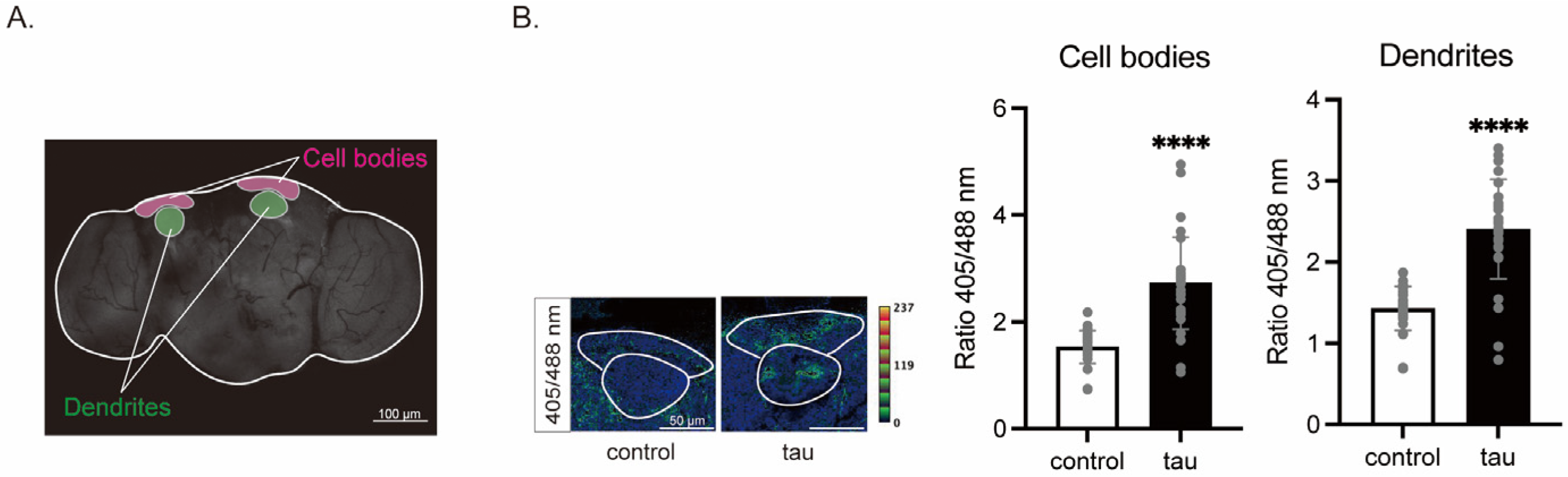
3.1.1. Tau Increases Mitochondrial ROS in Neurons
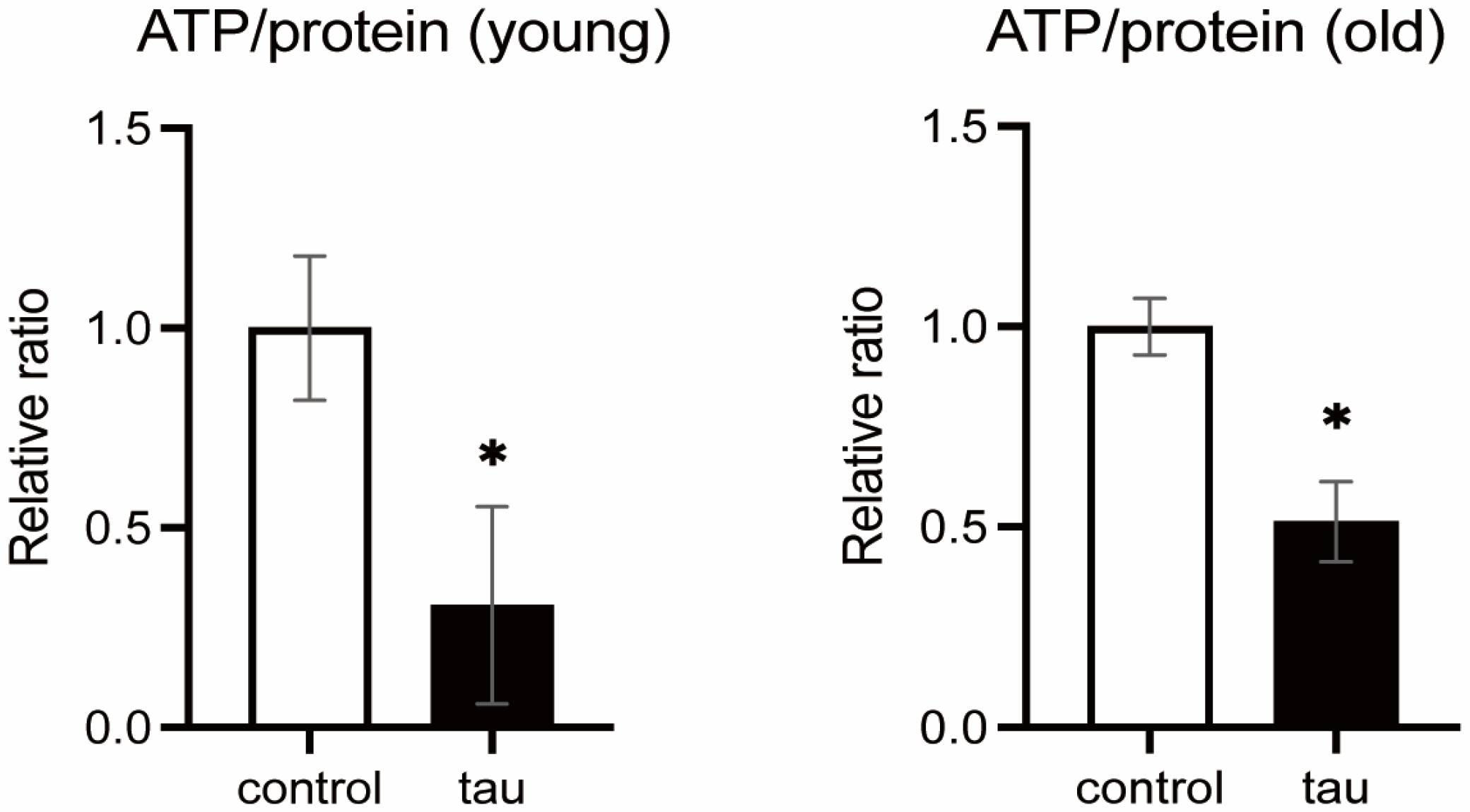
3.1.2. Tau Expression Reduces ATP Levels
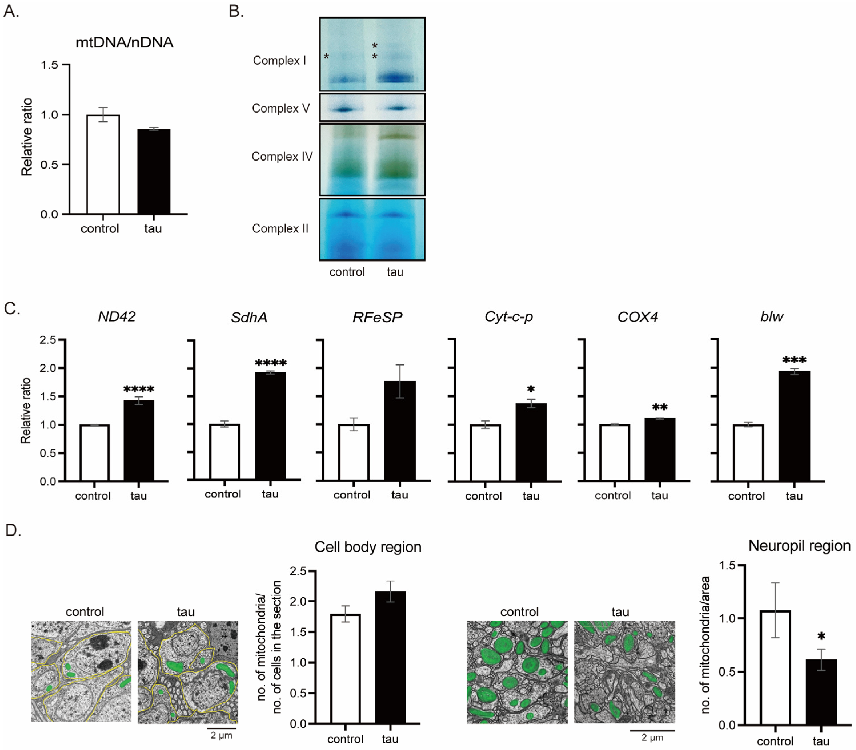
3.1.3. Tau Does Not Reduce the Number of Mitochondria
3.2. Effects of 5-ALA/SFC Feeding on Tau Toxicity
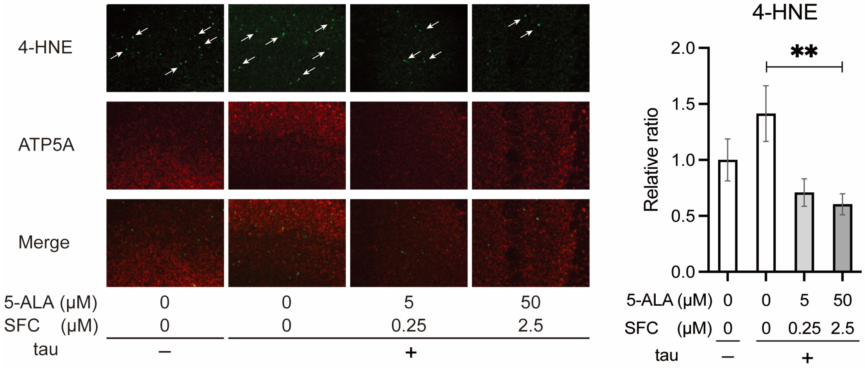
3.2.1. 5-ALA/SFC Reduces Oxidative Damage Caused by Tau
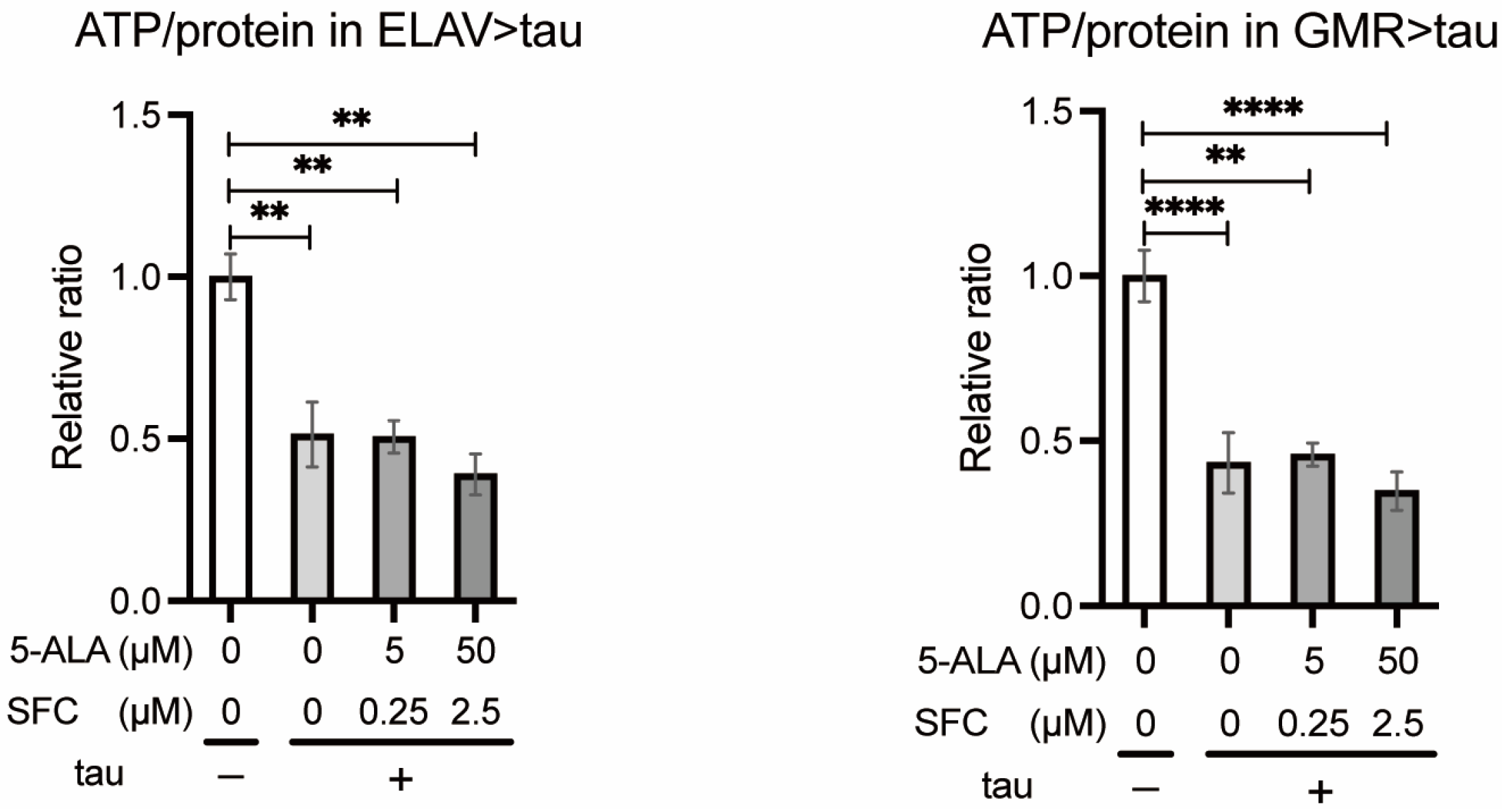
3.2.2. 5-ALA/SFC Does Not Increase ATP Levels in Tau Flies
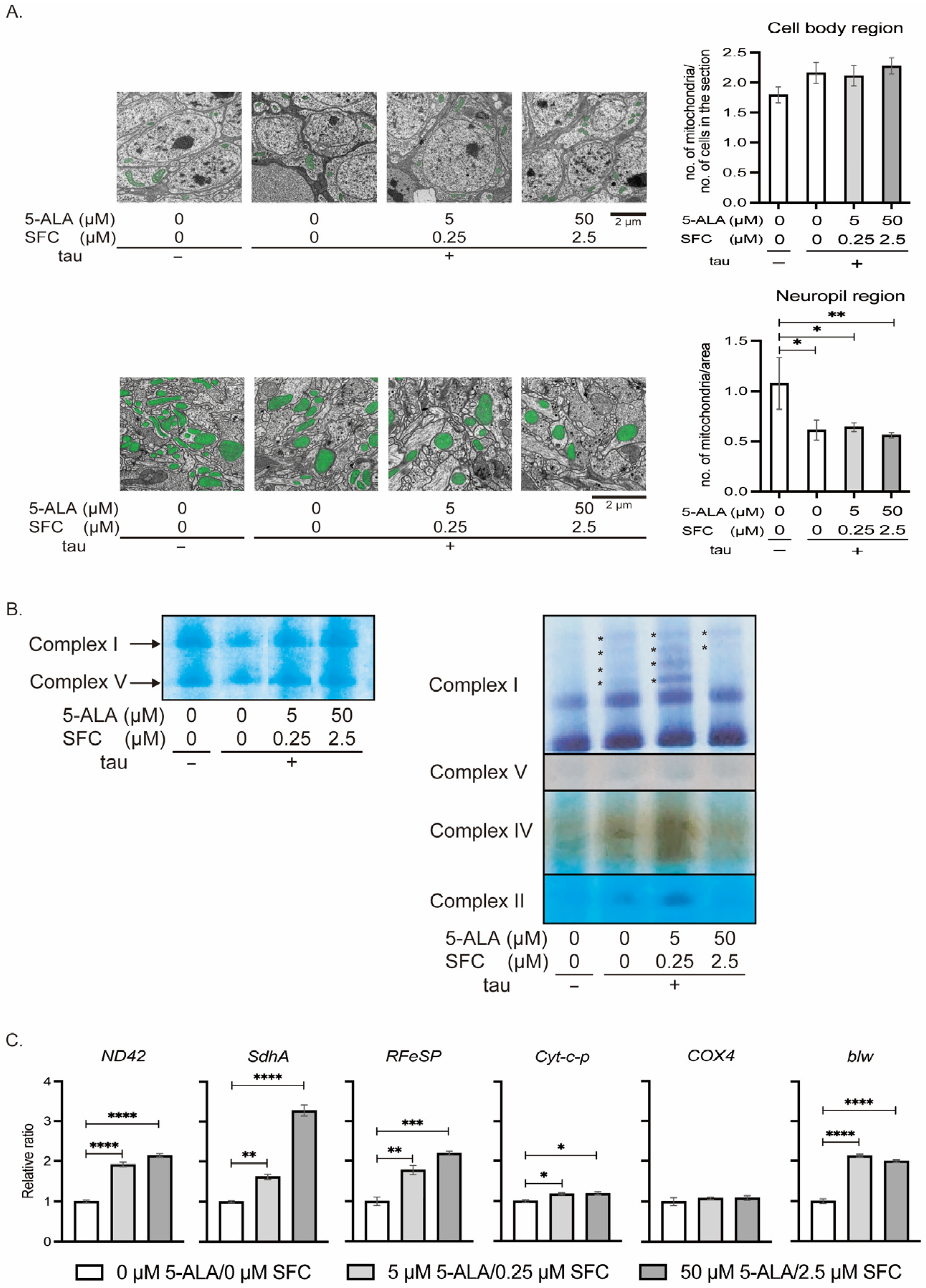
3.2.3. 5-ALA/SFC Does Not Correct Mitochondrial Distribution and OXPHOS Activities in Tau Flies
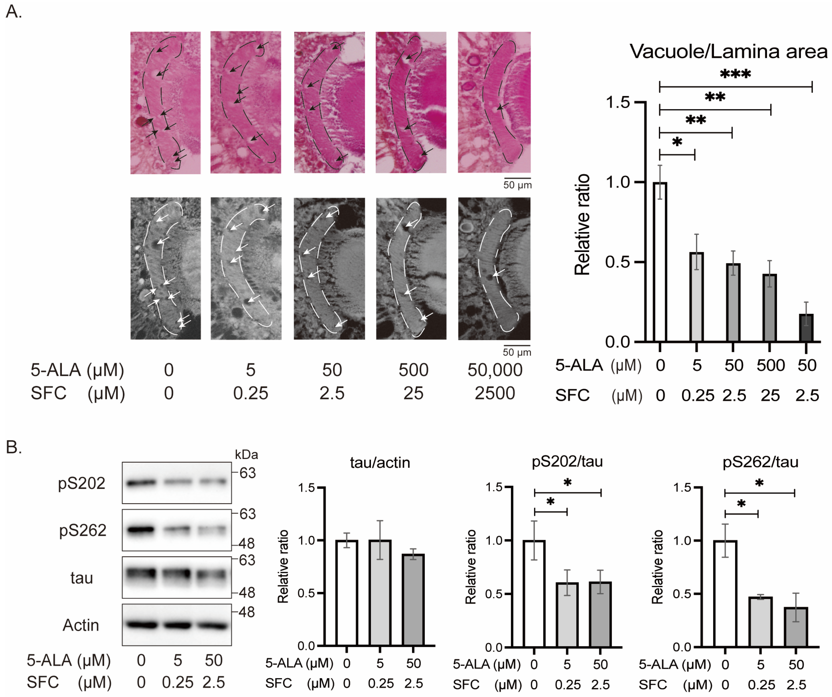
3.2.4. 5-ALA/SFC Reduces Tau Phosphorylation at Disease-Associated Sites and Mitigates Tau-Induced Neurodegeneration
4. Discussion
4.1. Tau Directly Modifies OXPHOS Complexes
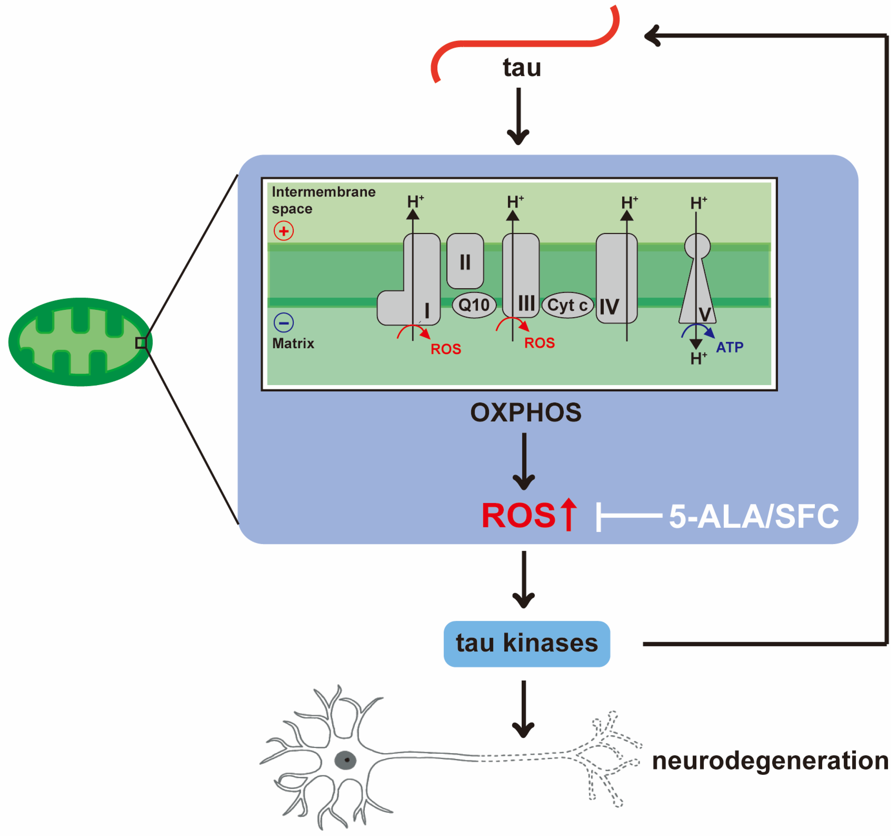
4.2. 5-ALA/SFC Reduces Oxidative Damage
4.3. 5-ALA/SFC Attenuates Tau Phosphorylation and Toxicity
5. Conclusions
Author Contributions
Funding
Institutional Review Board Statement
Informed Consent Statement
Data Availability Statement
Acknowledgments
Conflicts of Interest
References
- 2025 Alzheimer’s disease facts and figures. Alzheimer’s Dement. 2025, 21, e70235. [CrossRef]
- Eckert, A.; Schmitt, K.; Götz, J. Mitochondrial dysfunction—The beginning of the end in Alzheimer’s disease? Separate and synergistic modes of tau and amyloid-β toxicity. Alzheimer’s Res. Ther. 2011, 3, 15. [Google Scholar] [CrossRef]
- Iqbal, K.; Liu, F.; Gong, C.-X.; Grundke-Iqbal, I. Tau in Alzheimer Disease and Related Tauopathies. Curr. Alzheimer Res. 2010, 7, 656–664. [Google Scholar] [CrossRef]
- Wegmann, S.; Biernat, J.; Mandelkow, E. A current view on Tau protein phosphorylation in Alzheimer’s disease. Curr. Opin. Neurobiol. 2021, 69, 131–138. [Google Scholar] [CrossRef]
- Rankin, C.A.; Sun, Q.; Gamblin, T.C. Pseudo-phosphorylation of tau at Ser202 and Thr205 affects tau filament formation. Mol. Brain Res. 2005, 138, 84–93. [Google Scholar] [CrossRef] [PubMed]
- SantaCruz, K.; Lewis, J.; Spires, T.; Paulson, J.; Kotilinek, L.; Ingelsson, M.; Guimaraes, A.; DeTure, M.; Ramsden, M.; McGowan, E.; et al. Tau Suppression in a Neurodegenerative Mouse Model Improves Memory Function. Science 2005, 309, 476–481. [Google Scholar] [CrossRef]
- Moreira, P.I.; Carvalho, C.; Zhu, X.; Smith, M.A.; Perry, G. Mitochondrial dysfunction is a trigger of Alzheimer’s disease pathophysiology. Biochim. Biophys. Acta BBA Mol. Basis Dis. 2010, 1802, 2–10. [Google Scholar] [CrossRef]
- Liao, J.; He, W.; Li, L.; Wang, J.; Gong, L.; Zhang, Q.; Lin, Z. Mitochondria in brain diseases: Bridging structural-mechanistic insights into precision-targeted therapies. Cell Biomater. 2025, 1, 100016. [Google Scholar] [CrossRef]
- Wang, W.; Zhao, F.; Ma, X.; Perry, G.; Zhu, X. Mitochondria dysfunction in the pathogenesis of Alzheimer’s disease: Recent advances. Mol. Neurodegener. 2020, 15, 30. [Google Scholar] [CrossRef] [PubMed]
- Balaban, R.S.; Nemoto, S.; Finkel, T. Mitochondria, Oxidants, and Aging. Cell 2005, 120, 483–495. [Google Scholar] [CrossRef]
- Murali Mahadevan, H.; Hashemiaghdam, A.; Ashrafi, G.; Harbauer, A.B. Mitochondria in Neuronal Health: From Energy Metabolism to Parkinson’s Disease. Adv. Biol. 2021, 5, 2100663. [Google Scholar] [CrossRef]
- Liu, Y.J.; Sulc, J.; Auwerx, J. Mitochondrial genetics, signalling and stress responses. Nat. Cell Biol. 2025, 27, 393–407. [Google Scholar] [CrossRef]
- Wu, Z.; Tantray, I.; Lim, J.; Chen, S.; Li, Y.; Davis, Z.; Sitron, C.; Dong, J.; Gispert, S.; Auburger, G.; et al. MISTERMINATE Mechanistically Links Mitochondrial Dysfunction with Proteostasis Failure. Mol. Cell 2019, 75, 835–848.e8. [Google Scholar] [CrossRef]
- Shinno, K.; Miura, Y.; Iijima, K.M.; Suzuki, E.; Ando, K. Axonal distribution of mitochondria maintains neuronal autophagy during aging via eIF2β. eLife 2025, 13, 95576. [Google Scholar] [CrossRef]
- Kuryata, O.; Akimov, O.; Riabushko, M.; Kostenko, H.; Kostenko, V.; Mishchenko, A.; Nazarenko, S.; Solovyova, N.; Kostenko, V. Therapeutic potential of 5-aminolevulinic acid in metabolic disorders: Current insights and future directions. iScience 2024, 27, 111477. [Google Scholar] [CrossRef]
- Gardner, L.C.; Smith, S.J.; Cox, T.M. Biosynthesis of delta-aminolevulinic acid and the regulation of heme formation by immature erythroid cells in man. J. Biol. Chem. 1991, 266, 22010–22018. [Google Scholar] [CrossRef]
- Kim, H.J.; Khalimonchuk, O.; Smith, P.M.; Winge, D.R. Structure, function, and assembly of heme centers in mitochondrial respiratory complexes. Biochim. Biophys. Acta BBA Mol. Cell Res. 2012, 1823, 1604–1616. [Google Scholar] [CrossRef]
- Shimura, M.; Nozawa, N.; Ogawa-Tominaga, M.; Fushimi, T.; Tajika, M.; Ichimoto, K.; Matsunaga, A.; Tsuruoka, T.; Kishita, Y.; Ishii, T.; et al. Effects of 5-aminolevulinic acid and sodium ferrous citrate on fibroblasts from individuals with mitochondrial diseases. Sci. Rep. 2019, 9, 10549. [Google Scholar] [CrossRef] [PubMed]
- Ota, U.; Hara, T.; Nakagawa, H.; Tsuru, E.; Tsuda, M.; Kamiya, A.; Kuroda, Y.; Kitajima, Y.; Koda, A.; Ishizuka, M.; et al. 5-aminolevulinic acid combined with ferrous ion reduces adiposity and improves glucose tolerance in diet-induced obese mice via enhancing mitochondrial function. BMC Pharmacol. Toxicol. 2017, 18, 7. [Google Scholar] [CrossRef] [PubMed]
- Zhao, M.; Zhu, P.; Fujino, M.; Nishio, Y.; Chen, J.; Ito, H.; Takahashi, K.; Nakajima, M.; Tanaka, T.; Zhao, L.; et al. 5-Aminolevulinic acid with sodium ferrous citrate induces autophagy and protects cardiomyocytes from hypoxia-induced cellular injury through MAPK-Nrf-2-HO-1 signaling cascade. Biochem. Biophys. Res. Commun. 2016, 479, 663–669. [Google Scholar] [CrossRef] [PubMed]
- Li, S.; Takahara, T.; Li, X.-K.; Fujino, M.; Sugiyama, T.; Tsukada, K.; Liu, C.; Kakuta, Y.; Nonomura, N.; Ito, H.; et al. 5-Aminolevulinic acid combined with ferrous iron ameliorate ischemia–reperfusion injury in the mouse fatty liver model. Biochem. Biophys. Res. Commun. 2016, 470, 900–906. [Google Scholar] [CrossRef] [PubMed]
- Nozawa, N.; Noguchi, M.; Shinno, K.; Tajima, M.; Aizawa, S.; Saito, T.; Asada, A.; Ishii, T.; Ishizuka, M.; Iijima, K.M.; et al. 5-Aminolevulinic acid and sodium ferrous citrate ameliorate muscle aging and extend healthspan in Drosophila. FEBS Open Bio 2022, 12, 295–305. [Google Scholar] [CrossRef]
- Nozawa, N.; Noguchi, M.; Shinno, K.; Saito, T.; Asada, A.; Ishii, T.; Takahashi, K.; Ishizuka, M.; Ando, K. 5-Aminolevulinic acid bypasses mitochondrial complex I deficiency and corrects physiological dysfunctions in Drosophila. Hum. Mol. Genet. 2023, 32, 2611–2622. [Google Scholar] [CrossRef]
- Omori, C.; Motodate, R.; Shiraki, Y.; Chiba, K.; Sobu, Y.; Kimura, A.; Nakaya, T.; Kondo, H.; Kurumiya, S.; Tanaka, T.; et al. Facilitation of brain mitochondrial activity by 5-aminolevulinic acid in a mouse model of Alzheimer’s disease. Nutr. Neurosci. 2017, 20, 538–546. [Google Scholar] [CrossRef]
- Wittmann, C.W.; Wszolek, M.F.; Shulman, J.M.; Salvaterra, P.M.; Lewis, J.; Hutton, M.; Feany, M.B. Tauopathy in Drosophila: Neurodegeneration Without Neurofibrillary Tangles. Science 2001, 293, 711–714. [Google Scholar] [CrossRef]
- Jackson, G.R.; Wiedau-Pazos, M.; Sang, T.-K.; Wagle, N.; Brown, C.A.; Massachi, S.; Geschwind, D.H. Human Wild-Type Tau Interacts with wingless Pathway Components and Produces Neurofibrillary Pathology in Drosophila. Neuron 2002, 34, 509–519. [Google Scholar] [CrossRef] [PubMed]
- Nishimura, I.; Yang, Y.; Lu, B. PAR-1 Kinase Plays an Initiator Role in a Temporally Ordered Phosphorylation Process that Confers Tau Toxicity in Drosophila. Cell 2004, 116, 671–682. [Google Scholar] [CrossRef]
- Iijima-Ando, K.; Sekiya, M.; Maruko-Otake, A.; Ohtake, Y.; Suzuki, E.; Bingwei, L.; Iijima, K.M. Loss of Axonal Mitochondria Promotes Tau-Mediated Neurodegeneration and Alzheimer’s Disease–Related Tau Phosphorylation Via PAR-1. PLoS Genet. 2012, 8, e1002918. [Google Scholar] [CrossRef] [PubMed]
- Iijima, K.; Gatt, A.; Iijima-Ando, K. Tau Ser262 phosphorylation is critical for Aβ42-induced tau toxicity in a transgenic Drosophila model of Alzheimer’s disease. Hum. Mol. Genet. 2010, 19, 2947–2957. [Google Scholar] [CrossRef]
- Albrecht, S.C.; Barata, A.G.; Großhans, J.; Teleman, A.A.; Dick, T.P. In Vivo Mapping of Hydrogen Peroxide and Oxidized Glutathione Reveals Chemical and Regional Specificity of Redox Homeostasis. Cell Metab. 2011, 14, 819–829. [Google Scholar] [CrossRef]
- Schneider, C.A.; Rasband, W.S.; Eliceiri, K.W. NIH Image to ImageJ: 25 years of image analysis. Nat. Methods 2012, 9, 671–675. [Google Scholar] [CrossRef]
- Costa, A.C.; Loh, S.H.Y.; Martins, L.M. Drosophila Trap1 protects against mitochondrial dysfunction in a PINK1/parkin model of Parkinson’s disease. Cell Death Dis. 2013, 4, e467. [Google Scholar] [CrossRef]
- Quiros, P.M.; Goyal, A.; Jha, P.; Auwerx, J. Analysis of mtDNA/nDNA Ratio in Mice. Curr. Protoc. Mouse Biol. 2017, 7, 47–54. [Google Scholar] [CrossRef] [PubMed]
- Kohda, M.; Tokuzawa, Y.; Kishita, Y.; Nyuzuki, H.; Moriyama, Y.; Mizuno, Y.; Hirata, T.; Yatsuka, Y.; Yamashita-Sugahara, Y.; Nakachi, Y.; et al. A Comprehensive Genomic Analysis Reveals the Genetic Landscape of Mitochondrial Respiratory Chain Complex Deficiencies. PLoS Genet. 2016, 12, e1005679. [Google Scholar] [CrossRef] [PubMed]
- Pareek, G.; Thomas, R.E.; Pallanck, L.J. Loss of the Drosophila m-AAA mitochondrial protease paraplegin results in mitochondrial dysfunction, shortened lifespan, and neuronal and muscular degeneration. Cell Death Dis. 2018, 9, 304. [Google Scholar] [CrossRef]
- Jha, P.; Wang, X.; Auwerx, J. Analysis of Mitochondrial Respiratory Chain Supercomplexes Using Blue Native Polyacrylamide Gel Electrophoresis (BN-PAGE). Curr. Protoc. Mouse Biol. 2016, 6, 1–14. [Google Scholar] [CrossRef]
- Oba, T.; Homma, D.; Limlingan, S.J.M.; Fukuchi, A.; Asada, A.; Saito, T.; Ando, K. A cell-penetrating peptide derived from SARS-CoV-2 protein Orf9b allosterically inhibits MARK4 activity and mitigates tau toxicity. Neurobiol. Dis. 2023, 188, 106334. [Google Scholar] [CrossRef] [PubMed]
- Sultanakhmetov, G.; Kato, I.; Asada, A.; Saito, T.; Ando, K. Microtubule-affinity regulating kinase family members distinctively affect tau phosphorylation and promote its toxicity in a Drosophila model. Genes. Cells 2024, 29, 337–346. [Google Scholar] [CrossRef]
- Gutscher, M.; Pauleau, A.-L.; Marty, L.; Brach, T.; Wabnitz, G.H.; Samstag, Y.; Meyer, A.J.; Dick, T.P. Real-time imaging of the intracellular glutathione redox potential. Nat. Methods 2008, 5, 553–559. [Google Scholar] [CrossRef]
- Esterbauer, H.; Schaur, R.J.; Zollner, H. Chemistry and biochemistry of 4-hydroxynonenal, malonaldehyde and related aldehydes. Free Radic. Biol. Med. 1991, 11, 81–128. [Google Scholar] [CrossRef]
- Buchholz, S.; Zempel, H. The six brain-specific TAU isoforms and their role in Alzheimer’s disease and related neurodegenerative dementia syndromes. Alzheimer’s Dement. 2024, 20, 3606–3628. [Google Scholar] [CrossRef] [PubMed]
- Buée, L.; Delacourte, A. Comparative Biochemistry of Tau in Progressive Supranuclear Palsy, Corticobasal Degeneration, FTDP-17 and Pick’s Disease. Brain Pathol. 1999, 9, 681–693. [Google Scholar] [CrossRef]
- Dias-Santagata, D.; Fulga, T.A.; Duttaroy, A.; Feany, M.B. Oxidative stress mediates tau-induced neurodegeneration in Drosophila. J. Clin. Investig. 2007, 117, 236–245. [Google Scholar] [CrossRef]
- Sealey, M.A.; Vourkou, E.; Cowan, C.M.; Bossing, T.; Quraishe, S.; Grammenoudi, S.; Skoulakis, E.M.C.; Mudher, A. Distinct phenotypes of three-repeat and four-repeat human tau in a transgenic model of tauopathy—ScienceDirect. Neurobiol. Dis. 2017, 105, 74–83. [Google Scholar] [CrossRef]
- Higham, J.P.; Malik, B.R.; Buhl, E.; Dawson, J.M.; Ogier, A.S.; Lunnon, K.; Hodge, J.J.L. Alzheimer’s Disease Associated Genes Ankyrin and Tau Cause Shortened Lifespan and Memory Loss in Drosophila. Front. Cell Neurosci. 2019, 13, 260. [Google Scholar] [CrossRef]
- Kerr, F.; Augustin, H.; Piper, M.D.W.; Gandy, C.; Allen, M.J.; Lovestone, S.; Partridge, L. Dietary restriction delays aging, but not neuronal dysfunction, in Drosophila models of Alzheimer’s disease. Neurobiol. Aging 2011, 32, 1977–1989. [Google Scholar] [CrossRef]
- Buhl, E.; Higham, J.P.; Hodge, J.J.L. Alzheimer’s disease-associated tau alters Drosophila circadian activity, sleep and clock neuron electrophysiology. Neurobiol. Dis. 2019, 130, 104507. [Google Scholar] [CrossRef]
- Leventhal, M.J.; Zanella, C.A.; Kang, B.; Peng, J.; Gritsch, D.; Liao, Z.; Bukhari, H.; Wang, T.; Pao, P.-C.; Danquah, S.; et al. An integrative systems-biology approach defines mechanisms of Alzheimer’s disease neurodegeneration. Nat. Commun. 2025, 16, 4441. [Google Scholar] [CrossRef]
- Frost, B.; Bardai, F.H.; Feany, M.B. Lamin Dysfunction Mediates Neurodegeneration in Tauopathies. Curr. Biol. 2016, 26, 129–136. [Google Scholar] [CrossRef] [PubMed]
- Frost, B.; Hemberg, M.; Lewis, J.; Feany, M.B. Tau promotes neurodegeneration through global chromatin relaxation. Nat. Neurosci. 2014, 17, 357–366. [Google Scholar] [CrossRef] [PubMed]
- Shulman, J.M.; Imboywa, S.; Giagtzoglou, N.; Powers, M.P.; Hu, Y.; Devenport, D.; Chipendo, P.; Chibnik, L.B.; Diamond, A.; Perrimon, N.; et al. Functional screening in Drosophila identifies Alzheimer’s disease susceptibility genes and implicates Tau-mediated mechanisms. Hum. Mol. Genet. 2014, 23, 870–877. [Google Scholar] [CrossRef]
- Castillo-Casaña, Y.; Arias, C.; Coria, R. Tau–Mitochondria Interactions in Neurodegeneration: Mechanisms and Therapeutic Potential. Cell Mol. Neurobiol. 2025, 46, 3. [Google Scholar] [CrossRef] [PubMed]
- Tracy, T.E.; Madero-Pérez, J.; Swaney, D.L.; Chang, T.S.; Moritz, M.; Konrad, C.; Ward, M.E.; Stevenson, E.; Hüttenhain, R.; Kauwe, G.; et al. Tau interactome maps synaptic and mitochondrial processes associated with neurodegeneration. Cell 2022, 185, 712–728.e14. [Google Scholar] [CrossRef] [PubMed]
- Maranzana, E.; Barbero, G.; Falasca, A.I.; Lenaz, G.; Genova, M.L. Mitochondrial Respiratory Supercomplex Association Limits Production of Reactive Oxygen Species from Complex I. Antioxid. Redox Signal. 2013, 19, 1469–1480. [Google Scholar] [CrossRef]
- Genova, M.L.; Lenaz, G. Functional role of mitochondrial respiratory supercomplexes. Biochim. Biophys. Acta BBA Bioenerg. 2014, 1837, 427–443. [Google Scholar] [CrossRef]
- Shimada, S.; Oosaki, M.; Takahashi, R.; Uene, S.; Yanagisawa, S.; Tsukihara, T.; Shinzawa-Itoh, K. A unique respiratory adaptation in Drosophila independent of supercomplex formation. Biochim. Biophys. Acta BBA Bioenerg. 2018, 1859, 154–163. [Google Scholar] [CrossRef]
- Bartolome, F.; Carro, E.; Alquezar, C. Oxidative Stress in Tauopathies: From Cause to Therapy. Antioxidants 2022, 11, 1421. [Google Scholar] [CrossRef] [PubMed]
- Steinhilb, M.L.; Dias-Santagata, D.; Fulga, T.A.; Felch, D.L.; Feany, M.B. Tau Phosphorylation Sites Work in Concert to Promote Neurotoxicity In Vivo. Mol. Biol. Cell 2007, 18, 5060–5068. [Google Scholar] [CrossRef]
- Chatterjee, S.; Sang, T.-K.; Lawless, G.M.; Jackson, G.R. Dissociation of tau toxicity and phosphorylation: Role of GSK-3β, MARK and Cdk5 in a Drosophila model. Hum. Mol. Genet. 2009, 18, 164–177. [Google Scholar] [CrossRef]
- Mondragón-Rodríguez, S.; Perry, G.; Zhu, X.; Moreira, P.I.; Acevedo-Aquino, M.C.; Williams, S. Phosphorylation of Tau Protein as the Link between Oxidative Stress, Mitochondrial Dysfunction, and Connectivity Failure: Implications for Alzheimer’s Disease. Oxidative Med. Cell. Longev. 2013, 940603. [Google Scholar] [CrossRef]
- Biernat, J.; Gustke, N.; Drewes, G.; Mandelkow, E.; Mandelkow, E. Phosphorylation of Ser262 strongly reduces binding of tau to microtubules: Distinction between PHF-like immunoreactivity and microtubule binding. Neuron 1993, 11, 153–163. [Google Scholar] [CrossRef]
- Yoshida, H.; Goedert, M. Phosphorylation of microtubule-associated protein tau by AMPK-related kinases—Yoshida—2012—Journal of Neurochemistry—Wiley Online Library. J. Neurochem. 2012, 120, 165–176. [Google Scholar] [CrossRef] [PubMed]
- Domise, M.; Didier, S.; Marinangeli, C.; Zhao, H.; Chandakkar, P.; Buée, L.; Viollet, B.; Davies, P.; Marambaud, P.; Vingtdeux, V. AMP-activated protein kinase modulates tau phosphorylation and tau pathology in vivo. Sci. Rep. 2016, 6, 26758. [Google Scholar] [CrossRef]
- Vingtdeux, V.; Davies, P.; Dickson, D.W.; Marambaud, P. AMPK is abnormally activated in tangle- and pre-tangle-bearing neurons in Alzheimer’s disease and other tauopathies. Acta Neuropathol. 2011, 121, 337–349. [Google Scholar] [CrossRef]
- Thornton, C.; Bright, N.J.; Sastre, M.; Muckett, P.J.; Carling, D. AMP-activated protein kinase (AMPK) is a tau kinase, activated in response to amyloid β-peptide exposure. Biochem. J. 2011, 434, 503–512. [Google Scholar] [CrossRef] [PubMed]
- Rabinovitch, R.C.; Samborska, B.; Faubert, B.; Ma, E.H.; Gravel, S.-P.; Andrzejewski, S.; Raissi, T.C.; Pause, A.; St-Pierre, J.; Jones, R.G. AMPK Maintains Cellular Metabolic Homeostasis through Regulation of Mitochondrial Reactive Oxygen Species. Cell Rep. 2017, 21, 1–9. [Google Scholar] [CrossRef] [PubMed]
- Iijima-Ando, K.; Zhao, L.; Gatt, A.; Shenton, C.; Iijima, K. A DNA damage-activated checkpoint kinase phosphorylates tau and enhances tau-induced neurodegeneration. Hum. Mol. Genet. 2010, 19, 1930–1938. [Google Scholar] [CrossRef]





| Figure 1 | control | elav-Gal4/Y;UAS-mito-roGFP2-Grx19/+;UAS-luciferase/+ |
| tau | elav-Gal4/Y;UAS-mito-roGFP2-Grx19/+;UAS-tau/+ | |
| Figure 2 | control | elav-Gal4/Y;UAS-tdTomato/+;+/+ |
| tau | elav-Gal4/Y;+/+;UAS-tau/+ | |
| Figure 3A | control | elav-Gal4/Y;+/+;+/+ |
| tau | elav-Gal4/Y;+/+;UAS-tau/+ | |
| Figure 3B | control | elav-Gal4/Y;+/+;+/+ |
| tau | elav-Gal4/Y;UAS-tau/CyO;+/+ | |
| Figure 3C | control | elav-Gal4/Y;+/+;+/+ |
| tau | elav-Gal4/Y;+/+;UAS-tau/+ | |
| Figure 3D | control | +/+;gmr-Gal4/+;+/+ |
| tau | +/+;gmr-Gal4/+;UAS-tau/+ | |
| Figure 4 | tau − | elav-Gal4/Y;+/+;+/+ |
| tau + | elav-Gal4/Y;+/+;UAS-tau/+ | |
| Figure 5 | tau − | elav-Gal4/Y;+/+;+/+ +/Y;gmr-Gal4/+;+/+ |
| tau + | elav-Gal4/Y;+/+;UAS-tau/+ +/Y;gmr-Gal4/+;UAS-tau/+ | |
| Figure 6A | tau − | +/+;gmr-Gal4/+;+/+ |
| tau + | +/+;gmr-Gal4/+;UAS-tau/+ | |
| Figure 6B | tau − | +/+;gmr-Gal4/+;+/+ |
| tau + | +/+;gmr-Gal4/+;UAS-tau/+ | |
| Figure 6C | +/+;gmr-Gal4/+;UAS-tau/+ | |
| Figure 7 | +/+;gmr-Gal4/+;UAS-tau/+ | |
| Figure 8 | +/+;gmr-Gal4/+;UAS-tau/+ |
Disclaimer/Publisher’s Note: The statements, opinions and data contained in all publications are solely those of the individual author(s) and contributor(s) and not of MDPI and/or the editor(s). MDPI and/or the editor(s) disclaim responsibility for any injury to people or property resulting from any ideas, methods, instructions or products referred to in the content. |
© 2026 by the authors. Licensee MDPI, Basel, Switzerland. This article is an open access article distributed under the terms and conditions of the Creative Commons Attribution (CC BY) license.
Share and Cite
Tamura, A.; Noguchi, M.; Nozawa, N.; Suzuki, E.; Ando, K. 5-ALA/SFC Mitigates Tau Toxicity via Lowering Oxidative Stress in a Drosophila Model of Tau Toxicity. Life 2026, 16, 725. https://doi.org/10.3390/life16050725
Tamura A, Noguchi M, Nozawa N, Suzuki E, Ando K. 5-ALA/SFC Mitigates Tau Toxicity via Lowering Oxidative Stress in a Drosophila Model of Tau Toxicity. Life. 2026; 16(5):725. https://doi.org/10.3390/life16050725
Chicago/Turabian StyleTamura, Arisa, Marie Noguchi, Naoko Nozawa, Emiko Suzuki, and Kanae Ando. 2026. "5-ALA/SFC Mitigates Tau Toxicity via Lowering Oxidative Stress in a Drosophila Model of Tau Toxicity" Life 16, no. 5: 725. https://doi.org/10.3390/life16050725
APA StyleTamura, A., Noguchi, M., Nozawa, N., Suzuki, E., & Ando, K. (2026). 5-ALA/SFC Mitigates Tau Toxicity via Lowering Oxidative Stress in a Drosophila Model of Tau Toxicity. Life, 16(5), 725. https://doi.org/10.3390/life16050725