1. Introduction
Lung cancer is a diverse disease that comprises many different variants. Lung cancer is primarily divided into two large groups: small-cell lung cancer (SCLC), which histologically is composed of neuroendocrine cells; and non-small-cell lung cancer (NSCLC), which is further sectioned based on its histology. NSCLC is mainly subdivided into squamous-cell carcinoma (SCC), adenocarcinoma, and large-cell carcinoma. An adenosquamous subtype has also been observed. NSCLC accounts for approximately 85% of all lung cancer cases, of which, the majority are classified as either squamous-cell carcinoma, or adenocarcinoma. Smoking is regarded as a major causative factor in the etiopathogenesis of all lung cancers. Nevertheless, adenocarcinoma is the most common type reported in non-smokers. Lung cancer is a leading cause of mortality worldwide, with late diagnosis contributing to its poor prognosis. Therefore, early diagnosis, with accurate tumor analysis, plays an integral role in establishing a proper treatment [
1,
2,
3].
Across the different approaches of treating lung cancer, immunotherapy has become a promising modality. By integrating immune checkpoint inhibitors (ICIs), immunotherapy modulates and antagonizes the interaction between neoplastic and cytotoxic T-cells by utilizing targeted monoclonal antibodies. ICIs have emerged as a critical breakthrough in oncology, and are now broadly implemented in clinical practice [
4,
5].
ICI therapy may target the PD-1/PD-L1 pathway. PD-1 and PD-L1 are proteins which are expressed on tumor cells and cytotoxic T-lymphocytes, respectively. The interaction between PD-1 and PD-L1 enables tumor cells to bypass recognition by the immune system. Disrupting this interaction, is a fundamental aspect of ICIs therapeutic implementation [
4,
5,
6].
The expression level of PD-L1, which is measured by immunohistochemistry, has been shown to be a predictive biomarker, with its expression level being an important factor. PD-L1 Tumor Proportion Score (TPS) may be reported as high expression (≥50%) or low expression (<50%), although other cutoffs can be made based on varying and supplementary thresholds (i.e., negative at <1%, intermediate at 1–49%). ICI therapy (e.g., pembrolizumab (Keytruda™) in patients with higher expression levels of PD-L1 has yielded positive results with better progression-free and overall survival in various types of malignancies [
6,
7,
8,
9,
10].
Immunotherapy, despite its promise, has unfortunately been linked to immune-related adverse events (irAEs). Adverse effects of ICI treatment are notably common, and range from mild (grades 1–2) to severe (grade 3 and above). Virtually any organ system can be affected. Common ICI-related irAEs include, but are not limited to, cutaneous (rash, pruritus), gastrointestinal (diarrhea, colitis), endocrine (hypothyroidism, hyperthyroidism), and musculoskeletal (muscle and joint pain). Less frequently, neurological and cardiac toxicities have been observed. While most irAEs resolve, less common manifestations may be permanent or contribute to long-term sequelae [
11,
12,
13,
14].
Cutaneous irAEs are the most common adverse effects associated with ICI therapy. They are typically the first to manifest. Cutaneous irAEs vary widely, and may present as pruritus, xerosis, maculopapular or psoriasiform eruptions, depigmentation and alopecia. Although most irAEs are usually mild (low grade), more severe and rarer irAEs, such as Stevens–Johnson syndrome (SJS)/toxic epidermal necrolysis (TEN), drug reaction with eosinophilia and systemic symptoms (DRESS), and bullous diseases, can still occur. Therefore, recognizing cutaneous irAEs is essential for providing optimal care, which may involve discontinuation of ICI agents or the initiation of immunosuppressive therapy [
14,
15,
16,
17].
The mechanisms underlying cutaneous irAEs are complex and poorly understood, but some plausible explanations may include: reduced central tolerance of CD4+ and CD8+ T-cells targeting neoplastic and normal tissues, activation of autoreactive B-lymphocytes inducing cytotoxic effects, abnormal and dysregulated discharge of pro-inflammatory mediators, specific human leukocyte antigen (HLA) variations, and accelerated concurrent drug-induced eruptions. An increased incidence of cutaneous irAEs has been linked to improved prognosis and longer survival. This is thought to stem from an enhanced anti-tumor response. Thus, irAEs manifestations may serve as a vital indicator of therapy effectiveness and outcomes [
16,
17,
18].
The objective of our study was to evaluate the impact of PD-L1 expression on the occurrence of cutaneous immune-related adverse events in patients with NSCLC undergoing immunotherapy, and to assess its influence on survival.
3. Results
We identified 285 patients who were treated at our centers, all of whom had complete follow-up data, including documented adverse events. The results showing that the median age was 66.8 years, ranging from 41 to 86 years. Of the participants, 33.3% (
n = 95) were female and 66.6% (
n = 190) were male. Histologically, 66.7% (
n = 190) of patients were diagnosed with adenocarcinoma, while 33.3% (
n = 95) had squamous-cell carcinoma. Regarding smoking status, 13.3% (
n = 38) of the patients were never smokers, 49.1% (
n = 140) were current smokers, and 37.7% (
n = 107) were past smokers. Performance status based on ECOG scores revealed that 20.7% (
n = 59) had a score of 0, 59.6% (
n = 170) had a score of 1, and 19.7% (
n = 56) had a score of 2 or higher. In terms of treatment, 79% (
n = 225) received chemo-immunotherapy, while 21% (
n = 60) were treated with only immunotherapy. Among those receiving immunotherapy, 71.2% (
n = 203) were administered pembrolizumab, and 28.8% (
n = 82) received the combination of ipilimumab plus nivolumab. PD-L1 expression levels were available for all patients: 35.8% (
n = 102) had PD-L1 expression < 1%, and 64.2% (
n = 183) had expression > 1%. Within the group with PD-L1 > 1%, 21.7% (
n = 62) had expression between 1–49%, and 42.5% (
n = 121) had PD-L1 levels greater than 50%, (
Table 1).
Regarding dermatological toxicity, we observed that 57 patients (20%) overall experienced rash. Among the 57 patients who experienced rash, 46 patients had grade 1, 7 patients had grade 2, and 4 patients had grade 3 reactions. No life-threatening cases (grade 4) were reported. In addition, 47 patients (16.5%) had pruritus all grade 1–2. When comparing these outcomes by PD-L1 status, we found that among patients with PD-L1 < 1%, 20 patients (19.6%) had a rash, while 37 patients (20.2%) with PD-L1 > 1% experienced rash. The difference between these groups was not statistically significant (
p = 0.91) (
Table 2). For the PD-L1 >1% subgroup, we found no statistically significant difference between the PD-L1 with staining of 1–49% and PD-L1 > 50% groups (
p = 0.083). Specifically, 13 patients (7.1%) with PD-L1 staining 1–49% had a rash, compared to 24 patients (13.1%) with PD-L1 staining > 50%, (
Table 2).
For pruritus, 9 patients (8.8%) with PD-L1 < 1% had this adverse effect, while 38 patients (20.2%) with PD-L1 > 1% experienced it, showing a significant statistical difference (
p = 0.0013). However, when comparing the subgroups within PD-L1 > 1%, there was no significant difference between the PD-L1 staining 1–49% group (14 patients, 7.1%) and the PD-L1 staining ≥ 50% group (24 patients, 13.1%), (
p = 0.206), (
Table 2 used Pearson chi-square test).
When comparing dermatological adverse events between patients who received immunotherapy alone and those who received a combination of treatment (chemo-immunotherapy), there was no significant difference observed in the incidence of rash. Specifically, 11 patients (18.3%) out of 60 who received only immunotherapy developed a rash, compared to 46 patients (20.4%) out of 225 who received chemo-immunotherapy (
p = 0.856), (
Table 3). Similarly, for pruritus, no significant difference was found between the two treatment groups. Pruritus occurred in 11 patients (18.3%) in the immunotherapy-only group and in 35 patients (15.6%) in the chemo-immunotherapy group (
p = 0.747), (
Table 3, used Pearson chi-square test). Notably, all grade 2 and 3 cases occurred in patients receiving combination chemo-immunotherapy.
Comparison between the two immunotherapy regimens—pembrolizumab and ipilimumab plus nivolumab—showed that rash occurred in 13.4% (11 patients) of those receiving ipilimumab plus nivolumab and in 21.1% (43 patients) of those receiving pembrolizumab. This difference was not statistically significant (
p = 0.181). Similarly, pruritus was reported in 9.8% (8 patients) of the ipilimumab plus nivolumab group compared to 18.7% (38 patients) in the pembrolizumab group, which also did not reach statistical significance (
p = 0.075), (
Table 4, used Pearson chi-square test).
When comparing rash incidence by gender, 22.1% of females (21 patients) experienced rash compared to 19% of males (36 patients), with no statistically significant difference (
p = 0.535). Similarly, pruritus was reported in 22% of males (19 patients) and 14.2% of females (27 patients), which also did not show a statistically significant difference (
p = 0.118), (
Table 5, used Pearson chi-square test).
In terms of age at the onset of symptoms, patients who developed a rash had a median age of 68.6 years, while those without a rash had almost the same ages with median age of 66.4 years; this difference was statistically significant (p = 0.033). In contrast, the median age of patients with pruritus was 67.7 years compared to 66.7 years in those without, which was not statistically significant (p = 0.51).
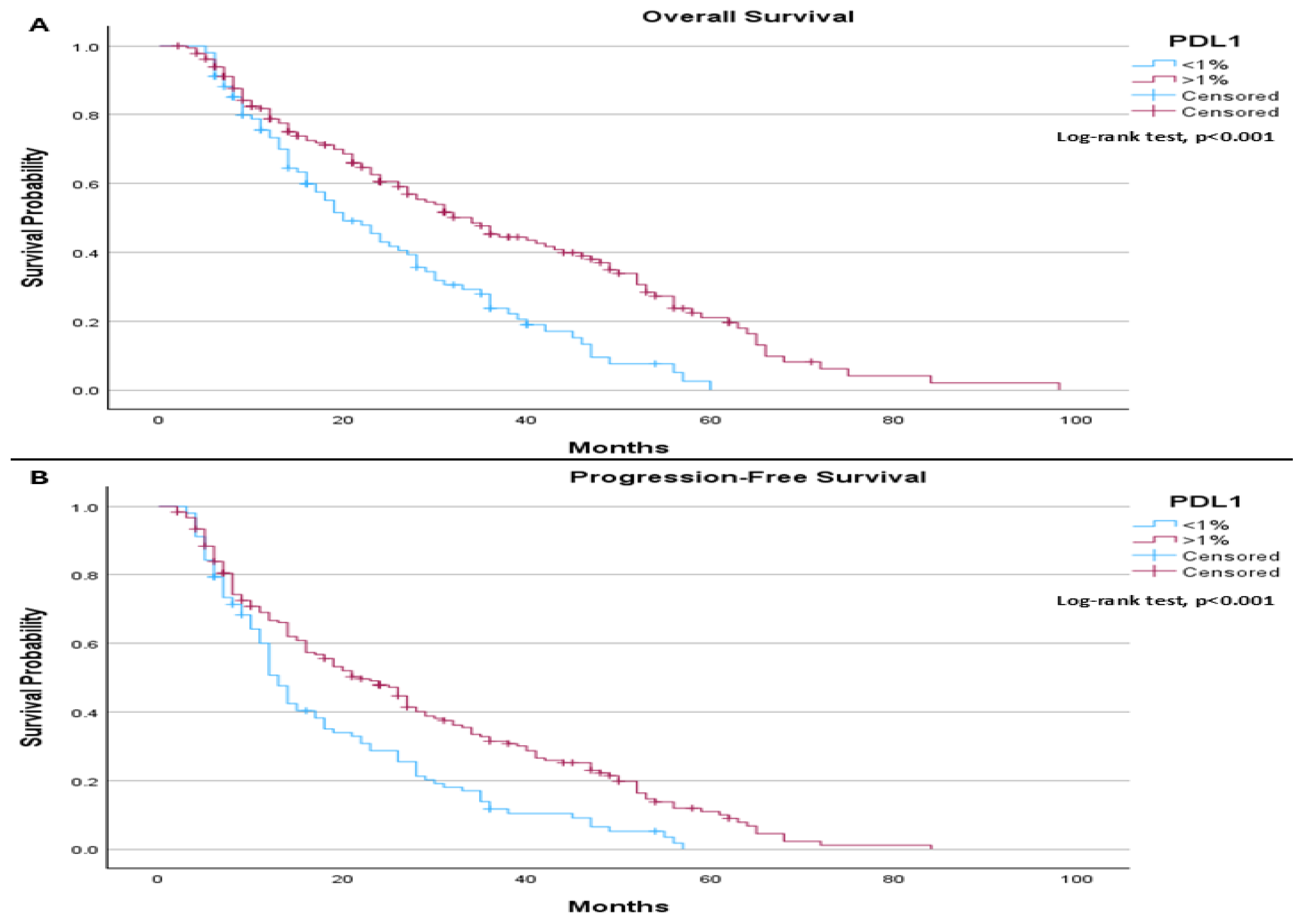
Median follow-up was 26.4 months, when comparing median overall survival based on PD-L1 status, a statistically significant difference was observed between the groups (
p < 0.001). Patients with PD-L1 expression < 1% (102 patients (35.8%)) had a median overall survival of 20.0 months, whereas those with PD-L1 expression ≥ 1% had a median overall survival of 34.0 months (
Figure 1A). A similar statistically significant difference was found in progression-free survival (
p < 0.001), with median values of 13.0 months for patients with PD-L1 < 1% and 22.0 months for those with PD-L1 ≥ 1% (183 patients (64.2%)) (
Figure 1B).
When evaluating median overall survival in relation to both PD-L1 status and the occurrence of rash as an adverse event, a statistically significant difference was found among the groups (p < 0.001). Patients with PD-L1 < 1% with rash OS is better than for without >1% (22 months vs. 19 months). In contrast, those with PD-L1 expression ≥ 1% had a median overall survival of 36.0 months without rash and 28.0 months with rash. A similar statistically significant difference was observed in progression-free survival (p < 0.001). For patients with PD-L1 < 1%, the median progression-free survival was 12.0 months without rash and 18.0 months with rash. Among those with PD-L1 ≥ 1%, progression-free survival was 20.0 months without rash and 27.0 months with rash.
When assessing median overall survival based on PD-L1 status and the presence of pruritus as an adverse event, a statistically significant difference was observed among the groups (p < 0.001). Patients with PD-L1 expression < 1% had a median overall survival of 19.0 months without pruritus and 23.0 months with pruritus. In comparison, those with PD-L1 expression ≥ 1% had a median overall survival of 32.0 months without pruritus and 48.0 months with pruritus. Similar statistically significant differences were noted in progression-free survival (p < 0.001). Median progression-free survival was 12.0 months for patients with PD-L1 < 1% without pruritus, 23.0 months for those with PD-L1 < 1% with pruritus, 19.0 months for PD-L1 ≥ 1% without pruritus, and 31.0 months for PD-L1 ≥ 1% with pruritus.
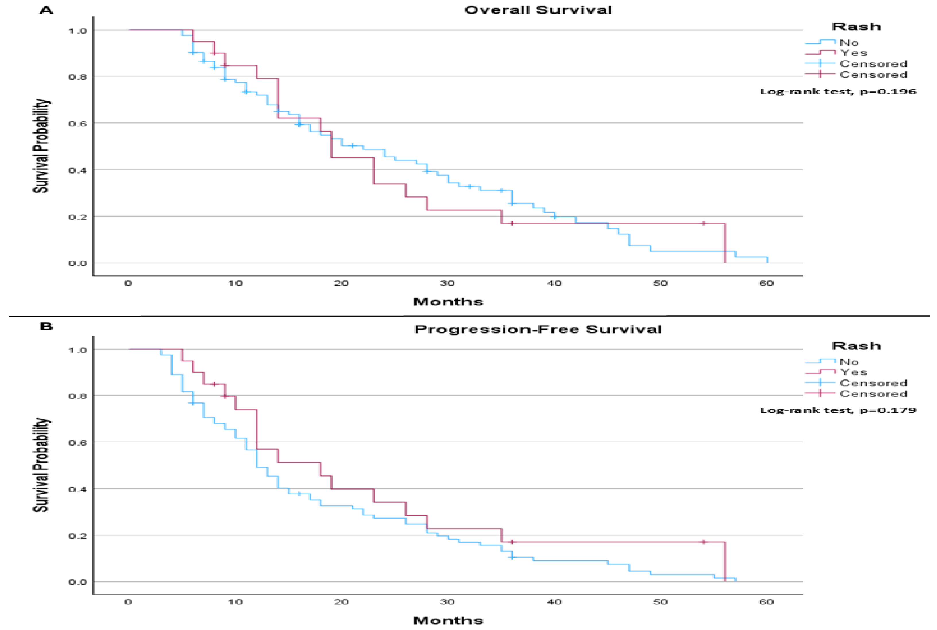
When comparing the survival outcomes according to rash and PD-L1 expression, the results show that, among patients with PD-L1 ≤ 1%, the median OS was 22.0 months in patients without rash compared to 19.0 months in those who developed rash. There was no statistically significant difference in OS between the groups (log-rank
p = 0.196;
Figure 2A). The median PFS was 12.0 months in patients without rash and 18.0 months in those with rash, without a statistically significant difference (log-rank
p = 0.179;
Figure 2B).
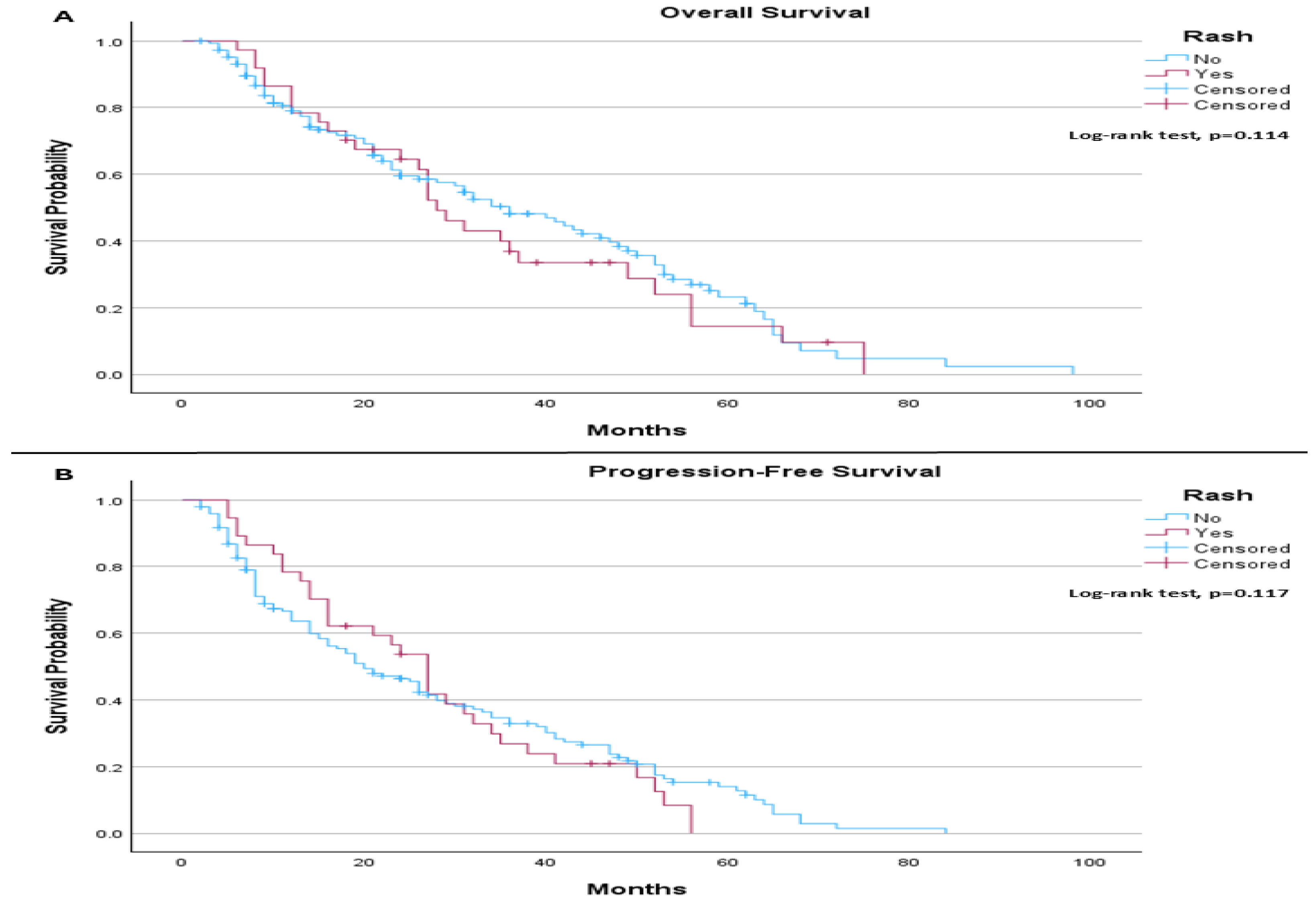
In the PD-L1 > 1% subgroup, the median OS was 36.0 months for patients without rash and 28.0 months for those with rash. The difference did not reach statistical significance (log-rank
p = 0.114;
Figure 3A). Median PFS was 20.0 months in patients without rash and 27.0 months in those with rash, with no statistically significant difference observed (log-rank
p = 0.117;
Figure 3B).
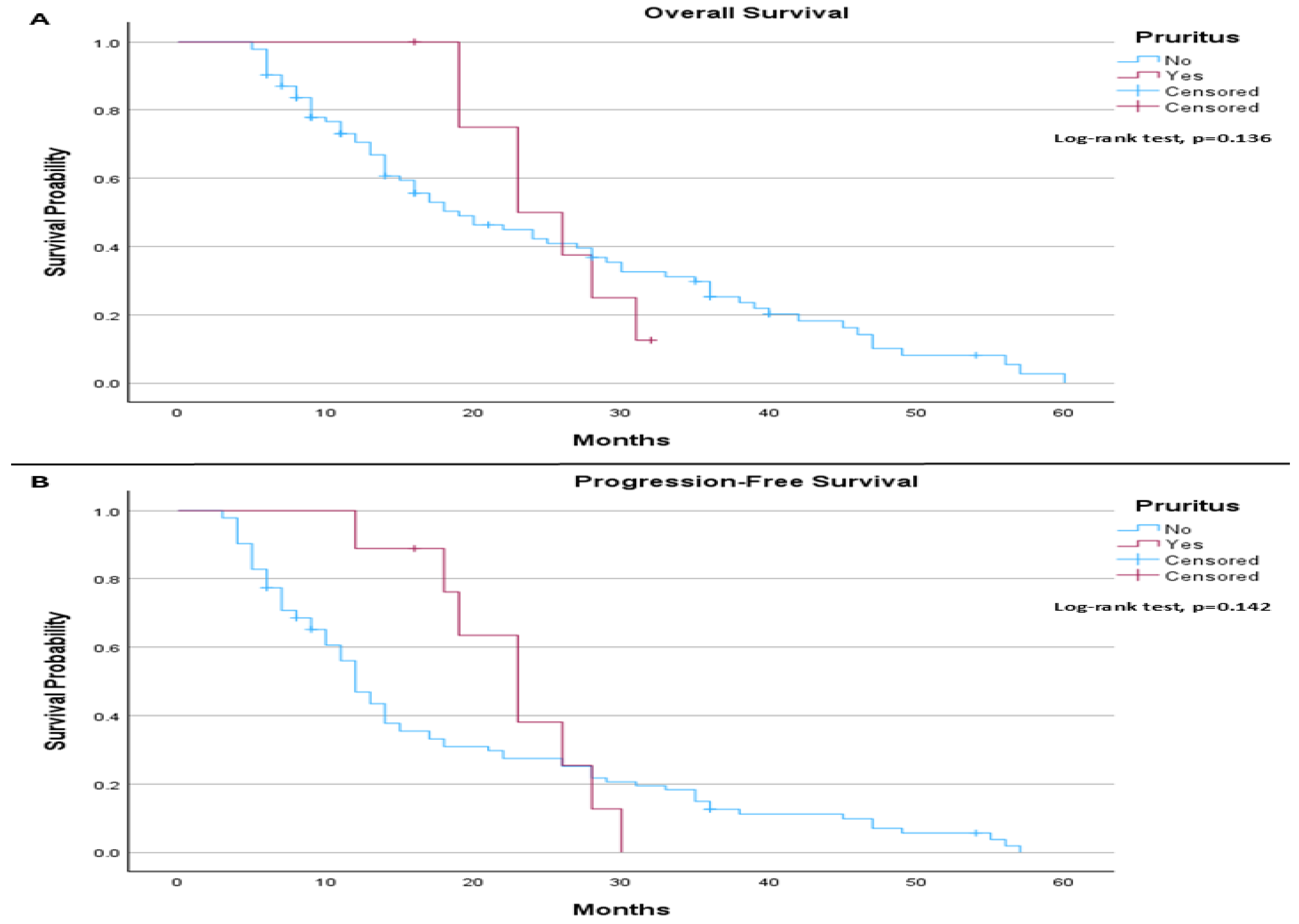
When comparing the survival outcomes according to pruritus and PD-L1 expression, we notice that, among patients with PD-L1 ≤ 1%, the median OS was 19.0 months in patients without pruritus and 23.0 months in those with pruritus. This difference was not statistically significant (log-rank
p = 0.136;
Figure 4A). Median PFS was 12.0 months in patients without pruritus and 23.0 months in those with pruritus, also without statistical significance (log-rank
p = 0.142;
Figure 4B).
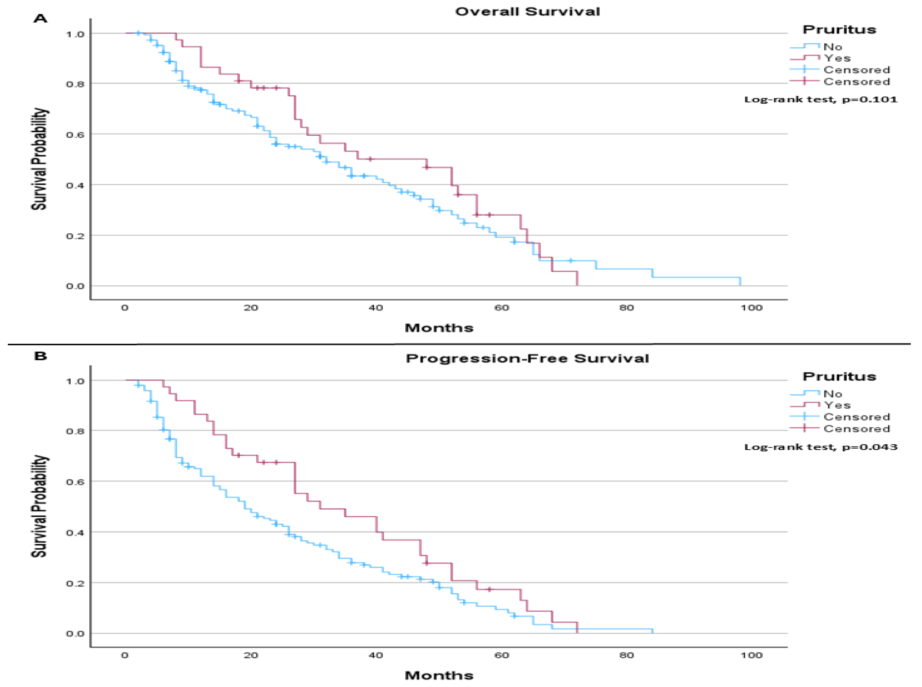
In the PD-L1 > 1% subgroup, the median OS was 32.0 months in patients without pruritus compared to 48.0 months in those with pruritus, without reaching statistical significance (log-rank
p = 0.101;
Figure 5A). However, median PFS was 19.0 months in patients without pruritus and 31.0 months in those with pruritus, demonstrating a statistically significant improvement in PFS among patients who developed pruritus (log-rank
p = 0.043;
Figure 5B).
In the time-dependent Cox regression analysis for OS, neither rash nor pruritus demonstrated a significant association with OS. Rash was not associated with improved or worsened survival (HR 1.12, 95% CI 0.82–1.54,
p = 0.47), and similarly, pruritus showed no statistically significant effect (HR 0.81, 95% CI 0.58–1.14,
p = 0.23). In contrast, PD-L1 expression ≥ 1% was significantly associated with improved OS (HR 0.62, 95% CI 0.48–0.80,
p < 0.001). Increasing age was associated with a modest but statistically significant increase in mortality risk (HR 1.02 per year, 95% CI 1.01–1.04,
p = 0.021). Poor performance status (ECOG ≥ 2) was also strongly associated with worse OS (HR 1.78, 95% CI 1.29–2.45,
p < 0.001), as was current smoking status (HR 1.34, 95% CI 1.01–1.78,
p = 0.043). Male sex, squamous histology, and receipt of chemo-immunotherapy were not significantly associated with OS, (
Table 6).
For PFS, rash was not significantly associated with outcomes (HR 0.97, 95% CI 0.74–1.28,
p = 0.84). However, pruritus was significantly associated with improved PFS (HR 0.68, 95% CI 0.50–0.92,
p = 0.014). PD-L1 expression ≥ 1% was again associated with improved PFS (HR 0.59, 95% CI 0.46–0.75,
p < 0.001). ECOG performance status ≥ 2 remained a strong predictor of worse PFS (HR 1.65, 95% CI 1.23–2.20,
p = 0.001). Age showed a non-significant trend toward worse PFS (HR 1.01 per year, 95% CI 1.00–1.03,
p = 0.08), while current smoking also demonstrated a borderline association with poorer PFS (HR 1.28, 95% CI 0.98–1.66,
p = 0.07). No significant associations were observed for sex, histology, or chemo-immunotherapy in relation to PFS, (
Table 7).
4. Discussion
Although immunotherapy has revolutionized the management of lung cancer—particularly NSCLC—a considerable proportion of patients still fail to achieve meaningful clinical benefit. Evaluating true clinical response to ICIs remains challenging for clinicians, and irAEs often necessitate treatment discontinuation, especially with combination regimens. The broad spectrum of irAEs, affecting multiple organ systems, further complicates the recognition of therapy-related toxicities. These challenges may be amplified in settings with limited access to specialized oncology care, potentially delaying diagnosis and intervention.
Cutaneous toxicities are among the most frequently reported irAEs, accounting for over 50% of cases, and are generally mild, rarely necessitating treatment discontinuation. Nonspecific maculopapular rashes, often accompanied by pruritus, typically appear within the first six weeks of therapy, usually involving less than 30% of the body surface area, with grade ≥ 3 events occurring in fewer than 5% of patients. Lichenoid eruptions are more common under PD-1 inhibitors, appearing in up to 30% of patients, whereas CTLA-4 inhibitors are less frequently associated with these reactions. The pathophysiology of cutaneous irAEs remains incompletely understood, but several immune-mediated mechanisms have been proposed. One leading hypothesis is cross-reactivity, in which checkpoint inhibition enhances T-cell responses against antigens shared by tumor cells and normal skin. In NSCLC, antigen-specific T-cell clones have been identified in both tumor tissue and skin lesions, supporting this mechanism. In addition, PD-1 and CTLA-4 blockade may promote Th1/Th17-driven inflammation and increased cytokine production, contributing to manifestations such as maculopapular, lichenoid, and psoriasiform eruptions, as well as pruritus. These mechanisms may help explain why cutaneous irAEs are among the most common and earliest toxicities seen during ICI therapy [
16].
In our cohort of 285 patients, dermatologic toxicities—specifically rash and pruritus—were observed in 20% and 16.14% of patients, respectively. Stratification by PD-L1 expression revealed statistically significant associations between PD-L1 status and the occurrence of these cutaneous events. Patients with PD-L1 ≥ 1% experienced longer median OS (34.0 months vs. 20.0 months) and PFS (22.0 vs. 13.0 months) compared to those with PD-L1 < 1%. When considering rash, OS ranged from 19.0 months (PD-L1 < 1% with rash) to 36.0 months (PD-L1 ≥ 1% without rash), while PFS varied from 12.0 to 27.0 months. Although rash was associated with a trend toward longer PFS, this did not translate into improved OS, suggesting that any association between rash and outcome should be interpreted cautiously given the small subgroup size and potential confounding factors.
Pruritus demonstrated an even stronger association with outcomes: patients with PD-L1 ≥ 1% who developed pruritus had the longest median OS (48.0 months) and PFS (31.0 months), compared to 19.0 and 12.0 months, respectively, for PD-L1 < 1% patients without pruritus.
Although the incidence of rash was only slightly higher in PD-L1 ≥ 1% patients (20.2%) compared to PD-L1 < 1% (19.6%; p = 0.91), pruritus occurred significantly more frequently in the PD-L1 ≥ 1% group (20.2% vs. 8.8%; p = 0.0013), supporting the hypothesis that higher PD-L1 expression may be associated with increased immune activation and skin toxicity. Subgroup analysis within PD-L1-positive patients (1–49% vs. >50%) did not reveal significant differences in rash (p = 0.083) or pruritus (p = 0.206), suggesting that PD-L1 positivity per se may influence dermatologic toxicity risk, but finer stratification does not substantially alter incidence.
No significant differences in cutaneous toxicities were observed between combination therapy (ipilimumab plus nivolumab) and monotherapy (pembrolizumab), despite prior reports suggesting higher rates with anti-CTLA-4 agents. This may reflect the predominance of monotherapy in our cohort, limiting statistical power, or differences in patient characteristics, treatment duration, and early toxicity management strategies [
23,
24]. Similarly, rates of rash and pruritus were comparable between patients receiving immunotherapy alone and those on chemo-immunotherapy, acknowledging that chemotherapy-related skin reactions could be misclassified as irAEs.
Our observed incidence of rash (20%) and pruritus (16.5%) aligns with major clinical trials, including KEYNOTE-189 [
23], KEYNOTE-407 [
24], and CheckMate 9LA [
25]. The low frequency of severe (grade ≥ 3) cutaneous events may be due to increased clinician experience with ICIs, proactive monitoring, early intervention, and patient education, which facilitate timely management and prevent progression to higher-grade toxicity [
26,
27,
28,
29].
For managing ICI-related cutaneous adverse events, treatment depends on the severity of the rash. In cases of grade 1 maculopapular rash, ICI therapy can generally be continued, with management focused on topical corticosteroids, oral antihistamines, emollients, and appropriate investigations to exclude other causes. For grade 2 toxicity, temporary interruption of ICI therapy should be considered, alongside the use of higher-potency topical steroids and supportive medications; systemic corticosteroids may be introduced if symptoms are refractory. Grade 3 rashes—characterized by involvement of more than 30% of the body surface area with moderate symptoms—require oral prednisone at 0.5–1 mg/kg in addition to topical and supportive treatments. Grade 4 toxicities are severe and potentially life-threatening, necessitating hospital admission and initiation of intravenous methylprednisolone at 1–2 mg/kg. When systemic steroids are used, they should be tapered gradually over at least four weeks once improvement is observed. For patients with grade 3 or higher toxicity, or those hospitalized, early dermatology consultation is essential to guide management and consider second-line therapies in cases of steroid-refractory toxicity [
27,
28,
29].
This study has several limitations. First, its retrospective design introduces the potential for selection bias and limits the ability to establish causal relationships. Furthermore, retrospective analyses are also prone to confounding, particularly time-on-treatment and immortal time bias, since patients with longer survival inherently have a greater opportunity to develop and report adverse events.
Second, the sample size—particularly within specific subgroups such as patients receiving combination chemo-immunotherapy—may have been insufficient to detect small but clinically meaningful differences in the incidence of cutaneous adverse events. Moreover, because rash and pruritus were analyzed separately, potential overlap between these cutaneous events could not be evaluated. In addition, attribution of cutaneous toxicities in patients receiving chemo-immunotherapy is inherently challenging in real-world retrospective data. Although patients were analyzed according to treatment modality (immunotherapy alone vs. chemo-immunotherapy), chemotherapy-related skin reactions cannot be fully excluded and may have been misclassified as immune-related events in some cases. Third, the grading and reporting of dermatologic toxicities relied on routine clinical documentation using CTCAE criteria, which may be subject to underreporting, incomplete capture of low-grade events, and inter-observer variability among treating clinicians.
Finally, the study population was derived from a single center, which may limit the generalizability of the findings to broader and more diverse patient populations.