1. Introduction
Assessment of lung function is central to diagnosing and managing respiratory diseases. Traditional pulmonary function tests such as spirometry and body plethysmography provide global measures of airflow, volumes, and volumes changes [
1,
2,
3,
4], but they cannot directly resolve the regional mechanical heterogeneity of the respiratory system. To probe mechanical properties beyond global indices, clinicians and researchers increasingly use oscillatory techniques that apply external perturbations to the respiratory system and record the resulting pressure–flow responses to estimate mechanical impedance [
5,
6,
7,
8,
9].
The forced oscillation technique (FOT) and its variants—including impulse oscillometry—were introduced to measure respiratory system impedance non-invasively by superimposing small-amplitude oscillations during spontaneous breathing [
5,
7,
10,
11]. In these methods, the applied oscillatory pressure or flow at multiple frequencies allows the computation of respiratory resistance and reactance as functions of frequency, which reflect elastic, resistive, and inertial components of lung and chest wall mechanics [
5,
6,
8,
12]. This frequency-dependent analysis offers advantages over traditional tests because it requires minimal subject effort and can detect abnormalities in lung mechanics that correlate with disease severity [
9,
11,
13,
14]. However, despite their clinical utility, current oscillatory and impedance measures typically yield summary indices that are limited in spatial resolution and often insensitive to underlying regional heterogeneity [
7,
15,
16,
17,
18].
Respiratory system mechanics has also been modeled as lumped networks of mechanical elements to interpret impedance measurements in healthy subjects and in disease states such as asthma, chronic obstructive pulmonary disease (COPD), and interstitial lung diseases [
12,
19,
20,
21,
22,
23]. These models often analogize the lung and chest wall to combinations of resistors, springs, and inertial elements (akin to electrical RLC circuits), enabling an estimation of effective mechanical parameters from measured data [
20,
23,
24]. Yet, existing models are typically applied on a case-by-case basis and usually do not leverage the full dynamical information contained in the frequency response across a broad range of actuations.
In many engineering fields, system-identification methods treat a physical system as a dynamical object, actively excite it with controlled, known inputs, and infer internal parameters from the measured response. Such approaches are foundational in structural health monitoring and vibration-based diagnostics because they exploit the entire dynamic response rather than isolated summary measures [
16,
18,
25,
26,
27]. In contrast, lung diagnostics has rarely adopted this system-identification paradigm explicitly, even though respiratory mechanics is fundamentally a dynamical system governed by differential equations relating tissue elasticity, mass, and damping [
5,
8,
16,
23,
25].
In parallel, imaging modalities such as chest radiography and computed tomography provide detailed structural information but are inherently static and do not directly quantify regional mechanical behavior [
15,
17,
18,
28]. Lung ultrasound is increasingly used as a bedside tool, but its diagnostic value relies largely on artefact interpretation at air–tissue interfaces rather than on direct measurement of tissue mechanics [
29]. Consequently, neither imaging nor standard functional tests offer a direct, model-based route to regional mechanical characterization of the lung–thorax system [
15,
17,
18,
28].
Pulmonary rehabilitation is a cornerstone of evidence-based care for chronic respiratory disease and is strongly recommended for patients with chronic obstructive pulmonary disease (COPD, bronchial asthma, etc., including those with an occupational component) and interstitial lung disease by contemporary clinical practice guidelines [
30,
31,
32,
33,
34]. In these programs, structured exercise training, education, and behavior change aim to improve exercise tolerance, symptoms, and health-related quality of life. However, patients entering the same rehabilitation program often exhibit markedly different underlying mechanical phenotypes, including regional fibrosis, emphysema, or chest wall abnormalities that are not captured by global spirometric indices alone [
30,
31,
32,
33]. The absence of a non-invasive, model-based method to quantify regional lung mechanics currently limits the ability to tailor rehabilitation intensity, modality, or monitoring to individual patients. A diagnostic framework that provides physically interpretable parameters, such as regional stiffness and coupling, could therefore inform personalized pulmonary rehabilitation by identifying mechanical targets, anticipating heterogeneous response, and supporting longitudinal tracking of mechanical adaptation.
Although the present work builds on decades of research on respiratory oscillometry [
5,
6,
7,
8,
9], the diagnostic framework proposed here is fundamentally distinct from conventional forced oscillation techniques. In standard FOT and impulse oscillometry [
5,
7,
10,
11,
13,
14], oscillatory pressure or flow is applied at the airway opening, and the response is summarized as respiratory system impedance, typically interpreted through global resistance and reactance indices or low-order compartment models. In contrast, the approach introduced here treats the lung–thorax complex explicitly as a coupled multi-degree-of-freedom mechanical system that is externally excited at the chest wall and identified through its full frequency-domain response. Rather than estimating impedance at a single boundary, the objective is to infer internal regional mechanical parameters by exploiting mode coupling and frequency-dependent dynamics [
15,
16,
19,
23,
25]. This distinction shifts the diagnostic focus from impedance characterization to mechanical system identification, enabling regional inference from externally measured surface responses.
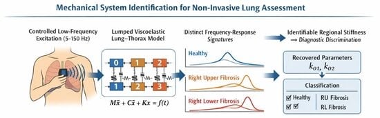
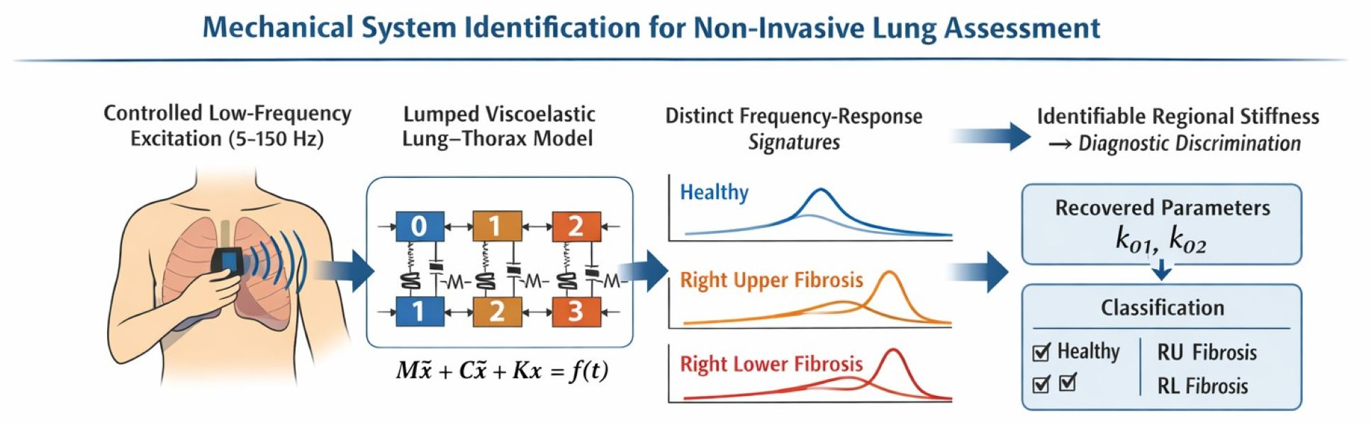
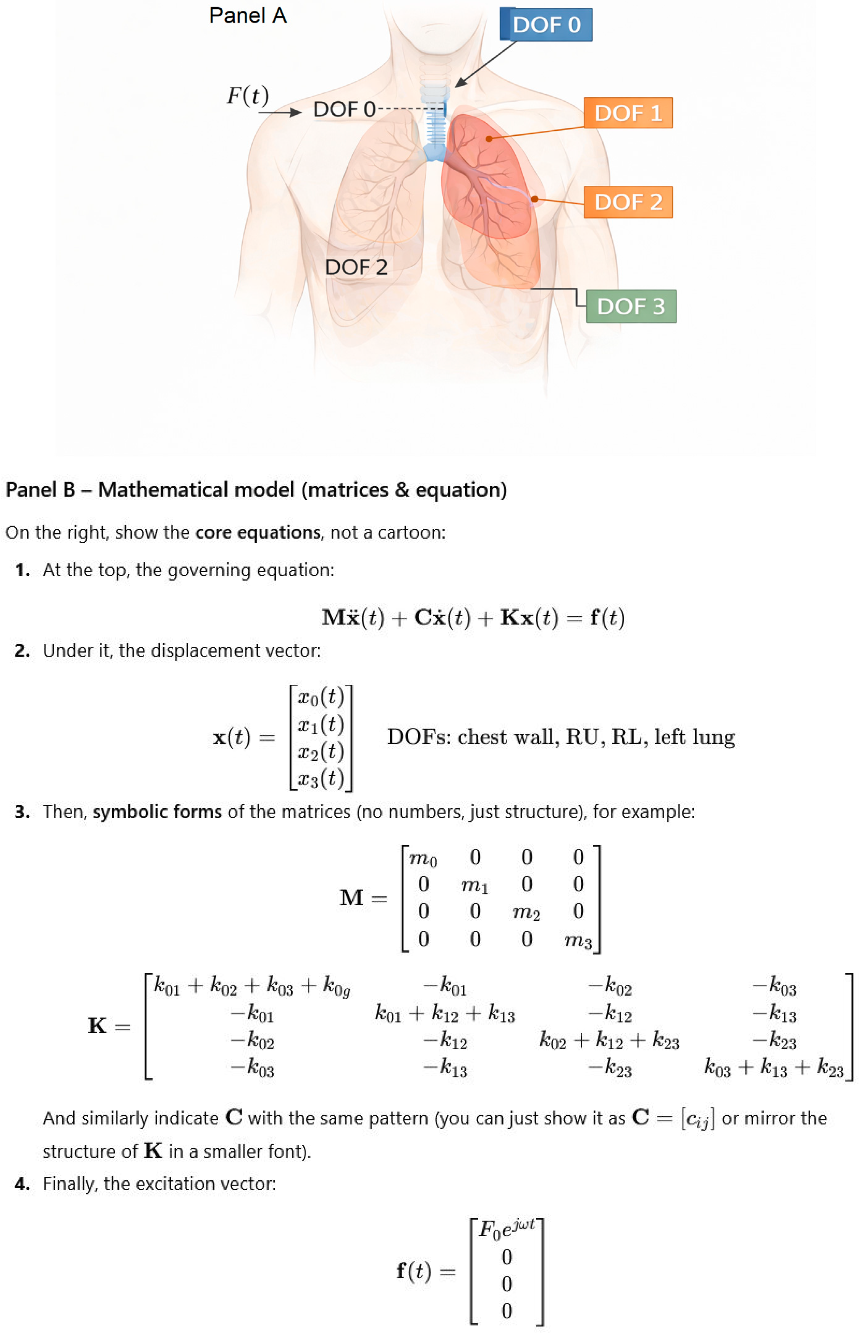
Here, we propose a conceptual diagnostic framework for the lung based on mechanical system identification and evaluate its feasibility using simulation. Instead of using static imaging or global indices, we treat the lung–thorax complex as a coupled viscoelastic dynamical system whose internal properties can be inferred from externally measured responses to controlled external excitation. Mechanical changes associated with pathology—such as regional stiffness alterations or heterogeneity—are reflected in frequency-domain response functions and, in principle, can be identified through inverse analysis. To demonstrate feasibility, we construct a multi-degree-of-freedom mechanical representation of the lung and show through simulation that pathological changes produce distinguishable signatures in the system response. This model establishes a new diagnostic conceptual framework whereby disease is inferred from dynamical behavior rather than from static snapshots or single scalar indices. In this sense, the proposed method shifts lung diagnostics from impedance measurement to inverse mechanical characterization.
3. Results
3.1. Frequency Response of the Mechanical Lung Model
The frequency response of the four-degree-of-freedom lung–thorax model was evaluated under harmonic excitation applied at the chest wall. Steady-state responses were computed over the low-frequency mechanical range relevant to the model assumptions.
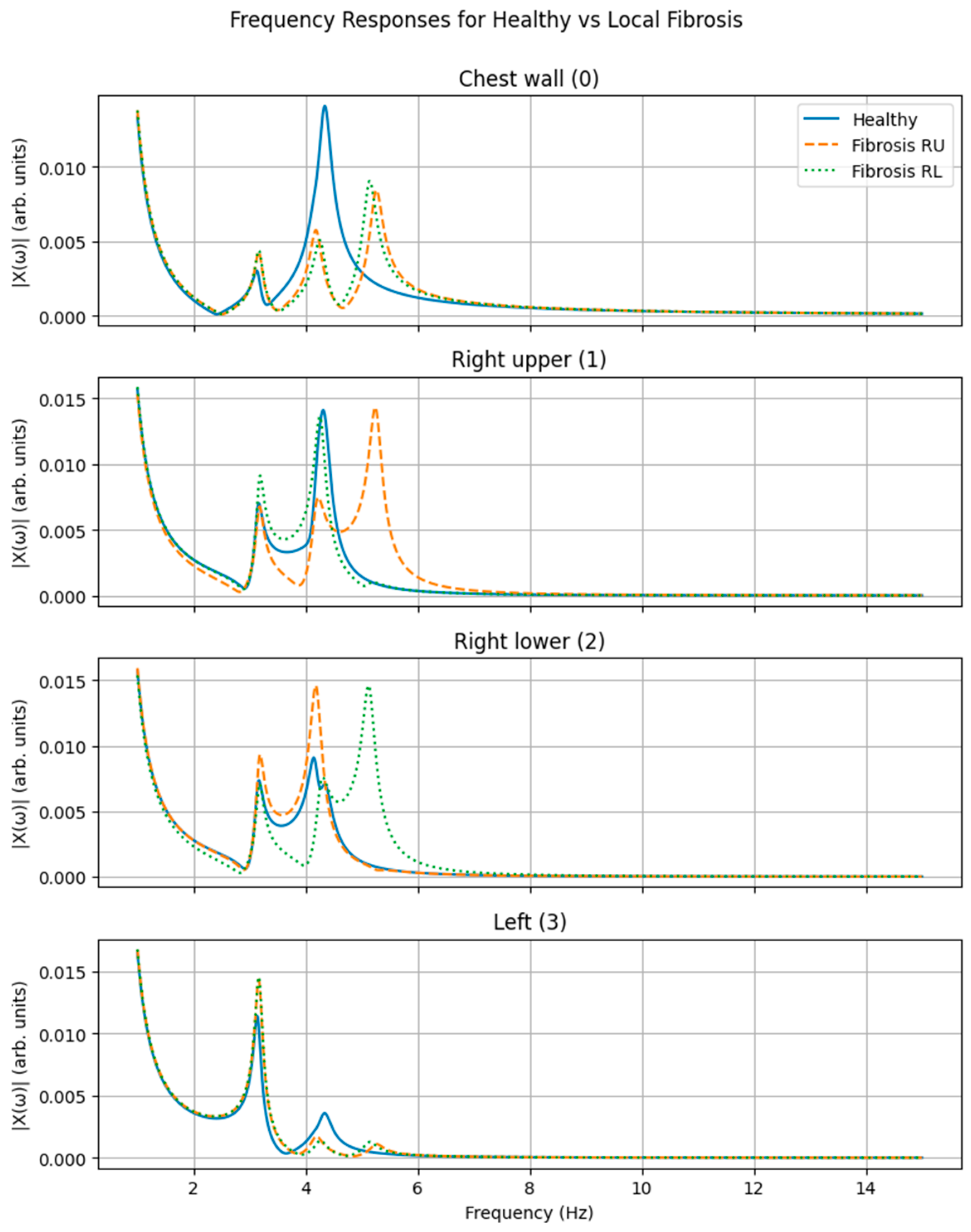
Figure 3 shows the magnitude of the frequency response functions (FRFs) for all degrees of freedom—chest wall, right upper lung region, right lower lung region, and left lung—for three simulated configurations: a healthy reference state, localized stiffness increases in the right upper lung region, and localized stiffness increase in the right lower lung region.
In the healthy configuration, the system exhibits smooth and continuous frequency-dependent responses across all compartments, with multiple resonance peaks reflecting the coupled dynamics of the chest wall and lung regions. When stiffness was selectively increased in the right upper lung region, distinct modifications were observed in the frequency response of the corresponding compartment, including shifts in resonance frequency and changes in response amplitude. These alterations were most pronounced in the right upper lung response but were also detectable in the chest wall response, despite excitation and measurement being applied externally.
Similarly, localized stiffening of the right lower lung region produced characteristic changes in the frequency response of the right lower compartment. The resulting response pattern differed from that observed for right upper lung stiffening, with distinct resonance shifts and amplitude redistribution. Responses of the remaining compartments exhibited comparatively smaller changes, preserving a different frequency signature.
Overall, the frequency-domain signatures associated with right upper and right lower stiffness perturbations were clearly distinguishable from one another and from the healthy reference configuration, indicating sensitivity of the global frequency response to localized mechanical alterations.
3.2. Inverse Recovery of Regional Stiffness Parameters
To assess the identifiability of internal mechanical parameters, inverse fitting was performed using simulated frequency response magnitude data corrupted with measurement noise. The inverse problem was formulated to recover selected chest–lung stiffness parameters while all other model parameters were held fixed.
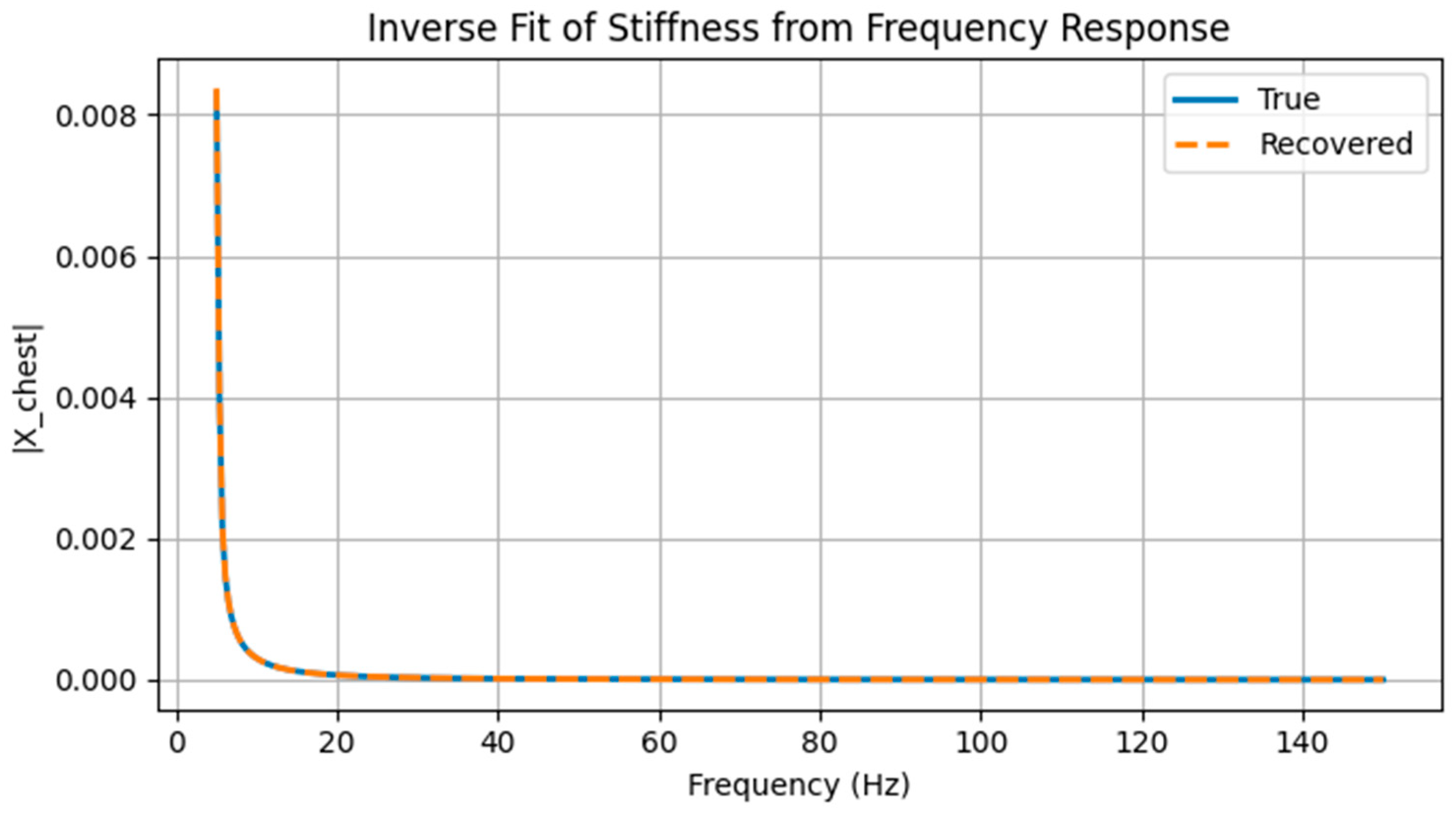
Figure 4 compares the chest wall frequency response magnitude obtained from simulated noisy measurements with the response predicted by the model using stiffness parameters recovered through nonlinear least-squares optimization. The recovered model closely reproduces the measured response across the analyzed frequency range, capturing both the overall amplitude and the frequency-dependent structure of the response.
The true stiffness values used to generate the synthetic measurements were k01 = 240 and k02 = 154 (arbitrary units), corresponding to chest–right-upper and chest–right-lower couplings, respectively. Inverse fitting yielded recovered values of k01 = 239.5 and k02 = 159.5. The close agreement between true and recovered parameters demonstrates that localized stiffness perturbations can be inferred from external frequency response measurements, even in the presence of moderate noise.
Although inverse estimation was performed using nonlinear least-squares optimization without explicit regularization, the problem remained well-conditioned within the examined parameter subspace, as confirmed by the sensitivity and Fisher-information analysis presented in
Section 3.3. In exploratory tests, the inclusion of a Tikhonov regularization term yielded parameter estimates within a few percent of the unregularized solution under moderate noise levels, indicating numerical stability for the simplified synthetic scenario. In practical experimental settings, where model mismatch and measurement uncertainty may be greater, regularized or Bayesian estimation strategies would likely be required to ensure stable parameter recovery.
3.3. Sensitivity and Identifiability of Regional Stiffness Parameters
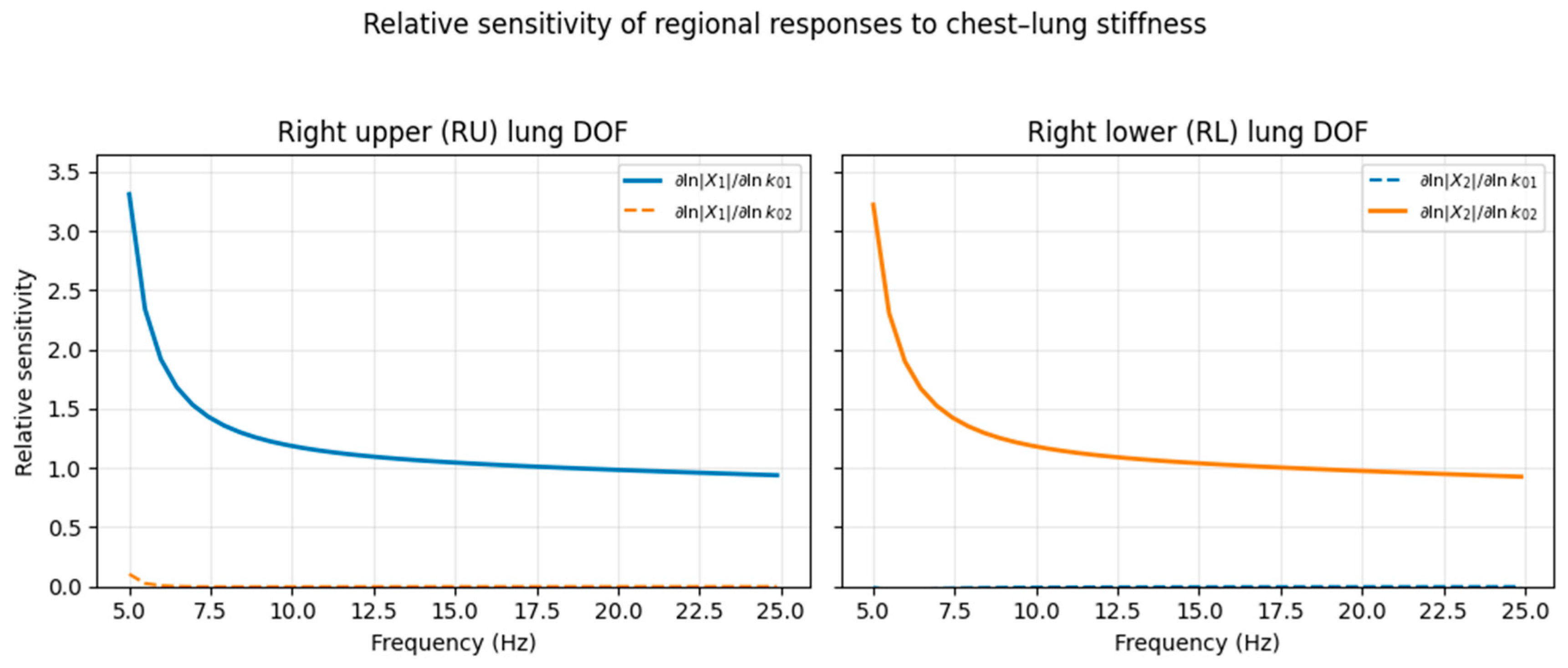
To assess whether regional chest–lung stiffness parameters are structurally identifiable from externally measured dynamics, a finite-difference sensitivity analysis was performed around the healthy baseline configuration. The chest–right-upper stiffness k01 and chest–right-lower stiffness k02 were perturbed individually by 1%, and the resulting changes in the multi-output frequency response magnitude ∣Xi(f)∣ were evaluated across the low-frequency band.
Sensitivities were expressed in dimensionless relative form as
which quantify the percentage change in response amplitude induced by a percentage change in stiffness.
Figure 5 shows the relative sensitivities for the right upper (RU) and right lower (RL) lung degrees of freedom over the 5–25 Hz band. The RU response exhibits strong sensitivity to
k01 with negligible dependence on
k02, whereas the RL response displays the opposite pattern. This directional dominance persists across the low-frequency range and indicates that the two regional stiffness parameters modulate distinct dynamical features of the system response.
To quantify identifiability more formally, sensitivities from all outputs and frequencies were stacked to form a global Jacobian matrix. The associated Fisher-information matrix had two strictly positive eigenvalues (λ1 = 1.75 × 10⁻⁹, λ2 = 8.91 × 10⁻¹⁰) and a low condition number (κ(F) ≈ 1.96), with modest correlation between sensitivity directions (ρ ≈ 0.29). These results demonstrate that the regional stiffness parameters are locally identifiable in the proposed multi-output configuration.
3.4. Discrimination of Pathological Conditions Using Response Features
To evaluate whether the observed frequency response differences are sufficient for diagnostic discrimination, frequency response data from multiple simulated realizations were analyzed using a classification pipeline. For each realization, frequency response magnitudes from all degrees of freedom were assembled into feature vectors and reduced in dimensionality prior to classification.
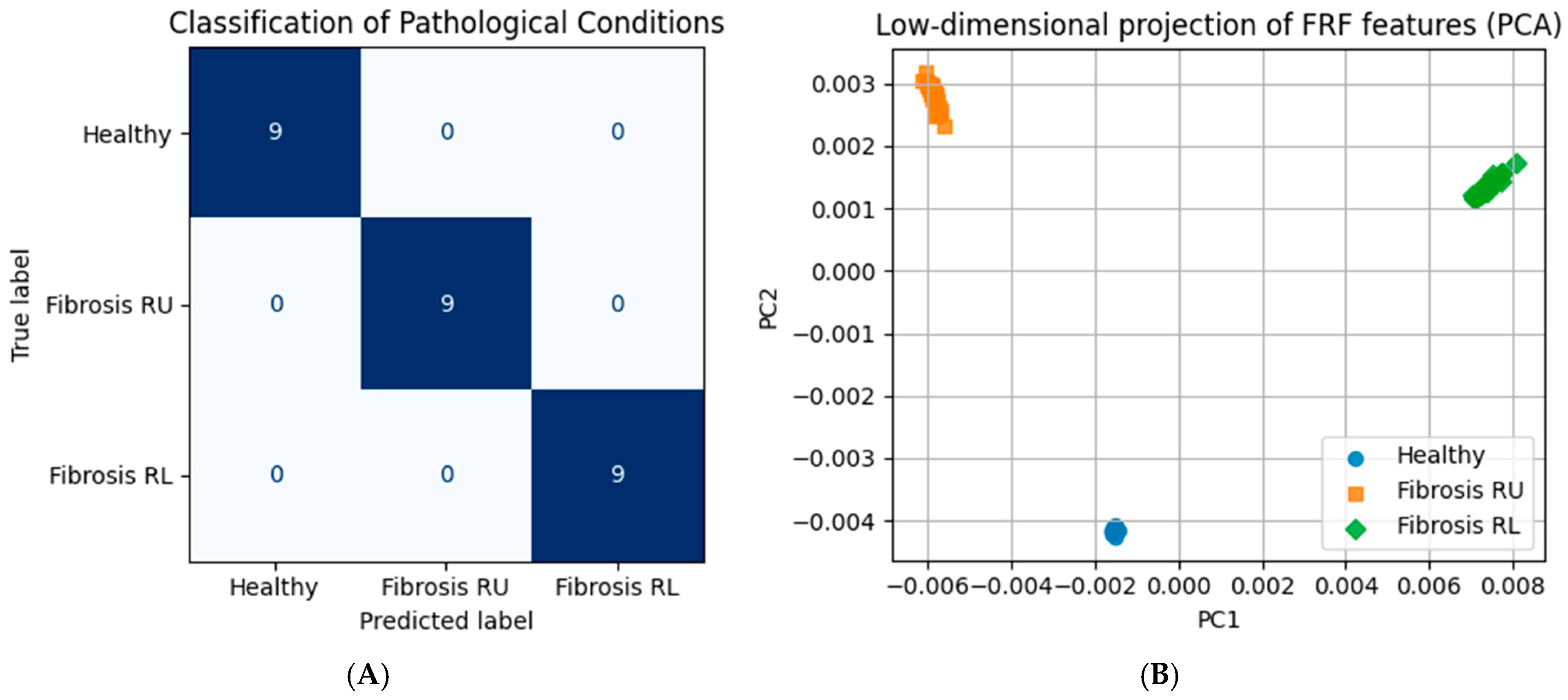
Figure 6A presents the confusion matrix summarizing classification performance for three simulated conditions: healthy lung, localized stiffness increase in the right upper lung region, and localized stiffness increase in the right lower lung region. Under the controlled simulation conditions considered here, all test samples were correctly classified, indicating complete separability of the three configurations in the chosen feature space.
Quantitative classification metrics are reported in
Table 3. Precision, recall, and F1-score were equal to 1.00 for all three classes.
To visualize the structure of the feature space independently of the classifier, the same frequency response features were projected onto the first two principal components.
Figure 6B shows the resulting low-dimensional representation, where each point corresponds to a single simulated realization. The three configurations form well-separated clusters in the PC1–PC2 plane, indicating that discriminative information is preserved in the dominant variance of the frequency response data.
To assess robustness beyond the idealized simulation conditions shown here, additional analyses were performed to quantify the effects of measurement noise and inter-subject mechanical variability on classification performance. These
Supplementary Experiments demonstrate that while variability reduces visual separability in low-dimensional projections, supervised classification using full frequency response features remains robust and well above chance levels across a wide range of noise and parameter perturbations (
Supplementary Figures S1–S3).
It should be emphasized that these results demonstrate theoretical discriminability under idealized modeling assumptions rather than expected diagnostic performance in experimental or clinical settings.
More challenging scenarios, including milder perturbations, mixed pathology patterns, and stronger inter-subject variability, will be important in future work to establish practical classification limits.
3.5. Summary of Key Results
Across all analyses, three principal results emerge. First, localized mechanical alterations produce distinct and reproducible signatures in the global frequency response of the lung–thorax system. Second, internal stiffness parameters associated with regional pathology can be accurately recovered through inverse analysis of externally measured frequency responses, even in the presence of measurement noise. Third, frequency response features enable reliable discrimination between different regional pathological configurations. Together, these results establish the feasibility of mechanical system identification as a basis for a non-invasive assessment of regional lung mechanics, with robustness to measurement noise and physiologically plausible parameter variability demonstrated in
Supplementary Analyses.
4. Discussion
4.1. From Simulation to Diagnostic Model
In this study, we introduced and evaluated a mechanical system-identification framework for lung assessment, in which internal mechanical properties are inferred from externally measured frequency responses. While the results presented here are based on simulated data, the significance of the work lies not in numerical performance alone, but in establishing the conceptual feasibility of a diagnostic model for the lung grounded in mechanical system identification.
Conventional lung diagnostics are largely divided between structural imaging modalities, which provide spatially resolved but static information, and global functional tests, which provide dynamic but spatially averaged measures. The approach presented here occupies a distinct conceptual space: the lung–thorax system is treated as a physical dynamical system, and diagnosis is performed by identifying internal mechanical parameters from its externally observable response. In this framework, pathology is not detected as an image or a scalar index, but as a change in the system’s dynamical signature.
The results demonstrate that localized alterations in lung stiffness generate distinct and recoverable changes in the frequency response of the system, even when excitation and measurement are applied exclusively at the chest wall. This demonstrates the theoretical feasibility of detecting regional mechanical changes from externally measured responses without direct internal access or imaging.
4.2. Relation to Existing Lung Diagnostic Methods
Current approaches to lung diagnostics can be broadly categorized into structural imaging techniques, such as chest radiography and computed tomography, and functional tests, such as spirometry and oscillometry. Structural imaging provides spatially resolved information on lung morphology, including consolidation, fibrosis patterns, or emphysema distribution, but it is inherently static and does not directly quantify regional mechanical behavior. Global pulmonary function tests, by contrast, quantify airflow and volume changes, yielding scalar indices such as FEV1 or total lung capacity that reflect overall respiratory performance but do not resolve regional heterogeneity.
Mechanical assessment of lung function has also been pursued using oscillatory techniques, most prominently the forced oscillation technique (FOT) and impulse oscillometry, which measure respiratory system impedance at the airway opening. These methods typically summarize the response in terms of resistance and reactance at a limited number of frequencies and are often interpreted using low-order or compartment-averaged models. While they provide valuable information about global mechanics and have demonstrated clinical utility, their ability to localize mechanical abnormalities to specific lung regions is limited.
The approach proposed in this work differs fundamentally from these existing methods. Rather than imaging anatomy or measuring airflow-based indices, the lung–thorax system is explicitly treated as a coupled multi-degree-of-freedom dynamical system, and diagnostic information is extracted from its low-frequency frequency response to external excitation. Internal mechanical properties, such as regional stiffness and coupling strength, are inferred through system identification using the full frequency response function, rather than through impedance summaries or image-derived features. In this framework, pathology manifests as a change in the system’s dynamical signature, not as a radiological pattern or a single global index.
The framework is also distinct from ultrasound imaging and ultrasound elastography. Conventional lung ultrasound relies on high-frequency wave propagation and image or artifact formation at air–tissue interfaces, whereas elastography seeks to reconstruct local stiffness from spatial strain patterns or shear wave propagation. Both approaches are challenged by the highly aerated and heterogeneous nature of lung tissue. In contrast, the present method operates in a low-frequency regime, does not require wave penetration into the parenchyma, and does not attempt to image tissue directly. Ultrasound, if employed, serves only as an external motion sensor at the chest wall, providing input to a mechanical system-identification model rather than acting as an imaging modality.
By focusing on mechanical dynamics rather than structure or airflow alone, the proposed framework introduces a complementary diagnostic dimension. It aims to recover regional mechanical properties from non-invasive surface measurements, thereby bridging the gap between global functional tests and structurally detailed but mechanically indirect imaging modalities.
Distinction from Forced Oscillation and Impedance-Based Techniques
Forced oscillation techniques and impulse oscillometry have demonstrated clinical utility by characterizing respiratory system impedance under small-amplitude oscillatory excitation. However, these methods typically apply excitation at the airway opening and interpret the response using scalar resistance and reactance measures or low-order compartment models that represent the lung as a largely homogeneous system. The framework presented here differs in both excitation strategy and diagnostic objective. Excitation is applied externally at the chest wall, and the lung–thorax system is modeled as a coupled multi-degree-of-freedom dynamical system. Diagnostic inference is performed by identifying internal mechanical parameters—such as regional stiffness—from the full frequency response function, rather than by summarizing impedance at the airway opening. As a result, the proposed approach is not an extension of oscillometry, but a shift toward vibration-based system identification analogous to methods used in structural health monitoring, adapted here for a non-invasive assessment of lung mechanics.
4.3. Physical Origin of Diagnostic Sensitivity
The ability to infer regional pathology from external measurements arises from fundamental properties of coupled dynamical systems. In such systems, local changes in stiffness or damping alter not only local responses but also global modal structure, leading to shifts in resonance frequencies, changes in mode shapes, and a redistribution of response energy across degrees of freedom.
In the lung–thorax system, mechanical coupling through the chest wall, pleura, and mediastinum ensures that regional alterations propagate to the surface. The frequency-domain analysis presented here shows that these effects are detectable even under small-amplitude excitation and in the presence of measurement noise. This is consistent with analogous approaches in structural health monitoring, where defects in inaccessible regions of a structure are identified through vibration-based analysis [
26,
27,
36,
37].
The clear separation of pathological configurations in the low-dimensional principal component space further indicates that diagnostic information is encoded in the dominant variance of the response, rather than in subtle or noise-sensitive features. This suggests that the approach may be robust to measurement variability and sensor limitations.
4.4. Identifiability and Model Order Considerations
The mechanical model employed in this study represents the lung–thorax system using a small number of effective degrees of freedom. This choice reflects a balance between anatomical relevance and the practical limits of inverse parameter identification from externally measured responses. In general, the number of internal mechanical parameters that can be reliably identified is constrained by the information content of the measured frequency response, which depends on excitation bandwidth, signal-to-noise ratio, and the strength of mechanical coupling between compartments.
The four-degree-of-freedom configuration adopted here should therefore be interpreted as a minimal identifiable model rather than a detailed anatomical decomposition. The chest wall degree of freedom provides a well-defined interface for external excitation and measurement, while the subdivision of the right lung into upper and lower effective compartments enables representation of clinically meaningful regional heterogeneity without excessive parameterization. The left lung is intentionally lumped to limit model complexity and preserve robustness of the inverse problem under realistic noise conditions.
Importantly, the generalized coordinates of the model correspond to effective modal motions rather than discrete anatomical structures. At the low frequencies considered in this study, the lung–thorax system behaves predominantly as a coupled viscoelastic structure, and its dynamics are governed by a small number of dominant modes. Under these conditions, localized changes in mechanical properties influence the global frequency response through shifts in resonance frequencies, mode shapes, and energy distribution across degrees of freedom, enabling regional inference even with a low-order representation.
As experimental resolution and measurement fidelity improve, higher-order models incorporating additional regional subdivisions may become identifiable. In practice, appropriate model order would be determined using standard system-identification criteria, including parameter sensitivity, stability of recovered estimates, and consistency of model fits across frequency. The present results demonstrate that even a low-order model suffices to recover localized stiffness alterations and discriminate between regional pathological configurations, thereby establishing a lower bound on the mechanical resolution achievable through non-invasive system identification of the lung–thorax system.
The explicit sensitivity and Fisher-information analysis presented in
Section 3.3 confirms that the regional stiffness parameters are locally identifiable from multi-output frequency responses and that the proposed model is not over-parameterized in the stiffness subspace relevant to simulated pathology.
An important next step will be to evaluate identifiability under simultaneous perturbations of stiffness, damping, and coupling parameters, where parameter cross-correlation and local minima may become more significant.
4.5. Implications for Pulmonary Rehabilitation
Pulmonary rehabilitation aims to improve exercise capacity, symptoms, and quality of life by targeting the integrated function of the lung–thorax system, peripheral muscles, and cardiovascular responses. In current clinical practice, enrollment and progression through rehabilitation programs are guided largely by global indices such as FEV1, diffusion capacity, exercise tests, and symptom scores. Although these measures are highly relevant, they do not explicitly quantify regional mechanical abnormalities or their distribution across the lung and chest wall. As a result, patients with distinct mechanical phenotypes may receive similar rehabilitation prescriptions, and heterogeneity in response to standardized programs is common.
Within this context, the present mechanical system-identification framework suggests a complementary route toward more individualized pulmonary rehabilitation. By recovering physically interpretable parameters, such as regional stiffness and inter-compartment coupling from non-invasive surface measurements, the model provides a structured representation of mechanical heterogeneity that could be used to stratify patients before rehabilitation, identify those with asymmetrical or highly localized stiffness patterns, and define mechanistic targets for intervention. In principle, longitudinal application of the same identification procedure could also be used to track changes in regional mechanics over time, offering an objective mechanical correlation of rehabilitation-induced adaptation alongside traditional functional outcomes.
Importantly, the current study does not evaluate pulmonary rehabilitation directly and should not be interpreted as evidence of rehabilitation efficacy. Rather, it establishes that regional mechanical alterations are, in principle, identifiable from external dynamics and that these identifiability properties create a natural bridge between mechanical modeling and rehabilitation planning. Future work will be required to link specific mechanical phenotypes and parameter trajectories to clinically observed rehabilitation responses, and to determine whether system-identification-based metrics can improve patient selection, tailoring of exercise prescriptions, or monitoring of long-term outcomes compared with conventional assessment alone.
4.6. Diagnostic Interpretation and Clinical Relevance
Within the proposed framework, diagnosis is naturally framed as parameter identification rather than pattern recognition. Regional stiffness estimates provide physically interpretable quantities that can be related to known pathological processes, such as fibrosis or emphysematous tissue destruction. Unlike purely data-driven classifiers, this parameter-based representation supports mechanistic interpretation and longitudinal tracking. Accordingly, the perfect classification observed in the present simulations should be understood as demonstrating the existence of diagnostically separable mechanical signatures under idealized conditions, rather than as a prediction of achievable classification accuracy in clinical practice. In particular, future studies should test the ability to detect subtle mechanical abnormalities and to distinguish mixed regional patterns under higher noise and model-mismatch conditions.
Moreover, the non-invasive nature of excitation and measurement suggests potential applicability in populations for whom conventional imaging may be impractical or undesirable, such as pediatric or critically ill patients. While the present study does not address clinical implementation directly, it establishes the physical plausibility of inferring regional lung mechanics from external measurements alone.
4.7. Limitations of the Present Study
Several limitations should be acknowledged. The mechanical model employed here is intentionally simplified, relying on a lumped-parameter representation and linearized dynamics. While appropriate for a proof-of-concept investigation, this abstraction does not capture the full anatomical complexity or nonlinear behavior of the lung. In addition, the chosen model order reflects a trade-off between spatial resolution and identifiability from surface-only measurements, and should be viewed as a minimal representation rather than a definitive anatomical partition. Accordingly, the present framework should be viewed as a proof-of-concept model applicable to passive low-frequency mechanical interrogation, rather than a complete physiological description of respiration under all operating conditions.
Additionally, airflow dynamics, active muscle contraction, and posture-dependent effects were neglected. These factors will need to be addressed in future extensions of the model. The noise model used in this study was also simplified, with additive uncorrelated Gaussian noise applied only to the magnitude of the frequency response. Phase noise, sensor bias, and correlated measurement artifacts were not explicitly modeled, representing a best-case experimental scenario. Incorporating more realistic noise characteristics and experimental uncertainties will be an important focus of future work.
Finally, all results were obtained using simulated data, and experimental validation remains an essential next step. These limitations, however, do not undermine the central contribution of the work, which is the establishment of identifiability and diagnostic feasibility within a physically grounded framework.
Future work will address these limitations through staged experimental validation, beginning with physical thoracic phantoms with tunable viscoelastic properties, followed by pilot measurements in human volunteers under controlled low-amplitude excitation. These studies will allow the calibration of the mechanical model, evaluation of realistic measurement noise, and assessment of parameter recovery under physiological variability.
Future model extensions should therefore incorporate combined perturbations in stiffness, damping, and coupling to better represent mixed or evolving pathological phenotypes.
4.8. Path Toward Experimental and Clinical Validation
Because all the results in the present study are simulation-based, experimental validation represents the essential next step toward translation.
The modeling framework presented here is well-suited for staged experimental validation. As an initial step, physical thoracic phantoms with tunable stiffness and damping properties could be constructed using layered viscoelastic materials and air-filled compartments to emulate regional lung mechanics. Such phantoms would enable a controlled evaluation of parameter recovery accuracy, sensitivity to stiffness contrast, excitation bandwidth selection, and sensor placement strategies under reproducible conditions.
From an instrumentation perspective, the required excitation amplitudes are modest. Order-of-magnitude estimates indicate that chest wall displacements on the scale of tens to hundreds of micrometers are sufficient to generate measurable frequency response signatures within the 5–150 Hz band range while remaining well within safe and comfortable limits for passive thoracic vibration. Broadband excitation across this frequency range could be delivered using a lightweight electromechanical shaker, voice-coil actuator, or instrumented impulse hammer applied over the sternum or anterior rib cage.
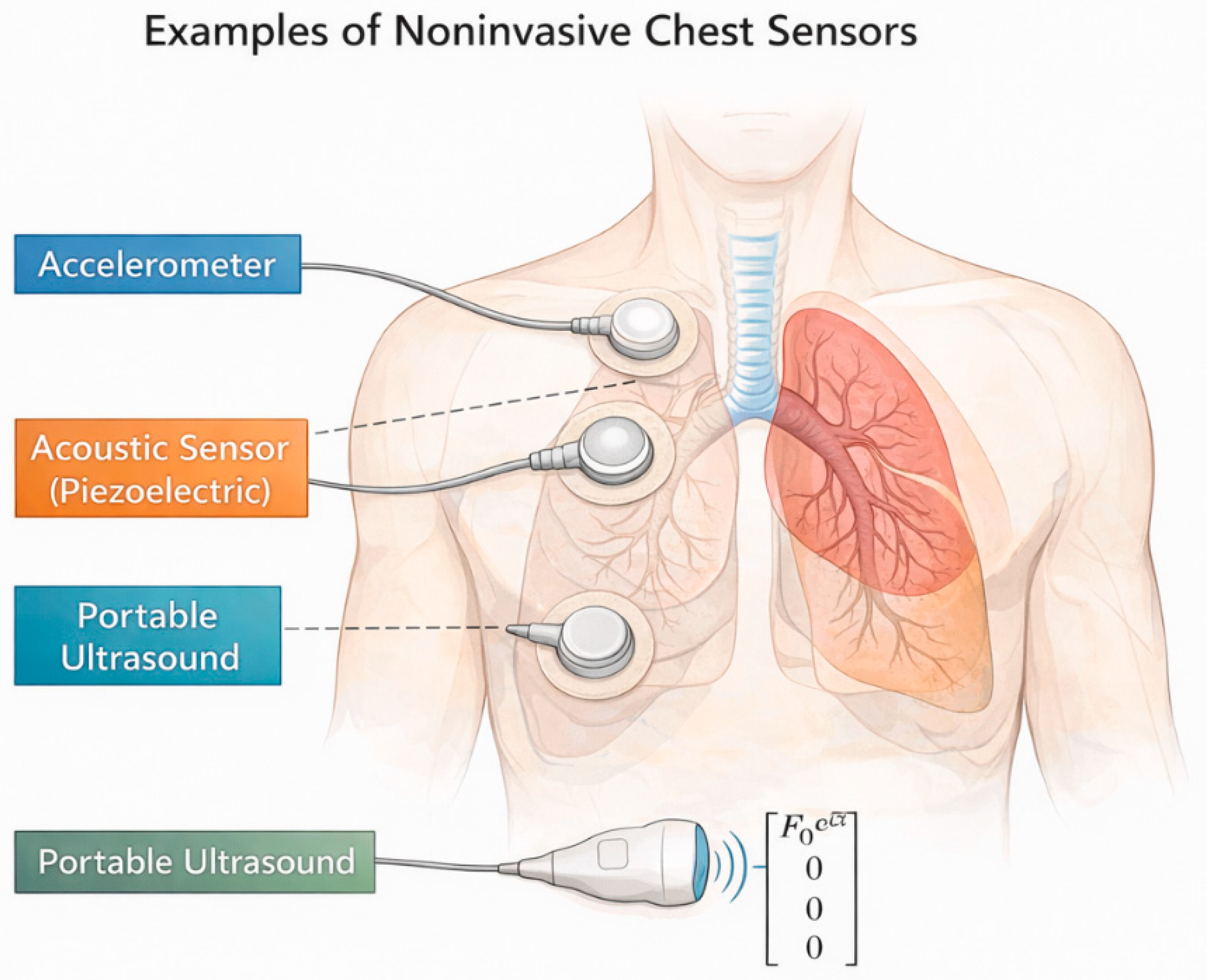
Surface response measurements could be obtained using contact accelerometers, piezoelectric acoustic sensors, or motion-tracking ultrasound probes with bandwidths exceeding 200 Hz. Commercial accelerometers with sensitivities in the range of 10–100 mV/g are capable of detecting sub-millimeter thoracic vibrations, suggesting that signal-to-noise ratios comparable to those assumed in the present simulations are achievable under controlled conditions. Because the proposed framework relies primarily on the structure of the frequency response rather than absolute displacement magnitude, moderate variability in sensor placement or baseline amplitude is not expected to compromise parameter identifiability.
Following phantom validation, pilot studies in human subjects under carefully controlled, low-amplitude excitation could be conducted using existing vibration or oscillatory technologies. Importantly, the framework does not require new imaging hardware but instead reframes externally measured mechanical response data within a physically grounded system-identification paradigm. This staged pathway—from controlled phantoms to feasibility testing in volunteers—provides a realistic route toward eventual clinical translation.
Future experimental studies should also compare alternative excitation protocols, including harmonic sweeps, broadband random excitation, and impulse-like inputs, in order to determine which waveform provides the best trade-off between identifiability, robustness, and ease of bedside implementation.
4.9. Implications and Outlook
By reframing lung diagnostics as a problem of mechanical system identification, this work opens a new avenue for the non-invasive assessment of regional lung mechanics. The proposed approach complements existing imaging and functional tests by providing access to physically interpretable mechanical properties that are not directly observable through static images or global pulmonary indices.
An important implication of this framework is its natural compatibility with data-driven analysis. Because the measured frequency responses and recovered model parameters are grounded in a physical description of lung mechanics, they form structured, low-dimensional features that are well-suited for machine-learning–based classification. Rather than applying artificial intelligence directly to raw signals or images, learning algorithms can operate on physics-informed representations, enabling the discrimination of disease-specific mechanical patterns while preserving interpretability.
Future work will focus on experimental validation, refinement of the mechanical model, and exploration of clinical use cases. In parallel, the integration of physics-based system identification with machine-learning classifiers may enable automated, robust recognition of pathological mechanical signatures and longitudinal tracking of disease progression. More broadly, the proposed framework illustrates how classical principles of dynamics and inverse problem theory, combined with modern data-driven methods, can be leveraged to develop new diagnostic models in medicine.
Potential clinical use cases include longitudinal monitoring after thoracic or upper-abdominal surgery, regional mechanical assessment on mechanically ventilated ICU patients, and tracking of mechanical adaptation during pulmonary rehabilitation in chronic respiratory disease. These scenarios are attractive because they may benefit from repeated non-invasive measurements and from physically interpretable markers of regional stiffness and coupling.
4.10. Clinical Perspective
What is new? This study introduces a diagnostic model that treats the lung–thorax system as a mechanical dynamical system and infers regional lung mechanical properties from externally measured frequency responses. Unlike conventional imaging or global pulmonary function tests, this approach identifies pathology through changes in the system’s dynamical signature rather than through static structural features or scalar indices.
What are the clinical implications? If validated experimentally, this framework could provide a non-invasive means of assessing regional lung mechanics using low-amplitude external excitation applied at the chest wall. Such an approach may complement existing diagnostic tools by offering physically interpretable information about tissue stiffness and heterogeneity, potentially enabling the earlier detection of mechanical alterations and longitudinal monitoring without reliance on imaging. In the setting of pulmonary rehabilitation, physics-based estimates of regional stiffness and coupling could help identify patients with distinct mechanical phenotypes, support more individualized prescription of rehabilitation intensity and modality, and provide an objective mechanical marker to accompany conventional functional and symptomatic outcomes.
What is the next step? Future work will focus on experimental validation using physical lung phantoms and human studies, refinement of the mechanical model, and evaluation of feasibility in clinical settings including in patients undergoing pulmonary rehabilitation.




 ,
,