Bioengineering Interventions to Enhance the Capacity of the Gut Microbiota in Controlling Food Allergies


Abstract
1. Introduction
2. Allergic Immune Modulation Through Gut Microbiota
3. Strategies to Enhance the Gut Microbiota for Management of Food Allergies

3.1. Prebiotics
3.1.1. Carbohydrate Additives

| Polysaccharides/Oligosaccharides | Food Allergy | Outcome | Reference |
|---|---|---|---|
| Chitosan | Shrimp tropomyosin, Ovalbumin, Peanut | Chitosan oligosaccharide (COS) alleviated shrimp tropomyosin allergy symptoms; reduced IgE, histamine, and Th2 cytokines; and enhanced the Th1 response. | [81,82,83] |
| Chitosan nanoparticles (carrying TGF-β DNA) increased intestinal TGF-β and reduced ovalbumin-induced allergy symptoms after oral delivery. | |||
| Chitin/Chitosan protected against peanut-induced reactions by reducing IgE levels and Th2 cytokines. | |||
| Red-edge tea polysaccharide (RETPS) | Ovalbumin | RETPS-3 and RETPS-4 from red-edge tea reduced Ovalbumin-specific IgE, histamine, and mast cell protease levels, alleviated allergic symptoms (itching, hypothermia, and diarrhea), and improved the gut microbiota. | [84] |
| Ulva-derived polysaccharides | Ovalbumin | Ulva-derived compounds alleviated allergic symptoms by lowering IgG1 and Ovalbumin-specific IgE, increasing protective IgG, promoting Th1 immunity, improving microbial balance, and strengthening the intestinal barrier. | [85] |
| Depolymerized sulfated galactans | Ovalbumin | Oral depolymerized sulfated galactans suppressed anaphylaxis and inflammatory markers, while gut tissues showed reduced epithelial damage, fewer inflammatory cells, and improved structure. | [86] |
| Fucoidan sulfated polysaccharide | Ovalbumin | Fucoidan suppressed allergic responses by reducing Ovalbumin-specific IgE, histamine, mast cell protease-1, and Th2 cytokines, while increasing IL-10 and TGF-β, and improving the gut microbiota. | [87] |
| Inulin | Ovalbumin | Inulin-based treatment reduced anaphylaxis and diarrhea, stabilized temperature, lowered Th2 markers (IL-4, IL-5, IL-13, IgE, and IgG1), enhanced Th1/regulatory responses, and restored ileal microbiota by increasing beneficial bacteria | [88] |
| Porphyra haitanensis sulfated polysaccharides | Ovalbumin | Marine sulfated polysaccharides (PHP40, PHP80) restored microbial balance, boosted beneficial bacteria (Bacillus, Enterococcus), suppressed harmful ones (Staphylococcus), and enhanced galactose metabolism and bile acid biosynthesis. | [99] |
| Aloe | Ovalbumin | Processed aloe reduced allergic symptoms by suppressing Th2 cells, eosinophils, and mast cells, while boosting regulatory T cells and IL-10. | [100] |
| Gracilaria lemaneiformis polysaccharide | Ovalbumin | Gracilaria lamaneiformis fermented with Lactobacillus acidophilus lowered Ovalbumin-specific IgE, histamine, and mast cell protease, suppressed Th2/IL-4, and boosted Treg cells and IL-10. | [101] |
| Fructo-oligosaccharides (FOSs) | Peanut | FOSs modulated DCs in peanut-allergic individuals, promoting immune tolerance rather than allergic inflammation. | [102] |
| Sulphated oligosaccharides | Ovalbumin | Sulfated oligosaccharides from Gracilaria lamaneiformis promote Tregs, suppressing Th2-driven IgE production and mast cell mediator release. | [103] |
| Combined intake of short fructan (1-kestose) and long fructan (inulin) | Ovalbumin | A combination of short- and long-chain fructans reduced allergy-related symptoms, such as diarrhea, and enhanced the gut microbiota. | [104] |
| Combination of short- and long-chain fructo-oligosaccharides (scFOSs/lcFOSs) | Peanut | scFOSs/lcFOSs reduced mast cell activity, boosted Tregs, and enhanced beneficial gut bacteria through prebiotic supplementation. | [105] |
| Lactulose | Cow milk | Synbiotic administration increased gut butyrate and markedly reduced allergic responses, including anaphylaxis, in mice. | [106] |
3.1.2. Polyphenols Additives
| Polyphenols | Food Allergy | Outcome | Reference |
|---|---|---|---|
| Bisdemethoxycurcumin (BMDC) | Ovalbumin | BDMC treatment reduced allergic symptoms (anaphylaxis, diarrhea), improved intestinal health, suppressed Th2 responses, enhanced Th1 and regulatory responses, and inhibited inflammatory pathways, including MAPK and NF-κB. | [108] |
| Resveratrol | Ovalbumin | Resveratrol reduced IgE levels and cytokines, including IL-4 and IL-13. | [118] |
| Theaflavins | Ovalbumin | Theaflavins help prevent food allergy symptoms. | [119] |
| Two dietary polyphenols, Epigallocatechin gallate (EGCG) and Chlorogenic acid (CA) | Tropomyosin | Modifying shrimp tropomyosin with EGCG or chlorogenic acid changes its structure. These changes make it harder for the immune system by reducing the avidity with which IgE and IgG antibodies bind to it, thereby lowering its allergenic potential. | [107] |
| Apple condensed tannins (ACTs) | Ovalbumin | Mice treated with ACT had significantly lower Ovalbumin-specific IgE and IgG1 levels, thereby inhibiting the immune system’s response to food proteins. | [111] |
| EGCG and chlorogenic acid | Arah1 (peanut) | Modified Ara h1 with EGCG and cholorogenic acid reduced peanut allergenicity. | [109] |
| Polyphenol aggregation | Peanut | Mice administered peanut–polyphenol mixtures, particularly at higher polyphenol doses, had lower IgE levels. | [120] |
3.1.3. Combination of Carbohydrates and Polyphenols
3.2. Probiotics Strategy
3.2.1. Generic Probiotics
3.2.2. Specific Probiotics
| Probiotic Strain | Targeted Food Allergy | Target Species | Outcome | Reference |
|---|---|---|---|---|
| Lactobacillus casei Zhang (LcZ) | Tropomyosin | Mouse | LcZ treatment reduced allergic symptoms, preserved gut integrity, and minimized tissue damage. | [164] |
| Bifidobacterium longum subsp. longum 51A (BL51A) | Ovalbumin | Mouse | BL51A-treated mice showed reduced weight loss and allergic symptoms. | [163] |
| Propionibacterium freudenreichii CIRM-BIA129 (Pf129) | Wheat gliadin | Mouse | Mice showed reduced anaphylaxis, lower allergen-specific IgE levels, serum mMCPT-1 levels, Th2 responses, and ILC2 activation, indicating decreased allergic inflammation. | [165] |
| Lactobacillus plantarum A56 | Ovalbumin | Mouse | Oral L. plantarum A56 reduced allergic symptoms and lung inflammation, restored intestinal villi, and improved microbial diversity. It suppressed Ovalbumin-specific IgE and IgG1 while increasing IgG2a, thereby inhibiting Th2 responses and supporting gut health. | [147] |
| Lactobacillus plantarum JC7 | Ovalbumin | Mouse | L. plantarum JC7 can prevent food allergies by correcting Th1/Th2 imbalances and modifying disordered intestinal microbiota. | [166] |
| Akkermansia muciniphila BAA-835 | Ovalbumin | Mouse | A. muciniphila BAA-835-treated mice showed reduced weight loss and lower IgE and IgG1 levels, indicating diminished allergic response. Histology revealed less inflammation and tissue damage, with a marked decrease in eosinophil- and neutrophil-associated enzyme activity, key drivers of allergic inflammation. | [167] |
| Lactobacillus rhamnosus GG | Peanut | Human | In children, combining peanut protein with probiotics reduced skin-test reactions and peanut-specific IgE, while increasing IgG4, which is associated with tolerance. Side effects were primarily mild (e.g., stomach pain, mild allergic reactions), although a few were moderate or severe (including anaphylaxis), which were managed successfully. No cases of eosinophilic esophagitis were reported. | [168] |
| Lactiplantibacillus plantarum HM-22 | α-lactalbumin (α-LA) | Mouse | Mice treated with this probiotic showed significant weight loss and increased levels of anti-inflammatory cytokines, which are associated with immune tolerance and suppression of allergic inflammation. | [148] |
| Lactiplantibacillus plantarum YIT 0132 (LP0132) | Cow milk allergy | Human | The probiotic showed a significant increase in β-lactoglobulin-specific IgG4, associated with tolerance development, and a notable reduction in IL-5 and IL-9, key cytokines that trigger allergic inflammation. | [149] |
| Lactobacillus rhamnosus GGA | Cow milk allergy | Human | Incorporating L. rhamnosus GGA into a cow milk-free diet resulted in a significant improvement in various gastrointestinal and behavioral symptoms in infants with cow milk allergy over four weeks. | [169] |
| Lactobacillus rhamnosus fermented milk (PFM) | Ovalbumin | Mouse | Feeding Lactobacillus rhamnosus fermented milk to mothers during the suckling period and to the offspring after weaning reduced clinical allergy in OVA-sensitized neonatal mice, lowered specific IgG/IgG1, and shifted immune responses from Th2 to Th1, with the strongest protection observed during the suckling period. | [170] |
3.2.3. Combination of Probiotics
| Probiotic/Synbiotic | Targeted Food Allergy | Target Species | Outcome | Reference |
|---|---|---|---|---|
| Probiotic mixture (P5: Lactococcus lactis KF140, Pediococcus pentosaceus KF159, Lactobacillus pentosus KF340, Lactobacillus paracasei 698, and Bacillus amyloliquefaciens 26N) | Ovalbumin | Mouse | P5 treatment significantly reduced Ovalbumin-specific IgE, suppressed Th2 and Th17 cytokines, and upregulated Th1 cytokines. This shift indicates restoration of the Th1/Th2 balance, a key factor in mitigating allergic responses. | [177] |
| Synbiotic (Anaerostipes caccae and Lactulose) | Cow milk allergy | Mouse | The synbiotic formulation restored colonic butyrate levels, protected against anaphylaxis, and promoted regulatory immune responses by suppressing inflammation and Th2 cytokines in mice. | [106] |
| Probiotic (Bifidobacterium animalis KV9, and Lactobacillus vaginalis) | Ovalbumin | Mouse | This combination of strains activated TLR4 via beneficial probiotics, thereby modulating immune responses. They upregulated MyD88 and IRF-1 in the spleen, thereby enhancing Th1 cells implicated in immune regulation and suppressing IRF-4, a transcription factor associated with allergic reactions. | [161,162] |
| Probiotic (Lactobacillus rhamnosus ŁOCK 0900, Lactobacillus rhamnosus ŁOCK 0908, and Lactobacillus casei ŁOCK 0918) | Cow milk allergy | Human | A 30% or more reduction in Atopic dermatitis (SCORAD scores) was observed primarily in children with allergy-related IgE antibodies. | [178] |
| Probiotic (Bifidobacterium longum KACC 91,563 and Enterococcus faecalis KACC 91532) | Ovalbumin | Mouse | B. longum KACC 91563 was more effective in improving the food allergy symptoms than Enterococcus faecalis KACC 91532, which showed no effect. B. longum released extracellular vesicles that selectively eliminated mast cells responsible for allergic reactions without disrupting overall immune function. | [179] |
| Probiotic (Bifidobacterium longum subsp. infantis, Lactobacillus acidophilus, Enterococcus faecalis, and Bacillus cereus) | Ovalbumin | Mouse | The probiotic mixture helped cesarean-section-born rats early in life by reducing allergic responses by calming overactive Th2 responses that drive allergic symptoms. | [180] |
| Probiotic (Lactobacillus acidophilus AD031, Bifidobacterium lactis AD011) | Ovalbumin | Mouse | B. lactis AD011 and L. acidophilus AD031 have shown the potential to prevent or reduce allergic reactions. | [181] |
| Probiotic (Lactobacillus rhamnosus GG and Bifidobacterium animalis spp. lactis BB-12) | General food allergy (Non-specific) | Human | The combination alleviated mild food allergy symptoms in young children by reducing digestive issues. Blood test showed decreased IL-17, increased IL-10, and reduced IgE level, with effects persisting for months after treatment. | [182] |
| Probiotic (Dahi containing Lactobacillus acidophilus LaVK2 and Bifidobacterium bifidum BbVK3) | Whey protein from cow’s milk | Mouse | Dietary probiotic Dahi reduced allergic reactions in whey protein-sensitized mice by shifting immunity from Th2 toward Th1 responses. | [183] |
3.2.4. Genetically Engineered Probiotics
| Recombinant Probiotics | Type of Food Allergen Expressed | Target Species | Outcome | Reference |
|---|---|---|---|---|
| Lactococcus lactis NZ3900/pNZ8149-NapA and L. lactis NZ3900/pNZ8149 | Ovalbumin | Mouse | Recombinant Lactococcus lactis supplementation reduced diarrhea in mice, lowered IgE, increased Ovalbumin-specific IgG, decreased IL-4, and boosted IFN-γ expression. | [188] |
| Lactococcus lactis | Ara h 2.02 | Mouse | Oral administration of Lactococcus lactis expressing Ara h 2.02 can suppress the allergic immune responses in sensitized mice. | [189] |
| Lactococcus lactis | Ara h2 | Human cells | The mimotopes elicited minimal allergic reactions and promoted a balanced immune response by increasing IFN-γ levels. | [190] |
| Lactococcus lactis-rm IL10 | β-lactoglobulin | Mouse | Engineered Lactococcus lactis that secretes IL-10 reduced anaphylaxis and lowered IgE/IgG1 levels; increased gut IgA and IL-10 production in Peyer’s patches and plasma, which helped in preventing allergic reactions and sensitization. | [191] |
| Lactococcus lactis | β-lactoglobulin | Mouse | Recombinant Lactococcus lactis delivering BLG restored immune balance and prevented cow milk allergy sensitization by reducing allergen-specific IgE and enhancing IgG2a and IFN-γ. | [192] |
| Lactococcus lactis MG1363 | Ara h2 | Human-derived serum/antibodies | Lactococcus lactis efficiently produces full-length, active Ara h 2 with natural-like immune reactivity, enabling safer, standardized allergen immunotherapy. | [193] |
| Lactococcus casei BL23 | β-lactoglobulin | Mouse | Recombinant probiotics induced IFN-γ (Th1) and mild IL-5 (Th2) responses. | [194] |
| Lactococcus lactis MG1363, Lactococcus lactis NZ9000 | Ara h2 | Mouse | Engineered Lactococcus lactis secreting Ara h 2 reduced IgE, enhanced IgG2a and IgA, and promoted Treg development. | [195] |
| Lactococcus lactis MG1363 | Ovalbumin | Mouse | Oral delivery of antigens via recombinant Lactococcus lactis promotes antigen-specific tolerance by inducing adaptive Tregs, offering a promising strategy for treating allergic diseases. | [196] |
| Heat-killed E. coli engineered to produce mutated Ara h1, 2,3 (HKE-MP123) | Peanut hypoallergen | Mouse | Rectal delivery of HKE-MP123 induced durable desensitization in peanut-allergic mice, most pronounced at higher doses, by suppressing Th2 and enhancing Th1/Treg responses. | [197] |
| Lactococcus lactis MG1363 NZ9000 | β-lactoglobulin | Mouse | Achieved safe mucosal immune activation with Lactococcus lactis delivering BLG, eliciting strong mucosal IgA without systemic IgE, indicating reduced allergenicity. | [198] |
3.2.5. Synbiotics
4. Clinical Trials
5. Safety, Risks and Future Aspects for Bioengineered Interventions for Food Allergy Management
6. Conclusions
Author Contributions
Funding
Institutional Review Board Statement
Informed Consent Statement
Data Availability Statement
Conflicts of Interest
Abbreviations
| AAAAI | American Academy of Allergy, Asthma & Immunology |
| ABC | ATP-binding cassette |
| ACAAI | American College of Allergy, Asthma, & Immunology |
| ACT | Apple condensed tannin |
| ACLY | ATP citrate lyase |
| AIT | Allergen-specific immunotherapy |
| AMPK | AMP-activated protein kinase |
| APC | Antigen-presenting cell |
| BMDC | Bisdemethoxycurcumin |
| BTK | Bruton’s tyrosine kinase |
| CA | Chlorogenic acid |
| CAZyme | Carbohydrate-associated enzyme |
| CMA | Cow’s milk allergy |
| CNS2 | Conserved non-coding sequence 2 |
| COS | Chitosan oligosaccharide |
| CREB | cAMP response element-binding protein |
| CRISPR | Clustered Regularly Interspaced Short Palindromic Repeats |
| DCs | Dendritic cells |
| DP | Degree of polymerization |
| EGCG | Epigallocatechin gallate |
| EHCF | Extensively hydrolyzed casein formula |
| ERK | Extracellular signal-regulated kinase |
| FDA | Food and Drug Administration |
| FOS | Fructo-oligosaccharide |
| FOXP3 | Forkhead box protein 3 |
| GOS | Galacto-oligosaccharide |
| GPCR | G protein-coupled receptor |
| HAT | Histone acetyltransferase |
| HDAC | Histone deacetylase |
| HIF1α | Hypoxia-inducible factor 1α |
| HLA | Human leukocyte antigen |
| HMO | Human milk oligosaccharide |
| 4-HPAA | 4-hydroxyphenylacetic acid |
| IgE | Immunoglobulin E |
| IgG | Immunoglobulin G |
| IL | Interleukin |
| IFN-γ | Interferon gamma |
| IP-10 | Interferon gamma-inducible protein-10 |
| IRAK-1 | Interleukin-1 receptor-associated kinase 1 |
| LAP | Latency-associated peptide |
| LAT | Linker for activation of T cells |
| MAPK | Mitogen-activated protein kinase |
| mMCP-1 | Mouse mast cell protease-1 |
| mTOR | Mechanistic target of rapamycin |
| NF-κB | Nuclear factor kappa B |
| NLRP3 | NOD-like receptor family pyrin domain-containing 3 |
| OIT | Oral immunotherapy |
| PAZyme | Polyphenol-associated enzyme |
| PBMC | Peripheral blood mononuclear cell |
| PGE2 | Prostaglandin E2 |
| PIK3D | Phosphatidylinositol 3-kinase delta |
| PPOIT | Probiotic and Peanut Oral Immunotherapy |
| RETPS | Red-edge tea polysaccharides |
| RMD | Raffinose/melibiose oligosaccharides |
| RPTOR | Regulatory-associated protein of mTOR |
| SCFA | Short-chain fatty acid |
| SlpB | Surface layer protein B |
| SLIT | Sublingual immunotherapy |
| STAT6 | Signal transducer and activator of transcription 6 |
| SYK | Spleen tyrosine kinase |
| Tfh | T follicular helper |
| TGF-β | Transforming growth factor beta |
| TIM4 | T-cell immunoglobulin and mucin domain containing 4 |
| TNF-α | Tumor necrosis factor alpha |
| TOLLIP | Toll-interacting protein |
| Tregs | Regulatory T cells |
| TSA | Trichostatin A |
| TSDR | Treg-specific demethylated region |
| ZO-1 | Zonula occludens-1 |
References
- Sicherer, S.H.; Sampson, H.A. Food allergy: Epidemiology, pathogenesis, diagnosis, and treatment. J. Allergy Clin. Immunol. 2014, 133, 291–307.e5. [Google Scholar] [CrossRef]
- Molina, M.A.F.; Kram, Y.E.; Lanser, B.J. The Global Burden of Food Allergy. Immunol. Allergy Clin. N. Am. 2025, 45, 325–337. [Google Scholar] [CrossRef]
- Guidelines for the Diagnosis and Management of Food Allergy in the United States: Report of the NIAID-Sponsored Expert Panel. Available online: https://pubmed.ncbi.nlm.nih.gov/21134576/ (accessed on 18 December 2025).
- Lloyd, M.; Loke, P.; Mack, D.P.; Sicherer, S.H.; Perkin, M.R.; Boyle, R.; Leung, A.S.Y.; Lee, B.W.; Levin, M.; Blumchen, K.; et al. Varying Approaches to Management of IgE-Mediated Food Allergy in Children Around the World. J. Allergy Clin. Immunol. Pract. 2023, 11, 1010–1027.e6. [Google Scholar] [CrossRef] [PubMed]
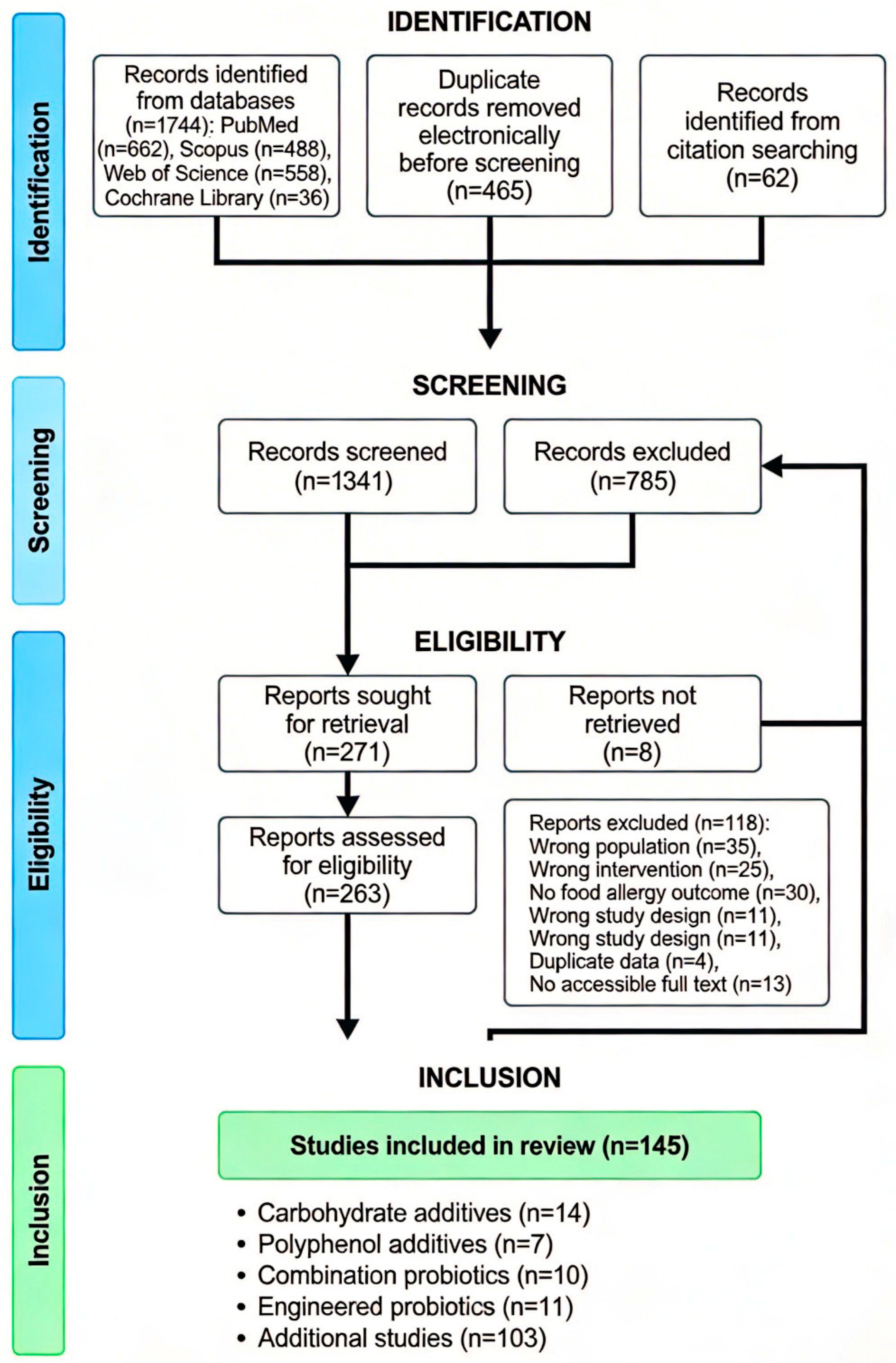
- Page, M.J.; McKenzie, J.E.; Bossuyt, P.M.; Boutron, I.; Hoffmann, T.C.; Mulrow, C.D.; Shamseer, L.; Tetzlaff, J.M.; Akl, E.A.; Brennan, S.E.; et al. The PRISMA 2020 statement: An updated guideline for reporting systematic reviews. BMJ 2021, 372, n71. [Google Scholar] [CrossRef] [PubMed]
- Sampson, H.A.; Aceves, S.; Bock, S.A.; James, J.; Jones, S.; Lang, D.; Nadeau, K.; Nowak-Wegrzyn, A.; Oppenheimer, J.; Perry, T.T.; et al. Food allergy: A practice parameter update-2014. J. Allergy Clin. Immunol. 2014, 134, 1016–1025.e43. [Google Scholar] [CrossRef]
- Bird, J.A.; Leonard, S.; Groetch, M.; Assa’AD, A.; Cianferoni, A.; Clark, A.; Crain, M.; Fausnight, T.; Fleischer, D.; Green, T.; et al. Conducting an Oral Food Challenge: An Update to the 2009 Adverse Reactions to Foods Committee Work Group Report. J. Allergy Clin. Immunol. Pract. 2020, 8, 75–90.e17. [Google Scholar] [CrossRef]
- Novick, B.E. Allergy testing. J. Med. Soc. N. J. 1992, 89, 117. [Google Scholar]
- Shaker, M.S.; Wallace, D.V.; Golden, D.B.; Oppenheimer, J.; Bernstein, J.A.; Campbell, R.L.; Dinakar, C.; Ellis, A.; Greenhawt, M.; Khan, D.A.; et al. Anaphylaxis-a 2020 practice parameter update, systematic review, and Grading of Recommendations, Assessment, Development and Evaluation (GRADE) analysis. J. Allergy Clin. Immunol. 2020, 145, 1082–1123. [Google Scholar] [CrossRef]
- Golden, D.B.; Wang, J.; Waserman, S.; Akin, C.; Campbell, R.L.; Ellis, A.K.; Greenhawt, M.; Lang, D.M.; Ledford, D.K.; Lieberman, J.; et al. Anaphylaxis: A 2023 practice parameter update. Ann. Allergy Asthma Immunol. 2024, 132, 124–176. [Google Scholar] [CrossRef] [PubMed]
- Pieretti, M.M.; Chung, D.; Pacenza, R.; Slotkin, T.; Sicherer, S.H. Audit of manufactured products: Use of allergen advisory labels and identification of labeling ambiguities. J. Allergy Clin. Immunol. 2009, 124, 337–341. [Google Scholar] [CrossRef]
- Sheth, S.S.; Waserman, S.; Kagan, R.; Alizadehfar, R.; Primeau, M.-N.; Elliot, S.; Pierre, Y.S.; Wickett, R.; Joseph, L.; Harada, L.; et al. Role of food labels in accidental exposures in food-allergic individuals in Canada. Ann. Allergy Asthma Immunol. 2010, 104, 60–65. [Google Scholar] [CrossRef]
- Landers, J.J.; O’Konek, J.J. Vaccines as therapies for food allergies. Adv. Pharmacol. 2021, 91, 229–258. [Google Scholar] [CrossRef]
- Durham, S.R.; Walker, S.M.; Varga, E.-M.; Jacobson, M.R.; O’Brien, F.; Noble, W.; Till, S.J.; Hamid, Q.A.; Nouri-Aria, K.T. Long-term clinical efficacy of grass-pollen immunotherapy. N. Engl. J. Med. 1999, 341, 468–475. [Google Scholar] [CrossRef]
- Nelson, H.S.; Lahr, J.; Rule, R.; Bock, A.; Leung, D. Treatment of anaphylactic sensitivity to peanuts by immunotherapy with injections of aqueous peanut extract. J. Allergy Clin. Immunol. 1997, 99, 744–751. [Google Scholar] [CrossRef] [PubMed]
- Oppenheimer, J.J.; Nelson, H.S.; Bock, S.A.; Christensen, F.; Leung, D.Y. Treatment of peanut allergy with rush immunotherapy. J. Allergy Clin. Immunol. 1992, 90, 256–262. [Google Scholar] [CrossRef] [PubMed]
- Bernstein, D.I.; Wanner, M.; Borish, L.; Liss, G.M.; Immunotherapy Committee, American Academy of Allergy, Asthma and Immunology. Twelve-year survey of fatal reactions to allergen injections and skin testing: 1990–2001. J. Allergy Clin. Immunol. 2004, 113, 1129–1136. [Google Scholar] [CrossRef]
- James, C.; Bernstein, D.I. Allergen immunotherapy: An updated review of safety. Curr. Opin. Allergy Clin. Immunol. 2017, 17, 55–59. [Google Scholar] [CrossRef]
- Shakya, A.K.; Ingrole, R.S.; Joshi, G.; Uddin, J.; Anvari, S.; Davis, C.M.; Gill, H.S. Microneedles coated with peanut allergen enable desensitization of peanut sensitized mice. J. Control Release 2019, 314, 38–47. [Google Scholar] [CrossRef]
- Landers, J.J.; Janczak, K.W.; Shakya, A.K.; Zarnitsyn, V.; Patel, S.R.; Baker, J.R.; Gill, H.S.; O’konek, J.J. Targeted allergen-specific immunotherapy within the skin improves allergen delivery to induce desensitization to peanut. Immunotherapy 2022, 14, 539–552. [Google Scholar] [CrossRef] [PubMed]
- PALISADE Group of Clinical Investigators. AR101 Oral Immunotherapy for Peanut Allergy. N. Engl. J. Med. 2018, 379, 1991–2001. [Google Scholar] [CrossRef]
- Brown, K.R.; Baker, J.; Vereda, A.; Beyer, K.; Burks, A.W.; du Toit, G.; Hourihane, J.O.; Jones, S.M.; Norval, D.; Dana, A.; et al. Safety of peanut (Arachis hypogaea) allergen powder-dnfp in children and teenagers with peanut allergy: Pooled summary of phase 3 and extension trials. J. Allergy Clin. Immunol. 2022, 149, 2043–2052.e9. [Google Scholar] [CrossRef]
- Wong, G.W.K. Options for Multiple Food Allergies—Food Avoidance or Pharmacologic Treatment? N. Engl. J. Med. 2024, 390, 946–948. [Google Scholar] [CrossRef]
- Skripak, J.M.; Nash, S.D.; Rowley, H.; Brereton, N.H.; Oh, S.; Hamilton, R.G.; Matsui, E.C.; Burks, A.W.; Wood, R.A. A randomized, double-blind, placebo-controlled study of milk oral immunotherapy for cow’s milk allergy. J. Allergy Clin. Immunol. 2008, 122, 1154–1160. [Google Scholar] [CrossRef] [PubMed]
- Burks, A.W.; Jones, S.M.; Wood, R.A.; Fleischer, D.M.; Sicherer, S.H.; Lindblad, R.W.; Stablein, D.; Henning, A.K.; Vickery, B.P.; Liu, A.H.; et al. Oral immunotherapy for treatment of egg allergy in children. N. Engl. J. Med. 2012, 367, 233–243. [Google Scholar] [CrossRef] [PubMed]
- Takahashi, M.; Soejima, K.; Taniuchi, S.; Hatano, Y.; Yamanouchi, S.; Ishikawa, H.; Irahara, M.; Sasaki, Y.; Kido, H.; Kaneko, K. Oral immunotherapy combined with omalizumab for high-risk cow’s milk allergy: A randomized controlled trial. Sci. Rep. 2017, 7, 17453. [Google Scholar] [CrossRef]
- Wood, R.A.; Togias, A.; Sicherer, S.H.; Shreffler, W.G.; Kim, E.H.; Jones, S.M.; Leung, D.Y.; Vickery, B.P.; Bird, J.A.; Spergel, J.M.; et al. Omalizumab for the Treatment of Multiple Food Allergies. N. Engl. J. Med. 2024, 390, 889–899. [Google Scholar] [CrossRef] [PubMed]
- Anagnostou, A.; Bird, J.A.; Chinthrajah, S.; Dribin, T.E.; Fleischer, D.M.; Kim, E.; Nowak-Wegrzyn, A.; Rachid, R.; Shaker, M.S.; Shreffler, W.; et al. The use and implementation of omalizumab as food allergy treatment: Consensus-based guidance and Work Group Report of the Adverse Reactions to Foods Committee of the American Academy of Allergy, Asthma & Immunology. J. Allergy Clin. Immunol. 2025, 155, 62–69.e1. [Google Scholar] [CrossRef]
- Noti, M.; Kim, B.S.; Siracusa, M.C.; Rak, G.D.; Kubo, M.; Moghaddam, A.E.; Sattentau, Q.A.; Comeau, M.R.; Spergel, J.M.; Artis, D. Exposure to food allergens through inflamed skin promotes intestinal food allergy through the thymic stromal lymphopoietin-basophil axis. J. Allergy Clin. Immunol. 2014, 133, 1390–1399.e6. [Google Scholar] [CrossRef]
- Cañas, J.A.; Núñez, R.; Cruz-Amaya, A.; Gómez, F.; Torres, M.J.; Palomares, F.; Mayorga, C. Epigenetics in Food Allergy and Immunomodulation. Nutrients 2021, 13, 4345. [Google Scholar] [CrossRef]
- Chun, Y.; Lee, J.H.; Bunyavanich, S. Epigenomic and epigenetic investigations of food allergy. Pediatr. Allergy Immunol. 2024, 35, e14065. [Google Scholar] [CrossRef]
- Canani, R.B.; Paparo, L.; Nocerino, R.; Cosenza, L.; Pezzella, V.; Di Costanzo, M.; Capasso, M.; Del Monaco, V.; D’Argenio, V.; Greco, L.; et al. Differences in DNA methylation profile of Th1 and Th2 cytokine genes are associated with tolerance acquisition in children with IgE-mediated cow’s milk allergy. Clin. Epigenet 2015, 7, 38. [Google Scholar] [CrossRef]
- Arzola-Martínez, L.; Ptaschinski, C.; Lukacs, N.W. Trained innate immunity, epigenetics, and food allergy. Front. Allergy 2023, 4, 1105588. [Google Scholar] [CrossRef]
- Paparo, L.; Nocerino, R.; Cosenza, L.; Aitoro, R.; D’aRgenio, V.; Del Monaco, V.; Di Scala, C.; Amoroso, A.; Di Costanzo, M.; Salvatore, F.; et al. Epigenetic features of FoxP3 in children with cow’s milk allergy. Clin. Epigenet 2016, 8, 86. [Google Scholar] [CrossRef] [PubMed]
- Ferraro, V.A.; Zanconato, S.; Carraro, S. The Epithelial Barrier Hypothesis in Food Allergies: The State of the Art. Nutrients 2025, 17, 1014. [Google Scholar] [CrossRef] [PubMed]
- Brown, S.J.; Asai, Y.; Cordell, H.J.; Campbell, L.E.; Zhao, Y.; Liao, H.; Northstone, K.; Henderson, J.; Alizadehfar, R.; Ben-Shoshan, M.; et al. Loss-of-function variants in the filaggrin gene are a significant risk factor for peanut allergy. J. Allergy Clin. Immunol. 2011, 127, 661–667. [Google Scholar] [CrossRef]
- Perkin, M.R.; Logan, K.; Tseng, A.; Raji, B.; Ayis, S.; Peacock, J.; Brough, H.; Marrs, T.; Radulovic, S.; Craven, J.; et al. Randomized Trial of Introduction of Allergenic Foods in Breast-Fed Infants. N. Engl. J. Med. 2016, 374, 1733–1743. [Google Scholar] [CrossRef] [PubMed]
- Choi, I.H.; Shin, Y.M.; Park, J.S.; Lee, M.S.; Han, E.H.; Chai, O.H.; Im, S.Y.; Ha, T.Y.; Lee, H.-K. Immunoglobulin E-dependent active fatal anaphylaxis in mast cell-deficient mice. J. Exp. Med. 1998, 188, 1587–1592. [Google Scholar] [CrossRef]
- Yamaguchi, M.; Lantz, C.S.; Oettgen, H.C.; Katona, I.M.; Fleming, T.; Miyajima, I.; Kinet, J.-P.; Galli, S.J. IgE enhances mouse mast cell Fc(epsilon)RI expression in vitro and in vivo: Evidence for a novel amplification mechanism in IgE-dependent reactions. J. Exp. Med. 1997, 185, 663–672. [Google Scholar] [CrossRef]
- Han, H.; Thelen, T.D.; Comeau, M.R.; Ziegler, S.F. Thymic stromal lymphopoietin-mediated epicutaneous inflammation promotes acute diarrhea and anaphylaxis. J. Clin. Investig. 2014, 124, 5442–5452. [Google Scholar] [CrossRef]
- Chang, P.V.; Hao, L.; Offermanns, S.; Medzhitov, R. The microbial metabolite butyrate regulates intestinal macrophage function via histone deacetylase inhibition. Proc. Natl. Acad. Sci. USA 2014, 111, 2247–2252. [Google Scholar] [CrossRef]
- De Filippis, F.; Paparo, L.; Nocerino, R.; Della Gatta, G.; Carucci, L.; Russo, R.; Pasolli, E.; Ercolini, D.; Canani, R.B. Specific gut microbiome signatures and the associated pro-inflamatory functions are linked to pediatric allergy and acquisition of immune tolerance. Nat. Commun. 2021, 12, 5958. [Google Scholar] [CrossRef]
- Hoskinson, C.; Dai, D.L.Y.; Del Bel, K.L.; Becker, A.B.; Moraes, T.J.; Mandhane, P.J.; Finlay, B.B.; Simons, E.; Kozyrskyj, A.L.; Azad, M.B.; et al. Delayed gut microbiota maturation in the first year of life is a hallmark of pediatric allergic disease. Nat. Commun. 2023, 14, 4785. [Google Scholar] [CrossRef]
- Yan, H.; Ajuwon, K.M. Butyrate modifies intestinal barrier function in IPEC-J2 cells through a selective upregulation of tight junction proteins and activation of the Akt signaling pathway. PLoS ONE 2017, 12, e0179586. [Google Scholar] [CrossRef] [PubMed]
- Johansson, M.E.V.; Larsson, J.M.H.; Hansson, G.C. The two mucus layers of colon are organized by the MUC2 mucin, whereas the outer layer is a legislator of host-microbial interactions. Proc. Natl. Acad. Sci. USA 2011, 108, 4659–4665. [Google Scholar] [CrossRef] [PubMed]
- Järvinen, K.M.; Konstantinou, G.N.; Pilapil, M.; Arrieta, M.; Noone, S.; Sampson, H.A.; Meddings, J.; Nowak-Węgrzyn, A. Intestinal permeability in children with food allergy on specific elimination diets. Pediatr. Allergy Immunol. 2013, 24, 589–595. [Google Scholar] [CrossRef]
- Johansson, M.E.; Jakobsson, H.E.; Holmén-Larsson, J.; Schütte, A.; Ermund, A.; Rodríguez-Piñeiro, A.M.; Arike, L.; Wising, C.; Svensson, F.; Bäckhed, F.; et al. Normalization of Host Intestinal Mucus Layers Requires Long-Term Microbial Colonization. Cell Host Microbe 2015, 18, 582–592. [Google Scholar] [CrossRef]
- Han, P.; Gu, J.-Q.; Li, L.-S.; Wang, X.-Y.; Wang, H.-T.; Wang, Y.; Chang, C.; Sun, J.-L. The Association Between Intestinal Bacteria and Allergic Diseases-Cause or Consequence? Front. Cell. Infect. Microbiol. 2021, 11, 650893. [Google Scholar] [CrossRef]
- Zhou, H.; Wang, L.; Lv, W.; Yu, H. Correction: The NLRP3 inflammasome in allergic diseases: Mechanisms and therapeutic implications. Clin. Exp. Med. 2025, 25, 124. [Google Scholar] [CrossRef] [PubMed]
- Lu, H.-F.; Zhou, Y.-C.; Hu, T.-Y.; Yang, D.-H.; Wang, X.-J.; Luo, D.-D.; Qiu, S.-Q.; Cheng, B.-H.; Zeng, X.-H. Unraveling the role of NLRP3 inflammasome in allergic inflammation: Implications for novel therapies. Front. Immunol. 2024, 15, 1435892. [Google Scholar] [CrossRef]
- Ma, M.; Li, G.; Qi, M.; Jiang, W.; Zhou, R. Inhibition of the Inflammasome Activity of NLRP3 Attenuates HDM-Induced Allergic Asthma. Front. Immunol. 2021, 12, 718779. [Google Scholar] [CrossRef]
- Chen, J.; Jia, S.; Xue, X.; Guo, C.; Dong, K. Gut microbiota: A novel target for exercise-mediated regulation of NLRP3 inflammasome activation. Front. Microbiol. 2024, 15, 1476908. [Google Scholar] [CrossRef]
- Yang, R.; Hu, X.; Xie, X.; Chen, H.; Fang, H.; Zhu, L.; Li, Z. Propionic Acid Targets the TLR4/NF-κB Signaling Pathway and Inhibits LPS-Induced Intestinal Barrier Dysfunction: In Vitro and In Vivo Studies. Front. Pharmacol. 2020, 11, 573475. [Google Scholar] [CrossRef]
- Yang, D.; Wang, Z.; Chen, Y.; Guo, Q.; Dong, Y. Interactions between gut microbes and NLRP3 inflammasome in the gut-brain axis. Comput. Struct. Biotechnol. J. 2023, 21, 2215–2227. [Google Scholar] [CrossRef]
- Heid, M.E.; Keyel, P.A.; Kamga, C.; Shiva, S.; Watkins, S.C.; Salter, R.D. Mitochondrial reactive oxygen species induces NLRP3-dependent lysosomal damage and inflammasome activation. J. Immunol. 2013, 191, 5230–5238. [Google Scholar] [CrossRef]
- Pierantonelli, I.; Rychlicki, C.; Agostinelli, L.; Giordano, D.M.; Gaggini, M.; Fraumene, C.; Saponaro, C.; Manghina, V.; Sartini, L.; Mingarelli, E.; et al. Author Correction: Lack of NLRP3-inflammasome leads to gut-liver axis derangement, gut dysbiosis and a worsened phenotype in a mouse model of NAFLD. Sci. Rep. 2017, 7, 17568. [Google Scholar] [CrossRef] [PubMed]
- Liu, X.-F.; Shao, J.-H.; Liao, Y.-T.; Wang, L.-N.; Jia, Y.; Dong, P.-J.; Liu, Z.-Z.; He, D.-D.; Li, C.; Zhang, X. Regulation of short-chain fatty acids in the immune system. Front. Immunol. 2023, 14, 1186892. [Google Scholar] [CrossRef]
- Wang, X.; He, G.; Peng, Y.; Zhong, W.; Wang, Y.; Zhang, B. Sodium butyrate alleviates adipocyte inflammation by inhibiting NLRP3 pathway. Sci. Rep. 2015, 5, 12676. [Google Scholar] [CrossRef] [PubMed]
- Yuan, X.; Wang, L.; Bhat, O.M.; Lohner, H.; Li, P.-L. Differential effects of short chain fatty acids on endothelial Nlrp3 inflammasome activation and neointima formation: Antioxidant action of butyrate. Redox Biol. 2018, 16, 21–31. [Google Scholar] [CrossRef]
- Feng, Y.; Wang, Y.; Wang, P.; Huang, Y.; Wang, F. Short-Chain Fatty Acids Manifest Stimulative and Protective Effects on Intestinal Barrier Function Through the Inhibition of NLRP3 Inflammasome and Autophagy. Cell. Physiol. Biochem. 2018, 49, 190–205. [Google Scholar] [CrossRef] [PubMed]
- Gong, Y.; Han, Z.; Wang, S.; Li, X.; Chen, X.; Yang, B. Progress in targeting the NLRP3 signaling pathway for inflammatory bowel disease (Review). Mol. Med. Rep. 2025, 32, 241. [Google Scholar] [CrossRef]
- Martino, D.; Neeland, M.; Dang, T.; Cobb, J.; Ellis, J.; Barnett, A.; Tang, M.; Vuillermin, P.; Allen, K.; Saffery, R. Epigenetic dysregulation of naive CD4+ T-cell activation genes in childhood food allergy. Nat. Commun. 2018, 9, 3308. [Google Scholar] [CrossRef]
- Canesso, M.C.C.; de Castro, T.B.R.; Nakandakari-Higa, S.; Lockhart, A.; Luehr, J.; Bortolatto, J.; Parsa, R.; Esterházy, D.; Lyu, M.; Liu, T.-T.; et al. Identification of antigen-presenting cell-T cell interactions driving immune responses to food. Science 2025, 387, eado5088. [Google Scholar] [CrossRef]
- Nagler-Anderson, C. Man the barrier! Strategic defences in the intestinal mucosa. Nat. Rev. Immunol. 2001, 1, 59–67. [Google Scholar] [CrossRef]
- Noval Rivas, M.; Burton, O.T.; Oettgen, H.C.; Chatila, T. IL-4 production by group 2 innate lymphoid cells promotes food allergy by blocking regulatory T-cell function. J. Allergy Clin. Immunol. 2016, 138, 801–811.e9. [Google Scholar] [CrossRef] [PubMed]
- Lebman, D.A.; Coffman, R.L. Interleukin 4 causes isotype switching to IgE in T cell-stimulated clonal B cell cultures. J. Exp. Med. 1988, 168, 853–862. [Google Scholar] [CrossRef] [PubMed]
- Sainte-Laudy, J.; Sabbah, A.; Vallon, C.; Guerin, J.C. Analysis of anti-IgE and allergen induced human basophil activation by flow cytometry. Comparison with histamine release. Inflamm. Res. 1998, 47, 401–408. [Google Scholar] [CrossRef]
- Nguyen, K.L.; Gillis, S.; MacGlashan, D.W. A comparative study of releasing and nonreleasing human basophils: Nonreleasing basophils lack an early component of the signal transduction pathway that follows IgE cross-linking. J. Allergy Clin. Immunol. 1990, 85, 1020–1029. [Google Scholar] [CrossRef]
- Sampson, H.A.; O’Mahony, L.; Burks, A.W.; Plaut, M.; Lack, G.; Akdis, C.A. Mechanisms of food allergy. J. Allergy Clin. Immunol. 2018, 141, 11–19. [Google Scholar] [CrossRef] [PubMed]
- Wei, Y.; Peng, J.; Wang, S.; Ding, Z.; Chen, G.; Sun, J. Probiotics and the Potential of Genetic Modification as a Possible Treatment for Food Allergy. Nutrients 2023, 15, 4159. [Google Scholar] [CrossRef]
- Zeng, L.; Qian, Y.; Cui, X.; Zhao, J.; Ning, Z.; Cha, J.; Wang, K.; Ge, C.; Jia, J.; Dou, T.; et al. Immunomodulatory role of gut microbial metabolites: Mechanistic insights and therapeutic frontiers. Front. Microbiol. 2025, 16, 1675065. [Google Scholar] [CrossRef]
- Gasaly, N.; de Vos, P.; Hermoso, M.A. Impact of Bacterial Metabolites on Gut Barrier Function and Host Immunity: A Focus on Bacterial Metabolism and Its Relevance for Intestinal Inflammation. Front. Immunol. 2021, 12, 658354. [Google Scholar] [CrossRef]
- Seo, S.-K.; Kwon, B. Immune regulation through tryptophan metabolism. Exp. Mol. Med. 2023, 55, 1371–1379. [Google Scholar] [CrossRef]
- Pan, X.; Chen, F.; Wu, T.; Tang, H.; Zhao, Z. Prebiotic oligosaccharides change the concentrations of short-chain fatty acids and the microbial population of mouse bowel. J. Zhejiang Univ. Sci. B 2009, 10, 258–263. [Google Scholar] [CrossRef]
- Arifuzzaman, M.; Won, T.H.; Yano, H.; Uddin, J.; Emanuel, E.R.; Hu, E.; Zhang, W.; Li, T.-T.; Jin, W.-B.; Grier, A.; et al. Dietary fiber is a critical determinant of pathologic ILC2 responses and intestinal inflammation. J. Exp. Med. 2024, 221, e20232148. [Google Scholar] [CrossRef]
- Fu, J.; Zheng, Y.; Gao, Y.; Xu, W. Dietary Fiber Intake and Gut Microbiota in Human Health. Microorganisms 2022, 10, 2507. [Google Scholar] [CrossRef]
- Furlani, F.; Sacco, P.; Decleva, E.; Menegazzi, R.; Donati, I.; Paoletti, S.; Marsich, E. Chitosan Acetylation Degree Influences the Physical Properties of Polysaccharide Nanoparticles: Implication for the Innate Immune Cells Response. ACS Appl. Mater. Interfaces 2019, 11, 9794–9803. [Google Scholar] [CrossRef]
- Park, M.J.; Ryu, H.S.; Kim, J.S.; Lee, H.K.; Kang, J.S.; Yun, J.; Kim, S.Y.; Lee, M.K.; Hong, J.T.; Kim, Y.; et al. Platycodon grandiflorum polysaccharide induces dendritic cell maturation via TLR4 signaling. Food Chem. Toxicol. 2014, 72, 212–220. [Google Scholar] [CrossRef]
- Liu, Q.-M.; Yang, Y.; Maleki, S.J.; Alcocer, M.; Xu, S.-S.; Shi, C.-L.; Cao, M.-J.; Liu, G.-M. Anti-Food Allergic Activity of Sulfated Polysaccharide from Gracilaria lemaneiformis is Dependent on Immunosuppression and Inhibition of p38 MAPK. J. Agric. Food Chem. 2016, 64, 4536–4544. [Google Scholar] [CrossRef]
- Huang, G.; Chen, X.; Huang, H. Chemical Modifications and Biological Activities of Polysaccharides. Curr. Drug Targets 2016, 17, 1799–1803. [Google Scholar] [CrossRef]
- Li, F.; Wang, L.; Jin, X.-M.; Yan, C.-H.; Jiang, S.; Shen, X.-M. The immunologic effect of TGF-beta1 chitosan nanoparticle plasmids on ovalbumin-induced allergic BALB/c mice. Immunobiology 2009, 214, 87–99. [Google Scholar] [CrossRef]
- Jiang, T.; Ji, H.; Zhang, L.; Wang, Y.; Zhou, H. Chitosan Oligosaccharide Exerts Anti-Allergic Effect against Shrimp Tropomyosin-Induced Food Allergy by Affecting Th1 and Th2 Cytokines. Int. Arch. Allergy Immunol. 2019, 180, 10–16. [Google Scholar] [CrossRef]
- Bae, M.-J.; Shin, H.S.; Kim, E.-K.; Kim, J.; Shon, D.-H. Oral administration of chitin and chitosan prevents peanut-induced anaphylaxis in a murine food allergy model. Int. J. Biol. Macromol. 2013, 61, 164–168. [Google Scholar] [CrossRef]
- Li, Y.; Lin, Y.-F.; Wang, S.-H.; Cheng, Z.; Liu, W.-M.; Zou, Z.-H.; Liu, G.-M.; Liu, Q.-M. Purified components of red-edge tea polysaccharide alleviate food allergy in mice by regulating intestinal homeostasis. Int. J. Biol. Macromol. 2025, 288, 138671. [Google Scholar] [CrossRef]
- Ou, J.-Y.; Wei, Y.-J.; Liu, F.-L.; Huang, C.-H. Anti-allergic effects of Ulva-derived polysaccharides, oligosaccharides and residues in a murine model of food allergy. Heliyon 2023, 9, e22840. [Google Scholar] [CrossRef]
- Liu, Y.; Ma, Y.; Chen, Z.; Zou, C.; Liu, W.; Yang, L.; Fu, L.; Wang, Y.; Liu, G.-M.; Cao, M.-J. Depolymerized sulfated galactans from Eucheuma serra ameliorate allergic response and intestinal flora in food allergic mouse model. Int. J. Biol. Macromol. 2021, 166, 977–985. [Google Scholar] [CrossRef]
- Mizuno, M.; Sakaguchi, K.; Sakane, I. Oral Administration of Fucoidan Can Exert Anti-Allergic Activity after Allergen Sensitization by Enhancement of Galectin-9 Secretion in Blood. Biomolecules 2020, 10, 258. [Google Scholar] [CrossRef]
- Han, K.; Xie, F.; Animasahun, O.; Nenwani, M.; Kitamoto, S.; Kim, Y.; Phoo, M.T.; Xu, J.; Wuchu, F.; Omoloja, K.; et al. Inulin-gel-based oral immunotherapy remodels the small intestinal microbiome and suppresses food allergy. Nat. Mater. 2024, 23, 1444–1455. [Google Scholar] [CrossRef]
- Liu, T.; Asif, I.M.; Liu, L.; Zhang, M.; Li, B.; Wang, L. Laminarin ameliorates iodoacetamide-induced functional dyspepsia via modulation of 5-HT3 receptors and the gut microbiota. Int. J. Biol. Macromol. 2024, 268, 131640. [Google Scholar] [CrossRef]
- Yu, X.; Gurry, T.; Nguyen, L.T.T.; Richardson, H.S.; Alm, E.J. Prebiotics and Community Composition Influence Gas Production of the Human Gut Microbiota. mBio 2020, 11, e00217-20. [Google Scholar] [CrossRef]
- Stiverson, J.; Williams, T.; Chen, J.; Adams, S.; Hustead, D.; Price, P.; Guerrieri, J.; Deacon, J.; Yu, Z. Prebiotic Oligosaccharides: Comparative Evaluation Using In Vitro Cultures of Infants’ Fecal Microbiomes. Appl. Environ. Microbiol. 2014, 80, 7388–7397. [Google Scholar] [CrossRef]
- Cheon, S.; Kim, G.; Bae, J.-H.; Lee, D.H.; Seong, H.; Kim, D.H.; Han, J.-S.; Lim, S.-Y.; Han, N.S. Comparative analysis of prebiotic effects of four oligosaccharides using in vitro gut model: Digestibility, microbiome, and metabolome changes. FEMS Microbiol. Ecol. 2023, 99, fiad002. [Google Scholar] [CrossRef]
- Masarweh, C.; Maldonado-Gomez, M.; Paviani, B.; Bhattacharya, M.; Weng, C.-Y.; Suarez, C.; Ehlers-Cheang, S.; Stacy, A.; Castillo, J.; Krishnakumar, N.; et al. Generation of novel prebiotic oligosaccharide pools from fiber drives biological insight in bacterial glycan metabolism. Appl. Environ. Microbiol. 2025, 91, e0207724. [Google Scholar] [CrossRef]
- Lee, D.H.; Seong, H.; Chang, D.; Gupta, V.K.; Kim, J.; Cheon, S.; Kim, G.; Sung, J.; Han, N.S. Evaluating the prebiotic effect of oligosaccharides on gut microbiome wellness using in vitro fecal fermentation. NPJ Sci. Food 2023, 7, 18. [Google Scholar] [CrossRef]
- Baba, Y.; Tsuge, D.; Aoki, R. Enhancement of carbohydrate metabolism by probiotic and prebiotic intake promotes short-chain fatty acid production in the gut microbiome: A randomized, double-blind, placebo-controlled crossover trial. Biosci. Biotechnol. Biochem. 2025, 89, 1191–1202. [Google Scholar] [CrossRef]
- van Trijp, M.P.; Rios-Morales, M.; Witteman, B.; Abegaz, F.; Gerding, A.; An, R.; Koehorst, M.; Evers, B.; van Dongen, K.C.; Zoetendal, E.G.; et al. Intraintestinal fermentation of fructo- and galacto-oligosaccharides and the fate of short-chain fatty acids in humans. iScience 2024, 27, 109208. [Google Scholar] [CrossRef]
- Barboza, M.; Sela, D.A.; Pirim, C.; LoCascio, R.G.; Freeman, S.L.; German, J.B.; Mills, D.A.; Lebrilla, C.B. Glycoprofiling bifidobacterial consumption of galacto-oligosaccharides by mass spectrometry reveals strain-specific, preferential consumption of glycans. Appl. Environ. Microbiol. 2009, 75, 7319–7325. [Google Scholar] [CrossRef]
- Rodriguez-Colinas, B.; Kolida, S.; Baran, M.; Ballesteros, A.O.; Rastall, R.A.; Plou, F.J. Analysis of fermentation selectivity of purified galacto-oligosaccharides by in vitro human faecal fermentation. Appl. Microbiol. Biotechnol. 2013, 97, 5743–5752. [Google Scholar] [CrossRef]
- Chen, P.; Huang, Y.; Zeng, H.; Zheng, M.; Guo, J. In vitro assessment of the effect of Porphyra haitanensis polysaccharides on the intestinal flora of allergic mice. Int. J. Biol. Macromol. 2025, 311, 143950. [Google Scholar] [CrossRef]
- Lee, D.; Kim, H.S.; Shin, E.; Do, S.-G.; Lee, C.-K.; Kim, Y.M.; Lee, M.B.; Min, K.Y.; Koo, J.; Kim, S.J.; et al. Polysaccharide isolated from Aloe vera gel suppresses ovalbumin-induced food allergy through inhibition of Th2 immunity in mice. Biomed. Pharmacother. 2018, 101, 201–210. [Google Scholar] [CrossRef]
- Chen, H.-Y.; Zhou, Y.-C.; Liu, Y.; Huang, J.-Y.; Liu, H.; Liu, C.-F.; Liu, W.-H.; Liu, G.-M.; Liu, Q.-M. Fermented Gracilaria lemaneiformis polysaccharides alleviate food allergy by regulating Treg cells and gut microbiota. Int. J. Biol. Macromol. 2024, 269, 132215. [Google Scholar] [CrossRef]
- Hayen, S.M.; Knulst, A.C.; Garssen, J.; Otten, H.G.; Willemsen, L.E.M. Fructo-Oligosaccharides Modify Human DC Maturation and Peanut-Induced Autologous T-Cell Response of Allergic Patients In Vitro. Front. Immunol. 2020, 11, 600125. [Google Scholar] [CrossRef]
- Liu, Q.; Zhou, Y.; Ma, L.; Gu, F.; Liao, K.; Liu, Y.; Zhang, Y.; Liu, H.; Hong, Y.; Cao, M.; et al. Sulfate oligosaccharide of Gracilaria lemaneiformis modulates type 1 immunity by restraining T cell activation. Carbohydr. Polym. 2022, 288, 119377. [Google Scholar] [CrossRef]
- Takahashi, H.; Fujii, T.; Yamakawa, S.; Yamada, C.; Fujiki, K.; Kondo, N.; Funasaka, K.; Hirooka, Y.; Tochio, T. Combined oral intake of short and long fructans alters the gut microbiota in food allergy model mice and contributes to food allergy prevention. BMC Microbiol. 2023, 23, 266. [Google Scholar] [CrossRef]
- Wagenaar, L.; Bol-Schoenmakers, M.; Giustarini, G.; Vonk, M.M.; van Esch, B.C.; Knippels, L.M.; Garssen, J.; Smit, J.J.; Pieters, R.H. Dietary Supplementation with Nondigestible Oligosaccharides Reduces Allergic Symptoms and Supports Low Dose Oral Immunotherapy in a Peanut Allergy Mouse Model. Mol. Nutr. Food Res. 2018, 62, e1800369. [Google Scholar] [CrossRef]
- Hesser, L.A.; Puente, A.A.; Arnold, J.; Ionescu, E.; Mirmira, A.; Talasani, N.; Lopez, J.; Maccio-Maretto, L.; Mimee, M.; Nagler, C.R. A synbiotic of Anaerostipes caccae and lactulose prevents and treats food allergy in mice. Cell Host Microbe 2024, 32, 1163–1176.e6. [Google Scholar] [CrossRef]
- Lv, L.; Qu, X.; Yang, N.; Liu, Z.; Wu, X. Changes in structure and allergenicity of shrimp tropomyosin by dietary polyphenols treatment. Food Res. Int. 2021, 140, 109997. [Google Scholar] [CrossRef]
- Wang, Y.; Zhang, P.; Zhang, J.; Hong, T. Bisdemethoxycurcumin attenuates OVA-induced food allergy by inhibiting the MAPK and NF-κB signaling pathways. Exp. Ther. Med. 2022, 23, 401. [Google Scholar] [CrossRef]
- He, W.; Zhang, T.; Velickovic, T.C.; Li, S.; Lyu, Y.; Wang, L.; Yi, J.; Liu, Z.; He, Z.; Wu, X. Covalent conjugation with (-)-epigallo-catechin 3-gallate and chlorogenic acid changes allergenicity and functional properties of Ara h1 from peanut. Food Chem. 2020, 331, 127355. [Google Scholar] [CrossRef]
- Lan, J.; Wang, K.; Chen, G.; Cao, G.; Yang, C. Effects of inulin and isomalto-oligosaccharide on diphenoxylate-induced constipation, gastrointestinal motility-related hormones, short-chain fatty acids, and the intestinal flora in rats. Food Funct. 2020, 11, 9216–9225. [Google Scholar] [CrossRef]
- Akiyama, H.; Sato, Y.; Watanabe, T.; Nagaoka, M.H.; Yoshioka, Y.; Shoji, T.; Kanda, T.; Yamada, K.; Totsuka, M.; Teshima, R.; et al. Dietary unripe apple polyphenol inhibits the development of food allergies in murine models. FEBS Lett. 2005, 579, 4485–4491. [Google Scholar] [CrossRef]
- Liang, A.; Leonard, W.; Beasley, J.T.; Fang, Z.; Zhang, P.; Ranadheera, C.S. Anthocyanins-gut microbiota-health axis: A review. Crit. Rev. Food Sci. Nutr. 2024, 64, 7563–7588. [Google Scholar] [CrossRef]
- Pasinetti, G.M.; Singh, R.; Westfall, S.; Herman, F.; Faith, J.; Ho, L. The Role of the Gut Microbiota in the Metabolism of Polyphenols as Characterized by Gnotobiotic Mice. J. Alzheimers Dis. JAD 2018, 63, 409–421. [Google Scholar] [CrossRef]
- Rodríguez-Daza, M.C.; Pulido-Mateos, E.C.; Lupien-Meilleur, J.; Guyonnet, D.; Desjardins, Y.; Roy, D. Polyphenol-Mediated Gut Microbiota Modulation: Toward Prebiotics and Further. Front. Nutr. 2021, 8, 689456. [Google Scholar] [CrossRef]
- Zhang, Y.; Yu, W.; Zhang, L.; Wang, M.; Chang, W. The Interaction of Polyphenols and the Gut Microbiota in Neurodegenerative Diseases. Nutrients 2022, 14, 5373. [Google Scholar] [CrossRef]
- Liu, X.; Alharbi, A.; Gibson, R.; Rodriguez-Mateos, A. (Poly)phenol-gut microbiota interactions and their impact on human health. Curr. Opin. Clin. Nutr. Metab. Care 2025, 28, 316–322. [Google Scholar] [CrossRef]
- Cao, X.; Wang, X.; Ren, Y.; Sun, Y.; Yang, Z.; Ge, J.; Ping, W. Lonicera caerulea L. polyphenols improve short-chain fatty acid levels by reshaping the microbial structure of fermented feces in vitro. Front. Microbiol. 2023, 14, 1228700. [Google Scholar] [CrossRef]
- Okada, Y.; Oh-Oka, K.; Nakamura, Y.; Ishimaru, K.; Matsuoka, S.; Okumura, K.; Ogawa, H.; Hisamoto, M.; Okuda, T.; Nakao, A. Dietary resveratrol prevents the development of food allergy in mice. PLoS ONE 2012, 7, e44338. [Google Scholar] [CrossRef]
- Ishimoto, K.; Konishi, Y.; Otani, S.; Maeda, S.; Ago, Y.; Hino, N.; Suzuki, M.; Nakagawa, S. Suppressive effect of black tea polyphenol theaflavins in a mouse model of ovalbumin-induced food allergy. J. Nat. Med. 2023, 77, 604–609. [Google Scholar] [CrossRef]
- Bansode, R.R.; Randolph, P.D.; Plundrich, N.J.; Lila, M.A.; Williams, L.L. Peanut protein-polyphenol aggregate complexation suppresses allergic sensitization to peanut by reducing peanut-specific IgE in C3H/HeJ mice. Food Chem. 2019, 299, 125025. [Google Scholar] [CrossRef]
- Cheng, B.; Feng, H.; Li, C.; Jia, F.; Zhang, X. The mutual effect of dietary fiber and polyphenol on gut microbiota: Implications for the metabolic and microbial modulation and associated health benefits. Carbohydr. Polym. 2025, 358, 123541. [Google Scholar] [CrossRef]
- Sheng, W.; Ji, G.; Zhang, L. Immunomodulatory effects of inulin and its intestinal metabolites. Front. Immunol. 2023, 14, 1224092. [Google Scholar] [CrossRef]
- Speckmann, B.; Ehring, E.; Hu, J.; Rodriguez Mateos, A. Exploring substrate-microbe interactions: A metabiotic approach toward developing targeted synbiotic compositions. Gut Microbes 2024, 16, 2305716. [Google Scholar] [CrossRef]
- Nemzer, B.V.; Al-Taher, F.; Kalita, D.; Yashin, A.Y.; Yashin, Y.I. Health-Improving Effects of Polyphenols on the Human Intestinal Microbiota: A Review. Int. J. Mol. Sci. 2025, 26, 1335. [Google Scholar] [CrossRef]
- Yip, W.; Hughes, M.R.; Li, Y.; Cait, A.; Hirst, M.; Mohn, W.W.; McNagny, K.M. Butyrate Shapes Immune Cell Fate and Function in Allergic Asthma. Front. Immunol. 2021, 12, 628453. [Google Scholar] [CrossRef]
- Meiners, F.; Ortega-Matienzo, A.; Fuellen, G.; Barrantes, I. Gut microbiome-mediated health effects of fiber and polyphenol-rich dietary interventions. Front. Nutr. 2025, 12, 1647740. [Google Scholar] [CrossRef]
- Shi, J.; Mao, W.; Song, Y.; Wang, Y.; Zhang, L.; Xu, Y.; Gu, H.; Yao, S.; Yao, Y.; Liu, Z.; et al. Butyrate alleviates food allergy by improving intestinal barrier integrity through suppressing oxidative stress-mediated Notch signaling. iMeta 2025, 4, e70024. [Google Scholar] [CrossRef]
- Yu, B.; Pei, C.; Peng, W.; Zheng, Y.; Fu, Y.; Wang, X.; Wang, W.; Wang, Z.; Chen, Y.; Wang, Q.; et al. Microbiota-derived butyrate alleviates asthma via inhibiting Tfh13-mediated IgE production. Signal Transduct. Target. Ther. 2025, 10, 181. [Google Scholar] [CrossRef]
- Morozumi, M.; Izumi, H.; Tsuda, M.; Tabata, F.; Nakamura, H.; Miyaji, K. Changes in and relationships between human milk oligosaccharides and microRNAs in milk-derived extracellular vesicles during the first 4 months of lactation. Front. Nutr. 2025, 12, 1694093. [Google Scholar] [CrossRef]
- Elzen, C.C.D.; Carvalho, A.; Bazan-Socha, S.; Jeurink, P.V.; Wygrecka, M.; Kool, M.; Garssen, J.; Potaczek, D.P.; Garn, H.; van Esch, B.C. Human milk oligosaccharides and polyphenols: Mechanisms, effects, and applications in allergies. J. Allergy Clin. Immunol. 2026, 157, 18–37. [Google Scholar] [CrossRef]
- Dębińska, A.; Sozańska, B. Dietary Polyphenols-Natural Bioactive Compounds with Potential for Preventing and Treating Some Allergic Conditions. Nutrients 2023, 15, 4823. [Google Scholar] [CrossRef]
- Zou, F.; Qiu, Y.; Huang, Y.; Zou, H.; Cheng, X.; Niu, Q.; Luo, A.; Sun, J. Effects of short-chain fatty acids in inhibiting HDAC and activating p38 MAPK are critical for promoting B10 cell generation and function. Cell Death Dis. 2021, 12, 582. [Google Scholar] [CrossRef]
- Rana, T.S.; Bansode, R.R.; Rana, J.P.; Williams, L.L. A systematic review: Polyphenol’s effect on food allergy via microbiome modulation. Front. Microbiol. 2025, 16, 1673472. [Google Scholar] [CrossRef]
- Osborn, L.J.; Schultz, K.; Massey, W.; DeLucia, B.; Choucair, I.; Varadharajan, V.; Banerjee, R.; Fung, K.; Horak, A.J.; Orabi, D.; et al. A gut microbial metabolite of dietary polyphenols reverses obesity-driven hepatic steatosis. Proc. Natl. Acad. Sci. USA 2022, 119, e2202934119. [Google Scholar] [CrossRef]
- Cristofori, F.; Dargenio, V.N.; Dargenio, C.; Miniello, V.L.; Barone, M.; Francavilla, R. Anti-Inflammatory and Immunomodulatory Effects of Probiotics in Gut Inflammation: A Door to the Body. Front. Immunol. 2021, 12, 578386. [Google Scholar] [CrossRef]
- Hemarajata, P.; Versalovic, J. Effects of probiotics on gut microbiota: Mechanisms of intestinal immunomodulation and neuromodulation. Ther. Adv. Gastroenterol. 2013, 6, 39–51. [Google Scholar] [CrossRef] [PubMed]
- Park, J.; Kim, M.; Kang, S.; Jannasch, A.; Cooper, B.; Patterson, J.; Kim, C. Short-chain fatty acids induce both effector and regulatory T cells by suppression of histone deacetylases and regulation of the mTOR-S6K pathway. Mucosal Immunol. 2015, 8, 80–93. [Google Scholar] [CrossRef] [PubMed]
- Kim, C.H. Complex regulatory effects of gut microbial short-chain fatty acids on immune tolerance and autoimmunity. Cell. Mol. Immunol. 2023, 20, 341–350. [Google Scholar] [CrossRef]
- Zhan, K.; Gong, X.; Chen, Y.; Jiang, M.; Yang, T.; Zhao, G. Short-Chain Fatty Acids Regulate the Immune Responses via G Protein-Coupled Receptor 41 in Bovine Rumen Epithelial Cells. Front. Immunol. 2019, 10, 2042. [Google Scholar] [CrossRef]
- Nagata, K.; Ando, D.; Ashikari, T.; Ito, K.; Miura, R.; Fujigaki, I.; Goto, Y.; Ando, M.; Ito, N.; Kawazoe, H.; et al. Butyrate, Valerate, and Niacin Ameliorate Anaphylaxis by Suppressing IgE-Dependent Mast Cell Activation: Roles of GPR109A, PGE2, and Epigenetic Regulation. J. Immunol. 2024, 212, 771–784. [Google Scholar] [CrossRef]
- Liu, Y.; Cao, X.; Liu, H.; Zhang, W. The crosstalk between probiotics and T cell immunity. Front. Immunol. 2025, 16, 1695840. [Google Scholar] [CrossRef]
- Gavzy, S.J.; Kensiski, A.; Lee, Z.L.; Mongodin, E.F.; Ma, B.; Bromberg, J.S. Bifidobacterium mechanisms of immune modulation and tolerance. Gut Microbes 2023, 15, 2291164. [Google Scholar] [CrossRef]
- Konieczna, P.; Akdis, C.A.; Quigley, E.M.M.; Shanahan, F.; O’Mahony, L. Portrait of an immunoregulatory Bifidobacterium. Gut Microbes 2012, 3, 261–266. [Google Scholar] [CrossRef]
- Ruiz, L.; Delgado, S.; Ruas-Madiedo, P.; Margolles, A.; Sánchez, B. Proteinaceous Molecules Mediating Bifidobacterium-Host Interactions. Front. Microbiol. 2016, 7, 1193. [Google Scholar] [CrossRef]
- Karczewski, J.; Troost, F.J.; Konings, I.; Dekker, J.; Kleerebezem, M.; Brummer, R.-J.M.; Wells, J.M. Regulation of human epithelial tight junction proteins by Lactobacillus plantarum in vivo and protective effects on the epithelial barrier. Am. J. Physiol. Gastrointest. Liver Physiol. 2010, 298, G851–G859. [Google Scholar] [CrossRef]
- Anderson, R.C.; Cookson, A.L.; McNabb, W.C.; Park, Z.; McCann, M.J.; Kelly, W.J.; Roy, N.C. Lactobacillus plantarum MB452 enhances the function of the intestinal barrier by increasing the expression levels of genes involved in tight junction formation. BMC Microbiol. 2010, 10, 316. [Google Scholar] [CrossRef]
- Duan, C.; Ma, L.; Qin, M.; Zhang, L.; Hu, S.; Liu, L.; Sun, Y.; Ma, F.; Li, D. Potential of Lactobacillus plantarum A56 in relieving food allergy through immunoregulation, antioxidation, and reshaping intestinal microbiota. J. Nutr. Biochem. 2024, 125, 109560. [Google Scholar] [CrossRef] [PubMed]
- Jiang, S.; Hou, Y.; Meng, L.; Pu, X.; Zhu, X.; Tuo, Y.; Qian, F.; Mu, G. Effect of Lactiplantibacillus plantarum HM-22 on immunoregulation and intestinal microbiota in α-lactalbumin-induced allergic mice. Food Funct. 2021, 12, 8887–8898. [Google Scholar] [CrossRef]
- Yamamoto-Hanada, K.; Sato, M.; Toyokuni, K.; Irahara, M.; Hiraide-Kotaki, E.; Harima-Mizusawa, N.; Morita, H.; Matsumoto, K.; Ohya, Y. Combination of heat-killed Lactiplantibacillus plantarum YIT 0132 (LP0132) and oral immunotherapy in cow’s milk allergy: A randomised controlled trial. Benef. Microbes 2023, 14, 17–30. [Google Scholar] [CrossRef] [PubMed]
- Pérez-Reytor, D.; Puebla, C.; Karahanian, E.; García, K. Use of Short-Chain Fatty Acids for the Recovery of the Intestinal Epithelial Barrier Affected by Bacterial Toxins. Front. Physiol. 2021, 12, 650313. [Google Scholar] [CrossRef]
- Zheng, L.; Kelly, C.J.; Battista, K.D.; Schaefer, R.; Lanis, J.M.; Alexeev, E.E.; Wang, R.X.; Onyiah, J.C.; Kominsky, D.J.; Colgan, S.P. Microbial-Derived Butyrate Promotes Epithelial Barrier Function through IL-10 Receptor-Dependent Repression of Claudin-2. J. Immunol. 2017, 199, 2976–2984. [Google Scholar] [CrossRef] [PubMed]
- Kaisar, M.M.M.; Pelgrom, L.R.; van der Ham, A.J.; Yazdanbakhsh, M.; Everts, B. Butyrate Conditions Human Dendritic Cells to Prime Type 1 Regulatory T Cells via both Histone Deacetylase Inhibition and G Protein-Coupled Receptor 109A Signaling. Front. Immunol. 2017, 8, 1429. [Google Scholar] [CrossRef]
- Abdulqadir, R.; Al-Sadi, R.; Gupta, Y.; Rawat, M.; Ma, T. Probiotic bacteria Bifidobacterium bifidum upregulation of intestinal epithelial tight junction barrier is mediated by TLR-2/TLR-6 receptor complex activation of occludin gene. npj Biofilms Microbiomes 2026, 12, 37. [Google Scholar] [CrossRef]
- Liu, M.; Xie, W.; Wan, X.; Deng, T. Clostridium butyricum protects intestinal barrier function via upregulation of tight junction proteins and activation of the Akt/mTOR signaling pathway in a mouse model of dextran sodium sulfate-induced colitis. Exp. Ther. Med. 2020, 20, 10. [Google Scholar] [CrossRef]
- Chelakkot, C.; Choi, Y.; Kim, D.-K.; Park, H.T.; Ghim, J.; Kwon, Y.; Jeon, J.; Kim, M.-S.; Jee, Y.-K.; Gho, Y.S.; et al. Akkermansia muciniphila-derived extracellular vesicles influence gut permeability through the regulation of tight junctions. Exp. Mol. Med. 2018, 50, e450. [Google Scholar] [CrossRef] [PubMed]
- Mo, C.; Lou, X.; Xue, J.; Shi, Z.; Zhao, Y.; Wang, F.; Chen, G. The influence of Akkermansia muciniphila on intestinal barrier function. Gut Pathog. 2024, 16, 41. [Google Scholar] [CrossRef]
- Zhou, L.; Zhang, M.; Wang, Y.; Dorfman, R.G.; Liu, H.; Yu, T.; Chen, X.; Tang, D.; Xu, L.; Yin, Y.; et al. Faecalibacterium prausnitzii Produces Butyrate to Maintain Th17/Treg Balance and to Ameliorate Colorectal Colitis by Inhibiting Histone Deacetylase 1. Inflamm. Bowel Dis. 2018, 24, 1926–1940. [Google Scholar] [CrossRef]
- Touch, S.; Godefroy, E.; Rolhion, N.; Danne, C.; Oeuvray, C.; Straube, M.; Galbert, C.; Brot, L.; Salgueiro, I.A.; Chadi, S.; et al. Human CD4+CD8α+ Tregs induced by Faecalibacterium prausnitzii protect against intestinal inflammation. JCI Insight 2022, 7, e154722. [Google Scholar] [CrossRef] [PubMed]
- Sokol, H.; Pigneur, B.; Watterlot, L.; Lakhdari, O.; Bermúdez-Humaran, L.G.; Gratadoux, J.-J.; Blugeon, S.; Bridonneau, C.; Furet, J.-P.; Corthier, G.; et al. Faecalibacterium prausnitzii is an anti-inflammatory commensal bacterium identified by gut microbiota analysis of Crohn disease patients. Proc. Natl. Acad. Sci. USA 2008, 105, 16731–16736. [Google Scholar] [CrossRef] [PubMed]
- Laforest-Lapointe, I.; Arrieta, M.-C. Patterns of Early-Life Gut Microbial Colonization during Human Immune Development: An Ecological Perspective. Front. Immunol. 2017, 8, 788. [Google Scholar] [CrossRef] [PubMed]
- Tian, X.; Liang, X.; He, H.; Cui, Q.; Liu, Q.; Fan, R.; Liu, T.; Yi, H.; Gong, P.; Wang, Q.; et al. Probiotics Alleviate Food Protein Allergy in Mice by Activating TLR4 Signaling Pathway. Mol. Nutr. Food Res. 2023, 67, e2200579. [Google Scholar] [CrossRef]
- Tian, X.; Fan, R.; He, H.; Cui, Q.; Liang, X.; Liu, Q.; Liu, T.; Lin, K.; Zhang, Z.; Yi, H.; et al. Bifidobacterium animalis KV9 and Lactobacillus vaginalis FN3 alleviated β-lactoglobulin-induced allergy by modulating dendritic cells in mice. Front. Immunol. 2022, 13, 992605. [Google Scholar] [CrossRef]
- Santos, S.S.; Miranda, V.C.; Trindade, L.M.; Cardoso, V.N.; Reis, D.C.; Cassali, G.D.; Nicoli, J.R.; Cara, D.C.; Martins, F.S. Bifidobacterium longum subsp. longum 51A Attenuates Signs of Inflammation in a Murine Model of Food Allergy. Probiotics Antimicrob. Proteins 2023, 15, 63–73. [Google Scholar] [CrossRef] [PubMed]
- Fu, L.; Xie, M.; Wang, C.; Qian, Y.; Huang, J.; Sun, Z.; Zhang, H.; Wang, Y. Lactobacillus casei Zhang Alleviates Shrimp Tropomyosin-Induced Food Allergy by Switching Antibody Isotypes through the NF-κB-Dependent Immune Tolerance. Mol. Nutr. Food Res. 2020, 64, e1900496. [Google Scholar] [CrossRef] [PubMed]
- Misme-Aucouturier, B.; Gagnaire, V.; LeCorre, E.; DeCarvalho, M.; Jan, G.; Bouchaud, G. Propionibacterium freudenreichii Prevents Food Allergy in Mice via the Surface Layer Protein SlpB. J. Agric. Food Chem. 2024, 72, 27495–27503. [Google Scholar] [CrossRef]
- Duan, C.; Ma, L.; Yu, J.; Sun, Y.; Liu, L.; Ma, F.; Li, X.; Li, D. Oral administration of Lactobacillus plantarum JC7 alleviates OVA-induced murine food allergy through immunoregulation and restoring disordered intestinal microbiota. Eur. J. Nutr. 2023, 62, 685–698. [Google Scholar] [CrossRef] [PubMed]
- Miranda, V.C.; Souza, R.O.; Quintanilha, M.F.; Gallotti, B.; Assis, H.C.; Faria, A.M.C.; Nicoli, J.R.; Cara, D.C.; Martins, F.S. A Next-Generation Bacteria (Akkermansia muciniphila BAA-835) Presents Probiotic Potential Against Ovalbumin-Induced Food Allergy in Mice. Probiotics Antimicrob. Proteins 2024, 16, 737–751. [Google Scholar] [CrossRef]
- Loke, P.; Hsiao, K.; Lozinsky, A.C.; Ashley, S.E.; Lloyd, M.; Pitkin, S.; Axelrad, C.J.; Jayawardana, K.S.; Tey, D.; Su, E.L.; et al. Probiotic peanut oral immunotherapy is associated with long-term persistence of 8-week sustained unresponsiveness and long-lasting quality-of-life improvement. Clin. Exp. Allergy 2022, 52, 806–811. [Google Scholar] [CrossRef]
- Basturk, A.; Isik, İ.; Atalay, A.; Yılmaz, A. Investigation of the Efficacy of Lactobacillus rhamnosus GG in Infants With Cow’s Milk Protein Allergy: A Randomised Double-Blind Placebo-Controlled Trial. Probiotics Antimicrob. Proteins 2020, 12, 138–143. [Google Scholar] [CrossRef]
- Saliganti, V.; Kapila, R.; Sharma, R.; Kapila, S. Feeding probiotic Lactobacillus rhamnosus (MTCC 5897) fermented milk to suckling mothers alleviates ovalbumin-induced allergic sensitisation in mice offspring. Br. J. Nutr. 2015, 114, 1168–1179. [Google Scholar] [CrossRef]
- Pinheiro, I.; Bolca, S.; Bossche, L.V.D.; Vanhove, W.; Van Ryckeghem, S.; Gottardi, D.; Laukens, D.; Possemiers, S. MH002, a Novel Butyrate-Producing Consortium of Six Commensal Bacterial Strains Has Immune-Modulatory and Mucosal-Healing Properties. Int. J. Mol. Sci. 2025, 26, 6167. [Google Scholar] [CrossRef]
- Reid, G. The scientific basis for probiotic strains of Lactobacillus. Appl. Environ. Microbiol. 1999, 65, 3763–3766. [Google Scholar] [CrossRef]
- Guest, J.F.; Fuller, G.W. Effectiveness of using an extensively hydrolyzed casein formula supplemented with Lactobacillus rhamnosus GG compared with an extensively hydrolysed whey formula in managing cow’s milk protein allergic infants. J. Comp. Eff. Res. 2019, 8, 1317–1326. [Google Scholar] [CrossRef]
- Voigt, J.; Lele, M. Lactobacillus rhamnosus Used in the Perinatal Period for the Prevention of Atopic Dermatitis in Infants: A Systematic Review and Meta-Analysis of Randomized Trials. Am. J. Clin. Dermatol. 2022, 23, 801–811. [Google Scholar] [CrossRef]
- Kukkonen, K.; Savilahti, E.; Haahtela, T.; Juntunen-Backman, K.; Korpela, R.; Poussa, T.; Tuure, T.; Kuitunen, M. Probiotics and prebiotic galacto-oligosaccharides in the prevention of allergic diseases: A randomized, double-blind, placebo-controlled trial. J. Allergy Clin. Immunol. 2007, 119, 192–198. [Google Scholar] [CrossRef]
- Rastin, M.; Mahmoudi, M.; Tabasi, N.; Kia, N.; Hajavi, J.; Esmaeili, S.-A. The Evaluation of the Effect of Tolerogenic Probiotics on the Maturation of Healthy Dendritic Cells versus Immature Dendritic Cells. Iran. J. Immunol. IJI 2023, 20, 26–35. [Google Scholar] [CrossRef] [PubMed]
- Shin, H.-S.; Eom, J.-E.; Shin, D.-U.; Yeon, S.-H.; Lim, S.-I.; Lee, S.-Y. Preventive Effects of a Probiotic Mixture in an Ovalbumin-Induced Food Allergy Model. J. Microbiol. Biotechnol. 2018, 28, 65–76. [Google Scholar] [CrossRef] [PubMed]
- Cukrowska, B.; Ceregra, A.; Maciorkowska, E.; Surowska, B.; Zegadło-Mylik, M.A.; Konopka, E.; Trojanowska, I.; Zakrzewska, M.; Bierła, J.B.; Zakrzewski, M.; et al. The Effectiveness of Probiotic Lactobacillus rhamnosus and Lactobacillus casei Strains in Children with Atopic Dermatitis and Cow’s Milk Protein Allergy: A Multicenter, Randomized, Double Blind, Placebo Controlled Study. Nutrients 2021, 13, 1169. [Google Scholar] [CrossRef]
- Kim, J.-H.; Jeun, E.-J.; Hong, C.-P.; Kim, S.-H.; Jang, M.S.; Lee, E.-J.; Moon, S.J.; Yun, C.H.; Im, S.-H.; Jeong, S.-G.; et al. Extracellular vesicle-derived protein from Bifidobacterium longum alleviates food allergy through mast cell suppression. J. Allergy Clin. Immunol. 2016, 137, 507–516.e8. [Google Scholar] [CrossRef]
- Jin, B.-Y.; Li, Z.; Xia, Y.-N.; Li, L.-X.; Zhao, Z.-X.; Li, X.-Y.; Li, Y.; Li, B.; Zhou, R.-C.; Fu, S.-C.; et al. Probiotic Interventions Alleviate Food Allergy Symptoms Correlated With Cesarean Section: A Murine Model. Front. Immunol. 2021, 12, 741371. [Google Scholar] [CrossRef] [PubMed]
- Kim, J.Y.; Choi, Y.O.; Ji, G.E. Effect of oral probiotics (Bifidobacterium lactis AD011 and Lactobacillus acidophilus AD031) administration on ovalbumin-induced food allergy mouse model. J. Microbiol. Biotechnol. 2008, 18, 1393–1400. [Google Scholar]
- Vorobieva, O.A.; Shih, E.V.; Drozdov, V.N.; Shikh, N.V. The results of the use of a combined probiotic (Lactobacillus rhamnosus GG and Bifidobacterium animalis spp. lactis BB-12) in children with gastrointestinal and skin manifestations of food allergy. Vopr. Pitan. 2023, 92, 79–86. [Google Scholar] [CrossRef]
- Shandilya, U.K.; Sharma, A.; Kapila, R.; Kansal, V.K. Probiotic Dahi containing Lactobacillus acidophilus and Bifidobacterium bifidum modulates immunoglobulin levels and cytokines expression in whey proteins sensitised mice. J. Sci. Food Agric. 2016, 96, 3180–3187. [Google Scholar] [CrossRef] [PubMed]
- Kurtz, C.; Denney, W.S.; Blankstein, L.; Guilmain, S.E.; Machinani, S.; Kotula, J.; Saha, S.; Miller, P.; Brennan, A.M. Translational Development of Microbiome-Based Therapeutics: Kinetics of E. coli Nissle and Engineered Strains in Humans and Nonhuman Primates. Clin. Transl. Sci. 2018, 11, 200–207. [Google Scholar] [CrossRef]
- Zhai, Q.; Feng, S.; Arjan, N.; Chen, W. A next generation probiotic, Akkermansia muciniphila. Crit. Rev. Food Sci. Nutr. 2019, 59, 3227–3236. [Google Scholar] [CrossRef]
- De Groote, M.A.; Frank, D.N.; Dowell, E.; Glode, M.P.; Pace, N.R. Lactobacillus rhamnosus GG bacteremia associated with probiotic use in a child with short gut syndrome. Pediatr. Infect. Dis. J. 2005, 24, 278–280. [Google Scholar] [CrossRef]
- Sanders, M.E.; Akkermans, L.M.; Haller, D.; Hammerman, C.; Heimbach, J.; Hörmannsperger, G.; Huys, G.; Levy, D.D.; Lutgendorff, F.; Mack, D.; et al. Safety assessment of probiotics for human use. Gut Microbes 2010, 1, 164–185. [Google Scholar] [CrossRef]
- Zhang, K.; Mirza, W.A.; Ni, P.; Yu, M.; Wang, C.; Wang, B.; Chang, S.; Yue, L.; Zhang, R.; Duan, G. Recombination Lactococcus lactis expressing Helicobacter pylori neutrophil-activating protein A attenuates food allergy symptoms in mice. FEMS Microbiol. Lett. 2021, 368, fnab034. [Google Scholar] [CrossRef]
- Chan, C.J.; Yong, Y.S.; Song Aa, L.; Abdul Rahim, R.; In, L.L.A.; Lim, R.L.H. Lactococcus lactis harbouring Ara h 2.02 alleviates allergen-specific Th2-associated responses in sensitized mice. J. Appl. Microbiol. 2020, 128, 862–874. [Google Scholar] [CrossRef] [PubMed]
- Lunder, M.; Luzar, J.; Ključevšek, T.; Berlec, A.; Štrukelj, B.; Kavalar, M.S.; Koren, A.; Korošec, P. Lactococcus lactis as a delivery system for surface displayed mimotopes of major peanut allergen Ara h 2. J. Immunol. Methods 2025, 543, 113921. [Google Scholar] [CrossRef] [PubMed]
- Frossard, C.P.; Steidler, L.; Eigenmann, P.A. Oral administration of an IL-10-secreting Lactococcus lactis strain prevents food-induced IgE sensitization. J. Allergy Clin. Immunol. 2007, 119, 952–959. [Google Scholar] [CrossRef]
- Adel-Patient, K.; Ah-Leung, S.; Creminon, C.; Nouaille, S.; Chatel, J.; Langella, P.; Wal, J. Oral administration of recombinant Lactococcus lactis expressing bovine beta-lactoglobulin partially prevents mice from sensitization. Clin. Exp. Allergy 2005, 35, 539–546. [Google Scholar] [CrossRef]
- Glenting, J.; Poulsen, L.K.; Kato, K.; Madsen, S.M.; Frøkiær, H.; Wendt, C.; Sørensen, H.W. Production of Recombinant Peanut Allergen Ara h 2 using Lactococcus lactis. Microb. Cell Factories 2007, 6, 28. [Google Scholar] [CrossRef]
- Hazebrouck, S.; Oozeer, R.; Adel-Patient, K.; Langella, P.; Rabot, S.; Wal, J.-M.; Corthier, G. Constitutive delivery of bovine beta-lactoglobulin to the digestive tracts of gnotobiotic mice by engineered Lactobacillus casei. Appl. Environ. Microbiol. 2006, 72, 7460–7467. [Google Scholar] [CrossRef] [PubMed]
- Ren, C.; Zhang, Q.; Wang, G.; Ai, C.; Hu, M.; Liu, X.; Tian, F.; Zhao, J.; Chen, Y.; Wang, M.; et al. Modulation of peanut-induced allergic immune responses by oral lactic acid bacteria-based vaccines in mice. Appl. Microbiol. Biotechnol. 2014, 98, 6353–6364. [Google Scholar] [CrossRef]
- Huibregtse, I.L.; Snoeck, V.; de Creus, A.; Braat, H.; de Jong, E.C.; van Deventer, S.J.; Rottiers, P. Induction of ovalbumin-specific tolerance by oral administration of Lactococcus lactis secreting ovalbumin. Gastroenterology 2007, 133, 517–528. [Google Scholar] [CrossRef] [PubMed]
- Li, X.-M.; Srivastava, K.; Grishin, A.; Huang, C.-K.; Schofield, B.; Burks, W.; Sampson, H.A. Persistent protective effect of heat-killed Escherichia coli producing “engineered,” recombinant peanut proteins in a murine model of peanut allergy. J. Allergy Clin. Immunol. 2003, 112, 159–167. [Google Scholar] [CrossRef] [PubMed]
- Chatel, J.M.; Langella, P.; Adel-Patient, K.; Commissaire, J.; Wal, J.M.; Corthier, G. Induction of mucosal immune response after intranasal or oral inoculation of mice with Lactococcus lactis producing bovine beta-lactoglobulin. Clin. Diagn. Lab. Immunol. 2001, 8, 545–551. [Google Scholar] [CrossRef]
- Berni Canani, R.; Paparo, L.; Nocerino, R.; Di Scala, C.; Della Gatta, G.; Maddalena, Y.; Buono, A.; Bruno, C.; Voto, L.; Ercolini, D. Gut Microbiome as Target for Innovative Strategies Against Food Allergy. Front. Immunol. 2019, 10, 191. [Google Scholar] [CrossRef]
- Pan, S.; Hsu, J.-C.; Hung, K.-T.; Ho, C.-J. Regulatory framework and challenges for live biotherapeutic products in Taiwan. J. Food Drug Anal. 2025, 33, 97–105. [Google Scholar] [CrossRef]
- Birchenough, G.M.H.; Johansson, M.E.V.; Gustafsson, J.K.; Bergström, J.H.; Hansson, G.C. New developments in goblet cell mucus secretion and function. Mucosal Immunol. 2015, 8, 712–719. [Google Scholar] [CrossRef]
- Ferris, M.M.; Subitoni Antonio, L.; Al-Sadi, R. Probiotics and the intestinal tight junction barrier function. Front. Cell Dev. Biol. 2025, 13, 1671152. [Google Scholar] [CrossRef]
- Lozinsky, A.C.; Loke, P.; Orsini, F.; O’sUllivan, M.; Prescott, S.L.; Gold, M.S.; Quinn, P.; DunnGalvin, A.; Tang, M.L. Study protocol of a multicentre, randomised, controlled trial evaluating the effectiveness of probiotic and peanut oral immunotherapy (PPOIT) in inducing desensitisation or tolerance in children with peanut allergy compared with oral immunotherapy (OIT) alone and with placebo (the PPOIT-003 study). BMJ Open 2020, 10, e035871. [Google Scholar] [CrossRef]
- Tang, M.L.K.; Ponsonby, A.-L.; Orsini, F.; Tey, D.; Robinson, M.; Su, E.L.; Licciardi, P.; Burks, W.; Donath, S. Administration of a probiotic with peanut oral immunotherapy: A randomized trial. J. Allergy Clin. Immunol. 2015, 135, 737–744.e8. [Google Scholar] [CrossRef]
- Hsiao, K.-C.; Ponsonby, A.-L.; Axelrad, C.; Pitkin, S.; Tang, M.L.K.; PPOIT Study Team. Long-term clinical and immunological effects of probiotic and peanut oral immunotherapy after treatment cessation: 4-year follow-up of a randomised, double-blind, placebo-controlled trial. Lancet Child Adolesc. Health 2017, 1, 97–105. [Google Scholar] [CrossRef]
- Tan, W.; Zhou, Z.; Li, W.; Lu, H.; Qiu, Z. Lactobacillus rhamnosus GG for Cow’s Milk Allergy in Children: A Systematic Review and Meta-Analysis. Front. Pediatr. 2021, 9, 727127. [Google Scholar] [CrossRef]
- Lazizi, S.; Labrosse, R.; Graham, F. Transitioning peanut oral immunotherapy to clinical practice. Front. Allergy 2022, 3, 974250. [Google Scholar] [CrossRef]
- Özçam, M.; Lin, D.L.; Gupta, C.L.; Li, A.; Gomez, J.C.; Wheatley, L.M.; Baloh, C.H.; Sanda, S.; Jones, S.M.; Lynch, S.V. Gut microbial bile and amino acid metabolism associate with peanut oral immunotherapy failure. Nat. Commun. 2025, 16, 6330. [Google Scholar] [CrossRef]
- Paparo, L.; Nocerino, R.; Bruno, C.; Di Scala, C.; Cosenza, L.; Bedogni, G.; Di Costanzo, M.; Mennini, M.; D’aRgenio, V.; Salvatore, F.; et al. Randomized controlled trial on the influence of dietary intervention on epigenetic mechanisms in children with cow’s milk allergy: The EPICMA study. Sci. Rep. 2019, 9, 2828. [Google Scholar] [CrossRef] [PubMed]
- Shibata, R.; Itoh, N.; Nakanishi, Y.; Kato, T.; Suda, W.; Nagao, M.; Iwata, T.; Yoshida, H.; Hattori, M.; Fujisawa, T.; et al. Gut microbiota and fecal metabolites in sustained unresponsiveness by oral immunotherapy in school-age children with cow’s milk allergy. Allergol. Int. 2024, 73, 126–136. [Google Scholar] [CrossRef] [PubMed]
- Bunyavanich, S.; Shen, N.; Grishin, A.; Wood, R.; Burks, W.; Dawson, P.; Jones, S.M.; Leung, D.Y.; Sampson, H.; Sicherer, S.; et al. Early-life gut microbiome composition and milk allergy resolution. J. Allergy Clin. Immunol. 2016, 138, 1122–1130. [Google Scholar] [CrossRef] [PubMed]
- Takahashi, M.; Taniuchi, S.; Soejima, K.; Hatano, Y.; Yamanouchi, S.; Kaneko, K. Erratum to: Two-weeks-sustained unresponsiveness by oral immunotherapy using microwave heated cow’s milk for children with cow’s milk allergy. Allergy Asthma Clin. Immunol. 2016, 12, 57. [Google Scholar] [CrossRef] [PubMed]
- Chernikova, D.A.; Zhao, M.Y.; Jacobs, J.P. Microbiome Therapeutics for Food Allergy. Nutrients 2022, 14, 5155. [Google Scholar] [CrossRef]
- Wein, T.; Hülter, N.F.; Mizrahi, I.; Dagan, T. Emergence of plasmid stability under non-selective conditions maintains antibiotic resistance. Nat. Commun. 2019, 10, 2595. [Google Scholar] [CrossRef] [PubMed]
- Kan, A.; Gelfat, I.; Emani, S.; Praveschotinunt, P.; Joshi, N.S. Plasmid Vectors for in Vivo Selection-Free Use with the Probiotic E. coli Nissle 1917. ACS Synth. Biol. 2021, 10, 94–106. [Google Scholar] [CrossRef] [PubMed]
- Dong, W.-R.; Xiang, L.-X.; Shao, J.-Z. Novel antibiotic-free plasmid selection system based on complementation of host auxotrophy in the NAD de novo synthesis pathway. Appl. Environ. Microbiol. 2010, 76, 2295–2303. [Google Scholar] [CrossRef]
- Peters, J.E. Targeted transposition with Tn7 elements: Safe sites, mobile plasmids, CRISPR/Cas and beyond. Mol. Microbiol. 2019, 112, 1635–1644. [Google Scholar] [CrossRef]
- Hollis, R.P.; Stoll, S.M.; Sclimenti, C.R.; Lin, J.; Chen-Tsai, Y.; Calos, M.P. Phage integrases for the construction and manipulation of transgenic mammals. Reprod. Biol. Endocrinol. RBE 2003, 1, 79. [Google Scholar] [CrossRef]
- Fogg, P.C.M.; Colloms, S.; Rosser, S.; Stark, M.; Smith, M.C.M. New applications for phage integrases. J. Mol. Biol. 2014, 426, 2703–2716. [Google Scholar] [CrossRef]
- Sales, T.T.; de Oliveira, M.A.; Florentino, L.H.; Lima, R.N.; Rech, E. There and turn back again: The application of phage serine integrases in eukaryotic systems. Front. Bioeng. Biotechnol. 2025, 13, 1478413. [Google Scholar] [CrossRef]
- Sheng, D.; Chen, X.; Li, Y.; Wang, J.; Zhuo, L.; Li, Y. ParC, a New Partitioning Protein, Is Necessary for the Active Form of ParA From Myxococcus pMF1 Plasmid. Front. Microbiol. 2020, 11, 623699. [Google Scholar] [CrossRef]
- Short, F.L.; Akusobi, C.; Broadhurst, W.R.; Salmond, G.P.C. The bacterial Type III toxin-antitoxin system, ToxIN, is a dynamic protein-RNA complex with stability-dependent antiviral abortive infection activity. Sci. Rep. 2018, 8, 1013. [Google Scholar] [CrossRef] [PubMed]
- Elison, G.L.; Acar, M. Scarless genome editing: Progress towards understanding genotype-phenotype relationships. Curr. Genet. 2018, 64, 1229–1238. [Google Scholar] [CrossRef] [PubMed]
- Reisch, C.R.; Prather, K.L.J. The no-SCAR (Scarless Cas9 Assisted Recombineering) system for genome editing in Escherichia coli. Sci. Rep. 2015, 5, 15096. [Google Scholar] [CrossRef] [PubMed]
- Lerner, A.; Matthias, T.; Aminov, R. Potential Effects of Horizontal Gene Exchange in the Human Gut. Front. Immunol. 2017, 8, 1630. [Google Scholar] [CrossRef]
- Stecher, B.; Denzler, R.; Maier, L.; Bernet, F.; Sanders, M.J.; Pickard, D.J.; Barthel, M.; Westendorf, A.M.; Krogfelt, K.A.; Walker, A.W.; et al. Gut inflammation can boost horizontal gene transfer between pathogenic and commensal Enterobacteriaceae. Proc. Natl. Acad. Sci. USA 2012, 109, 1269–1274. [Google Scholar] [CrossRef]
- Neil, K.; Allard, N.; Grenier, F.; Burrus, V.; Rodrigue, S. Highly efficient gene transfer in the mouse gut microbiota is enabled by the Incl2 conjugative plasmid TP114. Commun. Biol. 2020, 3, 523. [Google Scholar] [CrossRef]
- Kiss, J.; Szabó, M.; Hegyi, A.; Douard, G.; Praud, K.; Nagy, I.; Olasz, F.; Cloeckaert, A.; Doublet, B. Identification and Characterization of oriT and Two Mobilization Genes Required for Conjugative Transfer of Salmonella Genomic Island 1. Front. Microbiol. 2019, 10, 457. [Google Scholar] [CrossRef]
- Hegyi, A.; Szabó, M.; Olasz, F.; Kiss, J. Identification of oriT and a recombination hot spot in the IncA/C plasmid backbone. Sci. Rep. 2017, 7, 10595. [Google Scholar] [CrossRef]
- Humphrey, S.; Fillol-Salom, A.; Quiles-Puchalt, N.; Ibarra-Chávez, R.; Haag, A.F.; Chen, J.; Penadés, J.R. Bacterial chromosomal mobility via lateral transduction exceeds that of classical mobile genetic elements. Nat. Commun. 2021, 12, 6509. [Google Scholar] [CrossRef]
- Liu, G.; Li, X.; Guan, J.; Tai, C.; Weng, Y.; Chen, X.; Ou, H.Y. oriTDB: A database of the origin-of-transfer regions of bacterial mobile genetic elements. Nucleic Acids Res. 2025, 53, D163–D168. [Google Scholar] [CrossRef]
- Oh, J.-H.; Lin, X.B.; Zhang, S.; Tollenaar, S.L.; Özçam, M.; Dunphy, C.; Walter, J.; van Pijkeren, J.-P. Prophages in Lactobacillus reuteri Are Associated with Fitness Trade-Offs but Can Increase Competitiveness in the Gut Ecosystem. Appl. Environ. Microbiol. 2019, 86, e01922-19. [Google Scholar] [CrossRef]
- Liu, L.; Helal, S.E.; Peng, N. CRISPR-Cas-Based Engineering of Probiotics. BioDesign Res. 2023, 5, 0017. [Google Scholar] [CrossRef] [PubMed]
- Stirling, F.; Bitzan, L.; O’kEefe, S.; Redfield, E.; Oliver, J.W.; Way, J.; Silver, P.A. Rational Design of Evolutionarily Stable Microbial Kill Switches. Mol. Cell 2018, 72, 395. [Google Scholar] [CrossRef]
- Fraikin, N.; Goormaghtigh, F.; Van Melderen, L. Type II Toxin-Antitoxin Systems: Evolution and Revolutions. J. Bacteriol. 2020, 202, e00763-19. [Google Scholar] [CrossRef]
- Chandra, S.; Gupta, K.; Khare, S.; Kohli, P.; Asok, A.; Mohan, S.V.; Gowda, H.; Varadarajan, R. The High Mutational Sensitivity of ccdA Antitoxin Is Linked to Codon Optimality. Mol. Biol. Evol. 2022, 39, msac187. [Google Scholar] [CrossRef] [PubMed]
- Broto, A.; Gaspari, E.; Miravet-Verde, S.; Dos Santos, V.A.P.M.; Isalan, M. A genetic toolkit and gene switches to limit Mycoplasma growth for biosafety applications. Nat. Commun. 2022, 13, 1910. [Google Scholar] [CrossRef]
- Kato, Y.; Mori, H. Genetically stable kill-switch using “demon and angel” expression construct of essential genes. Front. Bioeng. Biotechnol. 2024, 12, 1365870. [Google Scholar] [CrossRef]
- Nguyen, N.; Wang, M.; Li, L.; Chan, C.T.Y. A genetic safeguard for eliminating target genes in synthetic probiotics in a gut environment. iScience 2025, 28, 113027. [Google Scholar] [CrossRef]
- Foo, G.W.; Leichthammer, C.D.; Saita, I.M.; Lukas, N.D.; Batko, I.Z.; Heinrichs, D.E.; Edgell, D.R. Intein-based thermoregulated meganucleases for containment of genetic material. Nucleic Acids Res. 2024, 52, 2066–2077. [Google Scholar] [CrossRef]
- Mecacci, S.; Torregrosa-Barragán, L.; Asin-Garcia, E.; Smith, R.W. Multilayered safety framework for living diagnostics in the colon. Front. Syst. Biol. 2023, 3, 1240040. [Google Scholar] [CrossRef] [PubMed]
- Dreher-Lesnick, S.M.; Stibitz, S.; Carlson, P.E. U.S. Regulatory Considerations for Development of Live Biotherapeutic Products as Drugs. Microbiol. Spectr. 2017, 5, (5):10.1128. [Google Scholar] [CrossRef]
- Cordaillat-Simmons, M.; Rouanet, A.; Pot, B. Live biotherapeutic products: The importance of a defined regulatory framework. Exp. Mol. Med. 2020, 52, 1397–1406. [Google Scholar] [CrossRef]
- Hoffmann, D.E.; Fraser, C.M.; Palumbo, F.; Ravel, J.; Rowthorn, V.; Schwartz, J. Probiotics: Achieving a better regulatory fit. Food Drug Law J. 2014, 69, 237–272, ii. [Google Scholar]
- Rouanet, A.; Bolca, S.; Bru, A.; Claes, I.; Cvejic, H.; Girgis, H.; Harper, A.; Lavergne, S.N.; Mathys, S.; Pane, M.; et al. Live Biotherapeutic Products, A Road Map for Safety Assessment. Front. Med. 2020, 7, 237. [Google Scholar] [CrossRef]
- Egger, E.; Tauer, C.; Cserjan-Puschmann, M.; Grabherr, R.; Striedner, G. Fast and antibiotic free genome integration into Escherichia coli chromosome. Sci. Rep. 2020, 10, 16510. [Google Scholar] [CrossRef] [PubMed]
- Peubez, I.; Chaudet, N.; Mignon, C.; Hild, G.; Husson, S.; Courtois, V.; De Luca, K.; Speck, D.; Sodoyer, R. Antibiotic-free selection in E. coli: New considerations for optimal design and improved production. Microb. Cell Factories 2010, 9, 65. [Google Scholar] [CrossRef] [PubMed]
- Vockley, J.; Sondheimer, N.; Puurunen, M.; Diaz, G.A.; Ginevic, I.; Grange, D.K.; Harding, C.; Northrup, H.; Phillips, J.A.; Searle, S.; et al. Efficacy and safety of a synthetic biotic for treatment of phenylketonuria: A phase 2 clinical trial. Nat. Metab. 2023, 5, 1685–1690. [Google Scholar] [CrossRef]
- Lee, J.Z.X.; Sit, J.K.C.; Leung, N.Y.H.; Chu, K.H.; Leung, P.S.C.; Leung, T.F.; Wai, C.Y.Y. Next-Generation Allergen-Specific Immunotherapy for Food Allergy. Clin. Rev. Allergy Immunol. 2025, 68, 93. [Google Scholar] [CrossRef] [PubMed]
- Rachid, R.; Umetsu, D.T. Immunological mechanisms for desensitization and tolerance in food allergy. Semin. Immunopathol. 2012, 34, 689–702. [Google Scholar] [CrossRef]
- Focke-Tejkl, M.; Valenta, R. Safety of engineered allergen-specific immunotherapy vaccines. Curr. Opin. Allergy Clin. Immunol. 2012, 12, 555–563. [Google Scholar] [CrossRef]
- Upton, J.E.M. Efficacy, effectiveness and other patient-centered outcomes of oral immunotherapy. J. Food Allergy 2022, 4, 28–33. [Google Scholar] [CrossRef]
- Fong, A.T.; Ahlstedt, S.; Golding, M.A.; Protudjer, J.L.P. The Economic Burden of Food Allergy: What We Know and What We Need to Learn. Curr. Treat. Options Allergy 2022, 9, 169–186. [Google Scholar] [CrossRef]
- Yamashita, K.; Nakamura, T.; Imai, T.; Honda, A.; Okada, Y.; Maeda, M.; Kamiya, T. Optimal period for achieving sustained unresponsiveness in peanut oral immunotherapy. Asia Pac. Allergy 2023, 13, 97–104. [Google Scholar] [CrossRef]
- Wang, S.; Zhan, Y.; Jiang, X.; Lai, Y. Engineering Microbial Consortia as Living Materials: Advances and Prospectives. ACS Synth. Biol. 2024, 13, 2653–2666. [Google Scholar] [CrossRef]
- Henry, L.P.; Bergelson, J. Applying ecological principles to microbiome engineering. Nat. Microbiol. 2025, 10, 2111–2121. [Google Scholar] [CrossRef]
- Chetty, A.; Blekhman, R. Multi-omic approaches for host-microbiome data integration. Gut Microbes 2024, 16, 2297860. [Google Scholar] [CrossRef] [PubMed]
- Duan, D.; Wang, M.; Han, J.; Li, M.; Wang, Z.; Zhou, S.; Xin, W.; Li, X. Advances in multi-omics integrated analysis methods based on the gut microbiome and their applications. Front. Microbiol. 2024, 15, 1509117. [Google Scholar] [CrossRef] [PubMed]
- Suárez-Fariñas, M.; Suprun, M.; Chang, H.L.; Gimenez, G.; Grishina, G.; Getts, R.; Nadeau, K.; Wood, R.A.; Sampson, H.A. Predicting development of sustained unresponsiveness to milk oral immunotherapy using epitope-specific antibody binding profiles. J. Allergy Clin. Immunol. 2019, 143, 1038–1046. [Google Scholar] [CrossRef]
- Zhu, H.; Tang, K.; Chen, G.; Liu, Z. Biomarkers in oral immunotherapy. J. Zhejiang Univ. Sci. B 2022, 23, 705–731. [Google Scholar] [CrossRef]
- Tremblay, B.L.; Bégin, P.; Gagnon-Brassard, F.; Boucher-Lafleur, A.-M.; Lavoie, M.; Madore, A.-M.; Lavoie, S.; Rochefort-Beaudoin, C.; Nuncio-Naud, C.; Morin, C.; et al. Zéro allergie research clinic: A clinical and research initiative in oral immunotherapy for managing IgE-mediated food allergy. Allergy Asthma Clin. Immunol. 2024, 20, 59. [Google Scholar] [CrossRef] [PubMed]
- Kaushik, A.; Dunham, D.; Han, X.; Do, E.; Andorf, S.; Gupta, S.; Fernandes, A.; Kost, L.E.; Sindher, S.B.; Yu, W.; et al. CD8+ T cell differentiation status correlates with the feasibility of sustained unresponsiveness following oral immunotherapy. Nat. Commun. 2022, 13, 6646. [Google Scholar] [CrossRef] [PubMed]
- Han, X.; Skatova, V.; Mikelov, A.; Ji, X.; Hoh, R.A.; Lee, J.-Y.; Cao, S.; Seastedt, H.; Schuetz, J.; Fernandes, A.; et al. Peanut allergy oral immunotherapy drives single-cell multi-omic changes in peanut-reactive T cells associated with sustained unresponsiveness. Nat. Immunol. 2025, 26, 2328–2342. [Google Scholar] [CrossRef] [PubMed]


Disclaimer/Publisher’s Note: The statements, opinions and data contained in all publications are solely those of the individual author(s) and contributor(s) and not of MDPI and/or the editor(s). MDPI and/or the editor(s) disclaim responsibility for any injury to people or property resulting from any ideas, methods, instructions or products referred to in the content. |
© 2026 by the authors. Licensee MDPI, Basel, Switzerland. This article is an open access article distributed under the terms and conditions of the Creative Commons Attribution (CC BY) license.
Share and Cite
Kumar, M.; Nalla, S.; Tripathy, J.N.; Shakya, A.K. Bioengineering Interventions to Enhance the Capacity of the Gut Microbiota in Controlling Food Allergies. Life 2026, 16, 433. https://doi.org/10.3390/life16030433
Kumar M, Nalla S, Tripathy JN, Shakya AK. Bioengineering Interventions to Enhance the Capacity of the Gut Microbiota in Controlling Food Allergies. Life. 2026; 16(3):433. https://doi.org/10.3390/life16030433
Chicago/Turabian StyleKumar, Manish, Shivani Nalla, Jatindra N. Tripathy, and Akhilesh Kumar Shakya. 2026. "Bioengineering Interventions to Enhance the Capacity of the Gut Microbiota in Controlling Food Allergies" Life 16, no. 3: 433. https://doi.org/10.3390/life16030433
APA StyleKumar, M., Nalla, S., Tripathy, J. N., & Shakya, A. K. (2026). Bioengineering Interventions to Enhance the Capacity of the Gut Microbiota in Controlling Food Allergies. Life, 16(3), 433. https://doi.org/10.3390/life16030433