Review

Cutaneous and Lymphangitic Infection Caused by Purpureocillium lilacinum in Immunocompromised Patients: A Case Report with a Narrative Review of the Literature

1
Unit of Infectious Diseases, City of Health and Sciences, 10100 Turin, Italy
2
Dermatology Clinic, Section of Dermatology, Department of Medical Sciences, University of Turin, 10100 Turin, Italy
3
Department of Medical Sciences, Infectious Diseases, University of Turin, 10100 Turin, Italy
4
Pathology Unit, Department of Oncology, University of Turin, 10100 Turin, Italy
5
Hepatology Unit, AOU Città della Salute e della Scienza di Torino, University of Turin, 10100 Turin, Italy
*
Author to whom correspondence should be addressed.
Shared first authorship.
Life 2025, 15(9), 1453; https://doi.org/10.3390/life15091453
Submission received: 31 July 2025 / Revised: 3 September 2025 / Accepted: 8 September 2025 / Published: 17 September 2025
(This article belongs to the Section Physiology and Pathology)

Abstract

Background: Purpureocillium lilacinum (P. lilacinum) is an emerging filamentous fungus known to cause opportunistic infections, particularly in immunocompromised patients. Formerly known as Paecilomyces lilacinus, this pathogen is widespread in the environment and can lead to a range of infections, from superficial skin lesions to invasive diseases. This article presents a case of deep cutaneous hyalohyphomycosis caused by P. lilacinum in a liver transplant patient, followed by a review of the literature focusing on new antifungal agents. Methods: We reported a brief case description followed by a narrative review of the literature regarding P. lilacinum cutaneous and lymphangitic infections in immunocompromised patients. Results: We conducted a review of the literature over the past 20 years, focusing on the clinical features, diagnostic challenges, and therapeutic outcomes of cutaneous and lymphangitic P. lilacinum infections in immunocompromised hosts. Conclusions: This review highlights the critical importance of early diagnosis through the analysis of biopsy samples using standard microbiological and histological techniques, complemented by innovative molecular biology methods. We also emphasise the role of appropriate antifungal treatment, despite the absence of an established standard of care, particularly in high-risk patients. Furthermore, we review and discuss the current lack of a standardised therapeutic regimen and the potential of novel antifungal agents as promising treatment options for P. lilacinum infections.

1. Introduction

Purpureocillium lilacinum (P. lilacinum), previously known as Paecilomyces lilacinus [1], is a filamentous fungus emerging as a human pathogen belonging to hyalohyphomycetes. It is characterised by hyaline septate hyphae (i.e., without pigment in the wall) observed in the tissue of the organs involved, especially skin, eye, lung, or multisite in systemic infections. P. lilacinum is ubiquitous and can be found in a wide range of habitats (for example, soil, forests, grassland, and deserts). It has been employed in farming as a biological control of parasitic infestation of plants [2]. P. lilacinum has also been described as a hospital indoor contaminant, but the real impact as a source of fungal infections is still not completely clear [3]. Many factors seem to play a role in the emerging importance of saprophytic fungi as human pathogens, including climate change and an increasingly immunosuppressed population. Many authors have hypothesised that global warming is expanding the geographic epidemiology of fungal disease and selecting species with adaptive thermotolerance for mammals [2]. An increasing number of human hyalohyphomycosis caused by this fungus has been described in the last twelve years. P. lilacinum is usually an opportunistic pathogen that affects the immunocompromised population. Patients suffering from haematological and oncological conditions seem to have the highest risk of infection, with acute leukaemia being a major risk factor. Moreover, solid organ transplant, diabetes mellitus, and chronic steroid treatment have also been described as common conditions in patients affected by P. liliacinum infections [4]. However, a rising number of case reports have described P. lilacinum infections among immunocompetent patients without clear risk factors for invasive fungal infection [5]. Interestingly, some activities or jobs, such as farming or gardening, might represent a particular risk for P. lilacinum infections [4]. Fungal infections may present from a superficial localised skin phenomenon to an invasive fungal infection. Purpureocillium lilacinum infections usually involve skin and soft tissues, but pulmonary involvement has also been described in many case reports [6]. Less common bloodstream infections (including rare cases of endocarditis), sinus infections, and disseminated infections have been reported [7]. Ocular involvement has also been described in the literature [8]. Immunosuppressed patients have a greater risk of invasive or disseminated manifestations. Cutaneous infections due to P. lilacinum represent a rare but potentially life-threatening condition in immunocompromised patients. To date, no standard treatment regimen has been established for Purpureocillium lilacinum infections. Voriconazole and posaconazole are reported as in vitro active agents and considered reasonable first-line options [9]. Terbinafine has occasionally been reported in the literature as a supportive agent in combination with azoles [4]. In contrast, echinocandins have shown variable susceptibility, while most strains demonstrate resistance to itraconazole and amphotericin B [10]. Herein we report a case of deep cutaneous hyalohyphomycosis caused by P. liliacinum with lymphangitic dissemination in a liver transplant patient. Moreover, we reviewed the cases previously reported in the literature among immunocompromised hosts in the last 20 years, aiming to describe clinical presentation, diagnosis, and treatment across the literature.

2. Materials and Methods

The current narrative review followed the Scale for the Assessment of Narrative Review Articles (SANRA) flow-chart [11] (Figure 1).
The main aim of this work was to summarise current evidence on cutaneous Purpureocillium lilacinum infection in immunocompromised patients, focusing on clinical characteristics and outcomes in this population. A search was run on Google Scholar and PubMed using the terms (‘Purpureocillium lilacinum’ [Mesh]) AND (‘Skin’ [Mesh]) AND (‘infection’ [Mesh]) in English. Results were limited to those published between 2004 and 2024. Studies were filtered for practice guidelines, guidelines, meta-analyses, systematic reviews, narrative reviews, case series, and case reports. In addition, we filtered for results including only humans, patients >18 years old, and immunosuppressed patients. Our search strategy permitted the identification of 185 papers, of which 98 were excluded following title and abstract evaluation. Then, the reviewers studied titles and abstracts. Subsequently, 21 papers were included. Researchers reviewed the summaries of all articles found and ultimately used data from the full articles to compile this review paper. Researchers assessed the inclusion of all titles and abstracts without language limitations in English. We duplicated other studies previously included and excluded papers with no methods described, along with papers not strictly related to the aim of the study, and filtered according to journal importance and the number of references. We performed descriptive statistics on the entire study population. Data were analysed using standard statistical methods. Variables were described with medians, absolute values, and rates. Moreover, we have described a case report of an immunocompromised patient admitted to our hospital for Purpureocillium lilacinum infection.

3. Case Report

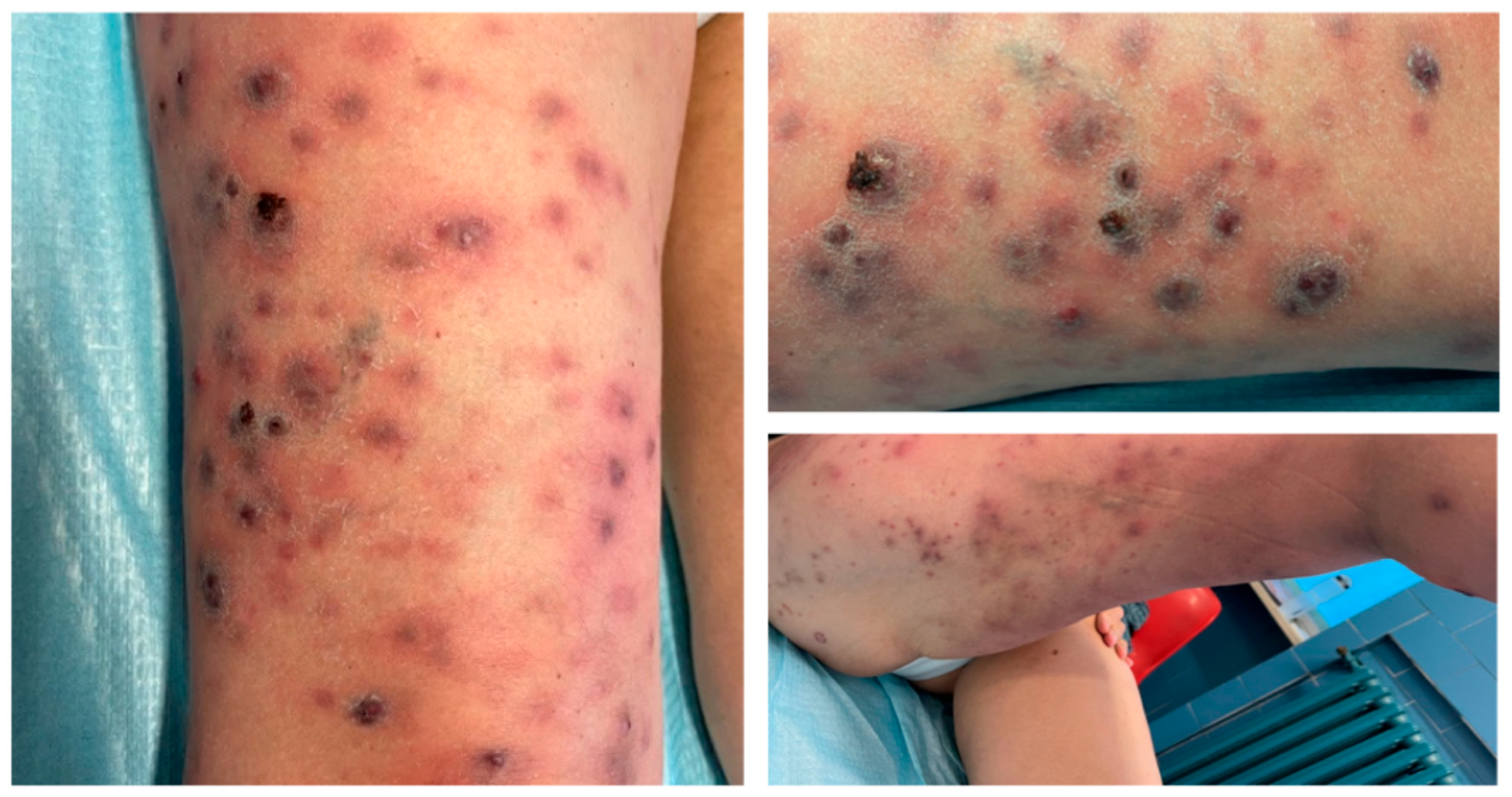
A 61-year-old female patient was referred to our department for the presence of erythematous-papulo-nodular lesions on her right leg, as reported in Figure 2.
She had a history of orthotopic liver transplant (OLT) because of autoimmune hepatitis. She underwent a first OLT in 2005 and subsequently a second OLT in 2010 for a graft failure. A third transplant was completed in 2019 for a new graft failure, and the patient was maintained on an immunosuppressive regimen with tacrolimus, mycophenolate mofetil, and oral steroids (i.e., prednisone 10 mg q24h). At the presentation, the patient complained about loss of weight (i.e., about 10 Kg in the last 12 months) and low-grade fever. The lesions were previously diagnosed as cutaneous herpes zoster, and the patient received a seven-day course of oral valacyclovir treatment (i.e., 1 g every 8 h) without improvement. Over the following weeks, lesions gradually spread to form dark violet papulo-nodular elements, some covered by sero-hemorrhagic crusts. Diffuse soft tissue swelling and redness, referred to as lymphedema, were associated with the lesions, as reported in Figure 3. A whole-body CT scan was performed, and no other organ involvement was ruled out.
Patient denied history of a recent trip abroad, animal bites, or tick exposure. Moreover, the patient reported that she lives in the countryside, owns two cats, and loves gardening. She reported a probable skin trauma on her right leg while gardening, some weeks before hospital admission. An empiric antibiotic therapy with doxycycline was started, according to the hypothesis of bacillary angiomatosis, with minimal improvement. Bartonella henselae and Human Herpesvirus 8 serologies and the PCR test were negative. QuantiFERON for Mycobacterium tuberculosis, serum galactomannan, and Cryptococcus antigen detection were not diagnostic. The blood count analysis found neutrophilic leukocytosis (13 × 109/L) and elevation of the inflammatory index (C-reactive protein 82.8 mg/L, normal range < 5 mg/L). Significant elevation of Beta-D-glucan values was registered in two non-consecutive samples (323 and >600 pg/mL, normal range < 7 pg/mL). Bacterial cultures were negative. Direct microscopic examination from the swab of the ulcerated nodule showed hyaline, septate hyphae and branched conidiophores with stripes. Sabouraud dextrose agar culture was positive for P. lilacinum. In parallel, Periodic acid–Schiff (PAS) and Grocott’s staining of skin biopsy identified broad-based hyphae and spores, including rare coccoid elements with a thick PAS-positive capsule, supporting the diagnosis of fungal infection (Figure 4).
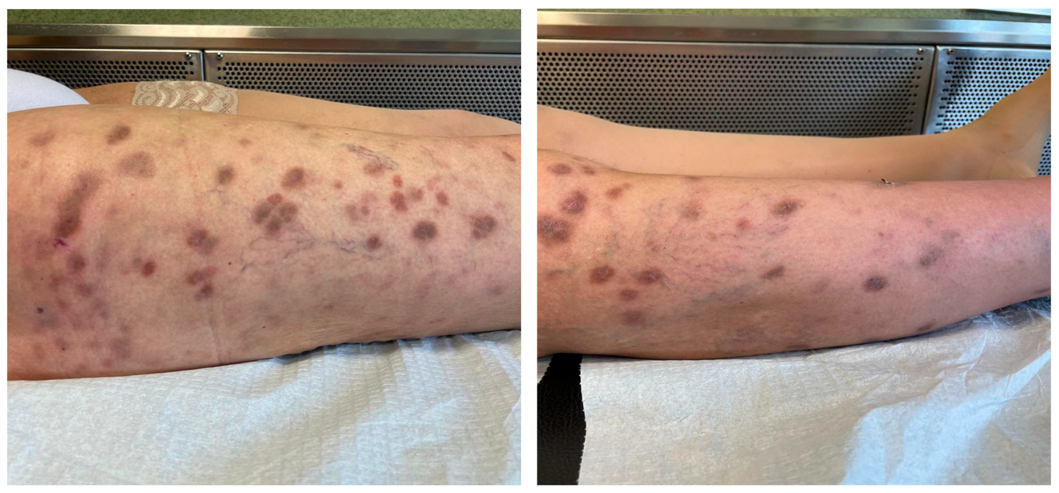
Antifungal susceptibility tests were not performed. The patient was hospitalised and treatment with terbinafine 250 mg/day plus voriconazole 400 mg/day was administered, with serial azole therapeutic drug monitoring (TDM) due to advanced cirrhosis. CT chest, nasal endoscopy, and blood cultures excluded secondary localisations. Therapy was continued based on clinical response and the reduction in Beta-D-glucan serum levels. Therapy with Voriconazole and terbinafine was withdrawn after nine months, following complete clinical response and healing of the lesions (Figure 5).
Therapy with voriconazole and terbinafine was administered for a total of 9 months, with progressive resolution of the skin lesions.

4. Results

The search strategy identified 185 articles, of which 21 studies were included for the final analysis, with a total of 21 patients, and the data are shown in Table 1.
Most of the patients were male (16–76.2%), with a median age of 59 years (range 28–84). Regarding risk factors for fungal infections, frequently reported conditions were steroid use (9–40.9%) and solid organ transplantation (9–40.9%), followed by haematological disorders (3–13.6%). One case involved a patient living with HIV. The majority of patients had no history of exposure or hobbies previously associated with Purpureocillium lilacinum infections in the literature.
More frequently, patients presented with nodular lesions (10–45.4%) or papules/pustules (9–40.9%). Three cases presented with erythematous purpuric plaques (13.6%), and one case reported another type of skin manifestation (0.5%).
Painful lesions were reported in around half of the patients (10–45.5%), in nine cases the skin manifestations were painless (40.9%), and in three cases it was not reported.
Regarding therapy, all patients received azoles at a certain point of the treatment (22–100%). Other molecules prescribed were liposomal amphotericin B (4–18.1%) and echinocandins (3–13.6%).
Terbinafine was prescribed in three cases (13.6%), always in combination with azoles.
In two cases, a surgical debridement of the skin lesion was performed (9%).
We found that in 11 cases, patients suffered from local dissemination of infection (50%), and in 5 cases (22.7%), they suffered from systemic involvement. Three patients died before completing treatment (13.6%). Data described in the studies reviewed are summarised in Table 2.

5. Discussion

In this study we have reported a case of P. lilacinum invasive infection in an OLT recipient, including a revision of the literature of systemic infections due to P. lilacinum in the immunocompromised population, focusing on cutaneous fungal involvement and presentations. Moreover, we have summarised new evidence regarding antifungal agents in the pipeline of treatment against P. lilacinum. Our case reported the history of a middle-aged female. Median age at diagnosis reported in the literature (ranging from 28 to 84 years old) was in line with that reported in our experience (61 years old), although that literature review showed a prevalence of patients of male sex [12,13,14,15,16,17,18,19,20,21,22,23,24,25,26,27,28,29,30,31,32]. Differences in sex have a determining importance on the prevalence and incidence of many infectious diseases. In an interesting recent review by Egger et al. on invasive fungal infections, notably, aspergillosis, mucormycosis, cryptococcosis, coccidiomycosis, histoplasmosis, and blastomycosis were overrepresented in males, concluding that biological sex should be further investigated as a predisposing factor for fungal infections [33]. In our case, the patient reported a probable skin trauma that had occurred when gardening some weeks before clinical presentation. P. lilacinum is a filamentous fungus widely distributed in the environment, including in soil, water, decaying vegetation, insects, and nematodes [1], and can cause human infections by inhalation or direct tissue inoculation [4]. Heutte et al. reported less frequent hospital-acquired infections due to contaminated medical devices, tattoo-related infections, or skin lotion contamination [3], but gardening and farming have been reported [12,13,14,15,16,17,18,19,20,21,22,23,24,25,26,27,28,29,30,31,32] as possible risk factors, such as for the patient in our case report.
Similarly to other fungal infections, immunocompromised patients are at higher risk of P. lilacinum infections. Cutaneous and subcutaneous infections represent the most commonly reported clinical manifestations of P. lilacinum in the literature [4], as in our case. However, particularly in immunocompromised patients, P. lilacinum can occasionally disseminate haematogenously, leading to severe invasive infections such as endocarditis, pneumonia, and central nervous system involvement [7]. We found that infections were mostly localised on the extremities and lesions were usually unilateral, reinforcing the hypothesis that skin inoculation could be an important means of infection, such as in the case we reported [12,13,14,15,16,17,18,19,20,21,22,23,24,25,26,27,28,29,30,31,32]. In the vast majority of cases, the skin findings were characterised by erythematous lesions, nodules, or papules, consistent with what was observed in the case we described [12,13,14,15,16,17,18,19,20,21,22,23,24,25,26,27,28,29,30,31,32]. Among immunocompromised patients, solid organ transplantation and chronic steroids therapy were the most-reported causes of immunosuppression (83%). In this group, deeper and painful skin lesions were observed, such as nodules, or even cellulitis and necrotic ulcerations because of angioinvasion. A bioptic approach was almost always the key to reaching a diagnosis. Definitive diagnosis was mainly established via fungal culture and histology. P. lilacinum grows rapidly on Sabouraud agar, ranging in colour from violet to bluish brown. Histological examination usually shows hyphae, phialides, and conidia, which may sporulate in infected tissues [9].
Moreover, molecular identification nowadays plays an important role in species identification. P. lilacinum’s reclassification in a genus apart from Paecilomyces, on the basis of phylogenic studies [1], may explain significant differences in resistance to most antifungals, including amphotericin B, fluconazole, itraconazole, flucytosine, and the echinocandins [9,10].
There has been no standard of care established up to now, besides that voriconazole or posaconazole should be a reasonable first-line treatment. Terbinafine was reported as a sparring treatment in combination with azoles in the literature reviewed [12,13,14,15,16,17,18,19,20,21,22,23,24,25,26,27,28,29,30,31,32]. Moreover, echinocandins reported a variable susceptibility, and most strains were resistant to itraconazole and amphotericin B [9,10].
Although 37% of patients have been treated with combination therapy in the literature, there is insufficient data to determine whether this is superior to monotherapy. The optimal duration of treatment remains undefined, and surgical debridement should be considered when feasible. Infections in immunocompromised patients are particularly challenging, and current evidence is insufficient to clearly identify the lesion characteristics that indicate resolution of the infectious process. Therefore, based on the available literature, it appears reasonable to continue treatment until all lesions have completely resolved. Resolution or improvement of immunosuppression and surgical source control improve the efficacy of medical treatment [12,13,14,15,16,17,18,19,20,21,22,23,24,25,26,27,28,29,30,31,32]. In our case, 1–3 Beta-D-glucan serology was used to monitor clinical response, but we found no evidence to support this practice in the current literature [12,13,14,15,16,17,18,19,20,21,22,23,24,25,26,27,28,29,30,31,32]. Mortality is around 13%, and with the limitations of a low number of studies, there does not seem to be a correlation with particular predisposing factors.
Recently, new antifungals have been studied. Ibrexafungerp is the first representative of a novel class of structurally distinct glucan synthase inhibitors: triterpenoids. Although it has shown excellent activity in vitro against Candida spp., Aspergillus spp., and some rare moulds, the activity against P. lilacinum is negligible [34]. Fosmagenopix is a new oral antifungal whose mechanism of action is targeting the GWT1 enzyme required for the localization of glycosylphosphatidylinositol-anchored mannoproteins in fungi. It has shown great activity in vitro against P. lilacinum, with MICs lower than those of posaconazole and voriconazole [35]. Olorofim selectively inhibits fungal dihydroorotate dehydrogenase (DHODH), a key enzyme in the pyrimidine biosynthesis pathway. For Olorofim, despite few data available, no in vitro efficacy has been observed in P. lilacinum clinical isolates, whereas MICs for P. variotii were low [34]. With regard to Rezafungine, a next-generation, broad-spectrum, and long-lasting echinocandin, there are no data in the literature concerning efficacy on P. lilacinum at this moment.
In conclusion, the review emphasises the critical need for early diagnosis and appropriate antifungal treatment, particularly in high-risk patients, accompanied by surgical debridement when feasible. Data on treatment duration and the superiority of combination therapy with respect to monotherapy are lacking, despite the fact that new, recently approved antifungals present efficacy and safety profiles that are promising for the treatment of this rare infection.

Author Contributions

Conceptualization, T.L., C.S. and P.F.; methodology, M.C. and G.A.; formal analysis, C.S., M.C., G.A. and T.L.; investigation and data curation, A.O., F.C., G.R., N.M., F.G. and R.S.; writing—original draft preparation, C.S., M.C., G.A. and T.L.; writing—review and editing, P.F., C.S., M.C., G.A. and T.L.; supervision, F.G.D.R., P.Q., S.R. and S.C. All authors have read and agreed to the published version of the manuscript.

Funding

This research received no external funding.

Institutional Review Board Statement

Ethical review and approval were waived for this study due to the narrative structure of the article, and informed consent for the patient was obtained.

Informed Consent Statement

Informed consent has been obtained from the patient.

Data Availability Statement

Data are available upon reasonable request.

Conflicts of Interest

The authors declare no conflicts of interest.

References

  1. Luangsa-Ard, J.; Houbraken, J.; van Doorn, T.; Hong, S.B.; Borman, A.M.; Hywel-Jones, N.L.; Samson, R.A. Purpureocillium, a New Genus for the Medically Important Paecilomyces lilacinus. FEMS Microbiol. Lett. 2011, 321, 141–149. [Google Scholar] [CrossRef]
  2. Nnadi, N.E.; Carter, D.A. Climate Change and the Emergence of Fungal Pathogens. PLoS Pathog. 2021, 17, e1009503. [Google Scholar] [CrossRef]
  3. Heutte, N.; André, V.; Dubos Arvis, C.; Bouchart, V.; Lemarié, F.; Legendre, P.; Votier, E.; Louis, M.Y.; Madelaine, S.; Séguin, V.; et al. Assessment of multi-contaminant exposure in a cancer treatment center: A 2-year monitoring of molds, mycotoxins, endotoxins, and glucans in bioaerosols. Environ. Monit. Assess. 2017, 189, 31. [Google Scholar] [CrossRef] [PubMed]
  4. Sprute, R.; Salmanton-García, J.; Sal, E.; Malaj, X.; Ráčil, Z.; Ruiz De Alegría Puig, C.; Falces-Romero, I.; Barać, A.; Desoubeaux, G.; Kindo, A.J.; et al. Invasive Infections with Purpureocillium lilacinum: Clinical Characteristics and Outcome of 101 Cases from FungiScope® and the Literature. J. Antimicrob. Chemother. 2021, 76, 1593–1603. [Google Scholar] [CrossRef]
  5. Saghrouni, F.; Saidi, W.; Ben Said, Z.; Gheith, S.; Ben Said, M.; Ranque, S.; Denguezli, M. Cutaneous Hyalohyphomycosis Caused by Purpureocillium lilacinum in an Immunocompetent Patient: Case Report and Review. Med. Mycol. 2013, 51, 664–668. [Google Scholar] [CrossRef] [PubMed]
  6. Abolghasemi, S.; Tabarsi, P.; Adimi, P.; Kiani, A.; Dolatshahi, S.; Mansouri, D. Pulmonary Paecilomyces in a diabetic patient. Tanaffos 2015, 14, 268–271. [Google Scholar] [PubMed] [PubMed Central]
  7. Khalique, Z.; Hatipoğlu, S.; Rosendahl, U.; Mohiaddin, R. Unusual Complicated Fungal Endocarditis in a Patient with Vascular Ehlers-Danlos Syndrome. Ann. Thorac. Surg. 2019, 107, e269–e271. [Google Scholar] [CrossRef]
  8. Chew, R.; Dorman, A.; Woods, M.L. Purpureocillium lilacinum Keratitis: A Case Series and Review of the Literature. Can. J. Ophthalmol. 2016, 51, 382–385. [Google Scholar] [CrossRef]
  9. Castelli, M.V.; Alastruey-Izquierdo, A.; Cuesta, I.; Monzon, A.; Mellado, E.; Rodriguez-Tudela, J.L.; Cuenca-Estrella, M. Susceptibility Testing and Molecular Classification of Paecilomyces spp. Antimicrob. Agents Chemother. 2008, 52, 2926–2928. [Google Scholar] [CrossRef]
  10. Drogari-Apiranthitou, M.; Mantopoulou, F.D.; Skiada, A.; Kanioura, L.; Grammatikou, M.; Vrioni, G.; Mitroussia-Ziouva, A.; Tsakris, A.; Petrikkos, G. In Vitro Antifungal Susceptibility of Filamentous Fungi Causing Rare Infections: Synergy Testing of Amphotericin B, Posaconazole and Anidulafungin in Pairs. J. Antimicrob. Chemother. 2012, 67, 1937–1940. [Google Scholar] [CrossRef]
  11. Baethge, C.; Goldbeck-Wood, S.; Mertens, S. SANRA—A Scale for the Quality Assessment of Narrative Review Articles. Res. Integr. Peer Rev. 2019, 4, 5. [Google Scholar] [CrossRef]
  12. Corbeddu, M.; Ferreli, C.; Cappai, R.; Ferraguti, P.; Atzori, L.; Pilloni, L.; Rongioletti, F. Fatal Hyalohyphomycosis with Cutaneous Involvement Caused by Purpureocillium Lilacinum in an Immunocompromised Patient with Bullous Pemphigoid. Acta Biomed. 2021, 92, e2021139. [Google Scholar] [CrossRef]
  13. Desoubeaux, G.; García, D.; Bailly, E.; Augereau, O.; Bacle, G.; De Muret, A.; Bernard, L.; Cano-Lira, J.F.; Garcia-Hermoso, D.; Chandenier, J. Subcutaneous Phaeohyphomycosis Due to Phialemoniopsis Ocularis Successfully Treated by Voriconazole. Med. Mycol. Case Rep. 2014, 5, 4–8. [Google Scholar] [CrossRef] [PubMed]
  14. Hall, V.C.; Goyal, S.; Davis, M.D.P.; Walsh, J.S. Cutaneous Hyalohyphomycosis Caused by Paecilomyces lilacinus: Report of Three Cases and Review of the Literature. Int. J. Dermatol. 2004, 43, 648–653. [Google Scholar] [CrossRef]
  15. Van Schooneveld, T.; Freifeld, A.; Lesiak, B.; Kalil, A.; Sutton, D.A.; Iwen, P.C. Paecilomyces lilacinus Infection in a Liver Transplant Patient: Case Report and Review of the Literature. Transpl. Infect. Dis. 2008, 10, 117–122. [Google Scholar] [CrossRef] [PubMed]
  16. Demitsu, T.; Nagashima, K.; Okabe, T.; Morisawa, Y.; Ishikawa, N.; Yagisawa, T.; Fukuta, H.; Ohtsuki, M.; Kano, R.; Harada, K. Subcutaneous Hyalohyphomycosis Due to Purpureocillium lilacinum in an Immunocompromised Patient after Renal Transplantation. J. Dermatol. 2017, 44, 725–726. [Google Scholar] [CrossRef]
  17. Nagamoto, E.; Fujisawa, A.; Yoshino, Y.; Yoshitsugu, K.; Odo, M.; Watanabe, H.; Igata, T.; Noguchi, H. Case of Paecilomyces lilacinus Infection Occurring in Necrotizing Fasciitis-Associated Skin Ulcers on the Face and Surrounding a Tracheotomy Stoma. Med. Mycol. J. 2014, 55, E21–E27. [Google Scholar] [CrossRef]
  18. Kitami, Y.; Kagawa, S.; Iijima, M. [A Case of Cutaneous Paecilomyces lilacinus Infection on the Face]. Nihon Ishinkin Gakkai Zasshi 2005, 46, 267–272. [Google Scholar] [CrossRef][Green Version]
  19. Keshtkar-Jahromi, M.; McTighe, A.H.; Segalman, K.A.; Fothergill, A.W.; Campbell, W.N. Unusual Case of Cutaneous and Synovial Paecilomyces lilacinus Infection of Hand Successfully Treated with Voriconazole and Review of Published Literature. Mycopathologia 2012, 174, 255–258. [Google Scholar] [CrossRef]
  20. Albert, R.; Lemaignen, A.; Desoubeaux, G.; Bailly, E.; Bernard, L.; Lacasse, M. Chronic Subcutaneous Infection of Purpureocillium lilacinum in an Immunocompromised Patient: Case Report and Review of the Literature. Med. Mycol. Case Rep. 2022, 38, 5–8. [Google Scholar] [CrossRef] [PubMed]
  21. Hilmarsdottir, I.; Thorsteinsson, S.B.; Asmundsson, P.; Bodvarsson, M.; Arnadottir, M. Cutaneous Infection Caused by Paecilomyces lilacinus in a Renal Transplant Patient: Treatment with Voriconazole. Scand. J. Infect. Dis. 2000, 32, 331–332. [Google Scholar] [CrossRef] [PubMed]
  22. Accetta, J.; Powell, E.; Boh, E.; Bull, L.; Kadi, A.; Luk, A. Isavuconazonium for the Treatment of Purpureocillium lilacinum Infection in a Patient with Pyoderma Gangrenosum. Med. Mycol. Case Rep. 2020, 29, 18–21. [Google Scholar] [CrossRef]
  23. Trinh, S.A.; Angarone, M.P. Purpureocillium lilacinum Tattoo-Related Skin Infection in a Kidney Transplant Recipient. Transpl. Infect. Dis. 2017, 19, e12689. [Google Scholar] [CrossRef]
  24. Lavergne, R.A.; Cassaing, S.; Nocera, T.; Pauwels, C.; Cointault, O.; Basse, G.; Lavayssière, L.; Berry, A.; Kamar, N.; Lamant, L.; et al. Simultaneous Cutaneous Infection Due to Paecilomyces lilacinus and Alternaria in a Heart Transplant Patient. Transpl. Infect. Dis. 2012, 14, E156–E160. [Google Scholar] [CrossRef]
  25. Ezzedine, K.; Belin, E.; Guillet, S.; D’Almeida, M.; Droitcourt, C.; Accocebery, I.; Milpied, B.; Jouary, T.; Malvy, D.; Taieb, A. Cutaneous Hyphomycosis Due to Paecilomyces lilacinus. Acta Derm. Venereol. 2012, 92, 156–157. [Google Scholar] [CrossRef]
  26. Ounissi, M.; Abderrahim, E.; Trabelsi, S.; Khaled, S.; Bezzine, H.; Ben Hamida, F.; Hedri, H.; Ben Abdallah, T.; Ben Maïz, H.; Kheder, A. Hyalohyphomycosis Caused by Paecilomyces lilacinus after Kidney Transplantation. Transplant. Proc. 2009, 41, 2917–2919. [Google Scholar] [CrossRef]
  27. Martin, C.A.; Roberts, S.; Greenberg, R.N. Voriconazole Treatment of Disseminated Paecilomyces Infection in a Patient with Acquired Immunodeficiency Syndrome. Clin. Infect. Dis. 2002, 35, 78–81. [Google Scholar] [CrossRef]
  28. Sotello, D.; Cappel, M.; Huff, T.; Meza, D.; Alvarez, S.; Libertin, C.R. Cutaneous Fungal Infection in an Immunocompromised Host. JMM Case Rep. 2017, 4, e005101. [Google Scholar] [CrossRef]
  29. Xia, X.J.; Liu, Z.H. Cutaneous Hyalophomycosis Caused by Purpureocillium lilacinum in an Autoimmune Hemolytic Anemia Patient with Previous Pulmonary Tuberculosis and Cutaneous Cryptococcosis Infection. Mycopathologia 2021, 186, 563–564. [Google Scholar] [CrossRef] [PubMed]
  30. Lemaigre, C.; Suarez, F.; Martellosio, J.P.; Barbarin, C.; Brunet, K.; Chomel, J.C.; Hainaut, E.; Rammaert, B.; Roblot, F.; Torregrosa-Diaz, J.M. Late Onset of Chronic Granulomatous Disease Revealed by Paecilomyces lilacinus Cutaneous Infection. J. Clin. Immunol. 2022, 42, 60–63. [Google Scholar] [CrossRef] [PubMed]
  31. Wang, Y.; Bao, F.; Lu, X.; Liu, H.; Zhang, F. Case Report: Cutaneous Mycosis Caused by Purpureocillium lilacinum. Am. J. Trop. Med. Hyg. 2023, 108, 693–695. [Google Scholar] [CrossRef] [PubMed]
  32. Chen, W.Y.; Lin, S.R.; Hung, S.J. Successful Treatment of Recurrent Cutaneous Purpureocillium lilacinum (Paecilomyces lilacinus) Infection with Posaconazole and Surgical Debridement. Acta Derm. Venereol. 2019, 99, 1313–1314. [Google Scholar] [CrossRef] [PubMed]
  33. Egger, M.; Hoenigl, M.; Thompson, G.R., 3rd; Carvalho, A.; Jenks, J.D. Let’s talk about sex characteristics—As a risk factor for invasive fungal diseases. Mycoses 2022, 65, 599–612. [Google Scholar] [CrossRef] [PubMed]
  34. Lamoth, F.; Alexander, B.D. Antifungal activities of SCY-078 (MK-3118) and standard antifungal agents against clinical non-Aspergillus mold isolates. Antimicrob. Agents Chemother. 2015, 59, 4308–4311. [Google Scholar] [CrossRef]
  35. Miyazaki, M.; Horii, T.; Hata, K.; Watanabe, N.A.; Nakamoto, K.; Tanaka, K.; Shirotori, S.; Murai, N.; Inoue, S.; Matsukura, M.; et al. In vitro activity of E1210, a novel antifungal, against clinically important yeasts and molds. Antimicrob. Agents Chemother. 2011, 55, 4652–4658. [Google Scholar] [CrossRef] [PubMed] [PubMed Central]
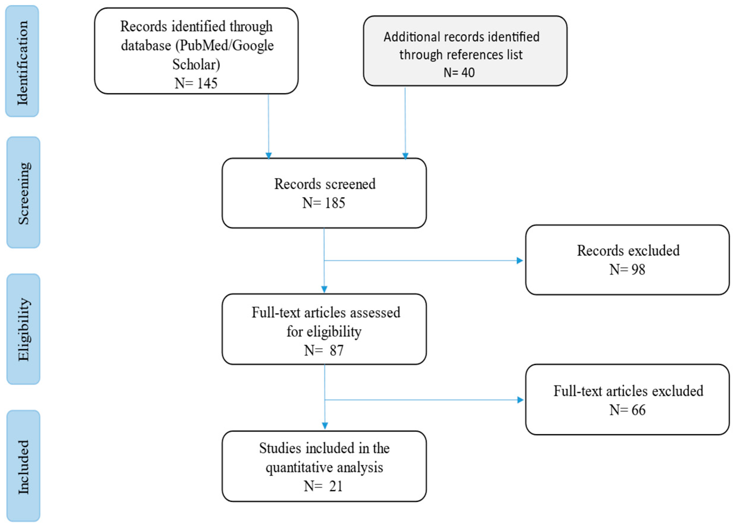
Figure 1. Flow chart of the studies revised in the narrative review.
Life 15 01453 g001
Figure 2. Multiple, monolateral, erythematous-papulo-nodular lesions on the right leg.
Life 15 01453 g002
Figure 3. Skin lesions were associated with diffuse soft tissue swelling and redness, referred to as lymphedema.
Life 15 01453 g003
Figure 4. Histopathological features: (A,B) Granulomatous dermatitis at incisional biopsy (haematoxylin–eosin stain, magnification 10×—(A) and 400×—(B)). (C) Fungal globose yeast-like structures and septate hyphae in the dermis (Grocott stain, magnification ×400).
Life 15 01453 g004
Figure 5. Progressive resolution of skin lesions under treatment after 3 months of therapy.
Life 15 01453 g005
Table 1. Literature regarding cutaneous infection due to P. lilacinum in immunocompromised hosts.
Age
Sex
ComorbiditiesHobbySkin LesionsPainSiteDiffusionTherapyOutcomeREF
179,
M
Diabetes mellitus; Pemphigus vulgarisGardeningErythematous pustule, warm, singlenoHand, monolateralBilateral leg
and ankle cellulitis, encephalic
Itraconazole
200 mg bid
Voriconazole
6 mg/kg every 12 h
Dead[12]
284,
M
Steroid therapy;
Diabetes mellitus
NASwelling and bursitis of the first metatarsophalangeal jointyesLower extremity, monolateralNoSurgery plus
voriconazole 200 mg bid
Recovered[13]
365,
M
Steroid therapy for temporal arteritisNAErythematous and oedematous area with papules and pustulesnoLower extremity, monolateralCellulitisItraconazole
200 bid plus surgery
Recovered[14]
456,
M
Liver transplantNAErythematous, tender single nodulenoKnee, monolateralMultiple nodules following lymphatic distributionVoriconazole 200 mg bid alone and in association with Terbinafine
Voriconazole 300 mg bid
Recovered[15]
572,
M
Renal transplantNAErythematous nodulenoForearms, monolateralMultiple reddish nodulesItraconazole
200 mg daily
Voriconazole
400 mg/day and Terbinafine 250 mg/day
Recovered[16]
628,
M
Bone marrow transplant;
Previous Pseudomonas aeruginosa necrotising fasciitis on the face
NoneErythematous papules and pustules, crusted at the margin of previous ulcersnoFace and site of closed tracheostomyMultiple necrotising ulcersItraconazole
200 bid
Micafungin
50 mg/day
LAMPHB
150 mg/day
Voriconazole
400 mg/day
Recovered[17]
774,
F
Immunosuppressed therapy for autoimmune haemolytic anaemiaNAErythematous brownish plaque, singleyesHemifaceMultiple
papules and abscesses around the plaque on the hemiface
Griseofulvin
500 mg/daily
Itraconazole
200 mg/day for 11 weeks, then
400 mg daily, 7 days a month
Recovered[18]
871,
F
Steroid therapy for giant cell arteritisNAErythematous nodulenoElbow, monolateralNAMicafungin
50 mg/day
Itraconazole
(dosage not available)
NA[19]
952,
M
Renal transplantGardeningOedematous and erythematous areanoLower extremity, monolateralCellulitis with multiple fistula orificesPosaconazole
300 mg/day
Surgery for local relapse
Recovered[20]
1059,
M
Renal transplantNAErythematous papules, pustules and small ulcersyesFoot and interdigital space, monolateralMultiple lesions on foot and leg, sepsis due to Candida spp. and Gram-negative bacteriaItraconazole
400–600 mg/day
Voriconazole
400 mg/day
LAMPHB
Dead[21]
1150,
M
CLL; previous pyoderma gangrenosumNAPurpuric nodules and a necrotic ulceryesLower extremity, monolateralExtended necrotic ulcerLAMPHB
6 mg/kg/day
Micafungin
150 mg/day
Voriconazole
400 mg bid
Isavuconazole IV
372 mg/day plus surgery
Recovered[22]
1233,
M
Renal transplant
Recent tattooErythematous papules, pustules clustered over tattoonoLeft forearm and shin, right ankleNoneVoriconazole
400 mg/day
Recovered[23]
1363,
M
Heart transplantation; Diabetes mellitusNAErythematous nodulesyesLeg and elbowMultiple necrotic ulcersVoriconazole
400 mg/day
Intralesional Voriconazole (1 mL)
followed by oral treatment with Terbinafine
250 mg a day the first month, then 125 mg/day
Recovered[24]
1460,
M
Pulse steroid therapy for rheumatoid arthritisNAErythematous nodules with pseudo-verrucous aspectNAHemifaceMultiple necrotic ulcers on face and handVoriconazole
400 mg/day
Posaconazole
400 mg/bid
NA[25]
1548,
F
Renal transplantNAErythematous oedematous areayesLower extremity, monolateralExtensive cellulitis with multiple ulcersItraconazole
400 mg/day
Voriconazole
(dosage non reported)
Recovered[26]
1640,
M
AIDSNAErythematous nodulesyesLower extremity, monolateralMultiple
ulcerated nodules
on entire leg and body
Itraconazole
200 mg bid
LAMPHB
5 mg/kg/day
Voriconazole
200–300 mg/bid
Dead[27]
1769,
M
Liver transplant recipientNone, lives with a dogErythematous nodules with pseudo-verrucous aspectyesHand, monolateralMultiple nodules tracking on arm and
axilla
Voriconazole
400 mg/day
Recovered[28]
1853,
M
Steroid therapy for autoimmune haemolytic
anaemia
NoneErythematous pustule and ulcerated nodulenoNeckNoneItraconazole
400 mg/day
Posaconazole
300 mg/die
Recovered[29]
1967,
F
X-linked chronic granulomatous diseaseSheep farmerErythematous nodules with pustulesyesUpper extremity (thumb)NoneVoriconazole
400 mg/day
Recovered[30]
2051,
F
Steroid therapy for nephrotic syndromeNARecurrent papules, pustules, and ulcerationNAFinger and forearm,
monolateral
NoneVoriconazole
(dosage not available)
Recovered[31]
21NA,
M
Steroid therapy for Evans’ SyndromeFarmerErythematous papules and pustulesyesForearm,
monolateral
Multiple ulcerated lesionsVoriconazole
400 mg/day then
Posaconazole
300 mg/day with surgery
Recovered[32]
NA: not reported; LAMPHB: liposomial amphotericin B.
Table 2. Characteristics of immunocompromised patients with cutaneous infections caused by P. lilacinum.
n (%)
Patient age
Years, mean (min-max)59 (28–84) years
Sex
Male16 (76.2)
Female6 (27.2)
Race/ethnicity
Caucasian9 (40.9)
Black/African American6 (27.2)
Asiatic7 (31.8)
Immunosuppression
Steroids9/22 (40.9)
Solid transplant9/22 (40.9)
Haematological condition3/22 (13.6)
HIV/AIDS1/22 (0.5)
Hobbies
Unknown/Other15/22 (68)
Farmer3/22 (13.6)
Gardening2/22 (9)
Skin lesions
Papules/pustules9/22 (40.9)
Nodules10/22 (45,4)
Erythematous purpuric plaques3/22, 13.6)
Other1/22 (0.5)
Localization
Upper extremities7/22 (31.8)
Lower extremities10/22 (45.4)
Pain associated
Yes10/22 (45,4)
No9/22 (40.9)
Local diffusion11/22 (50)
Systemic diffusion/secondary foci5/22 (22.7)
Treatment
Azoles22/22 (100)
Terbinafine3/22 (13.6)
Echinocandins3/22 (13.6)
Liposomal amphotericin4/22 (18.1)
Griseofulvin1/22 (0.5)
Surgical debridement2/22 (9)
Mortality3/22 (13.6)
Disclaimer/Publisher’s Note: The statements, opinions and data contained in all publications are solely those of the individual author(s) and contributor(s) and not of MDPI and/or the editor(s). MDPI and/or the editor(s) disclaim responsibility for any injury to people or property resulting from any ideas, methods, instructions or products referred to in the content.