Systematic Review

Viruses in Simuliidae: An Updated Systematic Review of Arboviral Diversity and Vector Potential

by
Alejandra Rivera-Martínez
1,
S. Viridiana Laredo-Tiscareño
1,
Jaime R. Adame-Gallegos
2,
Erick de Jesús de Luna-Santillana
3,*,
Carlos A. Rodríguez-Alarcón
1,
Julián E. García-Rejón
4,
Mauricio Casas-Martínez
5 and
Javier A. Garza-Hernández
1,*
1
Instituto de Ciencias Biomédicas, Universidad Autónoma de Ciudad Juárez, Juárez 32310, Chihuahua, Mexico
2
Facultad de Ciencias Químicas, Universidad Autónoma de Chihuahua, Chihuahua 31125, Chihuahua, Mexico
3
Laboratorio Medicina de la Conservación, Centro de Biotecnología Genómica del Instituto Politécnico Nacional, Reynosa 88710, Tamaulipas, Mexico
4
Laboratorio de Arbovirología, Centro de Investigaciones Regionales “Dr. Hideyo Noguchi”, Universidad de Yucatán, Mérida 97225, Yucatán, Mexico
5
Centro Regional de Investigación en Salud Pública, Instituto Nacional de Salud Pública, Tapachula 30700, Chiapas, Mexico
*
Authors to whom correspondence should be addressed.
Life 2025, 15(5), 807; https://doi.org/10.3390/life15050807
Submission received: 7 March 2025 / Revised: 9 May 2025 / Accepted: 13 May 2025 / Published: 19 May 2025
(This article belongs to the Section Epidemiology)

Abstract

:
Black flies (Diptera: Simuliidae) are important vectors of pathogens, including filarial nematodes, protozoans, and arboviruses, which significantly impact human and animal health. Although their role in arbovirus transmission has not been as thoroughly studied as that of mosquitoes and ticks, advances in molecular tools, particularly metagenomics, have enabled the identification of non-cultivable viruses, significantly enhancing our understanding of black-fly-borne viral diversity and their public and veterinary health implications. However, these methods can also detect insect-specific viruses (i.e., viruses that are unable to replicate in vertebrate hosts), which may lead to the incorrect classification of black flies as potential vectors. This underscores the need for further research into their ecological and epidemiological roles. This systematic review, conducted following the PRISMA protocol, compiled and analyzed evidence on arbovirus detection in Simuliidae from scientific databases. Several arboviruses were identified in these insects, including vesicular stomatitis virus New Jersey serotype (VSVNJ), Venezuelan equine encephalitis virus (VEEV), and Rift Valley fever virus. Additionally, in vitro studies evaluating the vector competence of Simuliidae for arboviruses such as dengue virus, Murray Valley encephalitis virus, and Sindbis virus were reviewed. These findings provide critical insights into the potential role of black flies in arbovirus transmission cycles, emphasizing their importance as vectors in both public and veterinary health contexts.

1. Introduction

Black flies (Diptera: Simuliidae) are significant vectors of public health importance due to their ability to transmit pathogens affecting both humans and animals, such as onchocerciasis and mansonellosis; however, their potential as vectors of medically important viruses remains largely understudied [1]. Improving our knowledge of their capacity to transmit arboviruses could have profound implications for public health, particularly in regions where these vectors are abundant and where associated diseases may be underreported or misdiagnosed [2]. This is attributable to several factors, including the technical challenges associated with collecting these insects from their often restricted and remote habitats, the difficulty of establishing and maintaining stable colonies under laboratory conditions, and the limited feasibility of conducting vector competence studies [3,4] (this issue is discussed in more detail in a subsequent section).
Traditionally, the detection of arboviruses in vectors has relied on virus isolation through cell culture and nucleic acid detection using molecular biology techniques [5]. While these methods are valuable, they come with limitations, particularly in identifying non-cultivable or low-prevalence viruses, which leads to an underestimation of viral diversity in blood-feeding vectors [6]. In recent years, the advent of next-generation sequencing (NGS) and metagenomics has provided a powerful alternative for more precise and comprehensive virus detection without the need for in vitro cultures [7].
Metagenomics has revolutionized pathogen research by enabling the identification of previously undetectable microorganisms, facilitating rapid genome sequencing, and uncovering pathogens of both human and veterinary importance. This technology has improved scientific expansion in fields such as taxonomy, microbial ecology, evolutionary biology, and, most notably, epidemiological surveillance and the discovery of novel zoonotic pathogens [8,9]. By allowing for the high-throughput, unbiased screening of biological samples, Metagenomics can reveal the full scope of viral diversity present within vector populations, providing insights into viral ecology and transmission dynamics [10]. Despite the rapid progress that has been made in applying metagenomic tools to study viral diversity in other hematophagous insects, such as mosquitoes and biting midges, the research focused on Simuliidae remains limited. This gap in knowledge underscores the need for further investigation, as black flies may harbor viruses of epidemiological relevance that have gone undetected due to methodological constraints in previous studies.
Therefore, the present systematic review aims to compile and critically analyze the available evidence on arbovirus detection in Simuliidae using molecular biology and metagenomic approaches. By providing a comprehensive overview of the current state of knowledge, this study seeks to enhance the understanding of viral diversity in these hematophagous dipterans and their potential role in the transmission of arboviruses relevant to humans, animals, and wildlife.

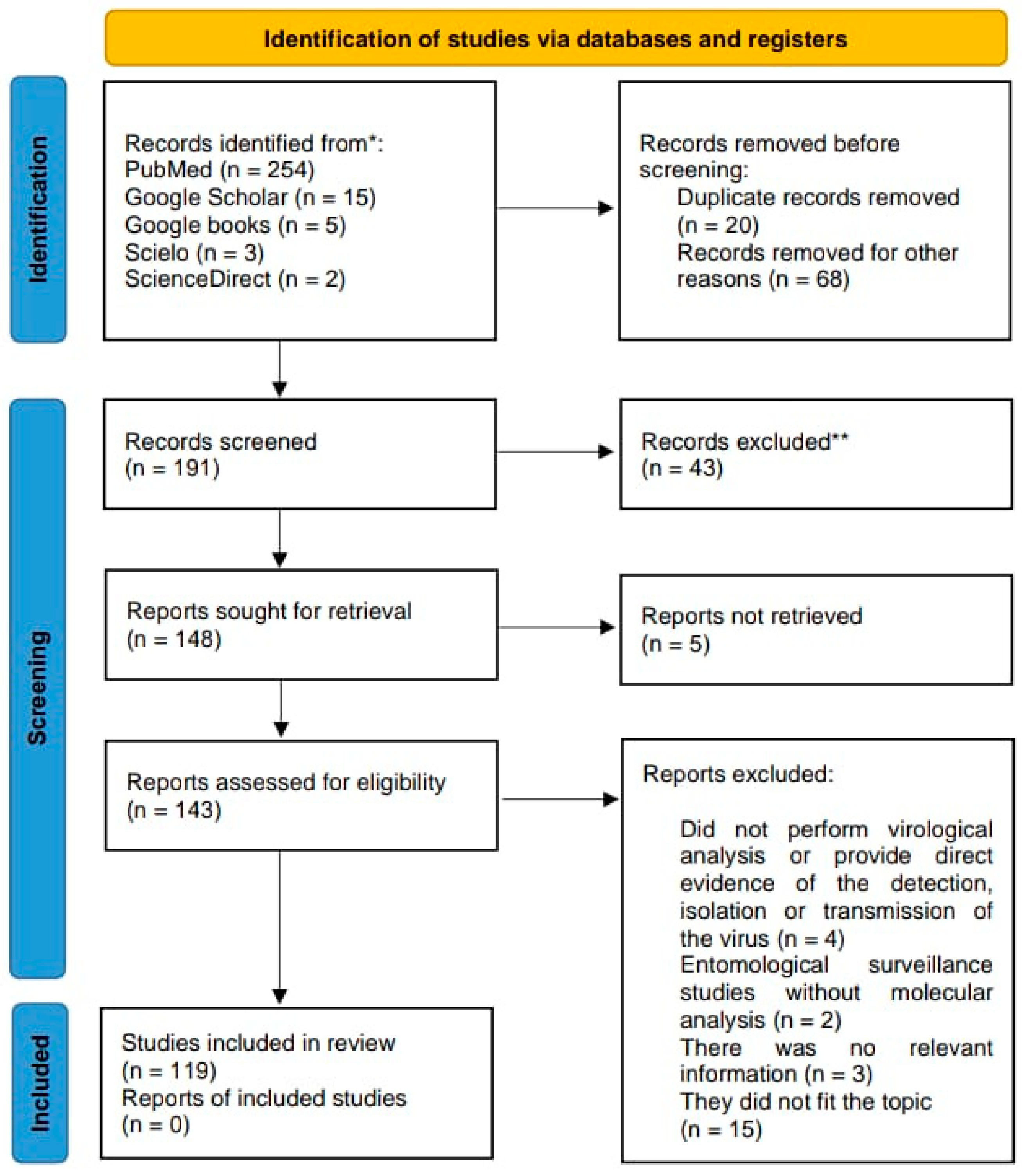
2. Materials and Methods

To conduct this systematic review, the PRISMA (Preferred Reporting Items for Systematic Reviews and Meta-Analyses.) protocol was followed, to ensure compliance with each step to identify, analyze, select, and incorporate the relevant literature [11] (Figure 1). The search was performed using academic search engines and databases such as PubMed, Google Scholar, Google Books, and ScienceDirect. Both English and Spanish terms were used, with a carefully selected set of keywords including “Simuliidae”, “Simulium”, “virome”, “virus”, “metagenomics”, “arbovirus”, “black flies”, “horizontal transmission”, “biological transmission”, “mechanical transmission”, “vector competence”, “vector competence barriers”, and “vector”. No restrictions were applied regarding publication years, allowing the inclusion of literature from the 20th century up to 5 May 2025.

3. Results Overview

3.1. Simuliidae as Vectors of Public-Health-Related Non-Viral Pathogens

The Simuliidae family, or black flies, includes approximately 2163 morphospecies of hematophagous dipterans globally [12]. Only females require blood meals, making them the primary vectors for pathogen transmission [13]. Host preferences vary, with species exhibiting zoophilic, anthropophilic, or opportunistic feeding behaviors. This behavior has significant medical and socioeconomic impacts, as high biting rates reduce tourism, cause animal mortality, and facilitate the spread of pathogens [3]. Notable pathogens transmitted by Simuliidae include Onchocerca volvulus, the causative agent of human onchocerciasis [14,15], and Mansonella ozzardii [1,16]. In veterinary medicine, Simuliidae are key vectors of Onchocerca spp., causing filariasis in livestock, dogs, wild ungulates [17], and equids [18]. They also transmit Dirofilaria ursi in black bears (Ursus americanus and Ursus thibetanus japonicus), with occasional human infections [19,20], and Splendidofilaria fallisensis in ducks and waterfowls [21]. Additionally, they spread protozoa such as Trypanosoma spp. and Leucocytozoon spp. in domestic and wild birds [22].
Simuliidae also contribute to public health challenges through severe blackfly hypersensitivity (SBH), a condition resulting from repeated bites in humans and animals [23,24]. SBH manifests as skin inflammation, intense itching, and prolonged rashes (Figure 2). Immune responses to salivary toxins can lead to systemic symptoms, including fever, headaches, and fatigue [25].

3.2. General Relevance of Arbovirosis in Vector-Borne Diseases

The term “arbovirus”, derived from arthropod-borne viruses, refers not to a taxonomic classification but to a group of viruses defined by their transmission mechanism [26]. These viruses rely on infected vertebrate hosts, who are bitten by hematophagous arthropods such as mosquitoes, ticks, Culicoides, and black flies, etc. Vectors acquire the virus during feeding, replicate it, and transmit it to new hosts, a process known as biological horizontal transmission [27,28]. Additional biological transmission routes include vertical (transovarial) transmission, in which larvae emerge from the eggs of infected females, and venereal transmission, where viremic females transmit the virus to males during mating [29,30].
Arboviral diseases have historically been recognized as a global public health concern, with a notable rise in cases during the late 20th and early 21st centuries due to their emergence and re-emergence [31]. Their spread is driven by climatic, ecological, and sociodemographic factors [32,33], as well as human activities that cause ecological disruptions [34]. These factors increase disease prevalence, particularly in socially disadvantaged regions with limited healthcare access and inadequate pathogen monitoring [35]. Urban expansion, population growth, and poor urban planning further exacerbate the spread. Migration and informal settlements heighten susceptibility to arboviruses, straining infrastructure, healthcare, and food systems [36]. These ecological and demographic shifts have a significant impact on arbovirus epidemiology.
In recent decades, the increasing prevalence and global spread of arboviruses have come to represent a significant public health threat, causing high morbidity and mortality in humans and animals [26]. Over 500 arboviruses have been documented (https://wwwn.cdc.gov/arbocat/ accessed on 5 July 2024); these are classified into nine families (Flaviviridae, Togaviridae, Peribunyaviridae, Phenuiviridae, Nairoviridae, Sedoreoviridae, Orthomyxoviridae, Rhabdoviridae, and Asfarviridae). Table 1 summarizes the key molecular characteristics of a representative sample of arbovirus families.

3.3. Detection and Isolation of Viruses in Simuliidae

Studies on the detection and isolation of arboviruses in Simuliidae remain limited and scarce. Nevertheless, the existing research has demonstrated that certain species within this family can act as vectors for arboviruses of significant public and veterinary health importance. Black flies have been implicated in the transmission of several arboviruses, including Leporipoxvirus (family Poxviridae), which primarily infects rabbits and squirrels [53,54], as well as various serotypes of vesicular stomatitis virus (VSV).
VSV, belonging to the genus Vesiculovirus within the family Rhabdoviridae, has a global distribution and is endemic to regions such as North America, Central America, northern South America, and parts of the southeastern United States, primarily affecting livestock, including horses, mules, donkeys, cattle, and pigs [55,56]. Consequently, it is considered the most important animal health arbovirus transmitted by Simuliidae. Notably, cases of VSV have been detected in livestock in regions of Mexico with high black fly prevalence [57,58]. These findings underscore the role of Simuliidae in the epidemiology of VSV and highlight the need for further research into their vector capacity for other arboviruses; the most recent findings in relation to this virus are discussed later in the text.
Several experimental studies have contributed to understanding the transmission dynamics of arboviruses by black flies. In 1934, Knowlton attempted to demonstrate the transmission of equine encephalitis virus from a horse to a guinea pig; however, the experiment did not yield conclusive evidence of successful transmission [59]. In contrast, Ferris et al. (1955) [60] demonstrated the ability of black flies to mechanically transmit VSV, although only for a short period following feeding on infected embryos. Transmission was effective only within the first 24 h, with no evidence of viral replication or an incubation period within the flies. As a result, their role as vectors was attributed to contamination of the mouthparts, classifying them as potential mechanical vectors, and not as biological vectors.
Historically, Cupp et al. (1992) [57] provided the first confirmed evidence of the biological transmission of an arbovirus by a member of the Simuliidae family. In their study, they evaluated the competence of Simulium vittatum Zetterstedt to act as a vector for the New Jersey serotype of vesicular stomatitis virus (VSVNJ). To achieve this, they infected female flies of this species via intrathoracic inoculation and oral exposure using a strain obtained during the 1982 VSVNJ outbreak in North America. Among the reported results, 70% of thoracically infected flies and 45% of orally infected flies secreted infectious virions in their saliva 10 days post-infection, indicating that VSVNJ exhibits positive vector competence. Further significant findings regarding the biological transmission of arboviruses occurred in 1953, when the Rift Valley fever virus was isolated from Simulium sp. collected near the Orange River in South Africa. This discovery suggested that, in addition to mosquitoes, other arthropods could play a role in the transmission of this arbovirus in nature [61,62].
Previously, Austin (1967) [63] evaluated the susceptibility for the biological and mechanical transmission of ten arboviruses in the blackfly Austrosimulium ungulatum through intrathoracic inoculation and feeding on viremic mice. The viruses examined included Semliki Forest virus (SFV), Whataroa (M78), Sindbis (MRM39), Bebaru (AMM2354), Murray Valley encephalitis (MVE), St. Louis encephalitis (SLE), Ntaya, Dengue I, Dengue II, and Batai. At serial post-infection intervals maintained at 22 °C, viral replication was assessed in the heads, salivary glands, and bodies of the black flies. The successful replication of the M78 virus was detected exclusively in the body from six days post-infection, with significantly higher viral titers compared to the baseline levels observed in control flies sacrificed immediately after infection. However, M78 was found to be refractory, indicating that viral dissemination or replication into the salivary glands did not occur, consequently, the virus was not transmitted. Similarly, the SFV, MRM39, AMM2354, Ntaya, and Chittagong viruses persisted for at least 10 days post-infection, whereas Dengue I, Dengue II, and SLE viruses were cleared before day 14 and were not detected in the heads (salivary glands). Therefore, it was determined that there is no possibility of the arboviruses tested being biologically transmitted.
However, the mechanical transmission of these arboviruses was also evaluated by feeding groups of A. ungulatum first on viremic suckling mice, and then on no viremic suckling mice. They found that A. ungulatum was able to transmit the M78 virus for up to 48 h via mechanical transmission under experimental conditions following the ingestion of viremic blood. These results support the hypothesis that, under specific conditions such as a high viral load and close contact with infected vertebrate hosts, black flies may contribute to the mechanical transmission of certain arboviruses, even if they are not competent biological vectors. Later, Sanmartín et al. (1973) [64] investigated the role of black flies in the transmission of Venezuelan equine encephalitis virus (VEEV) during outbreaks recorded in 1967 in three localities in Colombia. In two locations, high black fly activity was identified, particularly among Simulium exiguum, S. metallicum, S. callidum, S. paynei, and S. mexicanum. The virus was successfully isolated in eleven samples from those specimens collected during the biting of horses and humans by intracerebrally inoculating two-to-seven-day-old white mice or by inoculating Vero cells. Except for the S. paynei pools, all specimens were macerated immediately after collection. This introduces potential bias in the interpretation of results, leading to the erroneous incrimination of black flies as the true biological vectors, because the viral isolation involved recently ingested blood from viremic hosts rather than from an active infection within the black flies. In contrast, due to the low post-collection mortality of S. paynei, these specimens were not processed immediately but were kept alive for several days prior to analysis, suggesting possible virus replication within the insect. In subsequent studies, the vector competence of S. mexicanum and S. metallicum for enzootic and epizootic Colombian strains of VEEV was experimentally assessed [65]. Wild-caught flies fed on viremic guinea pigs were monitored for viral replication and tested for biological and mechanical transmission. No evidence of biological transmission or sustained viral replication was found, though occasional mechanical transmission occurred. These findings suggest that neither species plays a significant role in VEEV transmission in nature. Notably, Gibbs and Long (2013) [66] report that VEEV was isolated from various families of hematophagous Diptera, including Simuliidae, referencing a study by Mitchell et al. (1985) [67]. However, upon reviewing the original study by Mitchell et al. (1985) [67], which involved prospective arbovirus surveillance in three provinces of Argentina between 1977 and 1980, a total of 104 Simuliidae specimens were tested for viruses, and all pools were negative. Therefore, the conclusion by Long and Gibbs (2013) [66] appears to be incorrect.
A discovery by Sommerman (1977) [68] demonstrated the first evidence of snowshoe hare virus (SSH) virus in Simulium sp., suggesting a broader vector range and highlighting the need for further investigation into the role of black flies in arbovirus transmission. In this study, the SSH virus, a member of the California serogroup, was isolated from multiple mosquito species and one unidentified black fly collected in interior Alaska between 1970 and 1972. Then, during an arbovirus survey conducted in Colorado and Utah amid the 1982–1983 epizootic of vesicular stomatitis New Jersey virus (VSNJ) in the western United States, a bunyavirus sequence identified as Lorken virus (Bunyamwera group) was detected in a pool of Simulium bivittatum collected near a horse livestock area and adjacent mountain sagebrush and alfalfa pasture [69].
Chanteau et al. (1993) [70] conducted a study on Nuku-Hiva Island, in the Marquesas Archipelago of French Polynesia, to investigate the potential role of Simulium buissoni in the epidemiology of hepatitis B virus (HBV) within a holoendemic population. Although there was no evidence of viral replication within the black flies, HBV DNA was detected only in small amounts in a few black fly pools, with a low viral load. Additionally, a positive correlation was observed between the number of skin lesions caused by black fly bites and HBV infection prevalence—particularly among children living in rural areas. This supports the hypothesis that, in regions with high black fly density, HBV transmission might occur indirectly through bite-induced skin lesions, without requiring viral replication within the insects.
Multiple studies have investigated the role of black flies in the transmission of vesicular stomatitis viruses. Mead et al. (1997) [71] evaluated the vector competence of several black fly species in southern Arizona for different isolates of vesicular stomatitis VSNJV. Their findings demonstrated that S. vittatum served as a competent vector, while other species, including S. bivittatum and Simulium longithallum, were refractory to infection. Expanding on these finding, Mead et al. (1999) [72] provided experimental confirmation of VSNJV transmission by S. vittatum using a murine model. In this study, artificially infected colony-reared flies were allowed to feed on susceptible mice, all of which developed neutralizing antibodies by 21 days post-infection, confirming successful transmission.
Subsequently, Mead et al. (2000) [73] expanded their research to assess the competence of both colonized and wild-caught black fly species for the VSV-Indiana serotype. They demonstrated that both S. vittatum and Simulium notatum were competent vectors, as an infectious virus was detected in the saliva of exposed flies, suggesting their potential role in the transmission of this serotype. Further validation of biological transmission was achieved by Mead et al. (2004) [74], who confirmed VSNJV infection in domestic pigs following exposure to infected black flies, supporting the vectorial capacity of VSN in Simuliidae in natural host systems. Supplementary field evidence was later provided by Drolet et al. (2006) [75] during a VSNJV outbreak in Wyoming, where large numbers of black flies were collected and screened for viral RNA. S. bivittatum was identified as a potential vector, with viral RNA detected in two pools, suggesting its involvement in outbreak dynamics under natural conditions. Mead et al. (2009) [76] reported the first successful transmission of VSNJV to cattle by infected black flies, resulting in clinical disease. Infected flies fed at sites where VSNJV lesions are typically observed, such as the mouth and coronet band, leading to local viral replication and vesicular lesions in cattle. Similarly, Howerth et al. (2002) [77] conducted experiments to determine how black flies become infected with VSNJV. Their findings indicated that the virus initially infects the gut, but transmission to the salivary glands may be blocked in older flies, impairing their ability to transmit the virus. This study also highlighted the role of black fly age in determining vector competence.
Recently, Mesquita et al. (2017) [78] explored the role of rodents, specifically, juvenile and nestling deer mice (Peromyscus maniculatus), in VSNJV transmission. They exposed these mice to VSNJV-infected S. vittatum bites and observed the development of severe neurological symptoms in some juvenile mice within six to eight days post-inoculation. Interestingly, juvenile mice did not develop viremia, while nestling mice did, and they exhibited widespread viral antigen distribution in their central nervous systems. Furthermore, these viremic nestling mice could infect naive black flies, demonstrating that these rodents could act as potential reservoirs or amplifying hosts for VSNJV.
In both the U.S. and Mexico, recent studies on arbovirus transmission dynamics have assessed the presence of VSV in various species of hematophagous dipterans, including Simuliidae species. Through a molecular analysis, VSVNJ RNA was detected in black fly samples, confirming their role as vectors in the transmission of this arbovirus in the region [55,79]. Similarly, in 2023, Scroggs et al. [80] conducted a large-scale vector surveillance study during a VSNJV outbreak across California, Nevada, and Texas, US. Several specimens of dipterans were collected, including Culicoides and Simulium spp., detecting a 96% rate of RNA-positive pools in both species, representing the first report of VSNJV in several wild-caught Simulium spp. The study highlights the importance of vector surveillance in understanding VSNJV transmission dynamics and informs control efforts. McGregor et al. (2021) [81] contributed further evidence of black flies and biting midges as VSNJV vectors during a VSV outbreak in the United States. Their surveillance in Kansas identified Culicoides and Simulium spp. as important vectors, with positive pools detected for the VSV-Indiana serotype. This study also reported new vector species, expanding the list of potential VSNJV vectors and emphasizing the need for targeted surveillance.
Despite increased efforts regarding arbovirus surveillance in Simuliidae, few studies have documented the presence of other arboviruses associated with this group as potential vectors. For example, Reeves and Milby (1990) [82] examined thousands of Simulium spp. in California, USA, but they were unable to detect or isolate any arboviruses, supporting the notion that these flies may possess limited or no vector competence under certain ecological conditions. More recently, Cavallaro et al. (2018) [83] evaluated the effectiveness of partially submerged adhesive traps for the surveillance of adult black flies and the detection of arboviruses. The collected species were identified as S. vittatum and S. decorum. Although positive cases of West Nile virus were detected in Culex spp. mosquitoes collected within a 16 km radius of the study sites, tests conducted on black flies did not reveal the presence of West Nile virus or Eastern equine encephalitis virus. Additionally, during a severe outbreak of bluetongue disease in a flock of sheep in Bruneau, Owyhee County, Idaho, various hematophagous dipterans were collected, including 89 black flies that had been in direct contact with infected animals. However, the virus could not be isolated from any of the collected black flies [84].
In the past decade, researchers have hypothesized that endemic Simuliidae species in African onchocerciasis-endemic regions may play a critical role in transmitting an unidentified virus, referred as Nodding Syndrome Virus (NSV), potentially associated with O. volvulus. This virus is suspected to contribute to a newly emerging neurotropic syndrome affecting both humans and other animal species. The syndrome has garnered significant attention within the scientific community due to its debilitating effects and its potential link to onchocerciasis. Such an association could complicate disease control efforts and pose a substantial public health risk in affected regions [85]. However, the exact etiology of onchocerciasis-associated epilepsy remains a subject of ongoing debate [86].
The CDC Arbovirus Catalog (https://wwwn.cdc.gov/arbocat/ accessed on 20 September 2024) [87] records the isolation of the arboviruses mentioned previously, as well as the Jerry Slough virus (JSV), which is transmitted by Simulium sp. However, no further information regarding this virus is available. Table 2 details viruses of medical and veterinary significance associated with black flies, along with the methodologies employed in the studies.

3.4. Metagenomics of Viromes as a Tool for the Detection of Zoonotic Arboviruses

Over two-thirds of human pathogens are zoonotic in origin [88], with RNA viruses, including arboviruses, constituting the majority of them [89,90]. Historically, unconventional methods for detecting these viruses involved using sentinel animals exposed to arthropod bites in the wild, as exemplified by the discovery of Zika virus in Africa [91]. Post-infection, serological diagnostics or viral isolation in cell cultures were employed [92]. Advances in molecular biology have shifted the focus to viral genome detection using PCR and Sanger sequencing, enabling the identification of non-cultivable arboviruses [93]. However, these methods require prior knowledge of the viral genome, such as oligonucleotide design for targeted amplification.
Next-generation sequencing (NGS), or metagenomics, enables the high-throughput sequencing of all viral genomes in a sample (Figure 3), facilitating the discovery of novel viral species [94]. Since 2010, 86 studies on arbovirus identification in mosquitoes and ticks have been documented on PubMed, with a notable increase in mosquito virome research between 2016 and 2022. These studies identified over 2000 new viruses, including 112 in Chinese arthropods [95], 1445 RNA viruses in multiple Chinese provinces [96], 32 in Culex mosquitoes in California [97], and 101 in Culex and Aedes species in Yunnan, China [98]. A recent study in São Paulo, Brazil, identified 229 viral species in Aedes, Anopheles, and Culex mosquitoes, primarily from the orders Picornavirales, Nodamuvirales, and Sobelivirales [99]. In Mexico, metagenomic studies in Yucatán have revealed RNA and mosquito-specific viruses, including Uxmal virus (Aedes taeniorhynchus) and Mayapán virus (Psorophora ferox) [100]. Subsequent research identified Houston virus in Culex quinquefasciatus [101], Ek Balam virus (Tymoviridae) in C. quinquefasciatus [102], and novel rhabdoviruses in Culex mosquitoes in the U.S. [103].
These studies of RNA virus isolation in mosquitoes worldwide expand the diversity of viruses discovered through metagenomics. Metagenomic approaches applied to mosquitoes have become key tools for virological surveillance, allowing for the identification of both known and unknown viruses related to diseases transmitted by hematophagous dipterans [104]. However, despite these advances, only two studies of species of the Simuliidae family have been published, as detailed below.

3.5. Metagenomic Studies of Viruses in Simuliidae

In the available scientific literature, studies of the metagenomics of arboviruses in Simuliidae are limited. The first such study was conducted by Kraberger et al. (2019) [105] in New Zealand, where 40 Simulium specimens from a single site were analyzed. In this study, new viruses belonging to four viral families were identified: Genomoviridae (n = 9), Circoviridae (n = 1), Microviridae (n = 108), and other unclassified viruses (n = 20). However, no in vitro experiments were performed to determine whether any of these viruses have the capacity to infect and replicate in vertebrate cells, leaving their potential as arboviruses unknown. Subsequently, Kobayashi et al. (2020) [106] conducted a pilot study in Japan on the virome of certain Culicomorpha, analyzing only three specimens of Simulium aureohirtum. Despite the small sample size, three new viruses were detected, belonging to three viral taxa: Dicistroviridae, Nodaviridae, and an unclassified taxon. As in the previous study, no vector competence assays were conducted in vertebrate cells. In contrast, De Coninck et al. (2024) [107] analyzed the virome of black flies in Cameroon and detected 641 RNA viruses, identified based on the sequence of RNA-dependent RNA polymerase (RdRP), many of which corresponded to previously unknown viral lineages.
These studies demonstrate that metagenomics is a powerful tool for discovering new viruses and characterizing viromes in insects such as Simuliidae. The integration of this NGS methodology with molecular studies, viral isolation, and vector competence assays in vertebrate cells will allow for a better understanding of the relevant interactions, which will make significant contributions to the advancement of scientific knowledge.

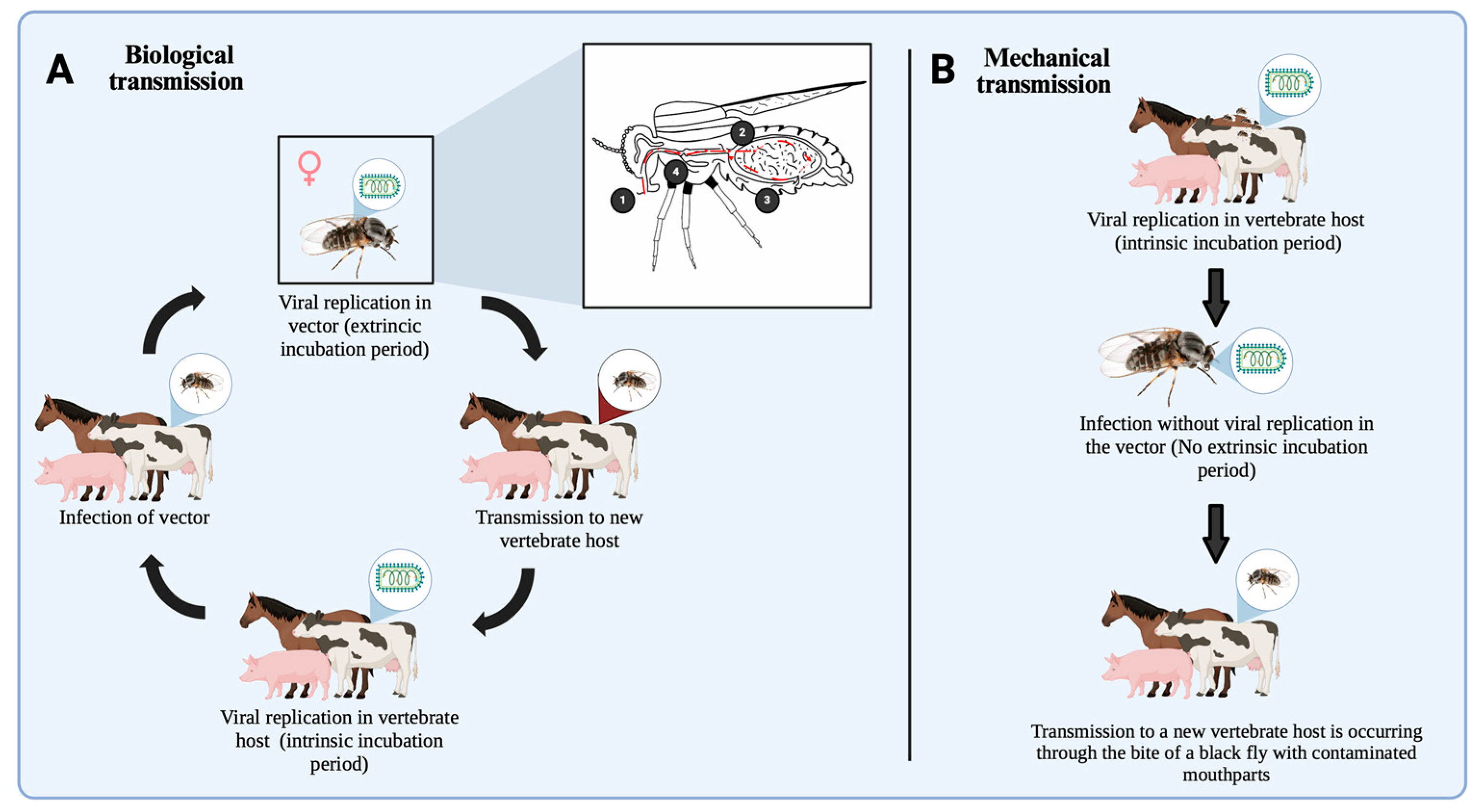
3.6. Exploring Causal and Casual Modes of Arbovirus Transmission Mechanisms in Simuliidae

Understanding the mechanisms of horizontal transmission (Figure 4) is fundamental to comprehending the epidemiological dynamics of the causal relationships within vector–pathogen–host interactions. Turell (1988) [108] defined this as the transfer of any virus from an infected arthropod vector to a vertebrate host, typically occurring during blood feeding.
In Simuliidae, the horizontal transmission of arboviruses occurs through two principal modes. Biological transmission (Figure 4A) involves the systemic infection of the vector, with viral dissemination and replication across vector competence barriers, ultimately leading to amplification in the salivary glands for transmission during subsequent blood feeding [109,110]. This process also involves several physiological mechanisms of the vector, including cellular immunity, microbial interactions, enzymes, iRNA, and the genetic background [111]. The alternative mode is mechanical (oral) transmission (Figure 4B), which entails the passive transfer of viral particles from one infected vertebrate host to another via contaminated mouthparts, without viral replication within the vector [112].
As described above, mechanical transmission has been experimentally demonstrated in vitro in A. ungulatum and S. venustum [60,63], both of which successfully transmitted vesicular stomatitis virus (VSV) to susceptible hosts 24–48 h after ingesting viremic blood. Although feasible, mechanical transmission is highly dependent on elevated viral title levels in the vertebrate host and is generally regarded as incidental or as resulting from casual interactions, with limited significance in natural virus maintenance [113]. However, under epidemic conditions, it may temporarily contribute to the amplification of viral spread [112]. In contrast, biological transmission entails a causal relationship between the vector and the arbovirus. For example, S. vittatum has been confirmed as a competent biological vector of VSV [55], with up to 45% of orally infected individuals harboring an infectious virus in their saliva following a 10-day extrinsic incubation period. This capacity for viral replication and subsequent transmission was further validated using murine and swine models, providing robust evidence of true vector competence [72].
Particularly relevant examples for examining whether arbovirus transmission is causal or incidental include RVFV and NSV. In the case of RVFV, Aedes mosquitoes (Culicidae) are well-established as the primary vectors; RVFV has been isolated from field-collected Simulium specimens in an endemic region of South Africa [61]. This observation has increased speculation about the potential role of black flies in the transmission dynamics of the RVFV. It suggests a possible, though unconfirmed, role of Simuliidae in RVFV transmission cycles, particularly during epizootic occurrences where multiple transmission routes may emerge under favorable environmental conditions [114]. Nonetheless, to date, no study has demonstrated the biological competence of Simuliidae for RVFV, and this association should therefore be interpreted with caution, as it may reflect incidental rather than causal involvement in transmission [115].
Another notable example comes from the emerging evidence in Central Africa, which suggests the possible circulation of a neurotropic virus, commonly referred to as NSV, associated with high black fly biting rates and onchocerciasis-related nodules in endemic regions [85,86,116]. Although a specific etiological agent has not been conclusively identified, one hypothesis proposes that O. volvulus, transmitted by Simulium spp., may harbor an endosymbiotic virus that is responsible for the observed neurological syndromes. In this scenario, black flies could serve as vectors for both the filarial parasite and the neurotropic virus, thereby contributing to the pathogenesis of NSV [117].
The evidence from both RVFV and NSV cases underscores the urgent need for integrated studies to better understand potential interactions between Simuliidae and arboviruses. In these cases, it is essential to distinguish between the mere detection of viral material and actual vector-mediated transmission. Molecular techniques such as RT-PCR or next-generation sequencing (NGS) can detect viral genetic material without proving that the virus can replicate or be transmitted to new hosts [118]. To establish Simuliidae as true biological vectors, laboratory experiments are needed to demonstrate viral replication, dissemination to the salivary glands, and successful transmission during blood feeding [119].
Clarifying the role of black flies in arbovirus transmission is important for both public health and scientific understanding. An inaccurate assessment or neglect of their potential role as vectors can compromise arbovirus surveillance efforts and limit the effectiveness of vector control programs. Therefore, the limited attention given to Simuliidae in arbovirology reflects a significant gap in our knowledge. Active viral monitoring in black fly populations, especially in areas where outbreaks occur without clearly identified vectors, combined with well-designed and rigorous laboratory studies, could be crucial to determining their true role in disease transmission and guiding more effective and inclusive control strategies.

4. Conclusions

This review of the arboviruses found in Simuliidae reveals that, despite advances made in virus identification in other vectors, this group remains relatively unexplored compared to mosquitoes and ticks. There are few arboviruses that have been shown to have vector competence in Simuliidae. The use of metagenomics has proven to be a powerful tool for revealing viral diversity in hematophagous vectors, enabling the detection of both known and unknown viruses, including insect-specific viruses with potential implications for arbovirus evolution and transmission dynamics. As this technology continues to be applied to species within the Simuliidae family, our understanding of viromes in vectors and their role in arbovirus transmission will expand. This approach will not only enrich our knowledge of emerging and re-emerging viruses but will also contribute to understanding the ecology, evolution, and potential zoonotic threats of viruses harbored by Simuliidae. Public health will also be directly impacted through improved surveillance and the development of more effective strategies for controlling vector-borne pathogens that cause diseases in humans and animals.

Supplementary Materials

The following supporting information can be downloaded at: https://www.mdpi.com/article/10.3390/life15050807/s1, Table S1: List of overall references for PRISMA analysis.

Author Contributions

Conceptualization, J.A.G.-H. and S.V.L.-T.; methodology, A.R.-M., J.A.G.-H. and S.V.L.-T.; software, A.R.-M.; validation, S.V.L.-T., J.R.A.-G., E.d.J.d.L.-S., C.A.R.-A., J.E.G.-R., M.C.-M. and J.A.G.-H.; formal analysis, A.R.-M., S.V.L.-T. and J.A.G.-H.; investigation, S.V.L.-T., J.R.A.-G., E.d.J.d.L.-S., C.A.R.-A., J.E.G.-R., M.C.-M. and J.A.G.-H.; resources, S.V.L.-T., J.R.A.-G., E.d.J.d.L.-S., C.A.R.-A., J.E.G.-R., M.C.-M. and J.A.G.-H.; data curation, A.R.-M., J.A.G.-H. and S.V.L.-T.; writing—original draft preparation, A.R.-M., J.A.G.-H. and S.V.L.-T.; writing—review and editing, S.V.L.-T., J.R.A.-G., E.d.J.d.L.-S., C.A.R.-A., J.E.G.-R., M.C.-M. and J.A.G.-H.; visualization, A.R.-M., J.A.G.-H. and S.V.L.-T.; supervision, J.A.G.-H.; project administration, J.A.G.-H.; funding acquisition, J.A.G.-H. and E.d.J.d.L.-S. All authors have read and agreed to the published version of the manuscript.

Funding

This study was supported by a grant of the project Secretaría de Ciencia, Humanidades, Tecnología e Innovación (Secihti) of Mexico CIENCIA DE FRONTERA 2023 (No. 419-24-23). It was also supported by postdoctoral scholarships from Secihti of Mexico (Stephanie Viridiana Laredo-Tiscareño scholarships, Nos. 740742, 769056, and 842817) and the Programa RIPI2019ICB45.

Institutional Review Board Statement

Not applicable.

Informed Consent Statement

Not applicable.

Data Availability Statement

Not applicable.

Acknowledgments

A.R.-M. thanks the Sistema Nacional de Posgrados SECIHTI for funding entry into the Master’s Science program in Animal Science at ICB-UACJ (scholarships No. 4034275; CVU No. 1338422), as well as its support in the successful completion of this systematic review. Finally, J.A.G.-H. thank my wife, S.V.L-T. for granting my wish to conceive my son (Alfonso Garza-Laredo, A.G.L.) and for encouraging me in writing this manuscript and my career.

Conflicts of Interest

The authors declare no conflicts of interest.

References

  1. Ta-Tang, T.H.; Crainey, J.L.; Post, R.J.; Luz, S.L.; Rubio, J.M. Mansonellosis: Current perspectives. Res. Rep. Trop. Med. 2018, 9, 9–24. [Google Scholar] [CrossRef] [PubMed]
  2. Smith, P.F.; Howerth, E.W.; Carter, D.; Gray, E.W.; Noblet, R.; Mead, D.G. Mechanical Transmission of Vesicular Stomatitis New Jersey Virus by Simulium vittatum (Diptera: Simuliidae) to Domestic Swine (Sus scrofa). J. Med. Entomol. 2009, 46, 1537–1540. [Google Scholar] [CrossRef] [PubMed]
  3. Adler, P.H.; Currie, D.C.; Madera, D.M. Las Moscas Negras (Simuliidae) de América Del Norte; Cornell University Press: Ítaca, NY, USA, 2004. [Google Scholar]
  4. Cunze, S.; Jourdan, J.; Klimpel, S. Ecologically and medically important black flies of the genus Simulium: Identification of biogeographical groups according to similar larval niches. Sci. Total Environ. 2024, 917, 170454. [Google Scholar] [CrossRef]
  5. Negredo Antón, A.I.; de Ory Manchón, F.; Sánchez-Seco Fariñas, M.P.; Franco Narváez, L.; Gegúndez Cámara, M.I.; Navarro Mari, J.M.; Tenorio Matanzo, A. Diagnóstico microbiológico de arbovirosis y robovirosis emergentes. Enfermedades Infecc. Y Microbiol. Clin. 2015, 33, 197–205. [Google Scholar] [CrossRef] [PubMed]
  6. Chiu, C.Y.; Miller, S.A. Clinical metagenomics. Nat. Rev. Genet. 2019, 20, 341–355. [Google Scholar] [CrossRef]
  7. Santiago-Rodriguez, T.M.; Hollister, E.B. Unraveling the viral dark matter through viral metagenomics. Front. Immunol. 2022, 13, 1005107. [Google Scholar] [CrossRef]
  8. Ghurye, J.S.; Cepeda-Espinoza, V.; Pop, M. Metagenomic Assembly: Overview, Challenges and Applications. Yale J. Biol. Med. 2016, 89, 353–362. [Google Scholar]
  9. Taş, N.; de Jong, A.E.; Li, Y.; Trubl, G.; Xue, Y.; Dove, N.C. Metagenomic tools in microbial ecology research. Curr. Opin. Biotechnol. 2021, 67, 184–191. [Google Scholar] [CrossRef]
  10. Pan, Y.F.; Zhao, H.; Gou, Q.Y.; Shi, P.B.; Tian, J.H.; Feng, Y.; Li, K.; Yang, W.H.; Wu, D.; Tang, G.; et al. Metagenomic analysis of individual mosquitos reveals the ecology of insect viruses. bioRxiv 2023. bioRxiv:2023.08.28.555221. [Google Scholar]
  11. Page, M.J.; McKenzie, J.E.; Bossuyt, P.M.; Boutron, I.; Hoffmann, T.C.; Mulrow, C.D.; Shamseer, L.; Tetzlaff, J.M.; Akl, E.A.; Brennan, S.E.; et al. The PRISMA 2020 statement: An updated guideline for reporting systematic reviews. BMJ 2021, 372, n71. [Google Scholar] [CrossRef]
  12. Adler, P.H.; Crosskey, R.W. World Blackflies (Diptera: Simuliidae): A Comprehensive Revision of the Taxonomic and Geographical Inventory [2019]. Available online: https://biomia.sites.clemson.edu/pdfs/blackflyinventory.pdf (accessed on 10 January 2025).
  13. Adler, P.H.; Cheke, R.A.; Post, R.J. Evolution, epidemiology, and population genetics of black flies (Diptera: Simuliidae). Infect. Genet. Evol. 2010, 10, 846–865. [Google Scholar] [CrossRef]
  14. Boatin, B.A.; Richards, F.O. Control of Onchocerciasis. Adv. Parasitol. 2006, 61, 349–394. [Google Scholar] [PubMed]
  15. World Health Organization (WHO). Elimination of human onchocerciasis: Progress report, 2023–2024. Wkly. Epidemiol. Rec. 2024, 41, 577–590. [Google Scholar]
  16. Shelley, A.J.; Coscarón, S. Simuliid blackflies (Diptera: Simuliidae) and ceratopogonid midges (Diptera: Ceratopogonidae) as vectors of Mansonella ozzardi (Nematoda: Onchocercidae) in northern Argentina. Mem. Inst. Oswaldo Cruz 2001, 96, 451–458. [Google Scholar] [CrossRef] [PubMed]
  17. Manzanell, R.; Stocker, A.S.; Deplazes, P.; Mathis, A. Morphological description and multilocus genotyping of Onchocerca spp. in red deer (Cervus elaphus) in Switzerland. Int. J. Parasitol. Parasites Wildl. 2022, 19, 273–284. [Google Scholar] [CrossRef] [PubMed]
  18. Sazmand, A.; Bahari, A.; Papi, S.; Otranto, D. Parasitic diseases of equids in Iran (1931–2020): A literature review. Parasit. Vectors 2020, 13, 586. [Google Scholar] [CrossRef]
  19. Michalski, M.L.; Bain, O.; Fischer, K.; Fischer, P.U.; Kumar, S.; Foster, J.M. Identification and phylogenetic analysis of Dirofilaria ursi (Nematoda: Filarioidea) from Wisconsin black bears (Ursus americanus) and its Wolbachia endosymbiont. J. Parasitol. 2010, 96, 412–419. [Google Scholar] [CrossRef]
  20. Yamada, M.; Shishito, N.; Nozawa, Y.; Uni, S.; Nishioka, K.; Nakaya, T. A combined human case of Dirofilaria ursi infection in dorsal subcutaneous tissue and Anisakis simplex sensu stricto (s.s.) infection in ventral subcutaneous tissue. Trop. Med. Health 2017, 45, 26. [Google Scholar] [CrossRef]
  21. Anderson, R.C. The simuliid vectors of Splendidofilaria fallisensis of ducks. Can. J. Zool. 1968, 46, 610–611. [Google Scholar] [CrossRef]
  22. Jumpato, W.; Tangkawanit, U.; Wongpakam, K.; Pramual, P. Molecular detection of Leucocytozoon (Apicomplexa: Haemosporida) in black flies (Diptera: Simuliidae) from Thailand. Acta Trop. 2019, 190, 228–234. [Google Scholar] [CrossRef]
  23. Rodríguez-Pérez, M.A.; Garza-Hernández, J.A.; Salinas-Carmona, M.C.; Fernández-Salas, I.; Reyes-Villanueva, F.; Real-Najarro, O.; Unnasch, T.R. The esperanza window trap reduces the human biting rate of Simulium ochraceum sl in formerly onchocerciasis endemic foci in Southern Mexico. PLOS Negl. Trop. Dis. 2017, 11, e0005686. [Google Scholar] [CrossRef]
  24. Ruiz-Arrondo, I.; Garza-Hernández, J.A.; Reyes-Villanueva, F.; Lucientes-Curdi, J.; Rodríguez-Pérez, M.A. Human-landing rate, gonotrophic cycle length, survivorship, and public health importance of Simulium erythrocephalum in Zaragoza, northeastern Spain. Parasites Vectors 2017, 10, 175. [Google Scholar] [CrossRef]
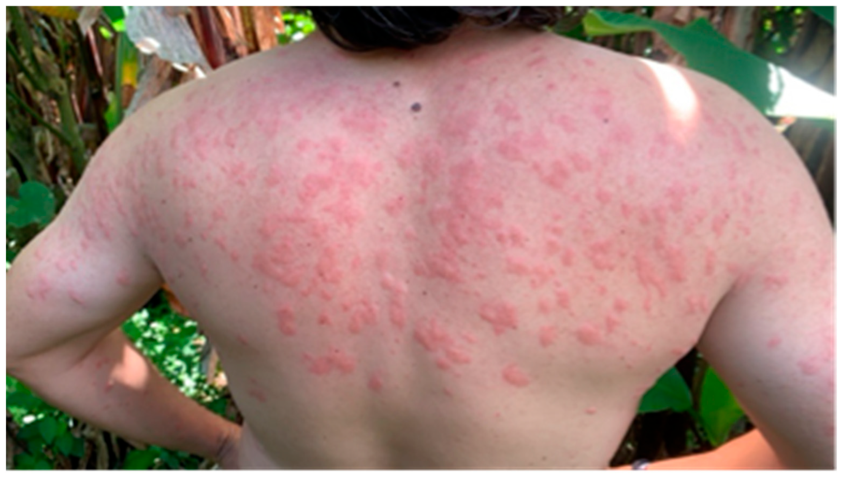
  25. Sitarz, M.; Buczek, A.M.; Buczek, W.; Buczek, A.; Bartosik, K. Risk of attacks by blackflies (Diptera: Simuliidae) and occurrence of severe skin symptoms in bitten patients along the Eastern border of the European Union. Int. J. Environ. Res. Public Health 2022, 19, 7610. [Google Scholar] [CrossRef] [PubMed]
  26. Young, P.R. Arboviruses: A Family on the Move. Adv. Exp. Med. Biol. 2018, 1062, 1–10. [Google Scholar] [PubMed]
  27. Kuno, G. Host range specificity of flaviviruses: Correlation with in vitro replication. J. Med. Entomol. 2007, 44, 93–101. [Google Scholar] [CrossRef] [PubMed]
  28. Hubálek, Z. History of Arbovirus Research in the Czech Republic. Viruses 2021, 13, 2334. [Google Scholar] [CrossRef]
  29. Kuno, G. Transmission of arboviruses without involvement of arthropod vectors. Acta Virol. 2001, 45, 139–150. [Google Scholar]
  30. Weaver, S.C.; Reisen, W.K. Present and future arboviral threats. Antivir. Res. 2010, 85, 328–345. [Google Scholar] [CrossRef]
  31. Gubler, D.J. Resurgent Vector-Borne Diseases as a Global Health Problem. Emerg. Infect. Dis. 1998, 4, 442. [Google Scholar] [CrossRef]
  32. Gratz, N.G. Emerging and Resurging Vector-Borne Diseases. Annu. Rev. Entomol. 1999, 44, 51. [Google Scholar] [CrossRef]
  33. Pavlovsky, E.N. On Natural Foci of Infection and Parasitic Diseases. Vestn. Akad. Nauk. SSSR 1939, 10, 98–108. [Google Scholar]
  34. Keesing, F.; Ostfeld, R.S. Impacts of Biodiversity and Biodiversity Loss on Zoonotic Diseases. Proc. Natl. Acad. Sci. USA 2021, 118, e2023540118. [Google Scholar] [CrossRef]
  35. de Oliveira, J.G.; Netto, S.A.; Francisco, E.O.; Vieira, C.P.; Variza, P.F.; Iser, B.P.M.; Lima-Camara, T.N.; Lorenz, C.; Prophiro, J.S. Aedes aegypti in Southern Brazil: Spatiotemporal Distribution Dynamics and Association with Climate and Environmental Factors. Trop. Med. Infect. Dis. 2023, 8, 77. [Google Scholar] [CrossRef] [PubMed]
  36. Kolimenakis, A.; Heinz, S.; Wilson, M.L.; Winkler, V.; Yakob, L.; Michaelakis, A.; Papachristos, D.; Richardson, C.; Horstick, O. The Role of Urbanisation in the Spread of Aedes Mosquitoes and the Diseases They Transmit—A Systematic Review. PLoS Negl. Trop. Dis. 2021, 15, e0009631. [Google Scholar] [CrossRef] [PubMed]
  37. Ciota, A.T. The role of co-infection and swarm dynamics in arbovirus transmission. Virus Res. 2019, 265, 88–93. [Google Scholar] [CrossRef]
  38. Snyder, J.; Jose, J.; Kuhn, R. The Togaviridae and the Flaviviridae; Elsevier eBooks; Elsevier: Amsterdam, The Netherlands, 2014. [Google Scholar]
  39. Xu, Z.; Peng, Y.; Yang, M.; Li, X.; Wang, J.; Zou, R.; Liang, J.; Fang, S.; Liu, Y.; Yang, Y. Simultaneous Detection of Zika, Chikungunya, Dengue, Yellow Fever, West Nile, and Japanese Encephalitis Viruses by a Two-Tube Multiplex Real-Time RT-PCR Assay. J. Med. Virol. 2022, 94, 2528–2536. [Google Scholar] [CrossRef]
  40. Latanova, A.; Starodubova, E.; Karpov, V. Flaviviridae Nonstructural Proteins: The Role in Molecular Mechanisms of Triggering Inflammation. Viruses 2022, 14, 1808. [Google Scholar] [CrossRef] [PubMed]
  41. Ryu, W. Other Positive-Strand RNA Viruses. In Molecular Virology of Human Pathogenic Viruses; Elsevier eBooks; Elsevier: Amsterdam, The Netherlands, 2017; pp. 177–184. [Google Scholar]
  42. Toribio, R.E. Arboviral Equine Encephalitides. Vet. Clin. N. Am. Equine Pract. 2022, 38, 299–321. [Google Scholar] [CrossRef]
  43. Chen, R.; Mukhopadhyay, S.; Merits, A.; Bolling, B.; Nasar, F.; Coffey, L.L.; Powers, A.; Weaver, S.C. ICTV Report Consortium. ICTV Virus Taxonomy Profile: Togaviridae. J. Gen. Virol. 2018, 99, 761–762. [Google Scholar] [CrossRef] [PubMed]
  44. AbudAbudurexiti, A.; Adkins, S.; Alioto, D.; Alkhovsky, S.V.; Avšič-Županc, T.; Ballinger, M.J.; Bente, D.A.; Beer, M.; Bergeron, É.; Blair, C.D.; et al. Taxonomy of the order Bunyavirales: Update 2019. Arch. Virol. 2019, 164, 1949–1965. [Google Scholar]
  45. Zhang, Y.; Liu, X.; Wu, Z.; Feng, S.; Lu, K.; Zhu, W.; Sun, H.; Niu, G. Oropouche Virus: A Neglected Global Arboviral Threat. Virus Res. 2024, 341, 199318. [Google Scholar] [CrossRef]
  46. Sasaya, T.; Palacios, G.; Briese, T.; Di Serio, F.; Groschup, M.H.; Neriya, Y.; Song, J.; Tomitaka, Y. ICTV Virus Taxonomy Profile: Phenuiviridae 2023. J. Gen. Virol. 2023, 104, e001990. [Google Scholar] [CrossRef] [PubMed]
  47. Kuhn, J.H.; Alkhovsky, S.V.; Avšič-Županc, T.; Bergeron, É.; Burt, F.; Ergünay, K.; Garrison, A.R.; Marklewitz, M.; Mirazimi, A.; Papa, A.; et al. ICTV Virus Taxonomy Profile: Nairoviridae 2024. J. Gen. Virol. 2024, 105, e002012. [Google Scholar] [CrossRef]
  48. Maclachlan, N.J.; Guthrie, A.J. Re-emergence of bluetongue, African horse sickness, and other orbivirus diseases. Vet. Res. 2010, 41, 35. [Google Scholar] [CrossRef]
  49. Vasilakis, N.; Tesh, R.B. Insect-Specific Viruses and Their Potential Impact on Arbovirus Transmission. Curr. Opin. Virol. 2015, 15, 69–74. [Google Scholar] [CrossRef]
  50. Lledó, L.; Giménez-Pardo, C.; Gegúndez, M.I. Epidemiological Study of Thogoto and Dhori Virus Infection in People Bitten by Ticks, and in Sheep, in an Area of Northern Spain. Int. J. Environ. Res. Public Health 2020, 17, 2254. [Google Scholar] [CrossRef]
  51. Gaudreault, N.N.; Madden, D.W.; Wilson, W.C.; Trujillo, J.D.; Richt, J.A. African Swine Fever Virus: An Emerging DNA Arbovirus. Front. Vet. Sci. 2020, 7, 215. [Google Scholar] [CrossRef] [PubMed]
  52. Walker, P.J.; Freitas-Astúa, J.; Bejerman, N.; Blasdell, K.R.; Breyta, R.; Dietzgen, R.G.; Fooks, A.R.; Kondo, H.; Kurath, G.; Kuzmin, I.V.; et al. ICTV Virus Taxonomy Profile: Rhabdoviridae 2022. J. Gen. Virol. 2022, 103, 001689. [Google Scholar] [CrossRef]
  53. Mykytowycz, R. The Transmission of Myxomatosis by Simulium melatum Wharton (Diptera: Simuliidae). CSIRO Wildl. Res. 1957, 2, 1–4. [Google Scholar] [CrossRef]
  54. Kalarani, I.B.; Thasneem, K.; Veerabathiran, R. Oncoviruses: Future Prospects of Molecular Mechanisms and Therapeutic Strategies. In Oncogenic Viruses; Elsevier eBooks; Academic Press: Cambridge, MA, USA, 2023; pp. 81–107. [Google Scholar]
  55. Young, K.I.; Valdez, F.; Vaquera, C.; Campos, C.; Zhou, L.; Vessels, H.K.; Hanley, K.A. Surveillance Along the Rio Grande During the 2020 Vesicular Stomatitis Outbreak Reveals Spatio-Temporal Dynamics of and Viral RNA Detection in Black Flies. Pathogens 2021, 10, 1264. [Google Scholar] [CrossRef]
  56. Pelzel-McCluskey, A.M. Vesicular Stomatitis Virus. Vet. Clin. N. Am. Equine Pract. 2023, 39, 147–155. [Google Scholar] [CrossRef]
  57. Cupp, E.W.; Maré, C.J.; Cupp, M.S.; Ramberg, F.B. Biological Transmission of Vesicular Stomatitis Virus (New Jersey) by Simulium vittatum (Diptera: Simuliidae). J. Med. Entomol. 1992, 29, 137–140. [Google Scholar] [CrossRef]
  58. Rodríguez, L.L. Emergence and Re-Emergence of Vesicular Stomatitis in the United States. Virus Res. 2002, 85, 211–219. [Google Scholar] [CrossRef] [PubMed]
  59. Knowlton, G.F. Insect transmission of equine encephalomyelitis studies. Bull. Utah Agric. Exp. Stn. 1934, 250, 47. [Google Scholar]
  60. Ferris, D.H.; Hanson, R.P.; Dickie, R.J.; Roberts, R.H. Experimental transmission of vesicular stomatitis virus by Diptera. J. Infect. Dis. 1955, 96, 184. [Google Scholar] [CrossRef] [PubMed]
  61. Van Velden, D.J.; Meyer, J.D.; Olivier, J.; Gear, J.H.; McIntosh, B. Rift Valley Fever Affecting Humans in South Africa: A Clinicopathological Study. S. Afr. Med. J. 1977, 51, 867–871. [Google Scholar]
  62. Palmer, R.W. Biological and Chemical Control of Blackflies (Diptera: Simuliidae) in the Orange River; Report to the Water Research Commission by the Onderstepoort Veterinary Institute; Water Research Commission: Pretoria, South Africa, 1995; pp. 1–10. [Google Scholar]
  63. Austin, F.J. The arbovirus vector potential of a simuliid. Ann. Trop. Med. Parasitol. 1967, 61, 189–199. [Google Scholar] [CrossRef]
  64. Sanmartí, N.C.; Mackenzi, E.R.B.; Trapid, O.H.; Barret, O.P.; Mullena, X.C.; Gutierrez, E.; Lesmes, S.C. Encefalitis Equina Venezolana en Colombia 1967. Bol. Ofic. Sanit. Panamer. 1973, 74, 108–137. [Google Scholar]
  65. Homan, E.J.; Zuluaga, F.N.; Yuill, T.M.; Lorbacher, H. Studies on the Transmission of Venezuelan Equine Encephalitis Virus by Colombian Simuliidae (Diptera). Am. J. Trop. Med. Hyg. 1985, 34, 799–804. [Google Scholar] [CrossRef]
  66. Gibbs, E.P.J.; Long, M.T. Equine Alphaviruses; Elsevier eBooks; Elsevier: Amsterdam, The Netherlands, 2013; pp. 191–197. [Google Scholar]
  67. Mitchell, C.J.; Monath, T.P.; Sabattini, M.S.; Cropp, C.B.; Daffner, J.F.; Calisher, C.H.; Jakob, W.L.; Christensen, H.A. Arbovirus Investigations in Argentina, 1977–1980. II. Arthropod Collections and Virus Isolations from Argentine Mosquitoes. Am. J. Trop. Med. Hyg. 1985, 34, 945–955. [Google Scholar] [CrossRef]
  68. Sommerman, K.M. Biting fly-arbovirus probe in interior Alaska (Culicidae) (Simuliidae)—(SSH: California complex) (Northway: Bunyamwera group). Mosq. News 1977, 37, 90–103. [Google Scholar]
  69. Kramer, W.L.; Jones, R.H.; Holbrook, F.R.; Walton, T.E.; Calisher, C.H. Isolation of arboviruses from Culicoides midges (Diptera: Ceratopogonidae) in Colorado during an epizootic of vesicular stomatitis New Jersey. J. Med. Entomol. 1990, 27, 487–493. [Google Scholar] [CrossRef] [PubMed]
  70. Chanteau, S.; Sechan, Y.; Moulia-Pelat, J.P.; Luquiaud, P.; Spiegel, A.; Boutin, J.P.; Roux, J.F. The Blackfly Simulium buissoni and Infection by Hepatitis B Virus on a Holoendemic Island of the Marquesas Archipelago in French Polynesia. Am. J. Trop. Med. Hyg. 1993, 48, 763–770. [Google Scholar] [CrossRef]
  71. Mead, D.G.; Mare, C.J.; Cupp, E.W. Vector Competence of Select Black Fly Species for Vesicular Stomatitis Virus (New Jersey Serotype). Am. J. Trop. Med. Hyg. 1997, 57, 42–48. [Google Scholar] [CrossRef] [PubMed]
  72. Mead, D.G.; Maré, C.J.; Ramberg, F.B. Bite Transmission of Vesicular Stomatitis Virus (New Jersey Serotype) to Laboratory Mice by Simulium vittatum (Diptera: Simuliidae). J. Med. Entomol. 1999, 36, 410–413. [Google Scholar] [CrossRef]
  73. Mead, D.G.; Ramberg, F.B.; Maré, C.J. Laboratory Vector Competence of Black Flies (Diptera: Simuliidae) for the Indiana Serotype of Vesicular Stomatitis Virus. Ann. N. Y. Acad. Sci. 2000, 916, 437–443. [Google Scholar] [CrossRef]
  74. Mead, D.G.; Gray, E.W.; Noblet, R.; Murphy, M.D.; Howerth, E.W.; Stallknecht, D.E. Biological Transmission of Vesicular Stomatitis Virus (New Jersey Serotype) by Simulium vittatum (Diptera: Simuliidae) to Domestic Swine (Sus scrofa). J. Med. Entomol. 2004, 41, 78–82. [Google Scholar] [CrossRef]
  75. Drolet, B.S.; Reeves, W.K.; Bennett, K.E.; Pauszek, S.J.; Bertram, M.R.; Rodriguez, L.L. Identical Viral Genetic Sequence Found in Black Flies (Simulium bivittatum) and the Equine Index Case of the 2006 U.S. Vesicular Stomatitis Outbreak. Pathogens 2021, 10, 929. [Google Scholar] [CrossRef] [PubMed]
  76. Mead, D.G.; Lovett, K.R.; Murphy, M.D.; Pauszek, S.J.; Smoliga, G.; Gray, E.W.; Noblet, R.; Overmyer, J.; Rodríguez, L.L. Experimental Transmission of New Jersey Vesicular Stomatitis Virus by Simulium vittatum to Cattle: Clinical Outcome Influenced by Feeding Site. J. Med. Entomol. 2009, 46, 866–872. [Google Scholar] [CrossRef]
  77. Howerth, E.W.; Mead, D.G.; Stallknecht, D.E. Immunolocalization of Vesicular Stomatitis Virus in Black Flies (Simulium vittatum). Ann. N.Y. Acad. Sci. 2002, 969, 340–345. [Google Scholar] [CrossRef]
  78. Mesquita, L.P.; Diaz, M.H.; Howerth, E.W.; Stallknecht, D.E.; Noblet, R.; Gray, E.W.; Mead, D.G. Pathogenesis of Vesicular Stomatitis New Jersey Virus Infection in Deer Mice (Peromyscus maniculatus) Transmitted by Black Flies (Simulium vittatum). Vet. Pathol. 2017, 54, 74–81. [Google Scholar] [CrossRef]
  79. Zhou, L.H.; Valdez, F.; Lopez Gonzalez, I.; Freysser Urbina, W.; Ocaña, A.; Tapia, C.; Zambrano, A.; Hernandez Solis, E.; Peters, D.P.C.; Mire, C.E.; et al. Vesicular Stomatitis Virus Transmission Dynamics Within Its Endemic Range in Chiapas, Mexico. Viruses 2024, 16, 1742. [Google Scholar] [CrossRef] [PubMed]
  80. Scroggs, S.L.P.; Swanson, D.A.; Steele, T.D.; Hudson, A.R.; Reister-Hendricks, L.M.; Gutierrez, J.; Shults, P.; McGregor, B.L.; Taylor, C.E.; Davis, T.M.; et al. Vesicular Stomatitis Virus Detected in Biting Midges and Black Flies during the 2023 Outbreak in Southern California. Viruses 2024, 16, 1428. [Google Scholar] [CrossRef] [PubMed]
  81. McGregor, B.L.; Rozo-Lopez, P.; Davis, T.M.; Drolet, B.S. Detection of Vesicular Stomatitis Virus Indiana from Insects Collected during the 2020 Outbreak in Kansas, USA. Pathogens 2021, 10, 1126. [Google Scholar] [CrossRef]
  82. Milby, M.M.; Reeves, W.C. Natural infection in arthropod vectors. In Epidemiology and Control of Mosquito-Borne Arboviruses in California, 1953–1987; Reeves, W.C., Ed.; California Mosquito and Vector Control Association: Sacramento, CA, USA, 1990; pp. 128–144. [Google Scholar]
  83. Cavallaro, M.C.; Risley, E.; Lockburner, P. Evaluation of Partially Submerged Sticky Traps on Lake Spillways for Adult Black Fly (Diptera: Simuliidae) Surveillance and Arbovirus Detection. J. Am. Mosq. Control Assoc. 2018, 34, 306–310. [Google Scholar] [CrossRef] [PubMed]
  84. Jones, R.H. Biting flies collected from recumbent bluetongue-infected sheep in Idaho. Mosq. News 1981, 41, 183. [Google Scholar]
  85. Colebunders, R.; Hendy, A.; Nanyunja, M.; Wamala, J.F.; van Oijen, M. Nodding Syndrome—A New Hypothesis and New Direction for Research. Int. J. Infect. Dis. 2014, 27, 74–77. [Google Scholar] [CrossRef]
  86. Arndts, K.; Kegele, J.; Ritter, M.; Prazeres da Costa, C.; Hoerauf, A.; Winkler, A.S. Active Infection with Onchocerca volvulus and the Linkage to Epilepsy/Nodding Syndrome. PLoS Negl. Trop. Dis. 2024, 18, e0012076. [Google Scholar] [CrossRef] [PubMed]
  87. Centers for Disease Control and Prevention (CDC). Arbovirus Catalog. Available online: https://wwwn.cdc.gov/arbocat/ (accessed on 12 May 2025).
  88. Cutler, S.J.; Fooks, A.R.; van der Poel, W.H. Public Health Threat of New, Reemerging, and Neglected Zoonoses in the Industrialized World. Emerg. Infect. Dis. 2010, 16, 1–7. [Google Scholar] [CrossRef]
  89. Woolhouse, M.; Scott, F.; Hudson, Z.; Howey, R.; Chase-Topping, M. Human Viruses: Discovery and Emergence. Philos. Trans. R. Soc. B 2012, 367, 2864–2871. [Google Scholar] [CrossRef]
  90. Kuno, G.; Mackenzie, J.S.; Junglen, S.; Hubálek, Z.; Plyusnin, A.; Gubler, D.J. Vertebrate Reservoirs of Arboviruses: Myth, Synonym of Amplifier, or Reality? Viruses 2017, 9, 185. [Google Scholar] [CrossRef]
  91. Kirya, B.G.; Okia, N.O. A Yellow Fever Epizootic in the Zika Forest, Uganda, 1972: Part 2—Serology in Monkeys. Trans. R. Soc. Trop. Med. Hyg. 1977, 71, 300–303. [Google Scholar] [CrossRef]
  92. Temmam, S.; Davoust, B.; Berenger, J.M.; Raoult, D.; Desnues, C. Viral Metagenomics on Animals as a Tool for the Detection of Zoonoses Prior to Human Infection? Int. J. Mol. Sci. 2014, 15, 10377–10397. [Google Scholar] [CrossRef]
  93. Ward, V.K.; Marriott, A.C.; Booth, T.F.; El-Ghorr, A.A.; Nuttall, P.A. Detection of an Arbovirus in an Invertebrate and a Vertebrate Host Using the Polymerase Chain Reaction. J. Virol. Methods 1990, 30, 291–300. [Google Scholar] [CrossRef] [PubMed]
  94. Zhang, Y.Z.; Shi, M.; Holmes, E.C. Using Metagenomics to Characterize an Expanding Virosphere. Cell 2018, 172, 1168–1172. [Google Scholar] [CrossRef]
  95. Li, C.-X.; Shi, M.; Tian, J.-H.; Lin, X.-D.; Kang, Y.-J.; Chen, L.-J.; Qin, X.-C.; Xu, J.; Holmes, E.C.; Zhang, Y.-Z. Unprecedented Genomic Diversity of RNA Viruses in Arthropods Reveals the Ancestry of Negative-Sense RNA Viruses. eLife 2015, 4, e05378. [Google Scholar] [CrossRef] [PubMed]
  96. Shi, M.; Lin, X.-D.; Tian, J.-H.; Chen, L.-J.; Chen, X.; Li, C.-X.; Qin, X.-C.; Li, J.; Cao, J.-P.; Eden, J.-S.; et al. Redefining the invertebrate RNA virosphere. Nature 2016, 540, 539–543. [Google Scholar] [CrossRef]
  97. Sadeghi, M.; Altan, E.; Deng, X.; Barker, C.M.; Fang, Y.; Coffey, L.L.; Delwart, E. Virome of >12 Thousand Culex Mosquitoes from Throughout California. Virology 2018, 523, 74–88. [Google Scholar] [CrossRef] [PubMed]
  98. Feng, Y.; Gou, Q.Y.; Yang, W.H.; Wu, W.C.; Wang, J.; Holmes, E.C.; Liang, G.; Shi, M. A Time-Series Meta-Transcriptomic Analysis Reveals the Seasonal, Host, and Gender Structure of Mosquito Viromes. Virus Evol. 2022, 8, veac006. [Google Scholar] [CrossRef] [PubMed]
  99. Guimarães, L.d.O.; Ribeiro, G.d.O.; da Couto, R.; Ramos, E.D.S.F.; Morais, V.d.S.; Telles-De-Deus, J.; Helfstein, V.C.; dos Santos, J.M.; Deng, X.; Delwart, E.; et al. Exploring Mosquito Virome Dynamics Within São Paulo Zoo: Insights into Mosquito-Virus-Environment Interactions. Front. Cell. Infect. Microbiol. 2025, 14, 1496126. [Google Scholar] [CrossRef]
  100. Charles, J.; Tangudu, C.S.; Hurt, S.L.; Tumescheit, C.; Firth, A.E.; Garcia-Rejon, J.E.; Machain-Williams, C.; Blitvich, B.J. Detection of Novel and Recognized RNA Viruses in Mosquitoes from the Yucatan Peninsula of Mexico Using Metagenomics and Characterization of Their In Vitro Host Ranges. J. Gen. Virol. 2018, 99, 1729. [Google Scholar] [CrossRef]
  101. Cigarroa-Toledo, N.; Baak-Baak, C.M.; Cetina-Trejo, R.C.; Cordova-Fletes, C.; Martinez-Nuñez, M.A.; Talavera-Aguilar, L.G.; Garcia-Rejon, J.E.; Torres-Chable, O.M.; Suzan, G.; Blitvich, B.J.; et al. Complete Genome Sequence of Houston Virus, a Newly Discovered Mosquito-Specific Virus Isolated from Culex quinquefasciatus in Mexico. Microbiol. Resour. Announc. 2018, 7, e00808-18. [Google Scholar] [CrossRef] [PubMed]
  102. Charles, J.; Tangudu, C.S.; Hurt, S.L.; Tumescheit, C.; Firth, A.E.; Garcia-Rejon, J.E.; Machain-Williams, C.; Blitvich, B.J. Discovery of a Novel Tymoviridae-Like Virus in Mosquitoes from Mexico. Arch. Virol. 2019, 164, 649–652. [Google Scholar] [CrossRef]
  103. Tangudu, C.S.; Hargett, A.M.; Laredo-Tiscareño, S.V.; Smith, R.C.; Blitvich, B.J. Isolation of a Novel Rhabdovirus and Detection of Multiple Novel Viral Sequences in Culex Species Mosquitoes in the United States. Arch. Virol. 2022, 167, 2577–2590. [Google Scholar] [CrossRef] [PubMed]
  104. Mirza, J.D.; de Oliveira Guimarães, L.; Wilkinson, S.; Rocha, E.C.; Bertanhe, M.; Helfstein, V.C.; de-Deus, J.T.; Claro, I.M.; Cumley, N.; Quick, J.; et al. Tracking Arboviruses, Their Transmission Vectors and Potential Hosts by Nanopore Sequencing of Mosquitoes. Microb. Genom. 2024, 10, 001184. [Google Scholar] [CrossRef]
  105. Kraberger, S.; Schmidlin, K.; Fontenele, R.S.; Walters, M.; Varsani, A. Unravelling the Single-Stranded DNA Virome of the New Zealand Blackfly. Viruses 2019, 11, 532. [Google Scholar] [CrossRef] [PubMed]
  106. Kobayashi, D.; Murota, K.; Faizah, A.N.; Amoa-Bosompem, M.; Higa, Y.; Hayashi, T.; Tsuda, Y.; Sawabe, K.; Isawa, H. RNA Virome Analysis of Hematophagous Chironomoidea Flies (Diptera: Ceratopogonidae and Simuliidae) Collected in Tokyo, Japan. Med. Entomol. Zool. 2020, 71, 225–243. [Google Scholar] [CrossRef]
  107. De Coninck, L.; Hadermann, A.; Ingletto, L.; Colebunders, R.; Njamnshi, K.G.; Njamnshi, A.K.; Mokili, J.L.; Fodjo, J.N.S.; Matthijnssens, J. Cameroonian Blackflies (Diptera: Simuliidae) Harbour a Plethora of (RNA) Viruses. Virus Evol. 2025, 11, veaf024. [Google Scholar] [CrossRef]
  108. Turell, M.J. Horizontal and vertical transmission of viruses by insect and tick vectors. In The Arboviruses; Maramorosch, K., Murphy, F.A., Eds.; CRC Press: Boca Raton, FL, USA, 2020; pp. 127–152. [Google Scholar]
  109. Hardy, J.L.; Houk, E.J.; Kramer, L.D.; Reeves, W.C. Intrinsic factors affecting vector competence of mosquitoes for arboviruses. Annu. Rev. Entomol. 1983, 28, 229–262. [Google Scholar] [CrossRef]
  110. Salazar, M.I.; Richardson, J.H.; Sánchez-Vargas, I.; Olson, K.E.; Beaty, B.J. Dengue virus type 2: Replication and tropisms in orally infected Aedes aegypti mosquitoes. BMC Microbiol. 2007, 7, 9. [Google Scholar] [CrossRef]
  111. Lewis, J.; Gallichotte, E.N.; Randall, J.; Glass, A.; Foy, B.D.; Ebel, G.D.; Kading, R.C. Intrinsic factors driving mosquito vector competence and viral evolution: A review. Front. Cell. Infect. Microbiol. 2023, 13, 1330600. [Google Scholar] [CrossRef]
  112. Kuno, G.; Chang, G.-J. Biological transmission of arboviruses: Reexamination of and new insights into components, mechanisms, and unique traits as well as their evolutionary trends. Clin. Microbiol. Rev. 2005, 18, 608–637. [Google Scholar] [CrossRef] [PubMed]
  113. Blanc, S.; Gutiérrez, S. The specifics of vector transmission of arboviruses of vertebrates and plants. Curr. Opin. Virol. 2015, 15, 27–33. [Google Scholar] [CrossRef] [PubMed]
  114. Hartman, A. Rift Valley fever. Clin. Lab. Med. 2017, 37, 285. [Google Scholar] [CrossRef]
  115. Pépin, M.; Bouloy, M.; Bird, B.H.; Kemp, A.; Paweska, J. Rift Valley fever virus (Bunyaviridae: Phlebovirus): An update on pathogenesis, molecular epidemiology, vectors, diagnostics and prevention. Vet. Res. 2010, 41, 61. [Google Scholar] [CrossRef]
  116. Hadermann, A.; Amaral, L.J.; Van Cutsem, G.; Fodjo, J.N.S.; Colebunders, R. Onchocerciasis-associated epilepsy: An update and future perspectives. Trends Parasitol. 2023, 39, 126–138. [Google Scholar] [CrossRef] [PubMed]
  117. Cambra-Pellejà, M.; Gandasegui, J.; Balaña-Fouce, R.; Muñoz, J.; Martínez-Valladares, M. Zoonotic implications of Onchocerca species on human health. Pathogens 2020, 9, 761. [Google Scholar] [CrossRef]
  118. Cassedy, A.; Parle-McDermott, A.; O’Kennedy, R. Virus detection: A review of the current and emerging molecular and immunological methods. Front. Mol. Biosci. 2021, 8, 637559. [Google Scholar] [CrossRef]
  119. Rozo-Lopez, P.; Drolet, B.S.; Londoño-Rentería, B. Vesicular stomatitis virus transmission: A comparison of incriminated vectors. Insects 2018, 9, 190. [Google Scholar] [CrossRef]
Figure 1. PRISMA analysis. Process of selecting scientific literature in a systematic review of arboviruses in Simuliidae using the PRISMA algorithm. The list of all records found with the mentioned keywords is given in the Supplement File S1. * Number of records identified from each database or register searched. ** Records excluded by a human; no automation tool was used.
Life 15 00807 g001
Figure 2. Severe black fly hypersensitivity in a person (pictured JRA-G), caused by multiple bites from S. ochraceum in Chiapas, Mexico, presenting a skin reaction with red spots and inflammation.
Life 15 00807 g002
Figure 3. Schematic representation of the metagenomic workflow for pathogen detection in hematophagous arthropods. Biological samples are processed through nucleic acid extraction and next-generation sequencing (NGS). Subsequently, bioinformatics analysis enables the identification of novel pathogens, including viruses, bacteria, and other microorganisms of interest to public and veterinary health. Image created in Biorender (https://www.biorender.com accessed on 15 April 2025).
Life 15 00807 g003
Figure 4. Mechanisms of horizontal transmission of arboviruses in Simuliidae. (A) In biological transmission, the ingested virus must cross via several physiological barriers within the black fly to establish a systemic infection. After ingestion, the virus must traverse the peritrophic matrix and penetrate the midgut epithelium (1), where it encounters the midgut infection barrier (MIB). If an infection is established in these cells, the virus must then overcome the midgut escape barrier (MEB) (2) to disseminate through the hemolymph and infect secondary tissues. Subsequently, it must reach the salivary gland infection barrier (SGIB) (3), and, finally, the salivary gland escape barrier (4), to reach the lumen and be secreted in the saliva. Once this process is completed, the virus is transmitted to a new vertebrate host during a subsequent blood meal, where viral replication occurs. (B) In mechanical transmission, the virus does not replicate within the vector. Transmission occurs passively via contaminated mouthparts, as the insect transfers viral particles directly from an infected host to another during feeding, without crossing physiological barriers or establishing infection in the black fly. Image created in Biorender (https://www.biorender.com accessed on 15 April 2025).
Life 15 00807 g004
Table 1. Molecular characteristics of the seven viral families that include a representative sample of relevant arboviruses of medical and veterinary importance.
* Viral Family (Order)Virion Size (nm)Nuclei Acid TypeNumber of SegmentsGenome SizeExample of ArbovirusReferences
Flaviviridae (Amarillovirales)40–60 nmssRNA +Not segmented9–13 kbDengue virus, Zika virus, Yellow fever virus,
Japanese encephalitis virus,
Saint Louis encephalitis virus, Murray Valley encephalitis virus
[37,38,39,40]
Togaviridae (Martellivirales)65–70 nmssRNA +Not segmented10–12 kbChikungunya virus,
Ross River virus, O’nyong-nyong virus,
Sindbis virus,
Barmah Forest virus,
Mayaro virus,
Western, Eastern, and Venezuelan equine encephalitis viruses
[37,41,42,43]
Peribunyaviridae (Elliovirales)80–120 nmssRNA −Segmented (3 segments)10.7–12.5 kbLa Crosse virus,
Oropouche virus,
Akabane virus
[30,44,45]
Phenuiviridae (Hareavirales)80–120 nmssRNA −Segmented (3 segments)8.1–25.1 kbRift Valley fever virus[46]
Nairoviridae (Hareavirales)80–120 nmssRNA −Segmented (3 segments)17.2–21.1 kbCrimean-Congo hemorrhagic fever virus[47]
Sedoreoviridae (Reovirales)60–85 nmdsRNASegmented (10–12 segments)18–30 kbBluetongue virus,
African horse sickness virus,
Equine encephalitis virus,
Epizootic hemorrhagic disease virus
[48,49]
Ortomixoviridae (Articulavirales)80–120 nmssRNA −Segmented (6–8 segments)10–15 kbThogoto virus
Dhori virus
[50]
Rhabdoviridae (Mononegavirales)100–180 nmssRNA −Not segmented10–16 kbVesicular stomatitis virus
Bovine Ephemeral Fever
[51]
Asfarviridae (Asfuvirales)175–215 nmdsDNANot segmented17–19 kbAfrican Swine Fever Virus[52]
* The taxonomic nomenclature is based on the most recent classification according to the International Committee on the Taxonomy of Viruses.
Table 2. Methodologies used for the detection, isolation, or diagnosis of arboviruses in Simuliidae.
Virus NameFamilyGenome Size and TypeMethodology Used in the ReportReference
Myxoma virusPoxviridae160 kb, dsDNAIn vivo studies[53]
Vesicular stomatitis viruses (VSV)
(Serotype New Jersey and Indiana)
Rhabdoviridae~11 kb, −ssRNACytopathic effect in Vero-M cells
Titration with fluorescent antibodies
Plaque assay
[57]
Plaque assay
Microplate neutralization
Virus re-isolation in cell culture
RT-PCR
[72]
RT-PCR
Sequencing
[55]
Real-time RT-PCR
Sequencing
[79]
Venezuelan equine encephalitis virus (VEEV)Togaviridae~11–12 kb, +ssRNACell culture
Complement fixation test (CFT)
[64]
Rift Valley fever virus (RVFV)Phenuiviridae~11.9 kb, −ssRNAVirus isolation in animal models
Histopathology of liver tissue
Complement fixation test (CFT)
Hemagglutination inhibition test (HI)
Electron microscopy
[61]
Snowshoe hare virus (SSHV)Peribunyaviridae~12–13 kb,
−ssRNA
Cell culture.
Serological test
[68]
Unclassified bunyaviruses Anteriormente Bunyaviridae~12–13 kb,
−ssRNA
Cell culture.
Plaque reduction neutralization test (PRNT)
[69]
Disclaimer/Publisher’s Note: The statements, opinions and data contained in all publications are solely those of the individual author(s) and contributor(s) and not of MDPI and/or the editor(s). MDPI and/or the editor(s) disclaim responsibility for any injury to people or property resulting from any ideas, methods, instructions or products referred to in the content.

