Article

Phenotypical and Molecular Characterization of Acinetobacter baumannii Isolated from Hospitalized Patients During the COVID-19 Pandemic in Brazil

by
Paula Araujo de Souza
1,2,*,
Milena Cristina Nunes dos Santos
2,
Rebeca Vitória da Silva Lage de Miranda
2,
Luciana Veloso da Costa
2,
Raphael Paiva Paschoal da Silva
3,
Cátia Aparecida Chaia de Miranda
4,
Greice Maria Silva da Conceição
5,
Stephen James Forsythe
6,
Maria Helena Simões Villas Bôas
1 and
Marcelo Luiz Lima Brandão
2
1
Laboratory of Microbiology of Food and Sanitizes, INCQS/Fiocruz, Rio de Janeiro 21040-900, Brazil
2
Laboratory of Microbiological Control, Bio-Manguinhos/Fiocruz, Rio de Janeiro 21040-900, Brazil
3
Hospital de Força Aérea do Galeão, Força Área Brasileira, Rio de Janeiro 21941-353, Brazil
4
Interdisciplinary Medical Research Laboratory, IOC/Fiocruz, Rio de Janeiro 21040-900, Brazil
5
Analytical Indicators and Data Systems Section, Bio-Manguinhos/Fiocruz, Rio de Janeiro 21040-900, Brazil
6
Foodmicrobe.com Ltd., Adams Hill, Keyworth, Nottingham NG12 5GY, UK
*
Author to whom correspondence should be addressed.
Life 2025, 15(4), 623; https://doi.org/10.3390/life15040623
Submission received: 21 December 2024 / Revised: 12 February 2025 / Accepted: 20 February 2025 / Published: 8 April 2025
(This article belongs to the Special Issue Diagnosis and Management of Microbial Infections)

Abstract

:
The goal of the present study was to analyze Acinetobacter baumannii strains isolated from hospitalized patients in the period of the COVID-19 pandemic by phenotypic and molecular methods and evaluate their antimicrobial resistance patterns and biofilm production. Forty-seven strains were isolated in 2021–2022 from a hospital in Brazil, and were identified by VITEK®2, MALDI-TOF/MS (VITEK MS® and MALDI Biotyper®), and 16S rRNA sequencing. Fourier-transform infrared (FTIR) spectroscopy was applied for typing and antimicrobial susceptibility testing (AST). In addition, biofilm formation and disinfectant tolerance tests were used. All methods accurately identified all the A. baumannii strains. FTIR typing identified 23 different profiles and 11 clusters, as well as differentiated between the strains from patients with and without COVID-19. Most strains exhibited resistance to the drugs tested, 91.5% of the strains were classified as XDR, 6.4% of the strains were MDR and only 1 strain was classified as non-MDR. Over half of the strains (n = 27, 57.4%) produced biofilms on polystyrene. Sodium hypochlorite (1.0%/15 min) was the best option for effective disinfection. Overall, this study will lay the foundation for further research on effective cleaning protocols for the eradication of A. baumannii biofilms, as well as the use of FTIR for pathogen surveillance in healthcare settings.

1. Introduction

Acinetobacter baumannii is considered to be a crucial threat to public health, especially owing to its high potential to obtain multidrug resistance phenotypes, enabling the emergence of multidrug-resistant (MDR), extensively drug-resistant (XDR) or even pandrug-resistant (PDR) strains [1,2,3,4,5]. According to the Food and Agriculture Organization of the United Nations (FAO) [6], it is estimated that by 2050, antimicrobial resistance could cause about 10 million deaths annually, resulting in a reduction of 2 to 3.5% in global Gross Domestic Product (GDP), a loss equivalent to USD 100 trillion.
A. baumannii is one of the most relevant opportunistic bacteria associated with healthcare-associated infections (HAIs) [7] and is listed in the critical group in the Bacterial Priority Pathogens List published by the WHO [8]. One of the main reasons why this microorganism is so hard to eradicate in the hospital environment is its ability to form a biofilm on medical equipment [9,10,11].
The typing of microorganisms in hospitals often relies on overpriced and laborious techniques that are restricted to retrospective research [12]. Therefore, the implementation of quick and accurate typing techniques into routine microbiology laboratories would accelerate the inspections and improve a hospital’s contamination control strategy and the traceability of microorganisms [13]. The Fourier-transform infrared (FTIR) spectroscopy is a technique that involves typing microorganisms within three hours [14]. This technique has been a successful tool for the surveillance of A. baumannii and for the detection of its clonal spreading in hospital outbreaks [13,14].
A. baumannii is often the cause of many clinically important hospital infections, such as pneumonia associated with the use of mechanical ventilation (VAP), septicemia, urinary tract infections and post-neurosurgical meningitis [15]. VAP has been considered a challenge due to its high mortality incidence [16]. VAP caused by A. baumannii was widely reported in patients with SARS-CoV-2 during the COVID-19 pandemic [5,17,18,19,20,21]. This could be attributed to the ability of this microorganism to produce biofilms on medical devices used for endotracheal intubation leading to an increase in A. baumannii–SARS-CoV-2 co-infections [17].
This study aimed to analyze A. baumannii strains isolated from hospitalized patients in the period of the COVID-19 pandemic using phenotypic and molecular techniques and evaluate its virulence factors.

2. Materials and Methods

2.1. Strains and Cultivation Conditions

Forty-seven isolates identified as A. baumannii complex using VITEK®2 (bioMérieux, Craponne, France), with a confidence of >99%, were isolated between June 2021 and March 2022 from a hospital in Rio de Janeiro State, Brazil. They were isolated from 33 patients, and from several sources: tracheal secretion (n = 16), blood (n = 11), rectal swab (n = 10), urine (n = 6), and tracheal swab (n = 4) (Table 1). The strains were isolated from patients with COVID-19 and without COVID-19 coinfection, and the COVID-19 tests were performed using AllplexTM 2019-nCoV Assay (version 2.1).
The strains were maintained at −70 °C in Brain Heart Infusion broth (BHI, Merck, Darmstadt, Germany) containing 20% glycerol (Merck, Darmstadt, Germany). Strains were deposited at the Coleção de Bactérias do Ambiente e Saúde (CBAS)/Fundação Oswaldo Cruz (Fiocruz). CBAS is affiliated with the World Federation for Culture Collections (WFCC) and registered as the World Data Centre for Microorganisms (WDCM) 958. The Pseudomonas aeruginosa ATCC 27853 strain was used as the positive control in the biofilm assays.

2.2. Identification by Matrix-Assisted Laser Desorption Ionization–Time of Flight Mass Spectrometry (MALDI-TOF MS) Proteomic Characterization

The strains were seeded on Sheep Blood Agar (SBA) (BioCen do Brasil, São Paulo, Brazil) at 32.5 ± 2.5 °C/48 h. MALDI Biotyper® (Bruker Daltonics, Bremen, Germany) and VITEK MS® systems (bioMérieux, Craponne, France) were used according to the manufacturer’s instructions to identify all isolates.
For the MALDI Biotyper®, a portion of the colony was suspended in 300 μL of sterile water and after vigorous homogenization, 900 μL of absolute ethanol (Merck KGaA, Darmstadt, Germany) was added. After another round of vigorous homogenization, the sample was centrifugated for 2 min. The supernatant was discarded and the centrifugation was performed again to ensure its complete removal. The pellet was dried at room temperature for five minutes with the tube cap open. Next, 50 μL of 70% formic acid was added and, after homogenization, 50 μL of acetonitrile was added to the pellet. After further centrifugation under the same conditions, one microlitre of supernatant was applied to the spot of the slide. After drying, 1 μL of Bruker HCCA was added to the spots. The calibration was performed using BTS spot with Flex Control v.3.4, according to manufacturer’s instructions. The results were evaluated using Bruker Species Entry List MBT Compass Library Revision K (2022), and the scores ≥ 2.00 were considered correctly identified at the species level with high confidence. For VITEK MS®, a portion of the colony was applied to the slide together with 1.0 µL of formic acid 70% and, after drying, 1.0 µL of al-pha-cyano-4-hydroxycinnamic acid matrix solution. The results were evaluated using the Saramis Premium (version 4.0.0.14) program, and the microorganism was considered correctly identified at the species level for results ≥ 75%.

2.3. Identification by 16S rRNA Gene Sequencing Analysis

The identification of the strains was accomplished by 16S rRNA gene sequencing using the MicroSEQ™ Full Gene 16S rDNA kit (Thermo Fisher Scientific, Waltham, MA, USA), according to the manufacturer’s instructions. DNA Star LaserGene SeqMan software v.7.0.0 was used for sequence processing, and the results were acquired from the following website: https://www.ezbiocloud.net/ (database update: 7 July 2021; last access: 23 February 2023). Only isolates with a species identification percentage ≥ 98.7% were considered valid [22]. All sequences were deposited at https://www.ncbi.nlm.nih.gov/ and the access numbers are provided in Table A1. The sequences of the strains A. baumannii ATCC 19606 (access number ACQB01000091), A. haemolyticus CIP64.3 (access number APQQ01000002), A. halotolerans R160 (access number KT032155), A. seifertii NIPH973 (access number KB851199), A. nosocomialis NIPH2119 (access number APOP01000014) and Moraxella lacunata NBRC 102154 (access number BCUK01000202) were included in the phylogenetic analysis. A neighbor-joining phylogenetic tree was built based on multiple alignments of nearly complete 16S rRNA gene sequences using the ClustalW algorithm with the software MEGA 11 by employing the Kimura 2-parameter model with 1000 bootstrap replicates [23].

2.4. Typing by FTIR Spectroscopy Analysis

Strains were seeded on TSA at 37 ± 1 °C/24 h. Samples were prepared in a room where the temperature was 22 ± 2 °C. A small portion of each colony was mixed well with ethanol 70% (50 μL), and 50 μL distilled water was added and mixed again. Fifteen microliters were dispensed into 3 spots onto a 96-well silicon microplate. The plate dried at room temperature and was introduced to the IR Biotyper® (Bruker Optics-Daltonics GmbH, Bremen, Germany). IRTS1 and IRTS2 standards were spotted in duplicate. The raw data were used to create a dendrogram to cluster the separation spectrum. A cut-off value was calculated by the program OPUS v.7.5 (Bruker Optics-Daltonics GmbH).

2.5. Evaluation of Antimicrobial Susceptibility Profile

A. baumannii strains were analyzed for antimicrobial susceptibility profiles using the Kirby–Bauer method. The antimicrobials tested included meropenem (MEM), imipenem (IPM), sulfamethoxazole–trimethoprim (SXT), piperacillin–tazobactam (TZP), amikacin (AMI), gentamicin (GEN), ciprofloxacin (CIP), ceftazidime (CAZ), cefoxitin (FOX), cefepime (CEF), ceftriaxone (CRO), tigecycline (TIG) and ampicillin–sulbactam (AMS). The results were interpreted following the Clinical and Laboratory Standards Institute [24].
Isolates were classified as multi-drug resistant (MDR), extensively drug-resistant (XDP) or pandrug-resistant (PDR) according to Magiorakos et al. [1].

2.6. Evaluation of Biofilm Formation and Its Tolerance to Disinfectants

The strains were tested for biofilm formation on the polystyrene surface of 96 well-plates (Falcon®, Jersey, NJ, USA) as described by Vasconcellos et al. [25]. The experiment was repeated in independent experiments (n = 3) for each strain in triplicate. The strains were grown on tubes containing 10 mL of BHI using sterilized inoculating loops and incubated at 37 °C/24 h with shaking. The negative control tube contained BHI only. P. aeruginosa ATCC 27853 was used as a control for biofilm production. Three wells of sterile 96-well polystyrene plates were filled with 200 µL of bacterial suspension each. The plates were covered and incubated at 22.5 ± 2.5 °C and 37 ± 2 °C for 48 h. Then, the contents of each plate were removed and rinsed 6x with water and air-dried for 45 min. After drying, the plates were stained with 200 µL of 0.41% crystal violet for 45 min. Then, the content of each plate was removed and rinsed 6x with running tap water and air-dried for 45 min. The crystal violet bound to the biofilm was dissolved with 200 µL of 96% ethanol (Merck, Darmstadt, Germany) for 10 min. One hundred and fifty microliters of each well was added to a new sterile 96-well polystyrene plate, and absorbance was determined at 600 nm by a microplate reader (Biomérieux, Reader 270, France). Strains were categorized, based on the cut-off optical density (ODC) compared with OD of the negative control, as non-adherent (-), weakly adherent (+), moderately adherent (++), and strongly adherent (+++) [26]. The final classification was the medium of all results.
Tolerance to disinfectants was evaluated for isolates classified as moderately or strongly adherent. The disinfectants tested were as follows: alcohol 70% (15 min) (Merck, Darmstadt, Germany), sodium hypochlorite (0.1% and 0.5%, 15 min) (Brasquímica, Belo Horizonte, Brazil), disinfectant based on the synergistic association between ammonium quaternary 5th generation and stabilized polymeric biguanide (10 min) (Mirax BG diluted 1:200 and 1:300 Hortolândia, Brazil), and peracetic acid 0.5% (10 min) (Divosan Forte VT6, Diversey®, Peróxidos do Brasil Ltd., Curitiba, Brazil) at the temperature of 37 ± 2 °C. Wilcoxon signed ranks test (R Core Team v.4.2.0, Vienna, Austria) was applied to verify the differences in biofilm formation and its tolerance to disinfectants. p-values < 0.05 were considered significant.

3. Results

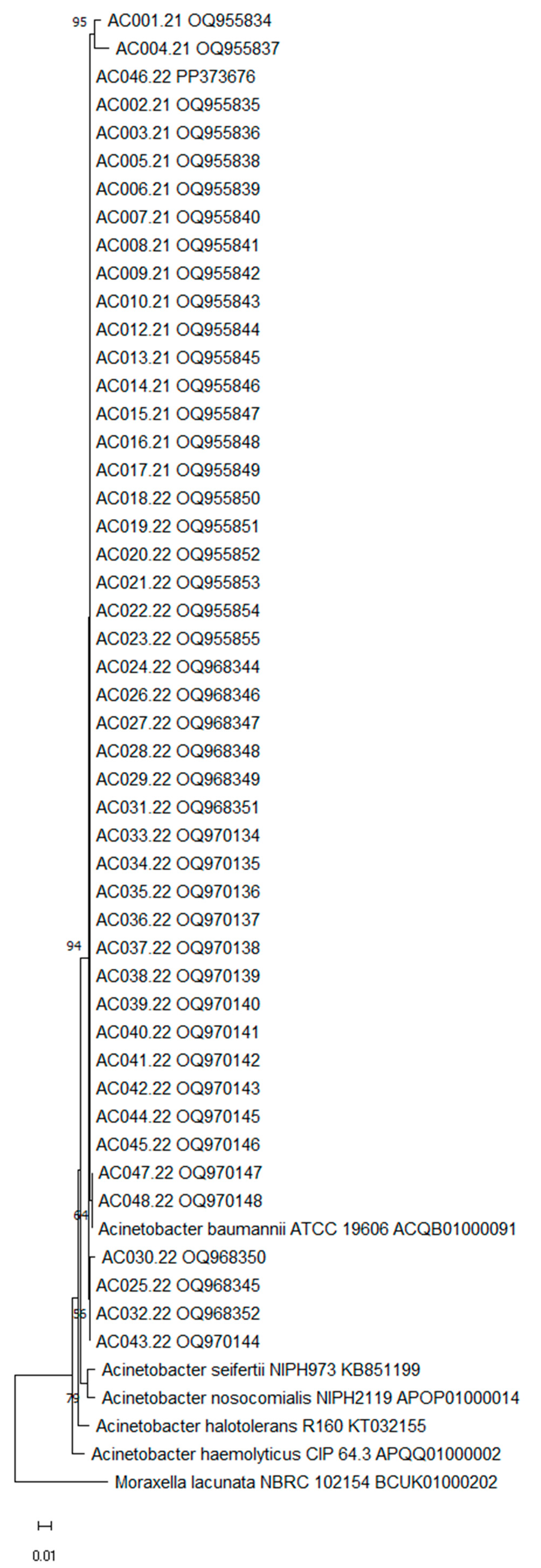
3.1. Identification and Typing

VITEK MS® and MALDI Biotyper® identified all isolates as A. baumannii with ≥80.1% confidence and a score of ≥2.02, respectively (Table 1). Similarly, 16S rRNA gene sequencing identified the strains as A. baumannii with a similarity ≥90.90% (Table A1). The neighbor-joining tree is presented in Figure 1.
According to FTIR spectroscopy, the strains showed 23 distinct profiles and formed 11 clusters with a 0.176 cut-off (Figure 2). The FTIR profile 18 formed the biggest cluster with six strains (AC019, AC023, AC027, AC029, AC031 and AC044) followed by profile 9 with five strains (AC008, AC014, AC020, AC025 and AC026) and profiles 7 (AC006, AC017, AC021 and AC032) and 20 (AC001, AC028, AC034 and AC035) with four strains each.
Both profiles 8 (AC002, AC005 and AC016) and 10 (AC009, AC010 and AC046) grouped three strains together. Finally, the FTIR profiles 5 (AC033 and AC036), 6 (AC041 and AC042), 14 (AC007 and AC039), 17 (AC012 and AC013) and 23 (AC015 and AC024) formed the smallest groups, clustering two strains each.
The FT-IR was also able to cluster most of the strains that were isolated from the same patient. Profile 9 grouped two strains (AC008 and AC014) from patient E. The same was observed in profile 17, where the strains AC012 and AC013, both from patient H, were clustered together. The strains AC028 and AC034, isolated from patient S, were clustered in FT-IR profile 20. Even though strain AC040 was also from patient S and was not clustered in profile 20, it was aligned side by side with the other strains from the same patient at the dendrogram. Finally, the strains AC033 and AC044, isolated from patient Z, were not grouped in the same cluster; however, they were grouped in profiles very close to each other, profiles 5 and 6, respectively.

3.2. Antimicrobial Susceptibility Profile

Regarding the antimicrobial susceptibility profile, the strains showed the same percentage of resistance (95.8%) and susceptibility (4.2%) to TZP, CAZ, CEF and MEM. The same occurred with SXT and GEN, where 93.6% of the strains were resistant and 6.4% were susceptible.
The same percentage of resistance (97.9%) was also observed to IPM, FOX and CIP, while 4.2% were susceptible to IPM and intermediately resistant to FOX and CIP. Regarding the TIG, 21.3% of the strains were resistant, while 36.2% were intermediate resistant, and finally, 42.5% showed susceptibility to this antibiotic.
The same percentage of resistance (87.3%) was observed for CRO, AMI and AMS. Concerning AMS, 4.2% of the strains were intermediately resistant and 8.5% were susceptible. For CRO, 10.6% were intermediately resistant and only 2.1% were susceptible. Finally, for AMI, 12.8% were susceptible.
Detailed information on the results of the antimicrobial susceptibility profile and Magiorakos et al. [1] classification can be observed in Table A2. Forty-three (91.5%) strains were classified as XDR, three (6.4%) strains were MDR and only the strain AC015 was classified as non-MDR. The antimicrobial TIG is not on Magiorakos et al. [1] list since it is a new drug, a glycylcycline class antibiotic. Nevertheless, it was used in the classification in the present study.

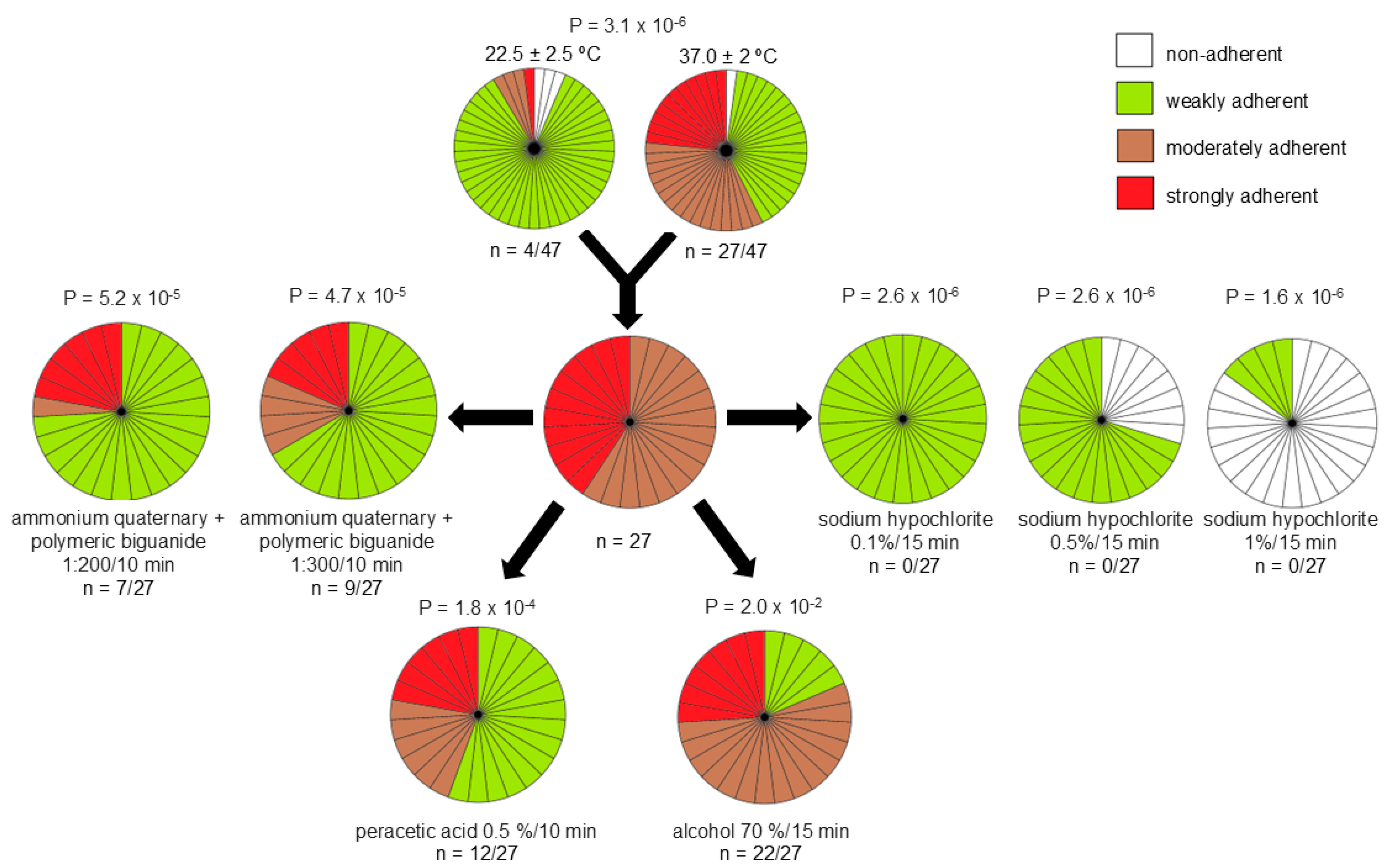
3.3. Biofilm Formation and Its Tolerance to Disinfectants

The biofilm production results can be found in Figure 3. A significant statistical dissimilarity between the temperatures of 22.5 ± 2.5 °C and 37 ± 2 °C was noticed (p = 3.2 × 10⁻⁶). At the temperature of 22.5 ± 2.5 °C, 6.4% (n = 3) of the strains formed moderately adherent biofilms, while one strain formed strongly adherent biofilms. At 37 ± 2 °C, 34.0% (n = 16) of the strains formed moderately adherent biofilmx and 23.4% (n = 11) of the strains formed strongly adherent biofilms.
The 27 (57.4%) strains categorized as moderately or strongly adherent were selected for the biofilm disinfectant tolerance test. Peracetic acid 0.5% was able to reduce the biofilm of fifteen (55.5%) strains to weakly adherent, while the alcohol 70% only reduced the biofilm of five (18.5%) strains. The ammonium quaternary 5th generation stabilized polymeric biguanide diluted 1:300 was able to reduce 18 (66.7%) strains to weakly adherent, while at 1:200 dilution, it reduced the biofilm formation of 20 (74.1%) strains. Exposure to 0.1% sodium hypochlorite for 15 min reduced all the strains to weakly adherent and the concentration of 0.5% reduced the biofilm of eight (29.6%) strains to non-adherent, while the concentration of 1.0% reduced twenty-three (85.2%) strains to non-adherent. Significant statistical values were observed in all disinfectant tolerance tests (p ≤ 1.8 × 10⁻⁴), with the exception of alcohol at 70% (p = 0.02).

4. Discussion

A. baumannii is one of the most prevalent pathogens in hospital-acquired nosocomial infections, and therefore its accurate identification is essential [3,15,17]. Correct bacteria identification is fundamental for establishing efficient disinfection methods, infection control strategies and for the correct use of antibiotics [2]. In this study, all three methods used for identification (VITEK MS®, MALDI Biotyper® and 16S rRNA sequencing) were able to identify all 47 strains to the Acinetobacter species level. Both MALDI TOF/MS systems used possess A. baumannii species in their libraries, and extraction protocols were sufficient to identify the strains with high confidence. These results were similar to previous reports using MALDI-TOF MS and 16S rRNA sequencing for A. baumannii identification [25,27]. Toh et al. [28] reported that MALDI-TOF MS was able to differentiate and identify 100% (n = 47) of the A. baumannii strains even from those that are closely related, such as A. pittii and A. calcoaceticus. Vasconcellos et al. [25] also reported that MALDI-TOF MS was sufficient to identify A. baumannii strains (n = 4) isolated from a pharmaceutical facility; however, it was not enough to differentiate other species of the Acinetobacter calcoaceticus–baumannii complex.
The FT-IR resulted in 23 distinct profiles and 11 clusters where the IR profiles 5, 6, 7, 8, 9, 10, 14, 17 and 23 clustered the strains related to co-infections with SARS-CoV-2, except for AC016 and AC020 (Figure 2). It was not observed that there was a relation between antimicrobial resistance and the FT-IR clusters, since only N-MDR strain was found (AC15), clustered with AC24, an MDR strain (Table A2). Regarding biofilm production, stronger biofilm producers’ strains were found belonging to clusters (n = 7) but also as singletons (n = 5). Nevertheless, the majority of moderate biofilm-producer strains were mainly found in clusters (n = 15/17).
The IR profiles 18 and 20 clustered mostly strains from patients not diagnosed with COVID-19, except for AC023, AC027 and AC035. Regarding cluster 4, the triplicate of the strains AC002 and AC016 showed twice due to the high similarity with the spectrum of AC005. This fact happens majorly due to the high clonality of A. baumannii strains, which suggests that these strains may belong to the same clone. These results corroborate those of the previous study where FTIR was able to discriminate all the P. aeruginosa strains and correctly clustered the strains related to co-infections with SARS-CoV-2 in one IR profile [29].
Other studies proved that IR Biotyper® is an accurate tool for real-time epidemiological investigation of multidrug-resistant A. baumannii nosocomial outbreaks. In the study of Martak et al. [13], the FT-IR was able to differentiate 10 from 11 STs of 20 A. baumannii strains from outbreaks that occurred in hospitals from 11 French cities, reporting a clustering concordance of 0.915. Guerrero-Lozano [14] compared FT-IR with multilocus sequence typing (MLST) and pulsed-field gel electrophoresis (PFGE) techniques for typing 17 nosocomial A. baumannii strains isolated from a hospital. The authors observed that the FTIR results were very consistent with those from MLST, being able to differentiate between three STs (ST 1, ST 2 and ST80). Regarding PFGE, FTIR could differentiate 3/4 of the pulsotypes. Lombardo et al. [30] compared the FT-IR and PFGE for typing 24 A. baumannii strains isolated from ICU, and three different hospitals in the metropolitan area of Bologna, and concluded that FT-IR was fully confirmed by PFGE results. In the future, MLST or PFGE could be performed with the strains isolated from the present study to confirm whether it corroborates with FT-IR results.
The AC015 and AC024 strains, grouped in the IR 23 profile, were isolated from patients with COVID-19 who were hospitalized at ICU-2 and these strains were the only ones susceptible to GEN and SXT. Furthermore, IR profiles 5 and 6 clustered XDR strains that were isolated in the same month from patients with COVID-19. The IR 17 profile clustered the AC012 and AC013 strains, which were isolated from the same patient, on the same day but at different sources and formed a weak biofilm at 22.5 ± 2.5 °C. The strains AC001 and AC004 (Patient A), AC006 and AC007 (Patient E), AC018, AC022 and AC023 (Patient M), AC019 and AC020 (Patient N), AC021 and AC026 (Patient O), AC036 and AC038 (Patient Y), AC042 and AC046 (Patient AC) did not cluster with the strains that were isolated from the specific patient. This could indicate that possibly there were different sources of contamination in the hospital.
Infections caused by A. baumannii have become increasingly severe due to their antimicrobial resistance to commonly used antibiotics, such as β-lactams, aminoglycosides and even carbapenems [31]. Colistin is also reported as a valuable therapeutic option. Unfortunately, it was not possible to test this drug in the present study. According to Table A2, the highest level of resistance was observed for IMP, CIP and FOX with 97.9%, followed by 95.8% for TZP, CAZ, CEF and MEM. Elevated levels (87.3%) of resistance were also observed for AMI, AMS and CRO.
A. baumannii resistance to carbapenems is highlighted by the WHO as a formidable global challenge due to the limited treatment options, which leads to severe nosocomial infections [8]. Sulphonamides can be used as alternatives in the treatment of CARB [32]; however, in this study, 93.6% of the isolates were resistant to SXT, which restricts even more the therapeutic options available. These data corroborate the results provided in the study of Al-Tamimi et al. [33], where 49.9% of the A. baumannii strains were resistant to SXT.
TIG is a unique glycylcycline class of antibiotics and is considered a last resort antibiotics in the therapy of illness caused by MDR bacteria [34,35,36]. An increase in resistance rates to TIG among Acinetobacter spp. has been reported [35,36,37], with a high resistance rate of 66% found in Israel [37]. Although TIG had the lowest rate of resistance (21.3%) in the present study, the FDA [38] reported that there is an increased risk of mortality with tigecycline in comparison to other drugs. Furthermore, tigecycline is not recommended for use with children and teenagers under 18 years old [35].
The incidence of MDR and XDR A. baumannii strains represents a challenge in clinical treatment, and results in increasing rates of mortality around the world [39]. In the present study, most of the strains (91.5%) were classified as XDR and 6.4% of the strains were MDR (Figure 2), regardless of the COVID-19 co-infection, since only the AC015 strain, classified as non-MDR, was isolated from a patient with COVID-19. However, the AC015 strain still represents a challenge to public health since it was able to form moderately and strongly adherent biofilms at temperatures of 22.5 ± 2.5 °C and 37 ± 2 °C, respectively.
A. baumannii co-infection in patients with SARS-CoV-2 was widely reported during the pandemic due to its capacity to produce biofilms on medical devices used in endotracheal intubation [40]. A. baumannii biofilms contribute to its persistence in the hospital environment and consequently increase the risk of infection by this microorganism. In the present study, 57.4% of the strains were able to form moderately or strongly adherent biofilms at 37 °C in polystyrene surfaces (Figure 3). These data corroborate the results of the study of Yang et al. [9], where 32.5 and 45.4% of A. baumannii strains isolated from a teaching hospital formed moderate and strong biofilms, respectively. Vasconcellos et al. [25] also reported that 47.4% of A. baumannii strains were able to form strongly adherent biofilms in polystyrene surfaces.
A. baumannii biofilm formation also has an important role in antibiotic resistance and disinfectant tolerance, which makes the choice of the most effective treatment or cleaning protocols even more limited [11]. In this study, none of the disinfectants tested were able to reduce all the A. baumannii strains to non-adherent. Even though 70% alcohol is largely used for disinfection of hospital surfaces, in the present study, it was not efficient in the biofilm reduction (Figure 3). The most effective disinfectant and the only one to reduce some biofilms to non-adherent was sodium hypochlorite, which in the concentrations of 0.5% and 1.0% reduced 29.6 and 85.12% of the strains, respectively, to non-adherent (Figure 3). However, it cannot be used on surfaces that are sensitive to corrosion. For that reason, other sanitizers and disinfectants and time exposure must be evaluated.

5. Conclusions

In conclusion, VITEK MS®, MALDI Biotyper® and 16S rRNA gene sequencing accurately identified all A. baumannii strains. FTIR was considered a useful tool for A. baumannii typing since it was able to differentiate the strains from patients with and without COVID-19. Most of the isolates were intermediate resistant or resistant to the antibiotics evaluated, while TIG was demonstrated to be the best treatment option for A. baumannii with the lowest rate of resistance. Almost all strains (n = 43) were classified as XDR, while three strains were MDR and only one strain was classified as non-MDR. Over half of the strains (n = 27) were able to form moderately or strongly adherent biofilm on stainless steel surfaces. It was observed that A. baumannii strains already possess these virulent characteristics regardless of COVID-19.

Author Contributions

Conceptualization, P.A.d.S., L.V.d.C., M.L.L.B., R.P.P.d.S. and M.H.S.V.B.; methodology, P.A.d.S., M.C.N.d.S., R.V.d.S.L.d.M., L.V.d.C., R.P.P.d.S., C.A.C.d.M. and M.L.L.B.; software, P.A.d.S., M.C.N.d.S., R.V.d.S.L.d.M., L.V.d.C., R.P.P.d.S., C.A.C.d.M. and M.L.L.B.; validation, P.A.d.S., M.C.N.d.S., R.V.d.S.L.d.M., L.V.d.C., R.P.P.d.S., C.A.C.d.M. and M.L.L.B.; formal analysis, P.A.d.S., M.C.N.d.S., P.A.d.S., S.J.F., C.A.C.d.M., M.H.S.V.B., G.M.S.d.C. and M.L.L.B.; investigation, P.A.d.S., M.C.N.d.S., R.V.d.S.L.d.M., L.V.d.C., R.P.P.d.S., C.A.C.d.M. and M.L.L.B.; resources, P.A.d.S., M.C.N.d.S., R.V.d.S.L.d.M., L.V.d.C., R.P.P.d.S., C.A.C.d.M. and M.L.L.B.; data curation, P.A.d.S., M.C.N.d.S., R.V.d.S.L.d.M., L.V.d.C., R.P.P.d.S., P.A.d.S., C.A.C.d.M., G.M.S.d.C. and M.L.L.B.; writing—original draft preparation, P.A.d.S.; writing—review and editing, L.V.d.C., R.P.P.d.S., C.A.C.d.M., S.J.F., M.H.S.V.B. and M.L.L.B.; visualization, P.A.d.S., M.C.N.d.S., P.A.d.S., S.J.F., M.H.S.V.B. and M.L.L.B.; supervision, M.L.L.B. and M.H.S.V.B.; project administration, M.L.L.B. and M.H.S.V.B.; funding acquisition, M.L.L.B. and M.H.S.V.B. All authors have read and agreed to the published version of the manuscript.

Funding

This research was funded in part by the Conselho Nacional de Desenvolvimento Científico e Tecnológico (CNPq): CNPq/MCTI/FNDCT No 18/2021-Faixa A-Grupos Emergentes Process n.º407747/2021-4, Fundação de Amparo à Pesquisa do Estado do Rio de Janeiro (FAPERJ): PROGRAMA E-13/2023 -AUXÍLIO BÁSICO À PESQUISA (APQ1)—2023/Ref. Proc. E-26/210.616/2024, and Coordenação de Aperfeiçoamento de Pessoal de Nível Superior—Brasil (CAPES)—Finance Code 001.

Institutional Review Board Statement

The present study was realized according to the Declaration of Helsinki and approved 15 February 2022 by the Hospital de Força Aérea do Galeão (HFAG) Ethics and Research Committee and is registered on Plataforma Brasil with CAAE code 55303721.0.0000.5250.

Informed Consent Statement

Informed consent was obtained from all subjects involved in the study.

Data Availability Statement

The data underlying this article are available in the article.

Acknowledgments

The authors are grateful to Bio-Manguinhos/Fiocruz, the Postgraduation Program in Biodiversity and Health Program of IOC/Fiocruz, CNPq, FAPERJ, and CAPES.

Conflicts of Interest

Author S.J. Forsythe was employed by the company Foodmicrobe.com Ltd. The remaining authors declare that the research was conducted in the absence of any commercial or financial relationships that could be construed as a potential conflict of interest.

Appendix A

Appendix A.1

Table A1. 16S rDNA sequencing analysis of Acinetobacter baumannii strains (n = 47).
Strain IDNCBI a Access NumberBase Pair Length (nt)Identification (%)
AC001/21OQ955834728A. baumannii (99.59)
AC002/21OQ955835842A. baumannii (99.88)
AC003/21OQ955836860A. baumannii (99.88)
AC004/21OQ955837704A. baumannii (97.28)
AC005/21OQ955838851A. baumannii (99.88)
AC006/21OQ955839974A. baumannii (99.90)
AC007/21OQ955840858A. baumannii (99.88)
AC008/21OQ9558411.030A. baumannii (99.90)
AC009/21OQ955842984A. baumannii (99.90)
AC010/21OQ955843940A. baumannii (99.90)
AC012/21OQ955844874A. baumannii (99.88)
AC013/21OQ955845980A. baumannii (99.90)
AC014/21OQ955846943A. baumannii (99.89)
AC015/21OQ955847922A. baumannii (99.89)
AC016/21OQ955848861A. baumannii (99.88)
AC017/21OQ955849986A. baumannii (99.90)
AC018/22OQ9558501.036A. baumannii (99.90)
AC019/22OQ955851945A. baumannii (99.89)
AC020/22OQ955852984A. baumannii (99.89)
AC021/22OQ955853860A. baumannii (99.88)
AC022/22OQ955854884A. baumannii (99.77)
AC023/22OQ955855845A. baumannii (99.88)
AC024/22OQ9683441.036A. baumannii (99.71)
AC025/22OQ9683451.034A. baumannii (99.90)
AC026.22OQ9683461.037A. baumannii (99.90)
AC027/22OQ968347873A. baumannii (99.88)
AC028/22OQ968348945A. baumannii (99.68)
AC029/22OQ9683491.034A. baumannii (99.90)
AC030/22OQ9683501.043A. baumannii (99.61)
AC031/22OQ9683511.043A. baumannii (99.90)
AC032/22OQ9683521.033A. baumannii (99.90)
AC033/22OQ9701341.031A. baumannii (99.90)
AC034/22OQ9701351.034A. baumannii (99.90)
AC035/22OQ9701361.042A. baumannii (99.90)
AC036/22OQ970137453A. baumannii (99.77)
AC037/22OQ9701381.031A. baumannii (99.90)
AC038/22OQ9701391.032A. baumannii (99.90)
AC039/22OQ9701401.033A. baumannii (99.90)
AC040/22OQ9701411.032A. baumannii (99.90)
AC041/22OQ9701421.020A. baumannii (90.90)
AC042/22OQ9701431.035A. baumannii (90.90)
AC043/22OQ9701441.034A. baumannii (90.90)
AC044/22OQ9701451.003A. baumannii (90.90)
AC045/22OQ9701461.034A. baumannii (90.90)
AC046/22PP3736761.306A. baumannii (90.90)
AC047/22OQ9701471.006A. baumannii (100)
AC048/22OQ9701481.003A. baumannii (100)
a National Center for Biotechnology Information.

Appendix A.2

Table A2. Antimicrobial susceptibility profile of Acinetobacter baumannii strains (n = 47).
StrainsAMSTZPFOXCAZCROCEFIPMMEMGENCIPTIGSXTAMIMagiorakos et al. [1] Classification
AC001.21I RRRRRRRRRSRRXDR
AC002.21RRRRRRRRRRSRRXDR
AC003.21RRRRRRRRRRSRRXDR
AC004.21RRRRRRRRRRSRRXDR
AC005.21RRRRRRRRRRSRSXDR
AC006.21RRRRIRRRRRRRRXDR
AC007.21RRRRIRRRRRRRRXDR
AC008.21RRRRIRRRRRRRRXDR
AC010.21RRRRRRRRRRSRRXDR
AC012.21RRRRRRRRRRSRRXDR
AC013.21RRRRRRRRRRIRRXDR
AC014.21SRRRIRRRRRRRRXDR
AC015.21SSRSISSSSISSSN-MDR
AC016.21RRRRRRRRRR SRRXDR
AC017.21RRRRRRRRRRIRRXDR
AC018.22RRRRRRRRRRIRSXDR
AC019.22RRRRRRRRRRIRRXDR
AC020.22RRRRRRRRRRIRRXDR
AC021.22RRRRRRRRRRIRRXDR
AC022.22SRRRRRRRRRSRRXDR
AC023.22RRRRRRRRRRSRRXDR
AC024.22RRRRRRRRSRISRMDR
AC025.22RRRRRRRRSRSRSMDR
AC026.22RRRRRRRRRRIRRXDR
AC027.22RRRRRRRRRRSRRXDR
AC028.22RRRRRRRRRRIRRXDR
AC029.22RRRRRRRRRRSRRXDR
AC030.22RRRRRRRRRRRSSXDR
AC031.22RRRRRRRRRRIRRXDR
AC032.22RRRRRRRRRRIRRXDR
AC033.22RRRRRRRRRRRRRXDR
AC034.22RRRRRRRRRRRRRXDR
AC035.22RRRRRRRRRRIRRXDR
AC036.22RRRRRRRRRRSRRXDR
AC037.22RRRRRRRRRRIRRXDR
AC038.22RRRRRRRRRRIRRXDR
AC039.22RRRRRRRRRRRRRXDR
AC040.22RRRRRRRRRRRRRXDR
AC041.22IRRRRRRRRRSRRXDR
AC042.22RRRRRRRRRRRRRXDR
AC043.22RRRRRRRRRRSRRXDR
AC044.22RRRRRRRRRRIRRXDR
AC045.22RRRRRRRRRRIRRXDR
AC046.22RRRRRRRRRRSRRXDR
AC047.22SSISSSRSRRSRSMDR
AC048.22RRRRRRRRRRIRRXDR
R: resistant; I: intermediate resistant; S: susceptible; MDR: multidrug-resistant; XDR: extensively drug-resistant; N-MDR: non-multidrug-resistant; meropenem (MEM); imipenem (IPM); sulfamethoxazole–trimethoprim (SXT); piperacillin–tazobactam (TZP); amikacin (AMI); gentamicin (GEN); ciprofloxacin (CIP); ceftazidime (CAZ); cefoxitin (FOX); cefepime (CEF); ceftriaxone (CRO); tigecycline (TIG); ampicillin–sulbactam (AMS).

References

  1. Magiorakos, A.-P.; Srinivasan, A.; Carey, R.B.; Carmeli, Y.; Falagas, M.E.; Giske, C.G.; Harbarth, S.; Hindler, J.F.; Kahlmeter, G.; Olsson-Liljequist, B.; et al. Multidrug-resistant, extensively drug-resistant and pandrug-resistant bacteria: An international expert proposal for interim standard definitions for acquired resistance. Clin. Microbiol. Infect. 2012, 18, 268–281. [Google Scholar] [CrossRef] [PubMed]
  2. Fitzpatrick, M.A.; Ozer, E.; Bolon, M.K.; Hauser, A.R. Influence of ACB complex genospecies on clinical outcomes in a U.S. hospital with high rates of multidrug resistance. J. Infect. 2015, 70, 144–152. [Google Scholar] [CrossRef] [PubMed]
  3. Lee, C.-R.; Lee, J.H.; Park, M.; Park, K.S.; Bae, I.K.; Kim, Y.B.; Cha, C.-J.; Jeong, B.C.; Lee, S.H. Biology of Acinetobacter baumannii: Pathogenesis, Antibiotic Resistance Mechanisms, and Prospective Treatment Options. Front. Cell. Infect. Microbiol. 2017, 7, 55–90. [Google Scholar] [CrossRef]
  4. Ibrahim, S.; Al-Saryi, N.; Al-Kadmy, I.M.S.; Aziz, S.N. Multidrug-resistant Acinetobacter baumannii as an emerging concern in hospitals. Mol. Biol. Rep. 2021, 48, 6987–6998. [Google Scholar] [CrossRef] [PubMed]
  5. Alenazi, T.A.; Bin Shaman, M.S.; Suliman, D.M.; Alanazi, T.A.; Altawalbeh, S.M.; Alshareef, H.; Lahreche, D.I.; Al-Azzam, S.; Araydah, M.; Karasneh, R.; et al. The Impact of Multidrug-Resistant Acinetobacter baumannii Infection in Critically Ill Patients with or without COVID-19 Infection. Healthcare 2023, 11, 487. [Google Scholar] [CrossRef] [PubMed]
  6. FAO (United Nations Food and Agriculture Organization). The FAO Action Plan on Antimicrobial Resistance 2016–2020; FAO: Rome, Italy, 2016. [Google Scholar]
  7. Ergonul, O.; Tokca, G.; Keske, Ş.; Donmez, E.; Madran, B.; Kömür, A.; Gönen, M.; Can, F. Elimination of healthcare-associated Acinetobacter baumannii infection in a highly endemic region. Int. J. Infect. Dis. 2022, 114, 11–14. [Google Scholar] [CrossRef]
  8. WHO. World Health Organization (WHO). WHO Bacterial Priority Pathogens List. WHO. 2024. Available online: https://www.who.int/publications/i/item/9789240093461 (accessed on 20 February 2025).
  9. Yang, C.-H.; Su, P.-W.; Moi, S.-H.; Chuang, L.-Y. Biofilm Formation in Acinetobacter baumannii: Genotype-Phenotype Correlation. Molecules 2019, 24, 1849. [Google Scholar] [CrossRef]
  10. Choudhary, M.; Shrivastava, R.; Vashistt, J. Acinetobacter baumannii Biofilm Formation: Association with Antimicrobial Resistance and Prolonged Survival under Desiccation. Curr. Microbiol. 2022, 79, 1–9. [Google Scholar] [CrossRef] [PubMed]
  11. Roy, S.; Chowdhury, G.; Mukhopadhyay, A.K.; Dutta, S.; Basu, S. Convergence of Biofilm Formation and Antibiotic Resistance in Acinetobacter baumannii Infection. Front. Med. 2022, 9, 793615. [Google Scholar] [CrossRef]
  12. Oho, M.; Nagasawa, Z.; Funashima, Y.; Ueda, O.; Watamabe, S.; Cui, L.; Miyamoto, H.; Sueoka, E. Correlation of Strain Classification with IR Biotyper and Molecular Epidemiological Method of Pseudomonas aeruginosa. Rinsho Biseibutshu Jinsoku Shindan Kenkyukai Shi 2021, 31, 29–40. [Google Scholar] [PubMed]
  13. Martak, D.; Valot, B.; Sauget, M.; Cholley, P.; Thouverez, M.; Bertrand, X.; Hocquet, D. Fourier-Transform InfraRed Spectroscopy Can Quickly Type Gram-Negative Bacilli Responsible for Hospital Outbreaks. Front. Microbiol. 2019, 10, 1440. [Google Scholar] [CrossRef]
  14. Guerrero-Lozano, I.; Galán-Sánchez, F.; Rodríguez-Iglesias, M. Fourier transform infrared spectroscopy as a new tool for surveillance in local stewardship antimicrobial program: A retrospective study in a nosocomial Acinetobacter baumannii outbreak. Braz. J. Microbiol. 2022, 53, 1349–1353. [Google Scholar] [CrossRef]
  15. Moubareck, C.A.; Halat, D.H. Insights into Acinetobacter baumannii: A review of microbiological, virulence, and resistance traits in a threatening nosocomial pathogen. Antibiotics 2020, 9, 119. [Google Scholar] [CrossRef] [PubMed]
  16. Čiginskienė, A.; Dambrauskienė, A.; Rello, J.; Adukauskienė, D. Ventilator-associated pneumonia due to drug-resistant Acinetobacter baumannii: Risk factors and mortality relation with resistance profiles, and independent predictors of in-hospital mortality. Medicina 2019, 55, 49. [Google Scholar] [CrossRef]
  17. Rangel, K.; Chagas, T.P.G.; De-Simone, S.G. Acinetobacter baumannii Infections in Times of COVID-19 Pandemic. Pathogens 2021, 10, 1006. [Google Scholar] [CrossRef] [PubMed]
  18. De Benedetto, I.; Lupia, T.; Shbaklo, N.; Bianchi, A.; Concialdi, E.; Penna, M.; Corcione, S.; De Rosa, F.G. Prognostic evaluation of Acinetobacter baumannii ventilator-associated pneumonia in COVID-19. Infez Med. 2022, 30, 570–576. [Google Scholar] [PubMed]
  19. Russo, A.; Gavaruzzi, F.; Ceccarelli, G.; Borrazzo, C.; Oliva, A.; Alessandri, F.; Magnanimi, E.; Pugliese, F.; Venditti, M. Multidrug-resistant Acinetobacter baumannii infections in COVID-19 patients hospitalized in intensive care unit. Infection 2021, 50, 83–92. [Google Scholar] [CrossRef]
  20. Novović, K.; Nedeljković, S.K.; Poledica, M.; Nikolić, G.; Grujić, B.; Jovčić, B.; Kojić, M.; Filipić, B. Virulence potential of multidrug-resistant Acinetobacter baumannii isolates from COVID-19 patients on mechanical ventilation: The first report from Serbia. Front. Microbiol. 2023, 14, 1094184. [Google Scholar] [CrossRef] [PubMed]
  21. Dai, Y.; Tao, S.; Ying, H.; Fang, Q.; Kong, J.; Guo, F.; Yang, Y.; Cao, P.; Zhou, Y.; Jin, W.; et al. Molecular pathogen profiling of COVID-19 coinfections. BMC Infect. Dis. 2024, 24, 1451. [Google Scholar] [CrossRef] [PubMed]
  22. Yoon, S.-H.; Ha, S.-M.; Kwon, S.; Lim, J.; Kim, Y.; Seo, H.; Chun, J. Introducing EzBioCloud: A taxonomically united database of 16S rRNA gene sequences and whole-genome assemblies. Int. J. Syst. Evol. Microbiol. 2017, 67, 1613–1617. [Google Scholar] [CrossRef]
  23. Tamura, K.; Stecher, G.; Kumar, S. MEGA11: Molecular Evolutionary Genetics Analysis Version 11. Mol. Biol. Evol. 2021, 38, 3022–3027. [Google Scholar] [CrossRef] [PubMed]
  24. Clinical and Laboratory Standards Institute (CLSI). Performance Standards for Antimicrobial Susceptibility Testing, 30th ed.; CLSI Supplement M100; CLSI: Berwyn, PA, USA, 2020. [Google Scholar]
  25. Vasconcellos, L.; Silva, S.V.; da Costa, L.V.; da Silva Lage de Miranda, R.V.; Reis, C.M.F.D.; da Silva Braga, L.M.P.; Silva, C.; Conceição, G.; Mattoso, J.; Silva, I.B.; et al. Phenotypical and molecular characterization of Acinetobacter spp. isolated from a pharmaceutical facility. Lett. Appl. Microbiol. 2023, 76, ovad101. [Google Scholar] [CrossRef]
  26. Stepanović, S.; Vuković, D.; Hola, V.; Bonaventura, G.D.; Djukić, S.; Ćirković, I.; Ruzicka, F. Quantification of biofilm in microtiter plates: Overview of testing conditions and practical recommendations for assessment of biofilm production by staphylococci. APMIS 2007, 115, 891–899. [Google Scholar] [CrossRef] [PubMed]
  27. Calderaro, A.; Chezzi, C. MALDI-TOF MS: A Reliable Tool in the Real Life of the Clinical Microbiology Laboratory. Microorganisms 2024, 12, 322. [Google Scholar] [CrossRef] [PubMed]
  28. Toh, B.E.; Paterson, D.L.; Kamolvit, W.; Zowawi, H.; Kvaskoff, D.; Sidjabat, H.; Wailan, A.; Peleg, A.Y.; Huber, C.A. Species identification within Acinetobacter calcoaceticusbaumannii complex using MALDI-TOF MS. J. Microbiol. Methods 2015, 118, 128–132. [Google Scholar] [CrossRef] [PubMed]
  29. de Souza, P.A.; Santos, M.C.S.d.; de Miranda, R.V.d.S.L.; da Costa, L.V.; da Silva, R.P.P.; de Miranda, C.A.C.; da Silva, A.P.R.; Forsythe, S.J.; Bôas, M.H.S.V.; Brandão, M.L.L. Evaluation of antimicrobial resistance patterns of Pseudomonas aeruginosa strains isolated among COVID-19 patients in Brazil typed by Fourier-transform infrared spectroscopy. Life 2024, 14, 1079. [Google Scholar] [CrossRef]
  30. Lombardo, D.; Cordovana, M.; Deidda, F.; Pane, M.; Ambretti, S. Application of Fourier Transform Infrared Spectroscopy for Real-Time Typing of Acinetobacter baumannii Outbreak in Intensive Care Unit. Futur. Microbiol. 2021, 16, 1239–1250. [Google Scholar] [CrossRef] [PubMed]
  31. Wu, H.-J.; Xiao, Z.-G.; Lv, X.-J.; Huang, H.-T.; Liao, C.; Hui, C.-Y.; Xu, Y.; Li, H.-F. Drug-resistant Acinetobacter baumannii: From molecular mechanisms to potential therapeutics (Review). Exp. Ther. Med. 2023, 25, 209. [Google Scholar] [CrossRef]
  32. Falagas, M.E.; Vardakas, K.Z.; Roussos, N.S. Trimethoprim/sulfamethoxazole for Acinetobacter spp.: A review of current microbiological and clinical evidence. Int. J. Antimicrob Agents 2015, 46, 231–241. [Google Scholar] [CrossRef]
  33. Al-Tamimi, M.; Albalawi, H.; Alkhawaldeh, M.; Alazzam, A.; Ramadan, H.; Altalalwah, M.; Alma’aitah, A.; Al Balawi, D.A.; Shalabi, S.; Abu-Raideh, J.; et al. Multidrug-resistant Acinetobacter baumannii in Jordan. Microorganisms 2022, 10, 849. [Google Scholar] [CrossRef] [PubMed]
  34. Dehbanipour, R.; Ghalavand, Z. Acinetobacter baumannii: Pathogenesis, virulence factors, novel therapeutic options and mechanisms of resistance to antimicrobial agents with emphasis on tigecycline. J. Clin. Pharm. Ther. 2022, 47, 1875–1884. [Google Scholar] [CrossRef]
  35. Yaghoubi, S.; Zekiy, A.O.; Krutova, M.; Gholami, M.; Kouhsari, E.; Sholeh, M.; Ghafouri, Z.; Maleki, F. Tigecycline antibacterial activity, clinical effectiveness, and mechanisms and epidemiology of resistance: Narrative review. Eur. J. Clin. Microbiol. Infect. Dis. 2022, 41, 1003–1022. [Google Scholar] [CrossRef]
  36. Hua, X.; He, J.; Wang, J.; Zhang, L.; Zhang, L.; Xu, Q.; Shi, K.; Leptihn, S.; Shi, Y.; Fu, X.; et al. Novel tigecycline resistance mechanisms in Acinetobacter baumannii mediated by mutations in adeS, rpoB and rrf. Emerg Microbes Infect 2021, 10, 1404–1417. [Google Scholar] [CrossRef] [PubMed]
  37. Reid, G.E.; Grim, S.A.; Aldeza, C.A.; Janda, W.M.; Clark, N.M. Rapid Development of Acinetobacter baumannii Resistance to Tigecycline. Pharmacother. J. Hum. Pharmacol. Drug Ther. 2007, 27, 1198–1201. [Google Scholar] [CrossRef] [PubMed]
  38. FDA. Drug safety communication: Increased risk of death with Tygacil (tigecycline) compared to other antibiotics used to treat similar infections (1 September 2010). In FDA Drug Safety Communication: FDA Warns of Increased Risk of Death with IV Antibacterial Tygacil (Tigecycline) and Approves New Boxed Warning; US Food and Drug Administration: Silver Spring, MD, USA, 2017. [Google Scholar]
  39. Shi, J.; Sun, T.; Cui, Y.; Wang, C.; Wang, F.; Zhou, Y.; Miao, H.; Shan, Y.; Zhang, Y. Multidrug resistant and extensively drug resistant Acinetobacter baumannii hospital infection associated with high mortality: A retrospective study in the pediatric intensive care unit. BMC Infect. Dis. 2020, 20, 597. [Google Scholar] [CrossRef] [PubMed]
  40. Loyola-Cruz, M.Á.; Durán-Manuel, E.M.; Cruz-Cruz, C.; Márquez-Valdelamar, L.M.; Bravata-Alcántara, J.C.; Cortés-Ortíz, I.A.; Cureño-Díaz, M.A.; Ibáñez-Cervantes, G.; Fernández-Sánchez, V.; Castro-Escarpulli, G.; et al. ESKAPE bacteria characterization reveals the presence of Acinetobacter baumannii and Pseudomonas aeruginosa outbreaks in COVID-19/VAP patients. Am. J. Infect Control 2023, 51, 729–737. [Google Scholar] [CrossRef]
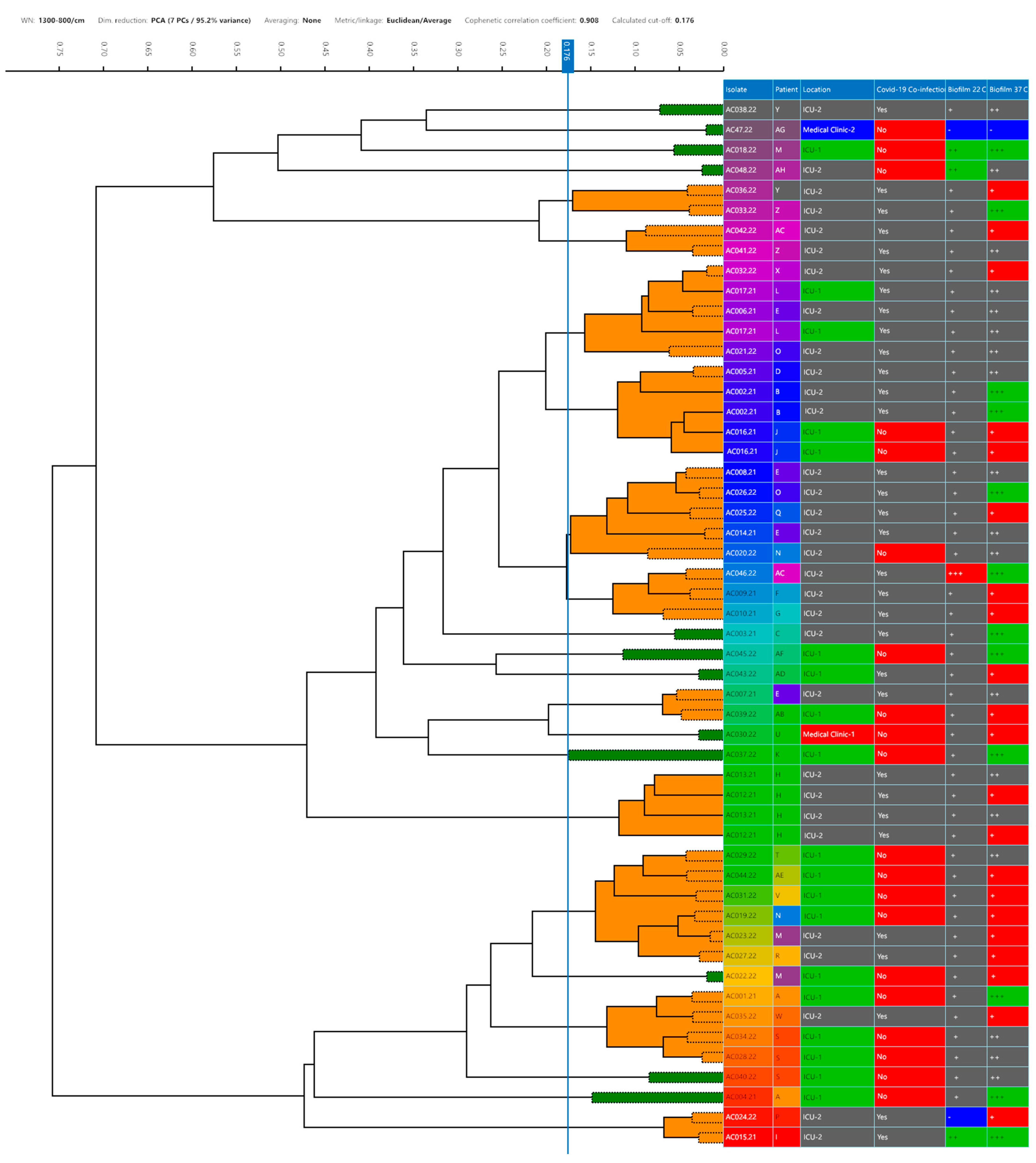
Figure 1. Neighbor-joining tree based on partial 16S rRNA gene sequences (549 bp) showing the phylogenetic position of the Acinetobacter baumannii strains evaluated in the present study (n = 47). The numbers at the nodes indicate the percentage of 1000 bootstrap replicates; only values > 50% are shown. A. seifertii, A. nosocomialis, A. halotolerans, A. haemolyticus and Moraxella lacunata was used as an outgroup. The scale bar represents 0.01 substitutions per nucleotide position. GenBank accession number is given.
Life 15 00623 g001
Figure 2. Dendrograms obtained by clustering FTIR spectra for Acinetobacter baumannii strains (n = 47). The vertical line represents the cut-off value. Green spectra indicate that the strain was a singleton, and orange spectra indicate a cluster formation. Different colors are used to differentiate: source; COVID-19 co-infection (yes or no); location and biofilm formation on stainless-steel surfaces (+ = positive; ‘-’= negative) and polystyrene (‘-‘ = non-adherent; + = weakly adherent; ++ = moderately adherent; and +++ = strongly adherent).
Life 15 00623 g002
Figure 3. Biofilm formation of Acinetobacter baumannii strains (n = 47) and assessment of biofilm sensitivity to disinfectants. Color legend: white—non-adherent; green—weak adherent; brown—moderate adherent; red—strong adherent. p-values are presented.
Life 15 00623 g003
Table 1. Details of Acinetobacter baumannii strains (n = 47) isolated in a Brazilian hospital during the COVID-19 pandemic.
Strain (CBAS a ID)SourcePatient IDHospital DepartmentCOVID-19 Co-InfectionIsolation DateVITEK® 2 Bionumber (Profile)MALDI-TOF MS b
VITEK MS® (%)MALDI Biotyper® (Score)
AC001/21 (936)Tracheal swabAICU c-1no21 June 0241010103500250 (I)A. baumannii (98.4)A. baumannii (2.43)
AC002/21(937)Tracheal swabBICU-2yes21 June 0201010103500210 (II)A. baumannii (99.9)A. baumannii (2.33)
AC003/21 (938)Tracheal swabCICU-2yes21 June 0241010103500352 (III)A. baumannii (99.9)A. baumannii (2.43)
AC004/21 (939)Tracheal swabAICU-1no21 June 0241010103500210 (IV)A. baumannii (95.0)A. baumannii (2.31)
AC005/21 (940)BloodDICU-2yes21 June 0201010103500352 (V)A. baumannii (99.9)A. baumannii (2.36)
AC006/21 (941)BloodEICU-2yes21 June 0201010103500312 (VI)A. baumannii (99.9)A. baumannii (2.28)
AC007/21 (942)BloodEICU-2yes21 June 0241010103500352 (III)A. baumannii (99.9)A. baumannii (2.04)
AC008/21 (943)BloodEICU-2yes21 June 0241010103500310 (VII)A. baumannii (81.7)A. baumannii (2.25)
AC009/21 (944)UrineFICU-2yes21 July 0201010103500210 (II)A. baumannii (99.9)A. baumannii (2.33)
AC010/21 (945)Tracheal secretionGICU-2Yes21 July 0201010103500210 (II)A. baumannii (99.9)A. baumannii (2.38)
AC012/21 (946)UrineHICU-2Yes21 July 0241010103500310 (VII)A. baumannii (99.9)A. baumannii (2.21)
AC013/21 (947)Tracheal secretionHICU-2Yes21 July 0201010103500352 (V)A. baumannii (99.9)A. baumannii (2.22)
AC014/21 (948)BloodEICU-2Yes21 July 0241010003500312 (VIII)A. baumannii (99.9)A. baumannii (2.32)
AC015/21 (949)BloodIICU-2Yes21 July 0201010103500312 (VI)A. baumannii (97.0)A. baumannii (2.29)
AC016/21 (950)BloodJICU-1No21 July 0241010103500310 (VII)A. baumannii (99.9)A. baumannii (2.26)
AC017/21 (951)BloodLICU-2Yes21 September 0241010103500210 (IV)A. baumannii (99.9)A. baumannii (2.15)
AC018/22 (952)Rectal swabMICU-1No22 January 0241010103500250 (I)A. baumannii (81.3)A. baumannii (2.25)
AC019/22 (953)Rectal swabNICU-1No22 January 0241010003500210 (IX)A. baumannii (83.0)A. baumannii (2.37)
AC020/22 (954)Tracheal secretionNICU-1No22 February 0241010103500212 (X)A. baumannii (99.4)A. baumannii (2.23)
AC021/22 (955)Tracheal secretionOICU-2Yes22 February 0241010103500352 (III)A. baumannii (99.9)A. baumannii (2.33)
AC022/22 (956)UrineMICU-1No22 January 0241010103500210 (IV)A. baumannii (99.9)A. baumannii (2.36)
AC023/22 (957)Tracheal secretionMICU-2Yes22 February 0241010103500210 (IV)A. baumannii (99.9)A. baumannii (2.02)
AC024/22 (958)Tracheal secretionPICU-2Yes22 February 0241010103500312 (XI)A. baumannii (99.9)A. baumannii (2.36)
AC025/22 (959)Tracheal secretionQICU-2Yes22 February 0241010003500310 (XII)A. baumannii (99.9)A. baumannii (2.32)
AC026/22 (960)BloodOICU-2Yes22 February 0241010103500310 (VII)A. baumannii (98.0)A. baumannii (2.41)
AC027/22 (961)BloodRICU-2Yes22 February 0201010103500210 (II)A. baumannii (99.9)A. baumannii (2.00)
AC028/22 (962)Tracheal secretionSICU-1no22 February 0201010003500210 (XIII)A. baumannii (99.9)A. baumannii (2.31)
AC029/22 (963)Tracheal secretionTICU-1no22 February 0241010103500310 (VII)A. baumannii (99.9)A. baumannii (2.24)
AC030/22 (964)Tracheal secretionUMedical clinic-1no22 February 0241010003500310 (XII)A. baumannii (99.9)A. baumannii (2.41)
AC031/22 (965)BloodVICU-1no22 February 0241010103500210 (IV)A. baumannii (88.8)A. baumannii (2.29)
AC032/22 (966)Rectal swabXICU-2yes22 February 0243051103500352 (XIV)A. baumannii (99.5)A. baumannii (2.46)
AC033/22 (967)Rectal swabZICU-2yes22 February 0201010003500210 (XIII)A. baumannii (99.9)A. baumannii (2.13)
AC034/22 (968)Tracheal secretionSICU-1no22 February 0241010103500210 (IV)A. baumannii (99.9)A. baumannii (2.44)
AC035/22 (969)Rectal swabWICU-2yes22 February 0241010003500210 (IX)A. baumannii (96.2)A. baumannii (2.20)
AC036/22 (970)UrineYICU-2yes22 February 0201010001500210 (XV)A. baumannii (99.9)A. baumannii (2.36)
AC037/22 (971)Tracheal secretionKICU-1no22 March 0241010103500352 (III)A. baumannii (96.4)A. baumannii (2.40)
AC038/22 (672)Tracheal secretionYICU-2yes22 March 0241010103500310 (VII)A. baumannii (99.9)A. baumannii (2.45)
AC039/22 (973)Tracheal secretionABICU-1no22 February 0241010103500310 (VII)A. baumannii (95.4)A. baumannii (2.07)
AC040/22 (974)Tracheal secretionSICU-1no22 February 0241010103500210 (IV)A. baumannii (91.9)A. baumannii (2.01)
AC041/22 (975)UrineZICU-2yes22 March 0241010103500210 (IV)A. baumannii (88.8)A. baumannii (2.49)
AC042/22 (976)Rectal swabACICU-2yes22 March 0241010003500210 (IX)A. baumannii (99.9)A. baumannii (2.17)
AC043/22 (977)Rectal swabADICU-2yes22 March 0241010103500310 (VII)A. baumannii (92.0)A. baumannii (2.25)
AC044/22 (978)Rectal swabAEICU-1no22 March 0201010003500210 (XIII)A. baumannii (99.9)A. baumannii (2.44)
AC045/22 (979)Rectal swabAFICU-1no22 March 0201010103500312 (VI)A. baumannii (91.8)A. baumannii (2.30)
AC046/22 (1030)Tracheal secretionACICU-2yes22 March 0201010103500210 (II)A. baumannii (92.0)A. baumannii (2.20)
AC047.22 (980)UrineAGMedical clinic-2no22 March 0241411103500353 (XVI)A. baumannii (99.9)A. baumannii (2.25)
AC048.22 (981)Rectal swabAHICU-2no22 March 0241010103500310 (VII)A. baumannii (99.9)A. baumannii (2.05)
a—Coleção de Bactérias do Ambiente e Saúde/Fundação Oswaldo Cruz; b—matrix-assisted laser desorption ionization–time of flight mass spectrometry performed; c—intensive care unit.
Disclaimer/Publisher’s Note: The statements, opinions and data contained in all publications are solely those of the individual author(s) and contributor(s) and not of MDPI and/or the editor(s). MDPI and/or the editor(s) disclaim responsibility for any injury to people or property resulting from any ideas, methods, instructions or products referred to in the content.

