Exploring the Lamina Terminalis: A Stepwise Anatomical Comparison of Pterional and Orbitozygomatic Craniotomy Approaches
Abstract
1. Introduction
2. Materials and Methods
3. Results
3.1. Pterional Craniotomy
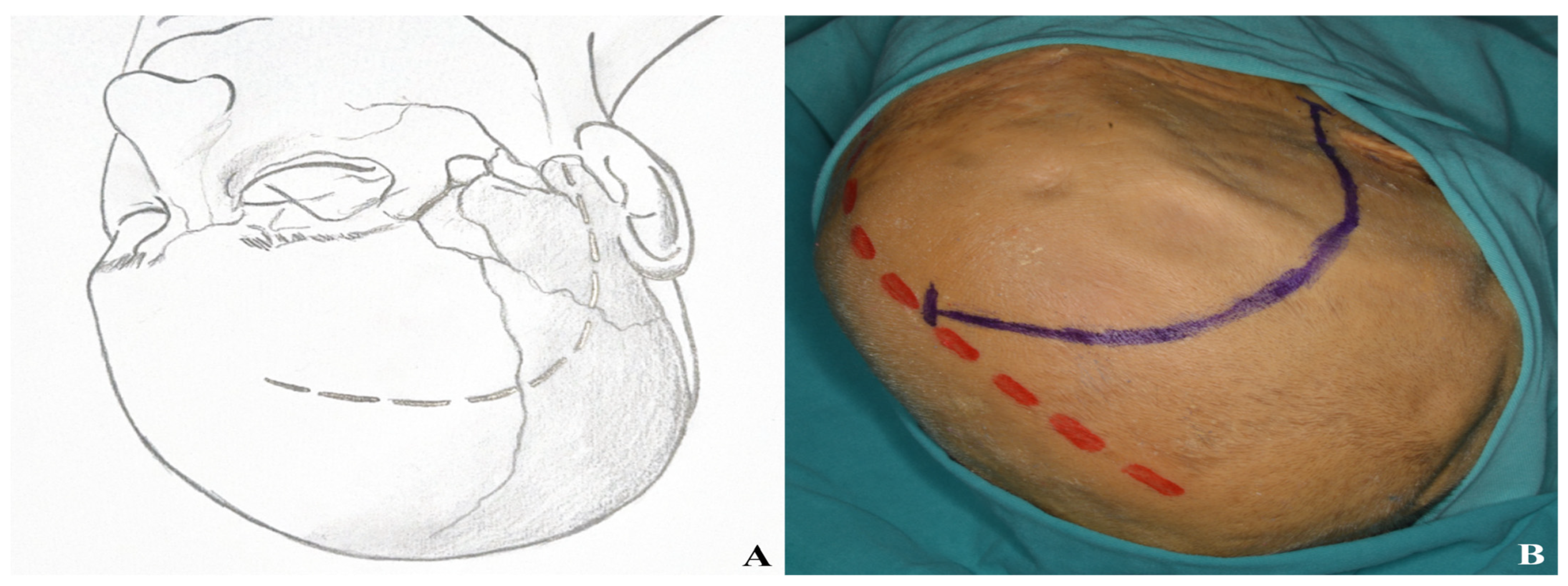
3.1.1. Incision
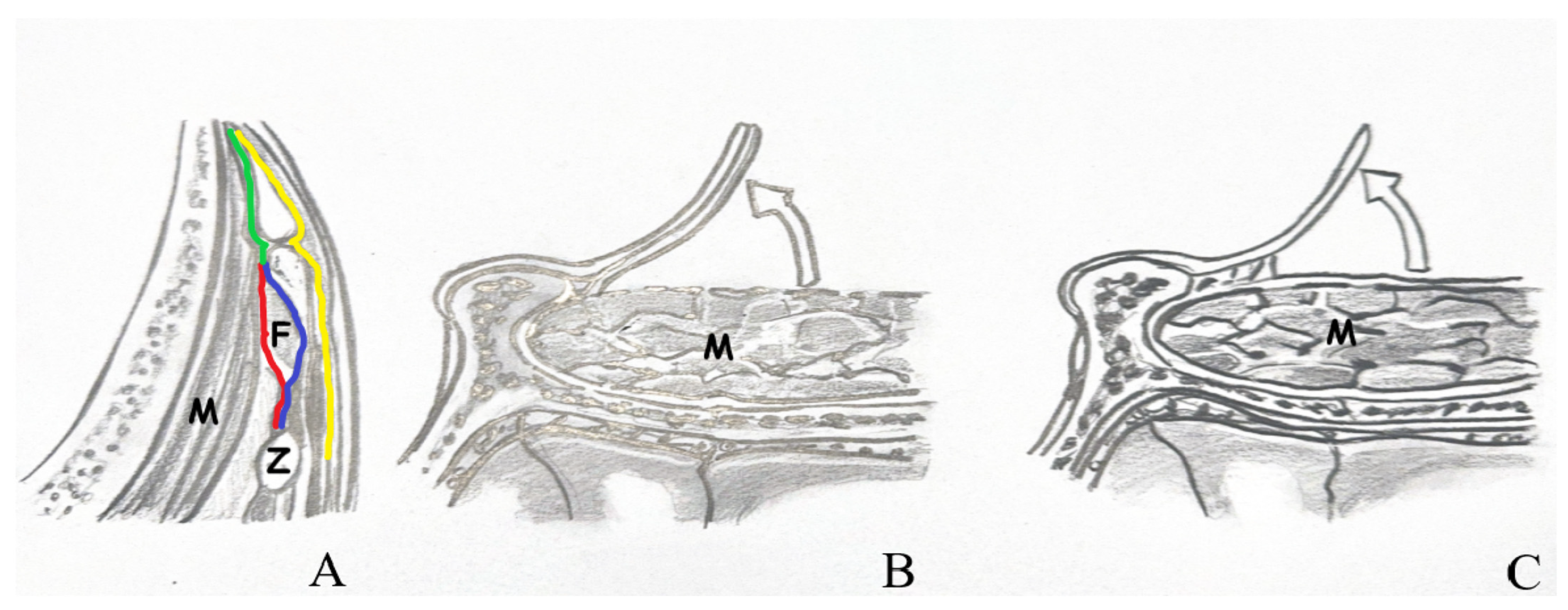
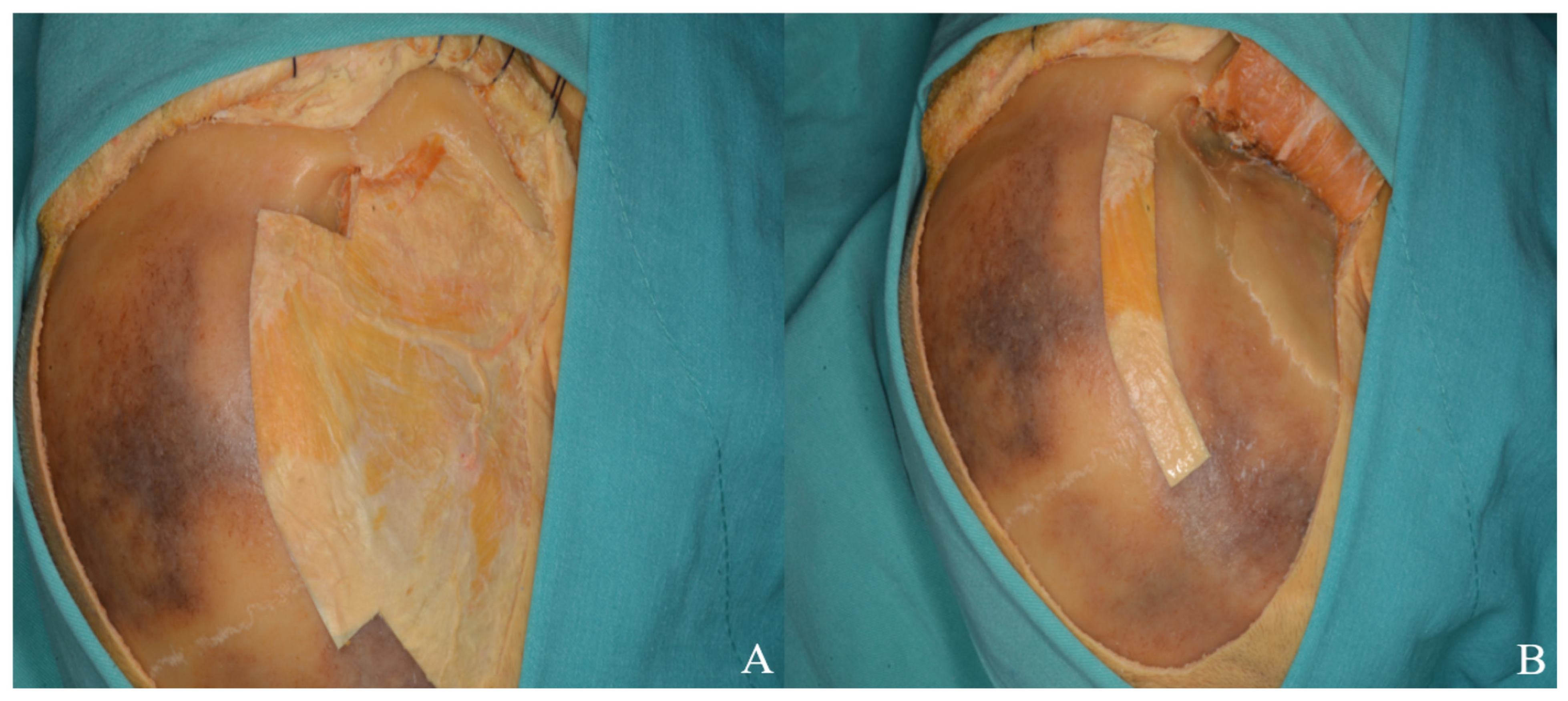
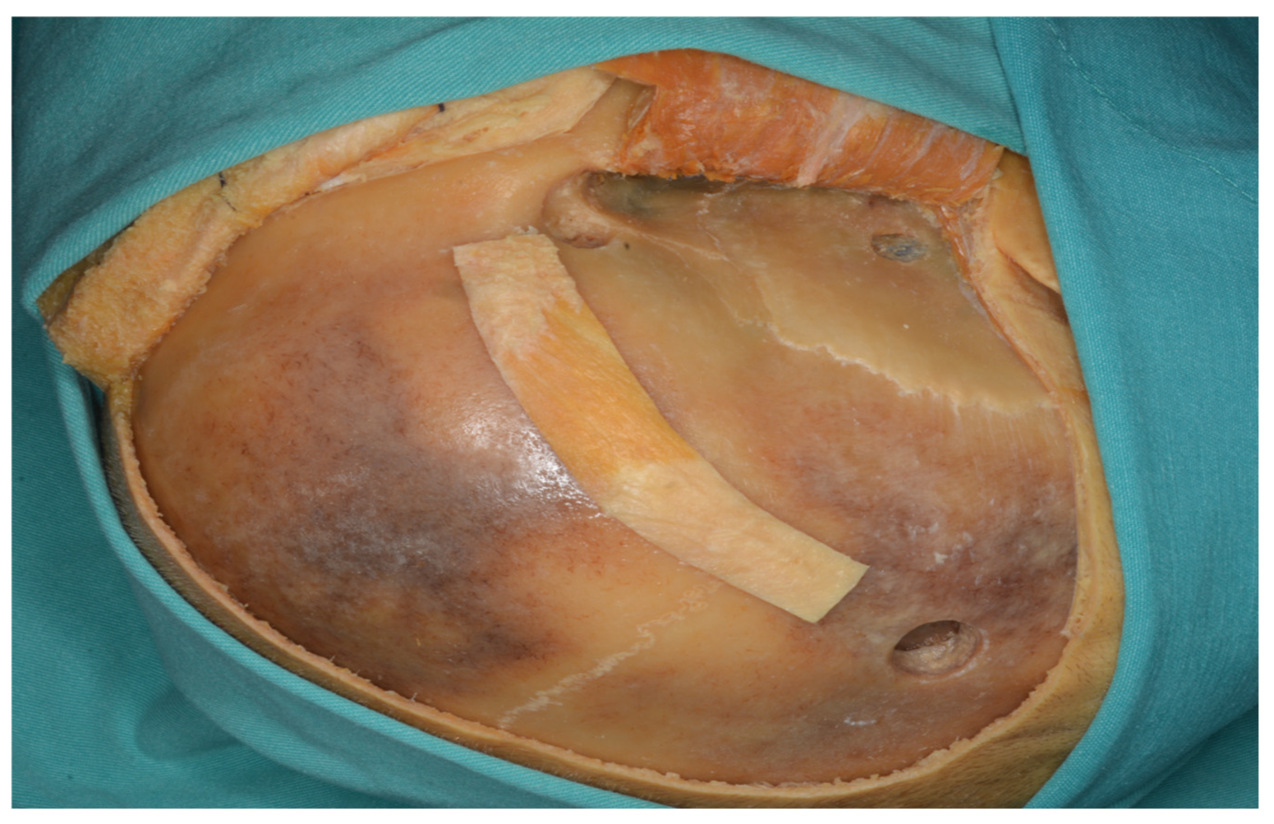
3.1.2. Interfacial, Subfascial Dissection Technique and Temporal Muscle Elevation
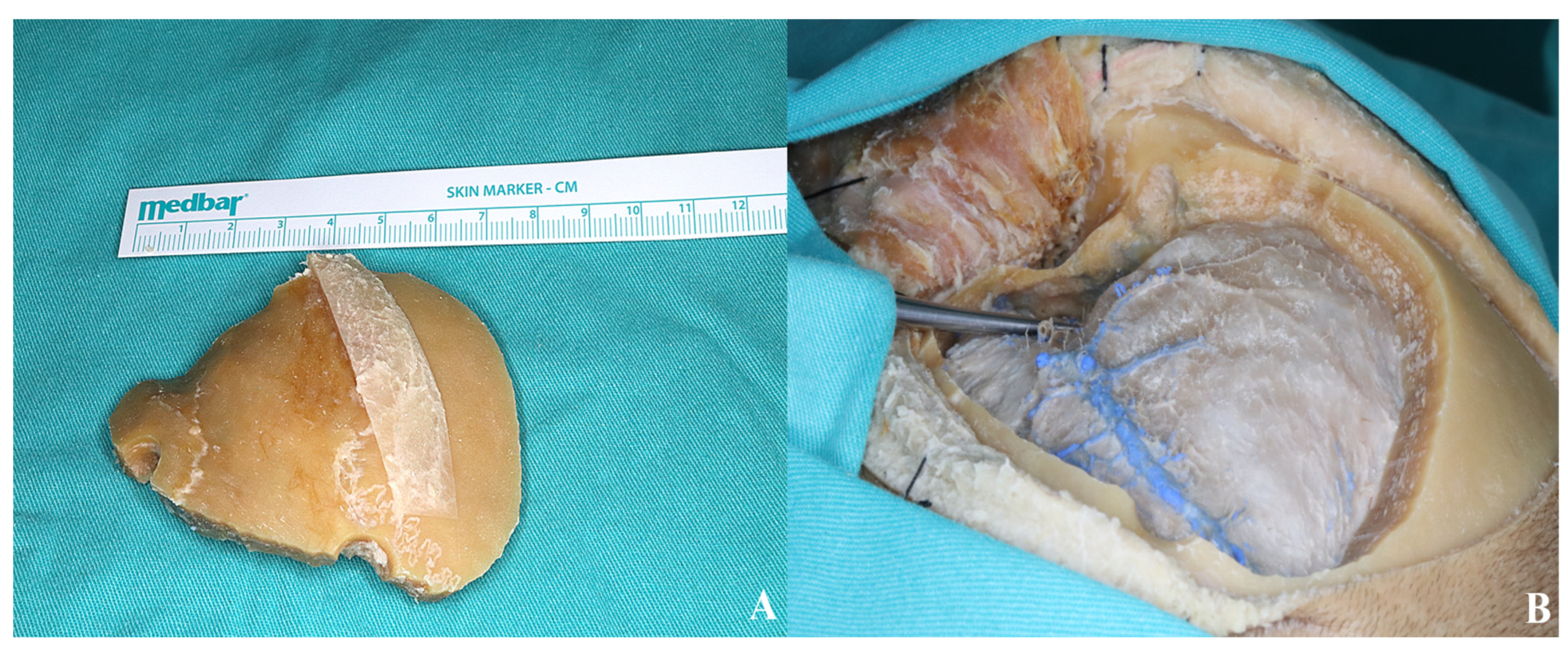
3.1.3. Craniotomy
3.1.4. Basal Drilling
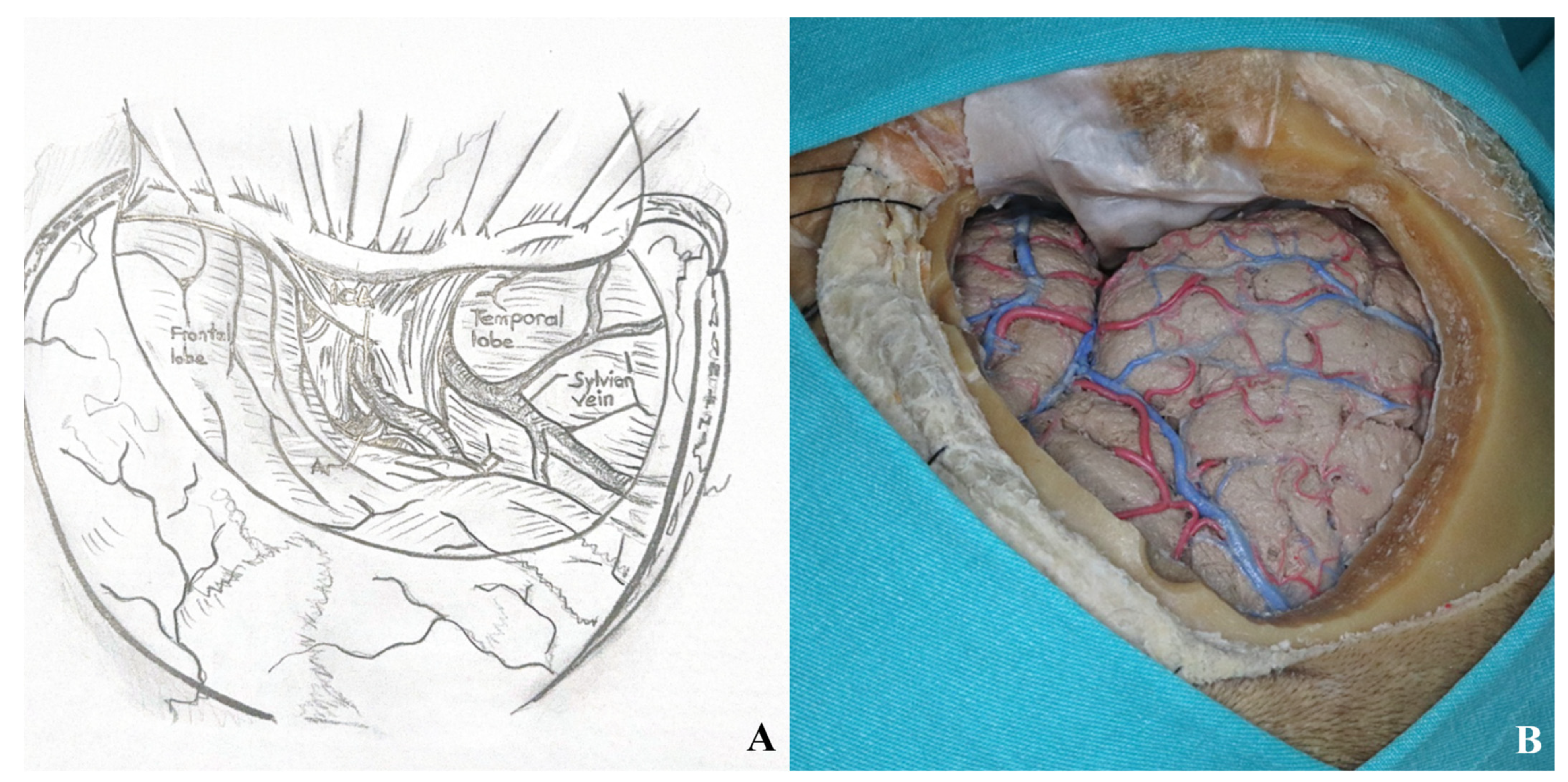
3.1.5. Dura Incision
3.2. Orbitozygomatic Approach
3.2.1. Incision
3.2.2. Temporal Muscle Elevation
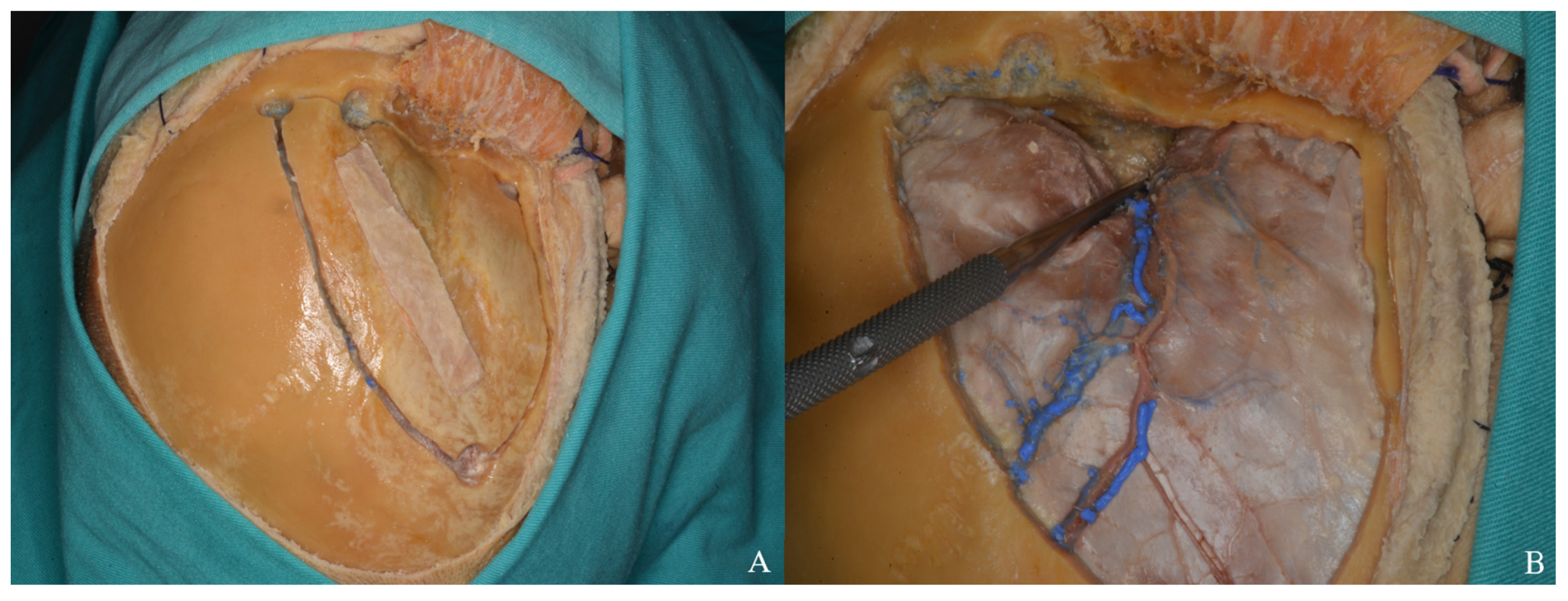
3.2.3. Periorbital Dissection
3.2.4. Craniotomy
- •
- One Piece Orbitozygomatic Craniotomy
- •
- •
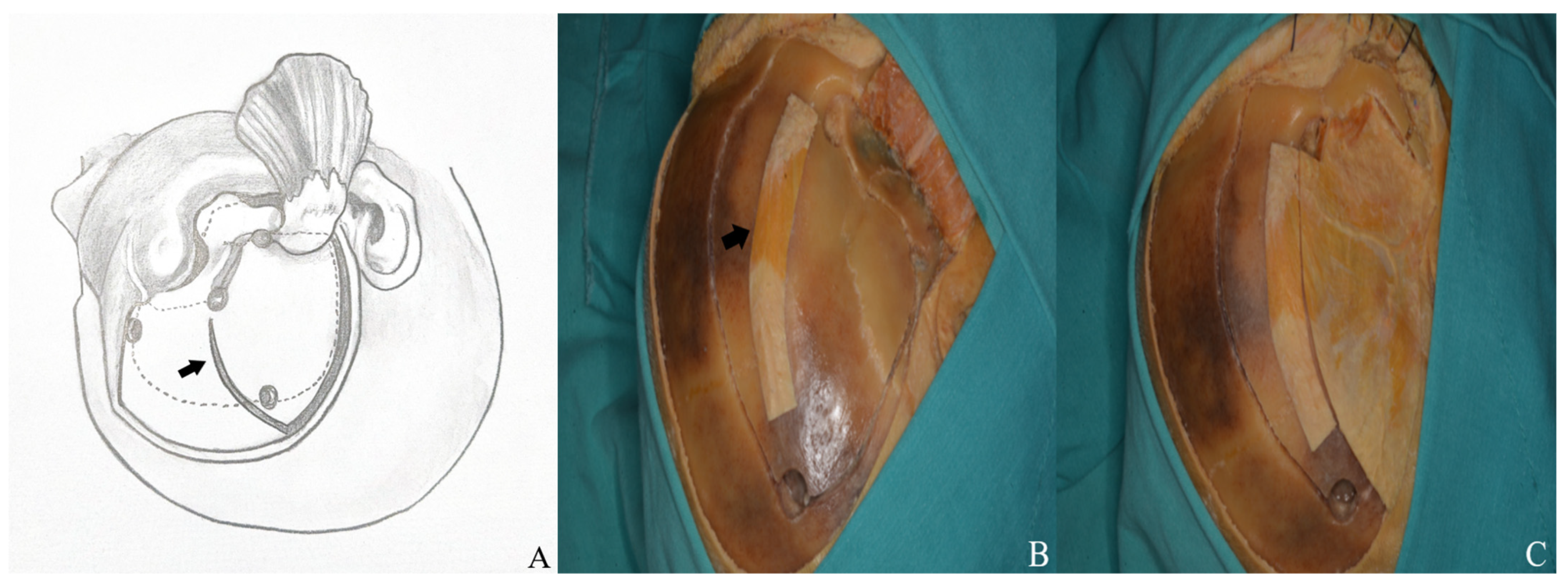
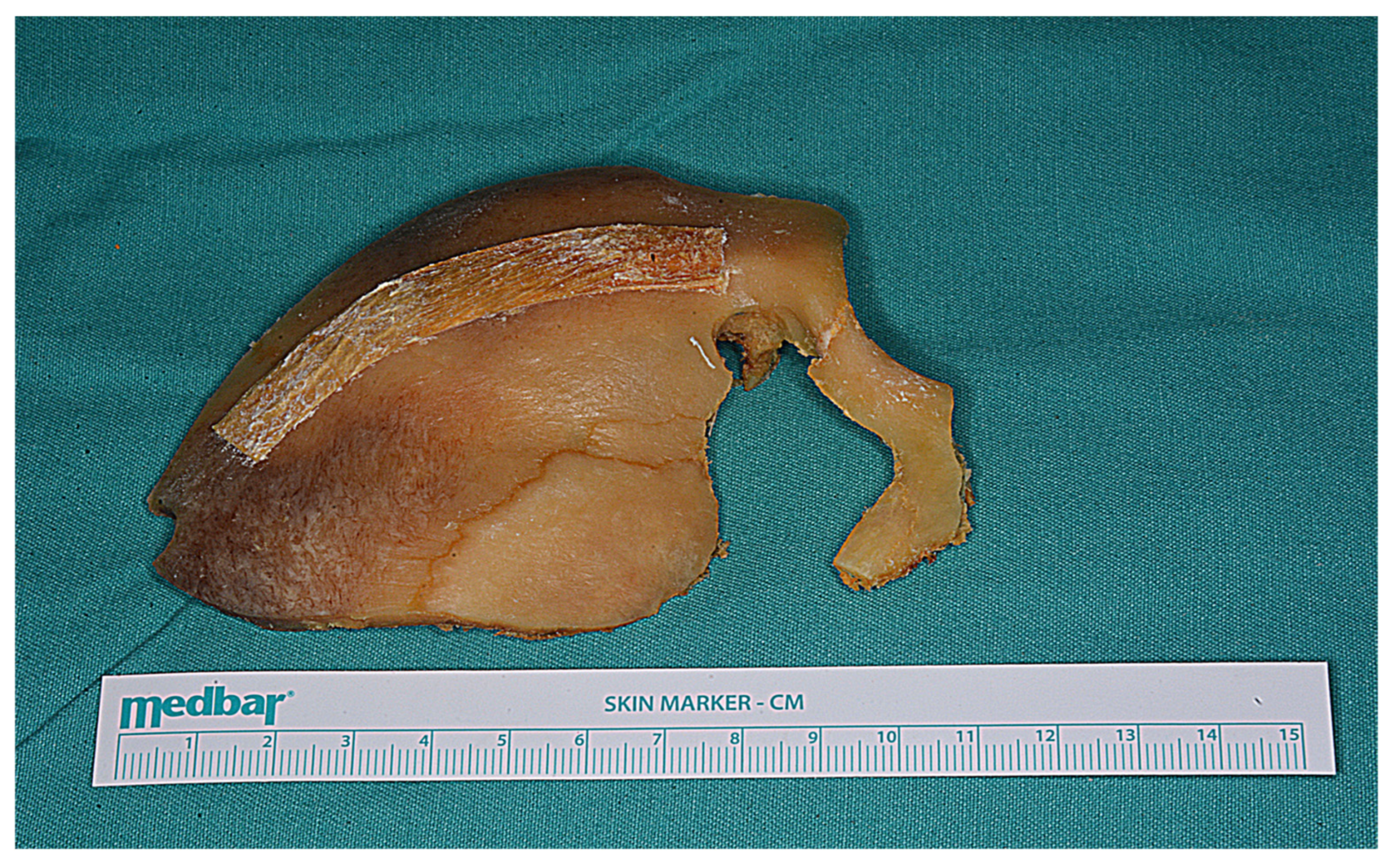
- The first extends from the periorbital aspect of the McCarty hole to the inferior orbital fissure.
- •
- The second runs from the zygomatic body to the inferior orbital fissure, ~1 cm below the frontal and temporal zygomatic limbs.
- •
- The third is made at the temporal root of the zygoma, anterior to the articular tubercle.
- •
- The fourth involves the orbital roof, including the supraorbital rim and infraorbital cut; the periorbita must be protected.
- •
- The fifth connects the anterior frontal and posterior temporal burr holes, passing anteriorly while preserving the supraorbital ridge.
- •
- The sixth links the McCarty hole to the posterior temporal burr hole.
- •
- Two Piece Orbitozygomatic Craniotomy
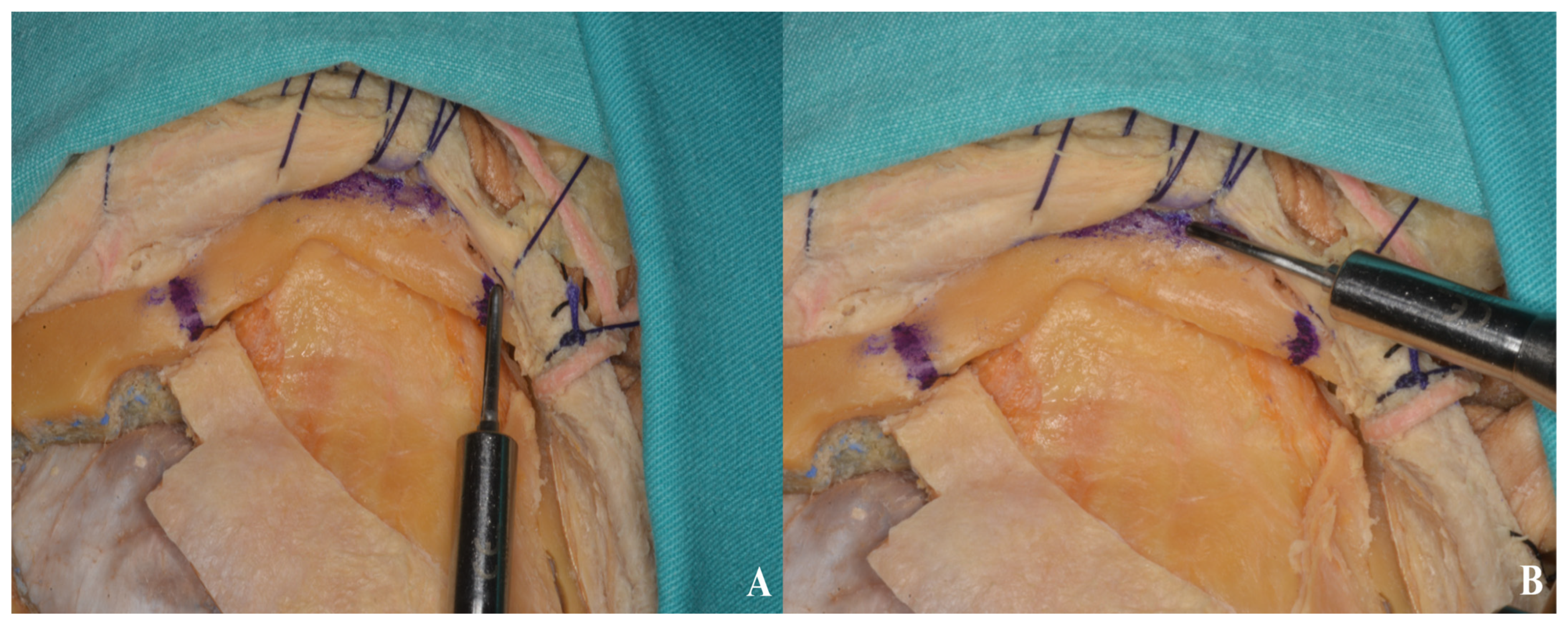
3.2.5. Basal Drilling
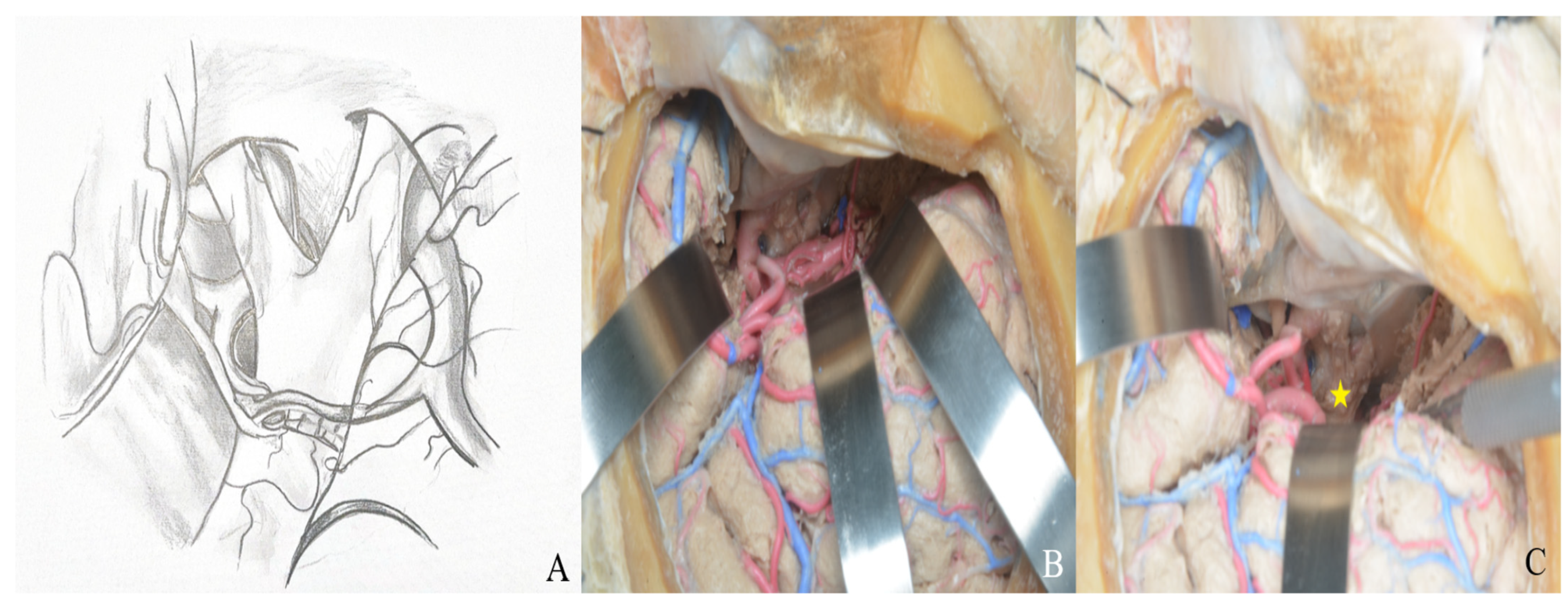
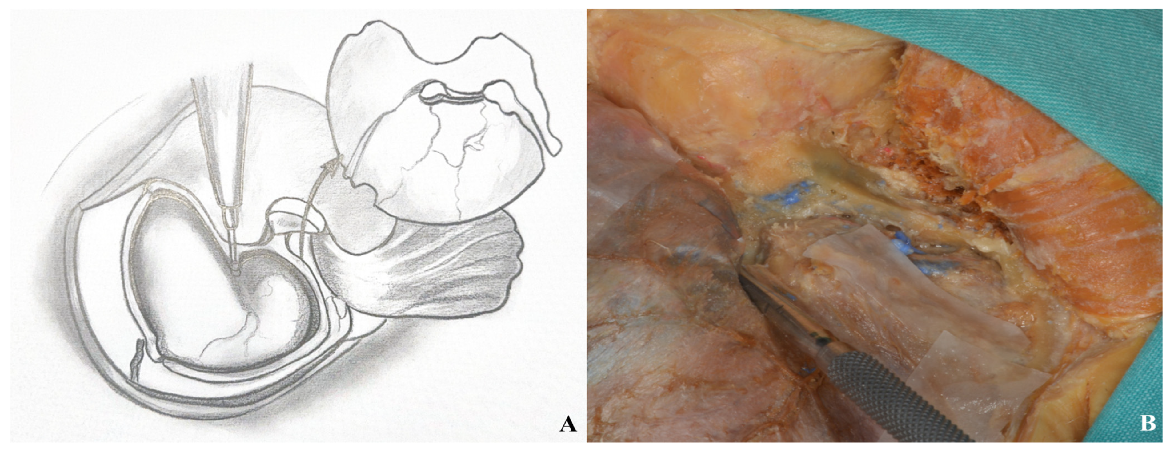
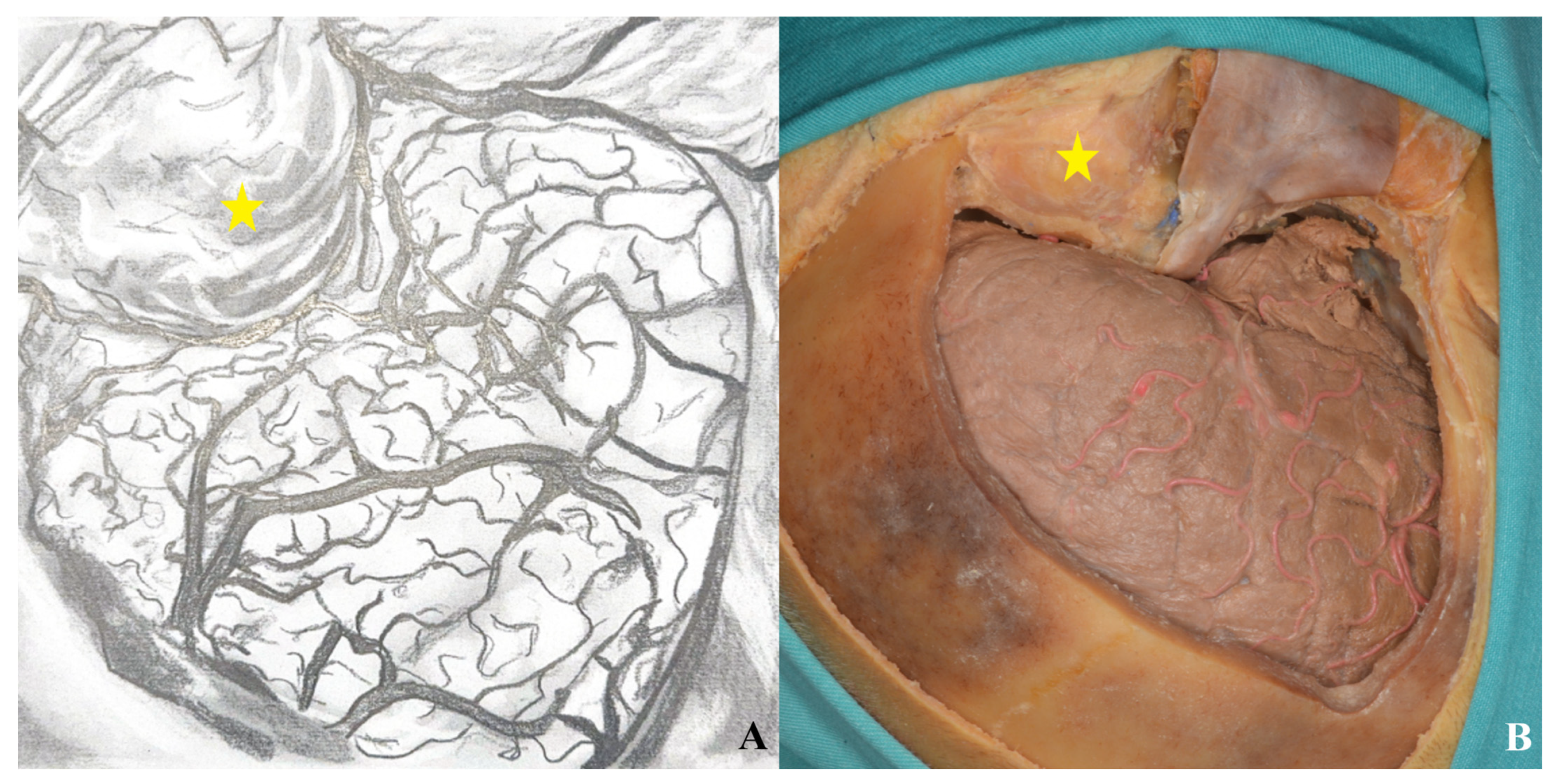
3.2.6. Dura Incision and Sylvian Fissure Opening
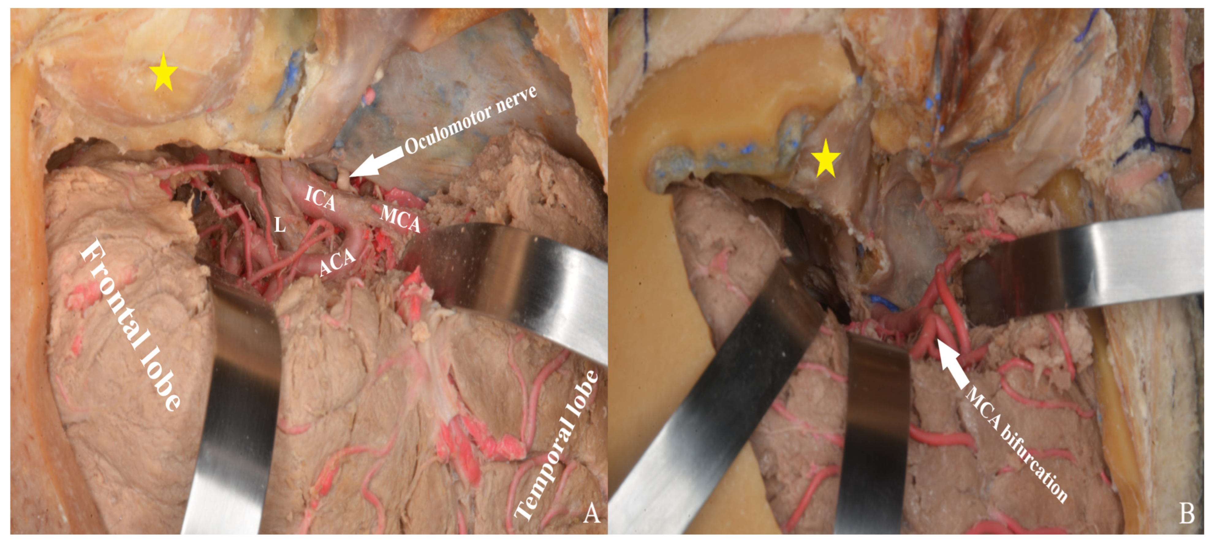
3.3. Lamina Terminalis
3.4. Lamina Terminalis Cistern
4. Discussion
5. Conclusions
Author Contributions
Funding
Institutional Review Board Statement
Informed Consent Statement
Data Availability Statement
Acknowledgments
Conflicts of Interest
References
- Pellerin, P.; Lesoin, F.; Dhellemmes, P.; Donazzan, M.; Jomin, M. Usefulness of the orbitofrontomalar approach associated with bone reconstruction for frontotemporosphenoid meningiomas. Neurosurgery 1984, 15, 715–718. [Google Scholar] [CrossRef] [PubMed]
- McArthur, L. An aseptic surgical access to the pituitary body and its neighborhood. J. Am. Med. Assoc. 1912, 58, 2009–2011. [Google Scholar] [CrossRef]
- Frazier, C. Choice of method in operations upon the pituitary body. Surg. Gynecol. Obstet. 1919, 29, 9–16. [Google Scholar]
- Frazier, C.H.I. An approach to the hypophysis through the anterior cranial fossa. Ann. Surg. 1913, 57, 145. [Google Scholar] [CrossRef]
- Dandy, W. A new hypophysis operation. Bull. Johns Hopkins Hosp. 1918, 29, 154. [Google Scholar]
- Yagmurlu, K.; Safavi-Abbasi, S.; Belykh, E.; Kalani, M.Y.S.; Nakaji, P.; Rhoton, A.L.; Spetzler, R.F.; Preul, M.C. Quantitative anatomical analysis and clinical experience with mini-pterional and mini-orbitozygomatic approaches for intracranial aneurysm surgery. J. Neurosurg. 2016, 127, 646–659. [Google Scholar] [CrossRef] [PubMed]
- Tubbs, R.S.; Nguyen, H.S.; Loukas, M.; Cohen-Gadol, A.A. Anatomic study of the lamina terminalis: Neurosurgical relevance in approaching lesions within and around the third ventricle. Child’s Nerv. Syst. 2012, 28, 1149–1156. [Google Scholar] [CrossRef] [PubMed]
- de Divitiis, O.; Angileri, F.F.; d’Avella, D.; Tschabitscher, M.; Tomasello, F. Microsurgical anatomic features of the lamina terminalis. Neurosurgery 2002, 50, 563–570. [Google Scholar]
- Mao, J.; Zhu, Q.; Ma, Y.; Lan, Q.; Cheng, Y.; Liu, G. Fenestration of lamina terminalis during anterior circulation aneurysm clipping on occurrence of shunt-dependent hydrocephalus after aneurysmal subarachnoid hemorrhage: Meta-analysis. World Neurosurg. 2019, 129, e1–e5. [Google Scholar] [CrossRef]
- Tomasello, F.; Angileri, F.F.; Cardali, S.; Conti, A. Letter to the Editor: Lamina terminalis fenestration. J. Neurosurg. 2014, 121, 219–221. [Google Scholar] [CrossRef]
- Akyuz, M.; Tuncer, R. The effects of fenestration of the interpeduncular cistern membrane arousted to the opening of lamina terminalis in patients with ruptured ACoA aneurysms: A prospective, comparative study. Acta Neurochir. 2006, 148, 725–732. [Google Scholar] [CrossRef]
- Yaşargil, M.G.; Kasdaglis, K.; Jain, K.K.; Weber, H.-P. Anatomical observations of the subarachnoid cisterns of the brain during surgery. J. Neurosurg. 1976, 44, 298–302. [Google Scholar] [CrossRef]
- Wang, S.-S.; Zheng, H.-P.; Zhang, F.-H.; Wang, R.-M. The microanatomical structure of the cistern of the lamina terminalis. J. Clin. Neurosci. 2011, 18, 253–259. [Google Scholar] [CrossRef]
- Kim, Y.-S.; Kim, J.-W.; Kim, W.-B.; Baek, B.-H.; Yoon, W.; Kim, T.-S.; Joo, S.-P. Combined Treatment of Large Fusiform A2 Aneurysm with End-to-Side Extended Superficial Temporal Artery–A3 Bypass Using Contralateral Superficial Temporal Artery Interposition Graft and Endovascular Aneurysm Trapping: A Case Report and Literature Review. J. Clin. Med. 2025, 14, 2927. [Google Scholar] [CrossRef]
- Takabayashi, K.; Shindo, T.; Kikuchi, T.; Takizawa, K. Neuroendocrine Carcinoma at the Sphenoid Sinus Misdiagnosed as an Olfactory Neuroblastoma and Resected Using High-Flow Bypass. Diagnostics 2022, 12, 1674. [Google Scholar] [CrossRef]
- Yoo, H.D.; Chung, S.Y.; Kim, S.M.; Park, K.S.; Ryu, S.J.; Kim, J.G. The Efficiency of FLAIR Images for Hemodynamic Change After STA-MCA Bypass with Moyamoya Disease and Symptomatic Steno-Occlusive Disorder. J. Clin. Med. 2025, 14, 3292. [Google Scholar] [CrossRef]
- Yi, K.H.; Oh, S.M. Lateral facial thread lifting procedure with temporal anchoring in deep temporal fascia: Anatomical perspectives of superficial temporal artery. Ski. Res. Technol. 2024, 30, e13587. [Google Scholar] [CrossRef]
- Chaddad-Neto, F.; Campos Filho, J.M.; Dória-Netto, H.L.; Faria, M.H.; Ribas, G.C.; Oliveira, E. The pterional craniotomy: Tips and tricks. Arq. De Neuro-Psiquiatr. 2012, 70, 727–732. [Google Scholar] [CrossRef]
- Yaşargil, M.; Fox, J.; Ray, M. The operative approach to aneurysms of the anterior communicating artery. In Advances and Technical Standards in Neurosurgery—Volume 2; Springer: Berlin/Heidelberg, Germany, 1975; pp. 113–170. [Google Scholar]
- Spetzler, R.F.; Lee, K.S. Reconstruction of the temporalis muscle for the pterional craniotomy. J. Neurosurg. 1990, 73, 636–637. [Google Scholar] [CrossRef]
- Coscarella, E.; Vishteh, A.G.; Spetzler, R.F.; Seoane, E.; Zabramski, J.M. Subfascial and submuscular methods of temporal muscle dissection and their relationship to the frontalis branch of the facial nerve. J. Neurosurg. 2000, 92, 877–880. [Google Scholar] [CrossRef]
- Jane, J.A.; Park, T.S.; Pobereskin, L.H.; Winn, R.H.; Butler, A.B. The supraorbital approach. Neurosurgery 1982, 11, 537–542. [Google Scholar] [CrossRef]
- Hakuba, A.; Liu, S.-s.; Shuro, N. The orbitozygomatic infratemporal approach: A new surgical technique. Surg. Neurol. 1986, 26, 271–276. [Google Scholar] [CrossRef]
- Al-Mefty, O. Supraorbital-pterional approach to skull base lesions. Neurosurgery 1987, 21, 474–477. [Google Scholar] [CrossRef]
- Zabramski, J.M.; Kiriş, T.; Sankhla, S.K.; Cabiol, J.; Spetzler, R.F. Orbitozygomatic craniotomy. J. Neurosurg. 1998, 89, 336–341. [Google Scholar] [CrossRef] [PubMed]
- Campero, A.; Martins, C.; Socolovsky, M.; Torino, R.; Yasuda, A.; Domitrovic, L.; Rhoton Jr, A. Three-piece orbitozygomatic approach. Oper. Neurosurg. 2010, 66, ons-E119–ons-E120. [Google Scholar] [CrossRef]
- Alaywan, M.; Sindou, M. Fronto-temporal approach with orbito-zygomatic removal surgical anatomy. Acta Neurochir. 1990, 104, 79–83. [Google Scholar] [CrossRef]
- Yasargil, M.G. Operative anatomy. In Microneurosurgery Vol I; Thieme Medical Publishers: New York, NY, USA, 1984. [Google Scholar]
- Meybodi, A.T.; Benet, A.; Rubio, R.R.; Yousef, S.; Mokhtari, P.; Preul, M.C.; Lawton, M.T. Comparative analysis of orbitozygomatic and subtemporal approaches to the basilar apex: A cadaveric study. World Neurosurg. 2018, 119, e607–e616. [Google Scholar] [CrossRef]
- da Silva, S.A.; Yamaki, V.N.; Solla, D.J.F.; de Andrade, A.F.; Teixeira, M.J.; Spetzler, R.F.; Preul, M.C.; Figueiredo, E.G. Pterional, pretemporal, and orbitozygomatic approaches: Anatomic and comparative study. World Neurosurg. 2019, 121, e398–e403. [Google Scholar] [CrossRef]
- Klironomos, G.; Dehdashti, A.R. Orbitozygomatic craniotomy and trans-sylvian approach for resection of a tuberculum sella meningioma with extension to the posterior fossa. Neurosurg. Focus 2017, 43, V13. [Google Scholar] [CrossRef] [PubMed]
- Abou-Al-Shaar, H.; Krisht, K.M.; Cohen, M.A.; Abunimer, A.M.; Neil, J.A.; Karsy, M.; Alzhrani, G.; Couldwell, W.T. Cranio-orbital and orbitocranial approaches to orbital and intracranial disease: Eye-opening approaches for neurosurgeons. Front. Surg. 2020, 7, 1. [Google Scholar] [CrossRef] [PubMed]
- Nanda, A.; Vannemreddy, P.S.; Vincent, D.A. Microsurgical and endoscopic approaches to the basilar bifurcation: Quantitative comparison of combined pterional/anterior temporal and orbitozygomatic extended approaches. Skull Base 2001, 11, 093–098. [Google Scholar] [CrossRef] [PubMed]
- Lee, J.-S.; Scerrati, A.; Zhang, J.; Ammirati, M. Quantitative analysis of surgical exposure and surgical freedom to the anterosuperior pons: Comparison of pterional transtentorial, orbitozygomatic, and anterior petrosal approaches. Neurosurg. Rev. 2016, 39, 599–605. [Google Scholar] [CrossRef] [PubMed]
- Luzzi, S.; Giotta Lucifero, A.; Spina, A.; Baldoncini, M.; Campero, A.; Elbabaa, S.K.; Galzio, R. Cranio-orbito-zygomatic approach: Core techniques for tailoring target exposure and surgical freedom. Brain Sci. 2022, 12, 405. [Google Scholar] [CrossRef] [PubMed]
- Carlstrom, L.P.; Graffeo, C.S.; Leonel, L.C.; Perry, A.; Link, M.J.; Peris-Celda, M. Anatomical step-by-step dissection of complex skull base approaches for trainees: Surgical anatomy of the frontotemporal and orbitozygomatic craniotomies. J. Neurol. Surg. Part B Skull Base 2024, 85, 370–380. [Google Scholar] [CrossRef]
- Efe, I.E.; Çinkaya, E.; Kuhrt, L.D.; Bruesseler, M.M.; Mührer-Osmanagic, A. Neurosurgical education using cadaver-free brain models and augmented reality: First experiences from a hands-on simulation course for medical students. Medicina 2023, 59, 1791. [Google Scholar] [CrossRef]







Disclaimer/Publisher’s Note: The statements, opinions and data contained in all publications are solely those of the individual author(s) and contributor(s) and not of MDPI and/or the editor(s). MDPI and/or the editor(s) disclaim responsibility for any injury to people or property resulting from any ideas, methods, instructions or products referred to in the content. |
© 2025 by the authors. Licensee MDPI, Basel, Switzerland. This article is an open access article distributed under the terms and conditions of the Creative Commons Attribution (CC BY) license (https://creativecommons.org/licenses/by/4.0/).
Share and Cite
Yilmaz, M.C.; Durmus, Y.E. Exploring the Lamina Terminalis: A Stepwise Anatomical Comparison of Pterional and Orbitozygomatic Craniotomy Approaches. Life 2025, 15, 1804. https://doi.org/10.3390/life15121804
Yilmaz MC, Durmus YE. Exploring the Lamina Terminalis: A Stepwise Anatomical Comparison of Pterional and Orbitozygomatic Craniotomy Approaches. Life. 2025; 15(12):1804. https://doi.org/10.3390/life15121804
Chicago/Turabian StyleYilmaz, Merih C., and Yunus E. Durmus. 2025. "Exploring the Lamina Terminalis: A Stepwise Anatomical Comparison of Pterional and Orbitozygomatic Craniotomy Approaches" Life 15, no. 12: 1804. https://doi.org/10.3390/life15121804
APA StyleYilmaz, M. C., & Durmus, Y. E. (2025). Exploring the Lamina Terminalis: A Stepwise Anatomical Comparison of Pterional and Orbitozygomatic Craniotomy Approaches. Life, 15(12), 1804. https://doi.org/10.3390/life15121804


