Article

Beyond Daily Values: Are Day-to-Day and Albumin-Adjusted Ratios of IL-6, PCT, and CRP Better Predictors of Ventilator-Associated Pneumonia?

1
Department for Interdisciplinary Intensive Medicine and Intermediate Care, Helios Dr. Horst Schmidt Hospital Wiesbaden, 65199 Wiesbaden, Germany
2
Department of Internal Intensive Care Medicine, Nuremberg Hospital, 90419 Nuremberg, Germany
3
Department for Neurosurgery, Helios Dr. Horst Schmidt Hospital Wiesbaden, 65199 Wiesbaden, Germany
4
Department of Anesthesiology, Intensive Care Medicine and Pain Medicine, Medical Faculty Mannheim, Heidelberg University, 68167 Mannheim, Germany
*
Author to whom correspondence should be addressed.
Life 2025, 15(11), 1697; https://doi.org/10.3390/life15111697
Submission received: 22 September 2025 / Revised: 24 October 2025 / Accepted: 30 October 2025 / Published: 1 November 2025
(This article belongs to the Special Issue Pathology, Diagnosis, and Treatments of Airway Diseases)

Abstract

Background: Ventilator-associated pneumonia (VAP) is a frequent complication in neurosurgical intensive care patients. This leads to prolonged mechanical ventilation, increased 30-day mortality rates, and extended hospital stays. However, early diagnosis remains a challenge. Biomarkers, such as IL-6, PCT, and CRP, are considered a cornerstone for recognizing VAP and initiating early treatment. Only a very limited number of studies have compared IL-6, PCT, and CRP, focusing on day-to-day dynamics and their albumin ratios. Therefore, we investigated whether, compared with their daily levels, the day-to-day dynamics and albumin-adjusted ratios of IL-6, PCT, and CRP offer improved diagnostic value for VAP. Second, we investigated these biomarkers in patients treated for VAP who did not meet the criteria for VAP. Methods: In this exploratory, matched case–control study, we investigated 171 neurosurgical ICU patients. Daily biomarker levels, their dynamics, and ratios to serum albumin were assessed beginning four days before VAP. Logistic regression and receiver operating curve (ROC) analyses were performed to evaluate the association between each biomarker and VAP. Results: IL-6 and its day-to-day dynamics demonstrated the largest differences between VAP patients and nonVAP patients (r = 0.631; r = 0.452) and were associated with VAP, yielding AUCs of 0.816 and 0.726, respectively. In contrast, for PCT, we did not demonstrate any associative utility, whereas CRP showed a significant, moderate effect size on the day of VAP occurrence (p = 0.015 *; r = 0.351). We could not demonstrate any superiority in the day-to-day dynamics or the albumin-adjusted ratios compared to the daily values. For patients who were treated for VAP without fulfilling the criteria for biomarkers, we did not observe any significant difference from nonVAP patients.

1. Introduction

Ventilator-associated pneumonia (VAP) is clinically characterized by new, persistent, or progressive infiltrates on chest X-rays, accompanied by two of the following criteria: (1) white blood cell count >10/nL or <4/nL, (2) fever >38 °C, or (3) purulent secretion [1]. Aside from burn patients, neurosurgical patients have the highest rate of VAP [2], which leads to prolonged mechanical ventilation, increased 30-day mortality rates, and extended hospital stays [3]. Moreover, it incurs additional costs of up to 40,000 US dollars per case, making it the second-most-expensive hospital-acquired infection [4].
In addition to clinical criteria, biomarkers are considered a cornerstone for recognizing VAP and initiating early treatment.
Procalcitonin (PCT) and C-reactive protein (CRP) are frequently used biomarkers in intensive care settings [5]. In some hospitals, IL-6 is also routinely used for this purpose [6]. Their clinical utility differs depending on their response time to an inflammatory stimulus and the extent of their increase [7,8,9]. While IL-6 is one of the fastest biomarkers for inflammation, peaking at 6 h after the stimulus [10], CRP reaches the maximum level with a delay of 36–50 h [11,12]. PCT responds at 4–6 h after exposure and peaks at 12–24 h [13].
For surgical and medical patients, the CRP and PCT levels might indicate intensive care unit (ICU) patients at risk of developing VAP 72 h after intubation [14,15].
Compared with other cytokines, IL-6 levels increase 24–48 h before clinical evidence of an inflammatory process is observed [7]. In a previous study of neurosurgical ICU patients, we showed that IL-6 levels increased 24 h before the diagnosis and treatment of VAP [16]. Among multiple biomarkers, IL-6 is the only one that distinguishes microbiologically confirmed VAP from suspected VAP [17].
Another difficulty in interpreting biomarkers is that their values vary widely across patients [18]. Therefore, day-to-day ratios might equalize these differences and thus might be more stable. In association with VAP, the CRP and PCT day-to-day dynamics were also tested, demonstrating superiority over CRP or PCT alone [19].
Other investigators have studied the predictive value of the CRP/albumin, PCT/albumin, and IL-6/albumin ratios, focusing on 30-day mortality rates and acute kidney injury [20,21,22]. Since most inflammatory markers, especially IL-6, downregulate albumin synthesis in the liver, their ratio might be of interest as a composite marker for VAP.
Thus, we hypothesized that day-to-day ratios rather than their daily values, as well as their albumin ratio, could further improve their performance as classifiers of VAP in neurosurgical ICU patients.
Second, we differentiated VAP cases into microbiologically confirmed cases (mcVAPs) and suspected but not microbiologically confirmed cases (suspVAPs) and assessed potential differences in biomarker levels between these groups in a secondary analysis.
Lastly, we included patients who received VAP treatment but did not meet the diagnostic criteria. Here, we compared biomarker values and clinical values to those of nonVAP patients. To address these questions, we designed this exploratory proof-of-concept study.

2. Materials and Methods

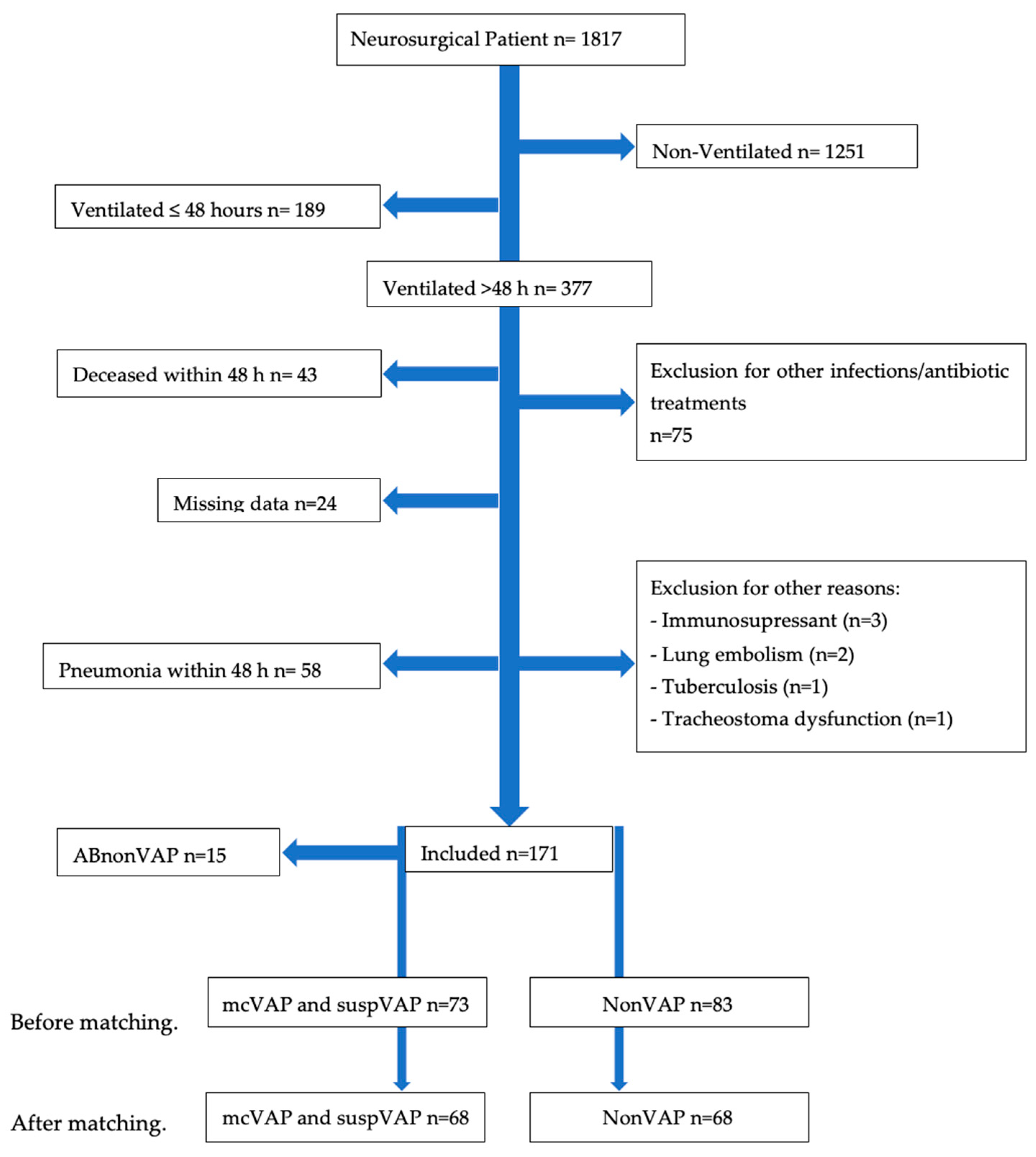
Neurosurgical patients admitted to the ICU of Helios Dr. Horst Schmidt Hospital, either through the emergency department or postoperatively after major neurosurgical procedures, were included in this study. The observation period spanned from 1 January 2020 to 31 December 2023. Patients meeting the following criteria were excluded: younger than 18 years; extubation or death within the first 48 h of ICU admission; incomplete baseline data on hours of ventilation, antibiotic treatment, or radiological confirmation; and infection or aspiration pneumonia on admission. Additionally, we excluded patients who received antibiotic treatment at admission or within the first day after admission, as well as those who received antibiotics for other infections within the observation time (Appendix A Figure A1, study flow diagram).
Ventilator-associated pneumonia (VAP) is defined as mechanical ventilation for at least 48 h, accompanied by new, persistent, or progressive infiltrates on chest X-rays, along with at least two of the following criteria: (1) white blood cell count > 10,000 or <4000/μL, (2) fever > 38 °C, or (3) purulent respiratory secretions. Additionally, as the CDC proposed, specific antibiotic treatment is mandatory.
We divided the VAP patients into microbiologically confirmed (mcVAP) and nonmicrobiologically confirmed (suspVAP) groups.
We matched controls among VAP-free patients one-to-one based on the number of ventilator days. For this purpose, we defined the day of VAP as described above and matched controls who underwent ventilation for at least this duration. Additionally, we compared groups with identical timeframes where VAP might have occurred.
From January 2020 to April 2022, serum IL-6 was the preferred daily marker; thereafter, PCT and CRP were recorded daily. Therefore, we compared two groups and matched within each group.
In addition to our cases and controls, we identified a third group, comprising patients who received a directed antibiotic for VAP without formally fulfilling the VAP definition (ABnonVAP).
The overall observation period began at admission and lasted up to 20 days postadmission or until discharge or death, whichever occurred first. The days before VAP diagnosis and the day of diagnosis were consecutively numbered as days −4 to −1 and 0, respectively. For the control group, day 0 was defined as the day corresponding to the one-to-one VAP match. For patients assigned to the ABnonVAP group, day 0 was defined as the day we started antibiotic treatment. The observation period from day −4 to day 0 was chosen to capture the dynamics of the biomarkers in the days preceding the clinical diagnosis of VAP. This period was expected to reflect the early pathophysiological changes preceding infection.
Ethics: The study was approved by Ethics Committee II of the Medical Faculty Mannheim, University of Heidelberg (Reference number: 2024–821).
Statistical analysis: We first examined the daily values of each inflammatory biomarker (IL-6, CRP, and PCT) from day −4 to day 0. We subsequently assessed their changes by calculating ratios between consecutive days (days −3/−4, −2/−3, −1/−2, and 0/−1) and their albumin ratios.
We then tested for differences, including effect sizes, and conducted logistic regression analyses, reporting odds ratios, and, where appropriate, receiver operating characteristic (ROC) curves. Metric variables are indicated as the mean [±SD] and/or median [IQR]. As needed, the group differences were tested with Student’s t test or the Mann–Whitney U test for metric variables and the chi-square test for categorical variables. Cohen’s d (d), rank-biserial correlation (r), or Cramer’s V (V) were used to determine the corresponding effect size [23]. Receiver operating characteristic (ROC) curves were used to determine the area under the ROC (AUC), sensitivity, and specificity for each parameter in predicting VAP, including the odds ratio (OR) and 95% confidence interval (CI). Statistical significance was assumed at an alpha level of 0.05 and indicated by an asterisk (*). To account for multiple testing, we applied the Benjamini–Hochberg correction to the biomarker p-values. Statistical analysis was performed with JASP 0.18.2, and matching and graph visualization were performed with Julius.ai (Caesar Labs, Inc., San Francisco, CA, USA).

3. Results

3.1. Population

A total of 1817 neurosurgical patients admitted to the ICU were screened for eligibility. Of these, 377 patients were ventilated for >48 h. The details are provided in the Appendix A Figure A1. After all the exclusion criteria were applied, 171 patients were eligible for further analysis, with 73 patients fulfilling the criteria for VAP, 83 patients in the nonVAP group, and 15 patients in the ABnonVAP group; after matching, 68 VAP patients, 45 of whom had mcVAP and 23 of whom had suspVAP, with a drop-out rate of 6.4% after matching, were included.
Appendix A Table A1 provides the matching details and quality checks, and Appendix A Table A2 shows the distributions between the IL-6 and PCT/CRP groups.

3.2. Baseline Mcvap/Suspvap vs. Nonvap

Age and SAPS scores are provided in Table 1. Significantly more men than women were diagnosed with mcVAP/suspVAP (69.1% vs. 30.9%, X2(1) = 6.795, p < 0.009, V = 0.224).
No significant differences were observed between the mcVAP/suspVAP and nonVAP groups for admission age, SAPS, diagnosis, comorbidities, or operative procedures.
Similarly, there were no significant differences in IL-6, WBC, PCT, CRP, the P/F ratio, or body temperature at admission between the groups. The details are provided in Table 2. Significant differences in length of stay and hours of ventilation were noted between nonVAP and mcVAP/suspVAP patients (length of stay: 399.81 h [SD 360.13] vs. 599.46 h [SD 335.57], p < 0.001, r = 0.576; hours of ventilation: 198.49 h [SD 165.23] vs. 380.10 h [SD 225.32], p < 0.001, r = 0.920).

3.3. Biomarker Values vs. Day-To-Day Ratios and Ratios to Albumin to Predict VAP

Differences in biomarker levels between mcVAP and suspVAP: During the observation period from day −4 to day 0, we did not detect differences in IL-6, CRP, or PCT levels between the mcVAP and suspVAP groups. Therefore, we combined mcVAP and suspVAP into a single group for the following analysis.
IL-6: Daily Levels: Median IL-6 levels were significantly higher in mcVAP/suspVAP patients than in nonVAP patients from day −1 to day 0.
On day −1, the median IL-6 level was 63.10 pg/mL (IQR: 71.00) in the mcVAP/suspVAP group and 46.35 pg/mL (IQR: 74.43) in the nonVAP group (p < 0.04, r = 0.255).
The greatest difference was observed on day 0, with median IL-6 levels of 109 pg/mL (IQR: 180.25) in the mcVAP/suspVAP group and 35.40 pg/mL (IQR: 35.80) in the nonVAP group (p < 0.001, r = 0.631).
For the day-to-day ratios, we observed significant differences in the IL-6 day-to-day ratios on days −1/−2 and 0/−1. On days −1/−2, the ratio was 1.84 (IQR: 1.92) in the mcVAP/suspVAP group and 0.93 (IQR: 1.42) in the nonVAP group (p = 0.013, r = 0.313). On days 0 and 1, the ratios were 1.42 (IQR: 1.85) and 0.74 (IQR: 0.81), respectively (p < 0.001, r = 0.452).
IL-6-to-albumin quotient: IL-6-to-albumin ratios were significantly greater in mcVAP/SuspulentVAP patients than in nonVAP patients on day 0, with borderline significance on day −1. On day −1, the ratio was 22.77 (IQR: 37.56) vs. 12.09 (IQR: 14.72) (p < 0.054, r = 0.257). On day 0, the ratio was 41.48 (IQR: 65.07) in the mcVAP/suspVAP group vs. 12.09 (IQR: 14.72) in the nonVAP group (p < 0.001, r = 0.257).
Logistic regression revealed that the IL-6 level on day 0 and the day 0/−1 ratio were significantly associated with VAP. The corresponding odds ratios (ORs) were 1.012 (p < 0.001, 95% CI: 1.004–1.020) for IL-6 on day 0 and 2.66 (p < 0.001, 95% CI: 1.390–5.091) for the day −1/−2 ratio, with AUCs of 0.816 and 0.726, respectively. The IL-6-to-albumin ratio significantly predicted VAP on day 0, with an OR of 1.065 (p < 0.001, 95% CI: 1.024–1.109) and an AUC of 0.864. Details are provided in Table 3, and the receiver operating curves are provided in Figure 1a.
PCT: Daily PCT levels were significantly different on day 0, with a median level of 0.24 ng/mL (IQR: 0.34) in the mcVAP/suspVAP group vs. 0.11 ng/mL (IQR: 0.19) in the nonVAP group (p = 0.004, r = 0.414). Day-to-day ratios: No statistically significant differences were found in the PCT day-to-day ratios from day −4 to day 0. PCT-to-albumin quotient: PCT-to-albumin ratio on day 0 was significantly different, with a median ratio of 0.08 (IQR: 0.14) in the mcVAP/suspVAP group and 0.04 (IQR: 0.05) in the nonVAP group (p = 0.004, r = 0). Logistic regression: In the logistic regression analysis, we did not identify significant differences in day levels, day-to-day ratios, or PCT-to-albumin ratios.
Table 1 provides further details, and the receiver operating curves are given in Figure 1b.
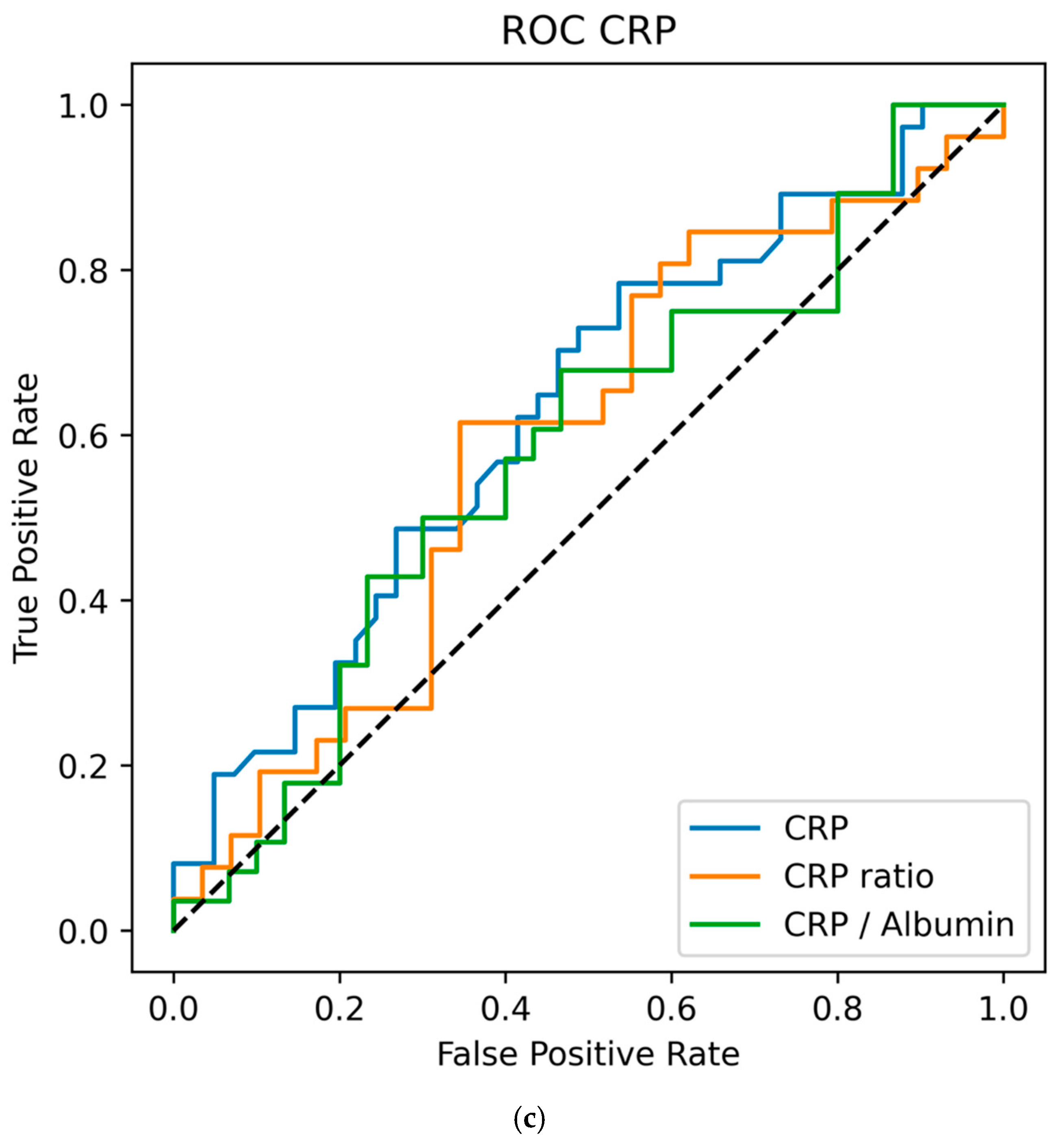
CRP: Daily CRP levels were significantly different on day 0, with a median level of 12.85 mg/L (IQR: 8.55) in the mcVAP/suspVAP group vs. 7.40 mg/L (IQR: 7.95) in the nonVAP group (p = 0.015, r = 0.351). Day-to-day ratios: No statistically significant differences were found for the CRP day-to-day ratios from day −4 to day 0. CRP-to-albumin quotient: In terms of the CRP-to-albumin ratio, we did not detect any statistically significant differences. Logistic regression analysis revealed that CRP values on day 0 were associated with VAP, with an OR of 1.093 (p = 0.012, 95% CI: 1.009–1.183) and an AUC of 0.676.
The details are provided in Table 4, and the receiver operating curves are provided in Figure 1c.
Adjustment for multiple testing: To control for multiple testing in biomarker analyses, we applied the Benjamini–Hochberg procedure (false discovery rate of 5%) to all biomarker-related p-values. This family included all tests comparing IL-6, PCT, and CRP across day −1 and day 0, their day-to-day ratios (−1/−2 and 0/−1), and their albumin-adjusted ratios. In total, 18 Mann–Whitney U and 18 logistic regression p-values were included in the correction. IL-6 on day 0 (both daily values and adjusted) and CRP on day 0 retained significance in both the Mann–Whitney U test and logistic regression. In contrast, the IL-6 level on day −1 lost significance after correction. Details are provided in Appendix A Table A1.

3.4. Antibiotic Treatment of Patients Who Do Not Meet the Criteria

Fifteen patients were given antibiotics for VAP without fulfilling the criteria for VAP. Considering those observed with PCT/CRP, 10 patients were treated without fulfilling the criteria for VAP, while under IL-6 observation, five patients were treated (χ2(1) = 7.190, p = 0.007, V = 0.270). In this group, we administered antibiotics 5.07 (SD 1.75) days after admission. Three of these patients had purulent secretions, whereas five had purulent secretions in the nonVAP group (χ2(1) = 5.412, p = 0.020, V = 0.255). None of the patients had infiltrates on the day when antibiotic therapy was started. On that day, differences between groups did not reach statistical significance (p > 0.05), although a numerical trend was observed in IL-6, CRP, the PaO2/FiO2 ratio, WBC count, and temperature. The details are provided in Table 5.

4. Discussion

Biomarkers play an important role in the early identification of infections, the timely initiation of antimicrobial therapy, and the evaluation of treatment response and duration. However, they seem inconsistent in diagnosing VAP. The predictive value of biomarkers in the context of VAP, therefore, remains a subject of ongoing investigation [24].
We designed an exploratory proof-of-concept study addressing one primary research question. We wanted to determine whether day-to-day ratios, or their ratios to albumin, had greater predictive value for VAP than the daily biomarker measures alone.
Given that inflammatory responses differ greatly between individuals [18] and that they downregulate albumin synthesis [21], we assumed that day-to-day and albumin ratios may increase diagnostic value for VAP.
Among all investigated biomarkers, IL-6 and its day-to-day ratio demonstrated the strongest and most consistent associations with VAP, with high effect sizes of 0.4–0.6 and AUC values above 0.8. Interestingly, IL-6 and its ratios were able to differentiate between VAP and nonVAP patients one day before VAP recognition. Despite this biological rationale, we could not observe any advantage in day-to-day ratios or in albumin adjustment. As for the day-to-day ratios, variability depends strongly on how stable the previous measurement was. Since IL-6 changes very quickly after even a minor stimulus, it can hide real differences rather than highlight them. For albumin adjustment, interindividual differences, e.g., non-inflammatory factors, might be a reason.
In contrast, PCT and CRP, as well as their respective albumin ratios and day-to-day changes, showed weaker significance in differentiating VAP patients from controls with only moderate predictive accuracy. Overall, we did not observe any improvement in discriminatory ability when the ratios were applied.
This might be due to a delayed response of CRP to an inflammatory stimulus. In terms of the values after the onset of VAP, CRP was still able to discriminate between VAP and nonVAP patients, especially on day 1 and day 2, when the median values were even higher than those on day 0 (14.3 mg/dL and 15.85 mg/dL vs. 12.60 mg/dL, respectively). For PCT, we detected very low values in all patients. One explanation might be that PCT responds differently depending on the type of bacteria, the infection site, and a certain delay [13]. However, our expectation of achieving more robust results when adjusting for albumin was not confirmed. This may be due to interindividual variability in albumin levels, which are affected by factors such as nutritional status.
To our knowledge, this was the first study to investigate day-to-day values and albumin ratios in VAP. In former studies, a combination of CRP and IL-6 showed the highest AUC of 0.79 compared to a wide panel of biomarkers for VAP [25]. Other investigators found that the CRP-to-albumin ratio was associated with community-acquired pneumonia in children [26]. In recent years, Pancreatic Stone Protein (PSP) and Pentraxin 3 (PT3) have emerged as biomarkers of interest in pneumonia. In a study by Ceccato et al., the kinetics of PSP were evaluated in serial measures, but no improvement was observed compared to PCT or CRP alone in the differentiation of VAP [25]. Also, Pentraxin 3 did not show superiority over the standard biomarker [26]. Therefore, we investigated the two most common biomarkers, PCT and CRP, in comparison with IL-6.
Second, we differentiated suspected VAP patients from microbiologically confirmed patients. In one-third of VAP patients, we could not identify a bacterium in tracheal or bronchial specimens.
In a recent update, the U.S. Centers for Disease Control (CDC) proposed differentiation. It includes worsening of oxygenation, a condition that is referred to as a “ventilator-associated event” (VAE). In addition to signs of inflammation plus the initiation of antimicrobial therapy, the CDC has proposed the name “ventilator-associated condition” (VAC). Upon microbiological confirmation, the CDC defines it as “infection-related ventilator-associated complication” (IVAC). This definition and the differentiations are, however, not intended for clinical diagnosis but rather for disease surveillance [27]. For clinical purposes, it is important to decide whether a patient requires directed antibiotic treatment. Therefore, our pragmatic approach was close to the CDC’s definition of VAC. Unless bedside techniques are available to identify bacterial infection faster and reliably, we believe that antibiotic treatment should not depend on microbiological confirmation. To address the problem of early bacterial detection, electronic nose sensor arrays and/or bed site nanopore sequencing might be promising approaches [28,29].
Third, our study revealed that some patients who received antibiotics did not meet the VAP criteria. This was particularly common in the CRP/PCT observation period compared with the IL-6 group (n = 10 (66.7%) vs. n = 5 (33.3%)). In previous studies, PCT was the focus of extensive research to guide the initiation or discontinuation of antibiotic therapy [13,30]. Our findings suggest that IL-6, known for its greater sensitivity and faster response time [10], may help refine antibiotic stewardship in ICUs.

Limitations

This study has several significant limitations.
First, the sample size is limited. This may have prevented the identification of further significant differences. Also, this was designed as a monocentric proof-of-concept study; the generalizability of our findings is limited. However, the monocentric design ensured standardized diagnostic and therapeutic procedures, thereby reducing inter-institutional bias and potentially strengthening the validity of our results.
Another limitation is that due to a change in institutional protocols, IL-6 and PCT/CRP were not measured simultaneously across all patients. This led to two distinct cohorts, which may have introduced a comparative bias that should be considered when interpreting the results, even though the baseline characteristics, including the SAPS score, did not differ.
Another relevant limitation is that, although IL-6 appears to be the most promising marker, its availability is limited due to its relatively high cost. It might therefore be useful to conduct prospective studies that assess cost-effectiveness and its effect on antibiotic prescription frequency, as well as other specific outcome data.

5. Conclusions

In this exploratory matched case–control study, we identified IL-6, including its day-to-day dynamics and albumin-adjusted values, as the most robust and consistent biomarker associated with VAP in neurosurgical ICU patients. These findings may be of interest for the development of automated predictive or AI-based models and should be validated in a large-scale, multicenter study.

Author Contributions

T.B. conceived the study, designed the methodology, performed the data analysis, and drafted the manuscript. C.R. contributed to data collection and interpretation. G.-S.H., J.K., H.A.L. and V.S.-L. contributed to the study design, statistical analysis, interpretation of data, and critical revision of the manuscript. All authors have read and agreed to the published version of the manuscript.

Funding

This study received no external funding. However, the publication was supported by the Helios Central Library (Helios Kliniken GmbH).

Institutional Review Board Statement

The study was conducted in accordance with the Declaration of Helsinki and approved by the Ethics Committee II of the Medical Faculty Mannheim, University of Heidelberg, Germany (reference number 2024-821, approval date: 4 April 2024). The requirement for informed consent was waived due to the retrospective design, in accordance with national legislation and institutional requirements.

Informed Consent Statement

Not applicable.

Data Availability Statement

The datasets generated and analyzed during this study are not publicly available due to institutional restrictions and data protection regulations. Anonymized data may be made available from the corresponding author upon reasonable request and with permission from the responsible institutions.

Conflicts of Interest

The authors declare no conflicts of interest.

Abbreviations

ABnonVAPPatients treated with antibiotics for VAP without fulfilling diagnostic criteria
AIArtificial Intelligence
AUCArea Under the Curve
CDCCenters for Disease Control and Prevention
CIConfidence Interval
CRPC-reactive Protein
D0Study days relative to VAP diagnosis
HRGHistidine-Rich Glycoprotein
ICUIntensive Care Unit
IL-6Interleukin-6
IQRInterquartile Range
IVACInfection-Related Ventilator-Associated Complication
LR p-valueLogistic Regression p-value
mcVAPMicrobiologically confirmed VAP
MW p-valueMann–Whitney U test p-value
NonVAPPatients without VAP (control group)
OROdds Ratio
PCTProcalcitonin
ROCReceiver Operating Characteristic (curve)
SAPSSimplified Acute Physiology Score
SDStandard Deviation
suspVAPSuspected but not microbiologically confirmed VAP
TempBody Temperature (°C)
USUnited States
VACVentilator-Associated Condition
VAEVentilator-Associated Event
VAPVentilator-Associated Pneumonia
WBCWhite Blood Cell count
PaO2/FiO2 ratio (P/F ratio)Ratio of arterial oxygen partial pressure to fraction of inspired oxygen

Appendix A

Table A1. Matching results.
CharacteristicUnmatched mcVAP + suspVAPUnmatched nonVAP Controlsp-ValueMatched mcVAP + suspVAPMatched nonVAP Controlsp-Value
Sample size (n)7383 6868
SAPS Score (SD)41.68 (11.07)38.45 (10.78)0.07942.55 (10.92)38.89 (10.94)0.064
Age (SD)65.36 (14.01)67.36 (13.93)0.37265.0 (14.39)67.22 (14.30)0.368
SAPS score and age before and after matching between VAP patients (mcVAP + suspVAP) and nonVAP controls. The values are given as the means (SDs).
Table A2. Number of patients within the biomarker groups.
CohortControlsmcVAPsuspVAPABnonVAPTotal
IL-6 (%)52 (31.1)32 (19.2)20 (12.0)5 (3.0)109 (65.3)
PCT/CRP (%)16 (9.6)13 (7.8)3 (1.8)10 (6.0)42 (25.1)
Total68 (40.7)45 (26.9)23 (13.8)15 (9.0)151 (100.0)
Distribution of patients across biomarker groups (IL-6 vs. PCT/CRP) and clinical VAP groups (controls, mcVAP, suspVAP, ABnonVAP). The percentages refer to the proportion of all included patients.
Table A3. Benjamini–Hochberg correction.
VariableMW_pLR_pMW_p_adjLR_p_adj
CRP Day −10.1770.8350.2120.835
Day −1/−2 Ratio0.6480.3720.6860.5904
CRP-Albumin-Ratio Day −10.6480.3720.6860.5904
CRP Day 00.0150.0120.0390.0432
Day 0/−1 Ratio0.1380.6540.2070.692470588
CRP to Albumin Day 00.1120.1990.2020.44775
PCT Day −10.1510.3550.2090.5904
PCT Day −1/−2 Ratio0.1770.0650.2120.167142857
PCT-Albumin-Ratio Day −10.7750.4630.7750.5904
PCT Day 00.0040.4360.0140.5904
PCT Day 0/−1 Ratio0.1260.4920.2060.5904
PCT to Albumin Day 00.0050.0180.0140.036
IL-6 Day −10.0400.5450.0900.613125
IL6 Day −1/−2 Ratio0.0130.0590.0390.167142857
IL-6-Albumin-Ratio Day −10.0540.4150.1080.5904
IL-6 Day 0<0.001<0.0010.0060.006
IL-6 Day 0/−1 Ratio<0.001<0.0010.0060.006
IL-6 to Albumin Day 0<0.001<0.0010.0060.006
p-value of Mann–Whitney U tests (MW_p), logistic regression analyses (LR_p), and Benjamini–Hochberg corrected p-values (MW_p_adj and LR_p_adj). The Benjamini–Hochberg procedure was applied only to the associated p-values.
Figure A1. Study flow diagram.
Life 15 01697 g0a1

References

  1. American Thoracic Society; Infectious Diseases Society of America. Guidelines for the Management of Adults with Hospital-acquired, Ventilator-associated, and Healthcare-associated Pneumonia. Am. J. Respir. Crit. Care Med. 2005, 171, 388–416. [Google Scholar] [CrossRef]
  2. Wu, V.K.S.; Fong, C.; Walters, A.M.; Lele, A.V. Prevalence, Clinical Characteristics, and Outcomes Related to Ventilator-Associated Events in Neurocritically Ill Patients. Neurocrit. Care 2020, 33, 499–507. [Google Scholar] [CrossRef] [PubMed]
  3. Melsen, W.G.; Rovers, M.M.; Groenwold, R.; Bergmans, D.C.; Camus, C.; Bauer, T.T.; Hanisch, E.; Klarin, B.; Koeman, M.; Krueger, W.A.; et al. Attributable mortality of ventilator-associated pneumonia: A meta-analysis of individual patient data from randomised prevention studies. Lancet Infect. Dis. 2013, 13, 665–671. [Google Scholar] [CrossRef]
  4. Zimlichman, E.; Henderson, D.; Tamir, O.; Fralnz, C.; Song, P.; Yamin, C.K.; Keohane, C.; Denham, C.R.; Bates, D.W. Health Care–Associated Infections. JAMA Intern. Med. 2013, 173, 2039–2046. [Google Scholar] [CrossRef]
  5. Simon, L.; Gauvin, F.; Amre, D.K.; Saint-Louis, P.; Lacroix, J. Serum Procalcitonin and C-Reactive Protein Levels as Markers of Bacterial Infection: A Systematic Review and Meta-analysis. Clin. Infect. Dis. 2004, 39, 206–217. [Google Scholar] [CrossRef]
  6. Fraunberger, P.; Wang, Y.; Holler, E.; Parhofer, K.G.; Nagel, D.; Walli, A.K.; Seidel, D. Prognostic value of interleukin 6, procalcitonin, and C-reactive protein levels in intensive care unit patients during first increase of fever. Shock 2006, 26, 10–12. [Google Scholar] [CrossRef]
  7. Boenisch, S.; Fae, P.; Drexel, H.; Walli, A.K.; Fraunberger, P. Are circulating levels of CRP compared to IL-6 and PCT still relevant in intensive care unit patients? J. Lab. Med. 2013, 37, 25–35. [Google Scholar] [CrossRef]
  8. Schmit, X.; Vincent, J.L. The time course of blood C-reactive protein concentrations in relation to the response to initial antimicrobial therapy in patients with sepsis. Infection 2008, 36, 213–219. [Google Scholar] [CrossRef]
  9. Rettig, T.C.D.; Verwijmeren, L.; Dijkstra, I.M.; Boerma, D.; Van De Garde, E.M.W.; Noordzij, P.G. Postoperative Interleukin-6 Level and Early Detection of Complications After Elective Major Abdominal Surgery. Ann. Surg. 2016, 263, 1207–1212. [Google Scholar] [CrossRef] [PubMed]
  10. Tanaka, T.; Narazaki, M.; Kishimoto, T. Il-6 in inflammation, Immunity, and disease. Cold Spring Harb. Perspect. Biol. 2014, 6, a016295. [Google Scholar] [CrossRef] [PubMed]
  11. Póvoa, P.; Coelho, L.; Almeida, E.; Fernandes, A.; Mealha, R.; Moreira, P.; Sabino, H. C-reactive protein as a marker of infection in critically ill patients. Clin. Microbiol. Infect. 2005, 11, 101–108. [Google Scholar] [CrossRef]
  12. Coelho, L.; Rabello, L.; Salluh, J.; Martin-Loeches, I.; Rodriguez, A.; Nseir, S.; Gomes, J.A.; Povoa, P. C-reactive protein and procalcitonin profile in ventilator-associated lower respiratory infections. J. Crit. Care 2018, 48, 385–389. [Google Scholar] [CrossRef]
  13. Schuetz, P.; Albrich, W.; Mueller, B. Procalcitonin for diagnosis of infection and guide to antibiotic decisions: Past, present and future. BMC Med. 2011, 9, 107. [Google Scholar] [CrossRef]
  14. Ding, H.G.; Zhou, H.F.; Diao, M.Y.; Xu, Y.; Pan, Q.M.; Shen, X.H. A novel biomarker of serum Histidine-Rich Glycoprotein (HRG) for diagnosing and predicting prognosis of ventilator-associated pneumonia (VAP): A pilot study. Eur. Rev. Med. Pharmacol. Sci. 2018, 22, 7920–7927. [Google Scholar] [CrossRef]
  15. Pelosi, P.; Barassi, A.; Severgnini, P.; Gomiero, B.; Finazzi, S.; Merlini, G.; d’Eril, G.M.; Chiaranda, M.; Niederman, M.S. Prognostic role of clinical and laboratory criteria to identify early ventilator-associated pneumonia in brain injury. Chest 2008, 134, 101–108. [Google Scholar] [CrossRef]
  16. Bexten, T.; Wiebe, S.; Brink, M.; Hinzmann, D.; Haarmeyer, G.-S. Early Detection of Ventilator-Associated Pneumonia in a Neurosurgical Patient: Do Biomarkers Help Us? Cureus 2025, 17, e78567. [Google Scholar] [CrossRef]
  17. Ramírez, P.; Ferrer, M.; Gimeno, R.; Tormo, S.; Valencia, M.; Piñer, R.; Menendez, R.; Torres, A. Systemic inflammatory response and increased risk for ventilator-associated pneumonia: A preliminary study. Crit. Care Med. 2009, 37, 1691–1695. [Google Scholar] [CrossRef]
  18. Lee, E.H.; Lee, K.H.; Song, Y.G.; Han, S.H. Discrepancy of C-Reactive Protein, Procalcitonin and Interleukin-6 at Hospitalization: Infection in Patients with Normal C-Reactive Protein, Procalcitonin and High Interleukin-6 Values. J. Clin. Med. 2022, 11, 7324. [Google Scholar] [CrossRef] [PubMed]
  19. Póvoa, P.; Martin-Loeches, I.; Ramirez, P.; Bos, L.D.; Esperatti, M.; Silvestre, J.; Gili, G.; Goma, G.; Berlanga, E.; Espasa, M.; et al. Biomarker kinetics in the prediction of VAP diagnosis: Results from the BioVAP study. Ann. Intensive Care 2016, 6, 32. [Google Scholar] [CrossRef] [PubMed]
  20. Chen, L.; Wu, X.; Qin, H.; Zhu, H. The PCT to Albumin Ratio Predicts Mortality in Patients with Acute Kidney Injury Caused by Abdominal Infection-Evoked Sepsis. Front. Nutr. 2021, 8, 584461. [Google Scholar] [CrossRef] [PubMed]
  21. Ranzani, O.T.; Zampieri, F.G.; Forte, D.N.; Azevedo, L.C.P.; Park, M. C-Reactive Protein/Albumin Ratio Predicts 90-Day Mortality of Septic Patients. PLoS ONE 2013, 8, e59321. [Google Scholar] [CrossRef] [PubMed]
  22. Oh, T.K.; Song, I.A.; Lee, J.H. Clinical usefulness of C-reactive protein to albumin ratio in predicting 30-day mortality in critically ill patients: A retrospective analysis. Sci. Rep. 2018, 8, 14977. [Google Scholar] [CrossRef] [PubMed]
  23. Lee, D.K. Introduction Alternatives to P value: Confidence interval and effect size KJA. Korean J. Anesthesiol. 2016, 69, 555–562. [Google Scholar] [CrossRef]
  24. Howroyd, F.; Chacko, C.; MacDuff, A.; Gautam, N.; Pouchet, B.; Tunnicliffe, B.; Weblin, J.; Gao-Smith, F.; Ahmed, Z.; Duggal, N.A.; et al. Ventilator-associated pneumonia: Pathobiological heterogeneity and diagnostic challenges. Nat. Commun. 2024, 15, 6447. [Google Scholar] [CrossRef] [PubMed]
  25. Ceccato, A.; Camprubí-Rimblas, M.; Bos, L.D.J.; Povoa, P.; Martin-Loeches, I.; Forné, C.; Areny-Balagueró, A.; Campaña-Duel, E.; Morales-Quinteros, L.; Quero, S.; et al. Evaluation of the Kinetics of Pancreatic Stone Protein as a Predictor of Ventilator-Associated Pneumonia. Biomedicines 2023, 11, 2676. [Google Scholar] [CrossRef]
  26. Ye, W.; Huang, Q.D.; Tang, T.Y.; Qin, G.Y. Diagnostic value of pentraxin 3 in respiratory tract infections: A meta-analysis. Medicine 2020, 99, e19532. [Google Scholar] [CrossRef]
  27. Centers for Disease Control and Prevention, National Center for Emerging and Zoonotic Infectious Diseases, Division of Healthcare Quality Promotion. Ventilator-Associated Event (VAE); CDC: Atlanta, GA, USA, 2024.
  28. Chen, C.Y.; Lin, W.C.; Yang, H.Y. Diagnosis of ventilator-associated pneumonia using electronic nose sensor array signals: Solutions to improve the application of machine learning in respiratory research. Respir. Res. 2020, 21, 45. [Google Scholar] [CrossRef]
  29. Pugh, J. The Current State of Nanopore Sequencing. In Nanopore Sequencing; Humana: New York, NY, USA, 2023; pp. 3–14. [Google Scholar] [CrossRef]
  30. Wirz, Y.; Meier, M.A.; Bouadma, L.; Luyt, C.E.; Wolff, M.; Chastre, J.; Tubach, F.; Schroeder, S.; Nobre, V.; Annane, D.; et al. Effect of procalcitonin-guided antibiotic treatment on clinical outcomes in intensive care unit patients with infection and sepsis patients: A patient-level meta-analysis of randomized trials. Crit. Care 2018, 22, 191. [Google Scholar] [CrossRef]
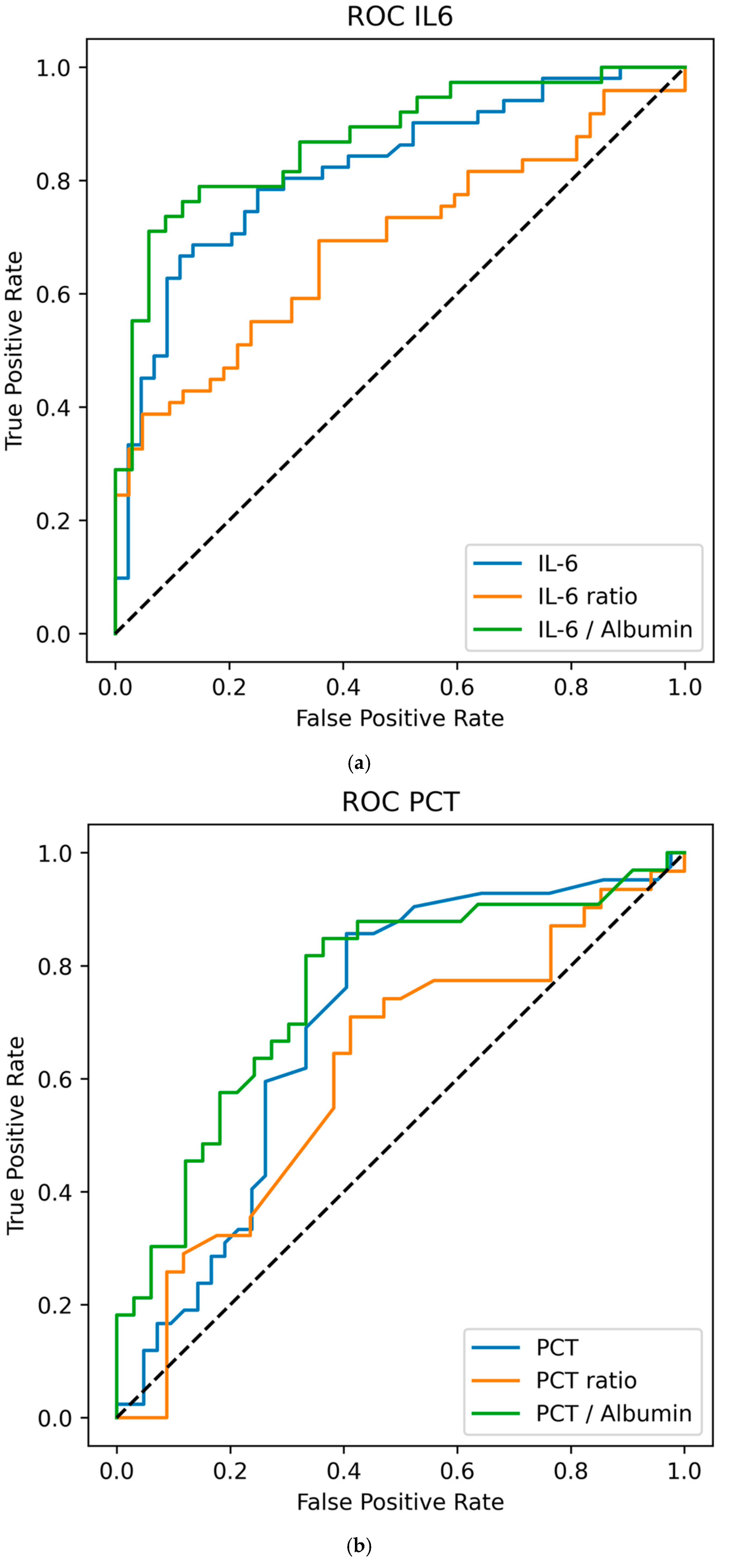
Figure 1. (a) ROC for IL-6 in VAP patients on day 0. Receiver operating characteristic (ROC) curves. IL-6 D0: Threshold = 26.26, sensitivity = 66.7%, specificity = 88.6%, AUC = 0.816. IL-6 D0/−1 ratio: threshold = 1.75, sensitivity = 38.8%, specificity = 95.2%, AUC = 0.726. IL-6/albumin D0: Threshold = 26.26, sensitivity = 71.1%, specificity = 94.1%, AUC = 0.864. (b) ROC for PCT in VAP patients on day 0. Receiver operating characteristic (ROC) curves for PCT. PCT D0: Threshold = 0.13, sensitivity = 85.7%, specificity = 59.5%; AUC = 0.707. PCT D0/−1 ratio: threshold = 0.90, sensitivity = 71.0%, specificity = 58.8%, AUC = 0.626 PCT/albumin D0: threshold = 0.04, sensitivity = 81.8%, specificity = 66.7%, AUC = 0.746. (c) ROC for CRP in VAP patients on day 0. ROC for CRP. CRP D0: Threshold = 8.50, sensitivity = 78.4%, specificity = 46.3%, AUC = 0.676. CRP D0/−1 ratio: Threshold = 1.05, sensitivity = 61.5%, specificity = 65.5%, AUC = 631. CRP/albumin D0: Threshold = 3.76, sensitivity = 67.9%, specificity = 53.3%, AUC = 636.
Life 15 01697 g001aLife 15 01697 g001b
Table 1. PCT levels and ratios on day −1 and day 0.
VariablenonVAP
Median (IQR)
mcVAP and suspVAP
Median (IQR)
MW p-Value
(W; r-Effect Size)
AUCOR (95% CI)LR p-Value
Day −1
PCT in ng/mLn = 39n = 44
Day −10.16 (0.32)0.24 (0.47)0.151 (616.00; 0.189)0.5951.017 (0.969–1.068)0.355
Day −1/−2 Ratio0.96 (0.38)1.00 (1.42)0.177 (321.00; 0.209)0.6051.231 (0.823–1.842)0.065
PCT to Albumin Ratio17.00 (29.30)22.77 (37.36)0.775 (491.00; 0.043)0.5211.033 (0.924–1.155)0.463
Day 0
PCT in ng/mLn = 31n = 42
Day 00.11 (0.19)0.24 (0.34)0.004 * (616.00; 0.414)0.7071.010 (0.978–1.043)0.461
Day 0/−1 Ratio0.757 (0.28)1.00 (0.44)0.126 (241.00; 0.252)0.6261.428 (0.516–3.952)0.492
PCT to Albumin0.04 (0.05)0.08 (0.15)0.005 (192.50; 0.474)0.7461.062(0.116–15.688)0.016
PCT levels, day-to-day ratios, and albumin ratios between nonVAP patients and VAP patients (mcVAP and suspVAP) on days −1 and 0 of VAP occurrence. The results are shown as medians with interquartile ranges (IQRs). The Mann–Whitney U test (MW p-value) indicates group differences, with effect sizes (r) reported. Predictive values are reported as odds ratios (ORs) with 95% confidence intervals (CIs), p-values (LR p-values), and area under the curve (AUC) values. * indicates statistical significance at p < 0.05.
Table 2. Baseline characteristics.
BaselinenonVAP n = 68mcVAP/suspVAP n = 68ABnonVAP n = 15Total n = 171
Age (years) (SD)67.22 (14.23)65.0 (14.39)60.0 (9.43)66.4 (13.93)
Sex F/M (% of total)36/32 (26/15)21/47 (14/32)14/11 (7/6)92/101 (48/52)
Length of stay in hours (SD)399.81 (360.13)599.46 (335.57)532.89 (424.82)502.29 (367.60)
Hours of ventilation in hours (SD)198.49 (165.23)380.10 (225.32)366.20(370.93)297.59 (236.05)
SAPS on admission [SD]38.71 (10.94)42.55 (10.02)41.22 (17.32)40.30 (11.11)
Diagnosis (% of Total)
Intracerebral bleeding55 (36.4)58 (38.3)15 (9.9)128 (84.8)
Traumatic head injury4 (2.6)3 (2.0)0 (0)7 (4.6)
Cerebral tumor3 (2.0)0 (0.5)0 (0)3 (2.0)
Malignant infarction6 (4.0)7 (4.6)0 (0)13 (8.6)
Comorbidity (% of Total)
Cardiovascular disease44 (29.1)45 (29.8)9 (6.0)98 (64.9)
Respiratory disease4 (2.4)3 (2.0)3 (2.0)10 (6.6)
Kidney disease1 (0.7)3 (2.0)2 (1.3)6 (4.0)
Substance abuse5 (3.3)9(6.0)2 (1.3)16 (10.6)
Other3(2.0)11 (7.3)3 (2.0)17 (11.3)
Baseline on day of ICU Admission
WBC (IQR)11.51 (6.280)10.34 (8.29)11.45 (7.29)10.8 (6.74)
IL-6 (IQR)40.80 (53.40)34.05 (97.73)34.85 (22.05)40.80 (69.30)
PCT (IQR)0.3 (0.35)0.11 (0.06)0.07 (0.05)0.11 (0.17)
CRP (IQR)0.20 (0.95)0.30 (2.15)0.45 (1.05)0.20 (1.55)
Baseline characteristics, diagnoses, comorbidities, and operative procedures of the study population. Data are presented as the means (standard deviations) for continuous variables and daily numbers (percentages of total) for categorical variables. IL-6, PCT, and CRP are presented within the IL-6 and PCT/CRP groups.
Table 3. IL-6 levels and ratios on day −1 and day 0.
VariablenonVAP
Median (IQR)
mcVAP and
suspVAP
Median (IQR)
MW p-Value
(W; r-Effect Size)
AUCOR (95% CI)LR p-Value
Day −1
IL-6 in pg/mLn = 40n = 52
Day −146.35 (63.10)63.10 (71.00)p = 0.040 * (739.50; 0.255)0.6271.001 (0.998–1.004)0.545
Day −1/−2 Ratio0.93 (1.42)1.84 (1.92)p = 0.013 * (627.00; 0.313)0.6561.289 (0.970–1.713)0.059
IL-6-Albumin Ratio17.00 (29.30)22.77 (37.36)p = 0.054 (551.50; 0.257)0.6291.004 (0.995–1.013)0.415
Day 0
IL-6 in pg/mLn = 39n = 53
Day 035.40 (35.80)109 (180.25)p < 0.001 * (359.50; 0.631)0.8161.012 (1.004–1.020)p < 0.001 *
Day 0/−1 Ratio0.74 (0.81)1.42 (1.85)p < 0.001 * (500.00; 0.452)0.7262.660 (1.390–5.091)p < 0.001 *
IL-6 to Albumin Ratio12.09 (14.72)41.48 (65.07)p < 0.001 * (151.50; 0.257)0.8641.065 (1.024–1.109)p < 0.001 *
IL-6/PCT/CRP levels, day-to-day ratios, and albumin ratios between nonVAP patients and VAP patients (mcVAP and suspVAP) on days −1 and 0 of VAP occurrence. The results are shown as medians with interquartile ranges (IQRs). The Mann–Whitney U test (MW p-value) indicates group differences, with effect sizes (r) reported. Predictive values are reported as odds ratios (ORs) with 95% confidence intervals (CIs), p-values (LR p-values), and area under the curve (AUC) values. * indicates statistical significance at p < 0.05.
Table 4. CRP levels and ratios on day −1 and day 0.
VariablenonVAP
Median (IQR)
mcVAP and
suspVAP
Median (IQR)
MW p Value
(W; r-Effect Size)
AUCOR (95% CI)LR p-Value
Day −1
CRP in mg/Ln = 34n = 41
Day −17.60 (7.50)9.65 (12.38)0.207 (517.00; 0.175)0.5881.058 (0.989–1.132)0.084
Day −1/−2 Ratio1.09 (1.12)1.42 (2.58)0.177 (242.00; 0.224)0.6121.005 (0.956–1.058)0.835
CRP to Albumin Ratio3.02 (3.38)3.07 (5.52)0.648 (435.50; 0.069)0.5350.982 (0.936–1.030)0.372
Day 0
CRP in mg/Ln = 32n = 37
Day 07.40 (7.95)12.85 (8.55)0.015 * (342.00; 0.351)0.6761.093 (1.009–1.183)0.012 *
Day 0/−1 Ratio0.94 (0.76)1.07 (0.78)0.138 (184.50; 0.262)0.6311.028 (0.909–1.162)0.654
CRP to Albumin3.19 (2.93)4.25 (2.96)0.112 (200.00; 0.273)0.6361.131 (0.926–1.382)0.199
CRP levels, day-to-day ratios, and albumin ratios between nonVAP patients and VAP patients (mcVAP and suspVAP) on days −1 and 0 of VAP occurrence. The results are shown as medians with interquartile ranges (IQRs). The Mann–Whitney U test (MW p-value) indicates group differences, with effect sizes (r) reported. Predictive values are reported as odds ratios (ORs) with 95% confidence intervals (CIs), p-values (LR p-values), and area under the curve (AUC) values. * indicates statistical significance at p < 0.05.
Table 5. Comparison of VAP vs. ABnonVAP.
VariablenonVAP (Median, IQR)ABnonVAP (Median, IQR)p-Value (W-Value, r-Effect Size)
Day 0n = 68n = 15
IL-6 (pg/mL)35.40 (35.80)42.80 (42.40)p = 0.941 (W = 95.00, r = 0.026)
PCT (ng/mL)0.11 (0.19)0.11 (0.15)p = 0.738 (W = 167.50, r = 0.069)
CRP (mg/L)7.40 (7.95)12.75 (6.03)p = 0.071 (W = 95.00, r = 0.387)
WBC (×109/L)9.30 (4.56)10.10 (4.15)p = 0.569 (W = 358.50, r = 0.098)
Temp (°C)37.20 (0.80)37.50 (0.45)p = 0.412 (W = 414.50, r = 0.136)
PaO2/FiO2347.00 (128.00)308.00 (147.00)p = 0.633 (W = 366.00, r = 0.084)
Comparison of relevant markers between nonVAP patients and those who were treated for VAP without fulfilling the criteria. The values are presented as medians (IQRs).
Disclaimer/Publisher’s Note: The statements, opinions and data contained in all publications are solely those of the individual author(s) and contributor(s) and not of MDPI and/or the editor(s). MDPI and/or the editor(s) disclaim responsibility for any injury to people or property resulting from any ideas, methods, instructions or products referred to in the content.