Article

In Situ Harvesting and Molecular Identification for the Germinating Species Diversity of Dinoflagellate Resting Cysts in Jiaozhou Bay, China

1
CAS Key Laboratory of Marine Ecology and Environmental Sciences, Institute of Oceanology, Chinese Academy of Sciences, Qingdao 266071, China
2
University of Chinese Academy of Sciences, Beijing 100049, China
3
Shandong Key Laboratory of Marine Ecological Restoration, Shandong Marine Resource and Environment Research Institute, Yantai 264006, China
4
China Laboratory for Marine Ecology and Environmental Science, Qingdao Marine Science and Technology Center, Qingdao 266237, China
5
Qingdao Innovation and Development Center, Harbin Engineering University, Qingdao 266000, China
6
Animal, Plant and Food Inspection Center of Nanjing Customs, Nanjing 210000, China
*
Authors to whom correspondence should be addressed.
Life 2025, 15(11), 1670; https://doi.org/10.3390/life15111670 (registering DOI)
Submission received: 20 September 2025 / Revised: 21 October 2025 / Accepted: 25 October 2025 / Published: 27 October 2025

Abstract

Dinoflagellate resting cysts are critical to dinoflagellate ecology, acting as a key seed source for initiating harmful algal blooms (HABs) through their germination. However, the in situ germination dynamics of these cysts remain poorly understood due to technical challenges. To overcome this, we utilized the Germlings Harvester (GEHA), an in situ germination device we designed, to collect water samples containing dinoflagellate cysts germinated from marine sediments in Jiaozhou Bay, China, after 5 and 20 days of incubation. By combining the GEHA with metabarcoding analysis targeting 28S rDNA-specific primers for dinoflagellates, we identified 44 dinoflagellate species spanning 31 genera, 18 families, and 7 orders. Of these, 12 species were linked to HABs or recognized as toxic, including Azadinium poporum, Alexandrium leei, Alexandrium pacificum, Akashiwo sanguinea, Karlodinium veneficum, Stoeckeria algicida, and Luciella masanensis. Additionally, five species were newly identified as cyst producers, and one symbiotic dinoflagellate, Effrenium voratum, was detected. Our results also found that germinated dinoflagellate species increased from 23 to 34 with extended incubation, and the ratio of mixotrophic to heterotrophic species was approximately 2:1 in the samples of in situ sediments and seawater outside GEHA, as well as across germination durations (Sg-5 d vs. Sg-20 d). These findings provide essential field evidence for the role of resting cysts in driving HAB formation in this region and highlight the efficacy of the GEHA-based approach for studying in situ cyst germination dynamics, offering a robust tool for monitoring, early warning, prevention, and forecasting of HABs.

1. Introduction

Dinoflagellates, pivotal primary producers in marine ecosystems, are also major contributors to harmful algal blooms (HABs) globally. Among HAB events, about 75% of them are caused by dinoflagellates [1], and dinoflagellates account for 40% of the species forming HABs globally [2]. In China, dinoflagellate blooms frequently occur in coastal waters, spanning the Bohai, Yellow, East China, and South China Seas. These blooms cause tremendous damage to fisheries, mariculture, tourism, and human health, as well as marine ecosystems [3]. The official report “Bulletin of China Marine Disaster” from 1989 [4] and several recent reviews [3] state that among the 17 major large-scale HAB-causing species in China’s coastal waters, 9 are dinoflagellates, accounting for approximately 53%. Of these, five species (e.g., Gymnodinium catenatum and Karenia mikimotoi) have caused mortality in marine animals or even humans due to bioaccumulation of toxins and transmission through the food chain [3].
About 200 out of approximately 2900 extant dinoflagellate species have been reported to produce resting cysts, and more than 20 of them form HABs [5,6]. The formation, dormancy, and germination of resting cysts are ecologically critical processes for dinoflagellates [7,8] because they are associated with genetic recombination [5,8]; termination of HABs [8]; resistance to adverse environments [9,10]; protection from viruses, grazers, and parasite attacks [8]; and geographic expansion of populations [11,12,13]. More importantly, resting cysts can germinate into vegetative cells under suitable conditions (e.g., warm spring/summer seasons, typically in the following year) after a dormancy period, which provides “inoculation” for new rounds of population growth and even initiates HABs [7,9,14]. Therefore, surveys on germination of dinoflagellate cysts are of vital importance for monitoring and forecasting of HABs.
Due to the complexity of field environments, however, it is challenging to replicate the germination conditions of dinoflagellate cysts in laboratory settings. Significant discrepancies of cyst germination dynamics exist between the laboratory and the field, e.g., Alexandrium spp. [10,15,16,17]. Consequently, conducting in situ germination experiments on cysts is essential and urgent. Internationally, previous research on in situ germination of resting cysts has primarily employed two types of devices. The first type involves placing an inverted apparatus on the seabed, which is typically deployed and retrieved by divers [18,19]. However, this design presents many limitations, including instability under ocean currents, considerable difficulties in collecting germinated water samples, and a high risk of contamination from the seawater outside of the devices. The second type, notably improved by Ishikawa et al. (2007) [15], consists of a two-chamber system: sediment is placed in the lower compartment, while filtered seawater is supplied to the upper section. After assembly, the device is deployed on the seabed to enable in situ cyst germination. This design represents a significant advancement over earlier devices and has served as a foundational tool for subsequent studies in this field. Nevertheless, it still exhibits certain shortcomings, such as complex setup procedures and a lack of an adjustable counterweight to accommodate varying hydrodynamic conditions across different environments, which may compromise the stability of the device on the seabed. Moreover, to date, no studies on the in situ germination of dinoflagellate cysts have been carried out in China. Previous studies on the in situ germination of dinoflagellate cysts have largely remained unexplored. These studies focus on detecting the germination in individual species, such as Alexandrium catenella, A. minutum, A. pacificum, Scrippsiella, and Gonyaulax, and species identification relies primarily on microscopes [15,16,17,18,19,20,21,22,23,24,25]. Obtaining a comprehensive inventory of all germinated dinoflagellate species in a certain environment may face significant challenges due to methodological limitations.
Jiaozhou Bay, a semi-enclosed bay, is situated on the south coast of the Shandong Peninsula, China, and connects to the northwest of the South Yellow Sea through a narrow channel, with a surface area of about 374 km2 and an average depth of only seven meters [26]. As the earliest, most comprehensively and systematically studied marine bay in China, the bay features a densely populated coastline with intensive industrial and agricultural activities [26,27]. Marine environments of the bay are influenced by high-intensity anthropogenic activities, including urbanization, terrestrial discharges, international shipping, large-scale engineering projects, and mariculture [27,28,29]. Since the 1990s, the bay has experienced frequent HABs, making it an ideal area for studying HAB outbreaks, and historical records indicate many dinoflagellate-related HABs in the bay (e.g., Noctiluca scintillans and Margalefidinium fulvescens) [28,30,31]. Morphological and molecular identification of sediment samples has detected 63 species of dinoflagellate cysts [8,32,33,34,35], whereas identification of water samples has identified 224 dinoflagellates [26,28,36] in Jiaozhou Bay. Notably, many species in the vegetative cell checklist have never appeared in prior cyst records, and some species laboratory-confirmed to produce cysts have not yet been detected in sediments.
In this study, we implemented the recently developed Germlings Harvester (GEHA), designed for the in situ monitoring of the germination of resting-stage cells of plankton from marine sediment [37], and combined it with metabarcoding analysis to study the in situ germination of dinoflagellate resting cysts. By coupling GEHA with metabarcoding analysis targeting dinoflagellate-specific 28S rDNA primers and key environmental parameters (e.g., temperature, salinity, and dissolved oxygen), we investigated the species diversity of in situ germinated dinoflagellate cysts. This approach provides foundational technical and data-driven insights for monitoring and early warning of HABs in the study region, underscoring the significant potential of the GEHA for broader applications in HAB research and management.

2. Materials and Methods

2.1. Sampling Site

The in situ sampling site of the GEHA was located at the southeast coast of Jiaozhou Bay, within the Qingdao Cruise Terminal (as shown in Figure 1). The area has an average water depth of 5 m, with coordinates at 120.314° E, 36.084° N. The water exchange between the area and the open sea is effective, with a half-water-exchange period of 5 d [38]. The distribution of and variation in water temperature have obvious seasonal characteristics. The lowest water temperature appears in January (2.9 °C) and the highest in September (25.5 °C) [39]. The germination experiments were conducted from April to May 2024, during which the temperature in the area ranged between 10 and 15 °C [39]. Characterized by well-developed tourism and severe eutrophication, the area serves as a representative site for monitoring the impact of the germination of dinoflagellate cysts on HABs in Jiaozhou Bay.

2.2. In Situ Sampling for the Germinated Dinoflagellate Cysts with GEHA

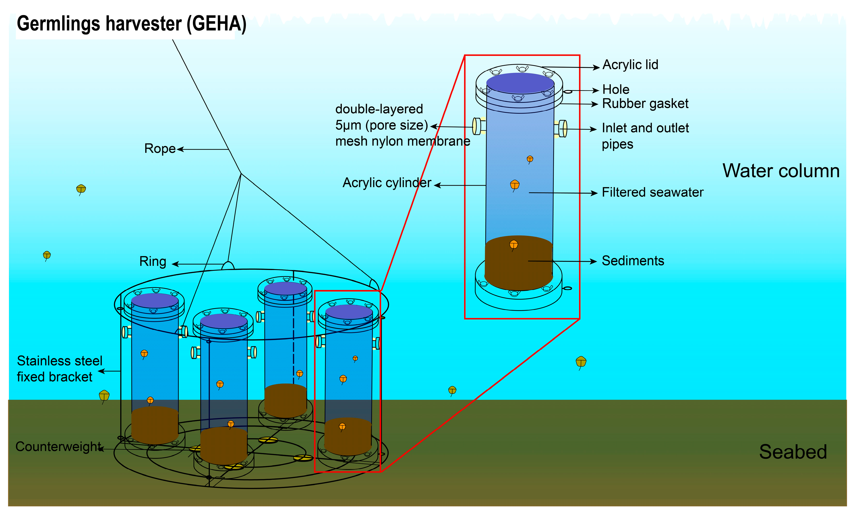
The GEHA consists of a stainless-steel fixed bracket and four acrylic cylinders. Each cylinder features a hollow structure and is positioned symmetrically with two inlet/outlet pipes at the upper part The pipes are wrapped with double-layered 5 μm mesh nylon membranes, ensuring the exchange of seawater inside and outside the cylinders while preventing contamination by external dinoflagellates on internally germinated dinoflagellates within the cylinders. The pore size of the mesh nylon membranes can be adjusted based on water environments and the cell size of the target species. The design and manufacturing of the GEHA have been submitted for patent application.
The schematic diagram of the GEHA is shown in Figure 2. In situ sediment samples, collected 30 m from the GEHA monitoring site, were packed into two sets of GEHAs. Then, in situ collected seawater, filtered through a 0.22 μm pore-size filter, was poured into the cylinders. After sealing the lids, GEHAs were slowly deployed on the seabed for the germination experiments. The two sets of GEHAs were retrieved after 5 days and 20 days. Seawater samples (4 L per cylinder) from the GEHA were collected via siphoning, while, for comparison (as control), 2 L of water samples were also taken, respectively, from the surface and bottom seawater from the location where the GEHA was deployed. Additionally, the environmental conditions of the seawater, such as temperature and dissolved oxygen (DO), were measured using the YSI (Yellow Springs Instrument, Yellow Springs, OH, USA) during GEHA deployment and retrieval. All of the above structures and handling procedures of the GEHA were comprehensively detailed in Shi et al. (2025) [37].

2.3. DNA Extraction, PCR Amplification, and Metabarcoding Sequencing

A pair of primers (DinoF primer, 5′-KACTTTGRRAAGAGAGTTAAAW-3′; DinoR, 5′-TCYGTGTTTCAAGACGGGTC-3′) was used to amplify about 400 bases of the partial 28S rDNA sequence, including the highly variable D2 domain, intended mainly for dinoflagellates [32]. Genomic DNA of water samples, including seawater samples in the GEHA and surface and bottom seawater samples surrounding the GEHA, was extracted from 0.4 μm hydrophilic polycarbonate membranes (47 mm diameter, Merck Millipore Ltd., Darmstadt, Germany); each filtered 2 L of water samples using a plant DNA extraction kit (Tiangen, Beijing, China) according to the kit manufacturer’s protocol. Genomic DNA of the sediment samples was extracted from the resting cysts using the Fast DNA SPIN Kit for Soil (MP Biomedicals, Santa Ana, CA, USA). The resting cysts in the sediment samples were concentrated from 32 g of sediment for each sediment sample using the sodium polytungstate (SPT) protocol [40] prior to DNA extraction. The quantity and quality of total DNA were analyzed with the ND-2000 NanoDrop spectrophotometer (Thermo Fisher Scientific, Somerset, NJ, USA).
All samples were sequenced with the NovaSeq-PE250 platform by LC-Bio Technology Co., Ltd. (Hangzhou, China). Paired-end sequencing reads were demultiplexed according to sample-specific barcode information, followed by barcode and primer sequence trimming using the software Cutadapt (v1.9) [41]. Subsequently, paired-end reads were merged into contiguous tags via overlap regions using the software FLASH (v1.2.8) [42]. Sequences containing ambiguous bases (>5% N-content) or that were shorter than the quality threshold according to the fqtrim (v0.94) and chimeric sequences were filtered using the software Vsearch (v2.3.4) [43]. High-resolution amplicon sequence variants (ASVs) and abundance tables were generated through DADA2 denoising [44], with taxonomic annotations assigned against the Nucleotide database. The raw sequencing data set was submitted to the NCBI Short Read Archive (SRA) database under accession number PRJNA1302703.

2.4. Statistical Analyses

ASVs with above 97% identities and 100% coverage with the reference sequences in the NCBI GenBank database were annotated to species levels. ASVs with lower coverage and identity were manually checked and corrected by blasting the individual sequences in the NCBI’s BLASTn (v2.16.0) [13,32,45]. Rarefaction curves were plotted for each sample, and an unweighted hierarchical clustering tree was generated with ASVs using QIIME2 [46]. All community diversity parameters (Shannon, Chao1, Good’s coverage, and Simpson index) were calculated with QIIME2 [46]. Venn diagrams, Sankey diagrams, and principal coordinates analysis were executed using the website Tutool (http://cloudtutu.com.cn/ (accessed on 26 May 2025)). Column, line, and pie charts were generated using the Origin software (Version 2021).

3. Results

3.1. Overview for the Metabarcoding Sequencing

A total of 16 samples were subjected to metabarcoding sequencing, categorized into three groups: (1) in situ sediment samples (S); (2) samples from the GEHA capturing germinated cysts (i.e., in situ sediment germination, Sg); and (3) surface seawater (Sw) and bottom seawater (Bw) samples collected from the GEHA deployment site (see Table S1 for detailed sample information). From these samples, 1,996,087 raw sequence tags were generated, with 1,885,489 valid tags retained after quality control. A total of 2006 amplicon sequence variants (ASVs) were assigned to dinoflagellates. Good’s coverage values exceeded 99% for all samples (Table S2), and rarefaction curves approached a plateau (Figure S1), indicating sufficient sequencing depth and robust representation of the dinoflagellate assemblages in this study.

3.2. Species Diversity of In Situ Germinated Dinoflagellate Cysts

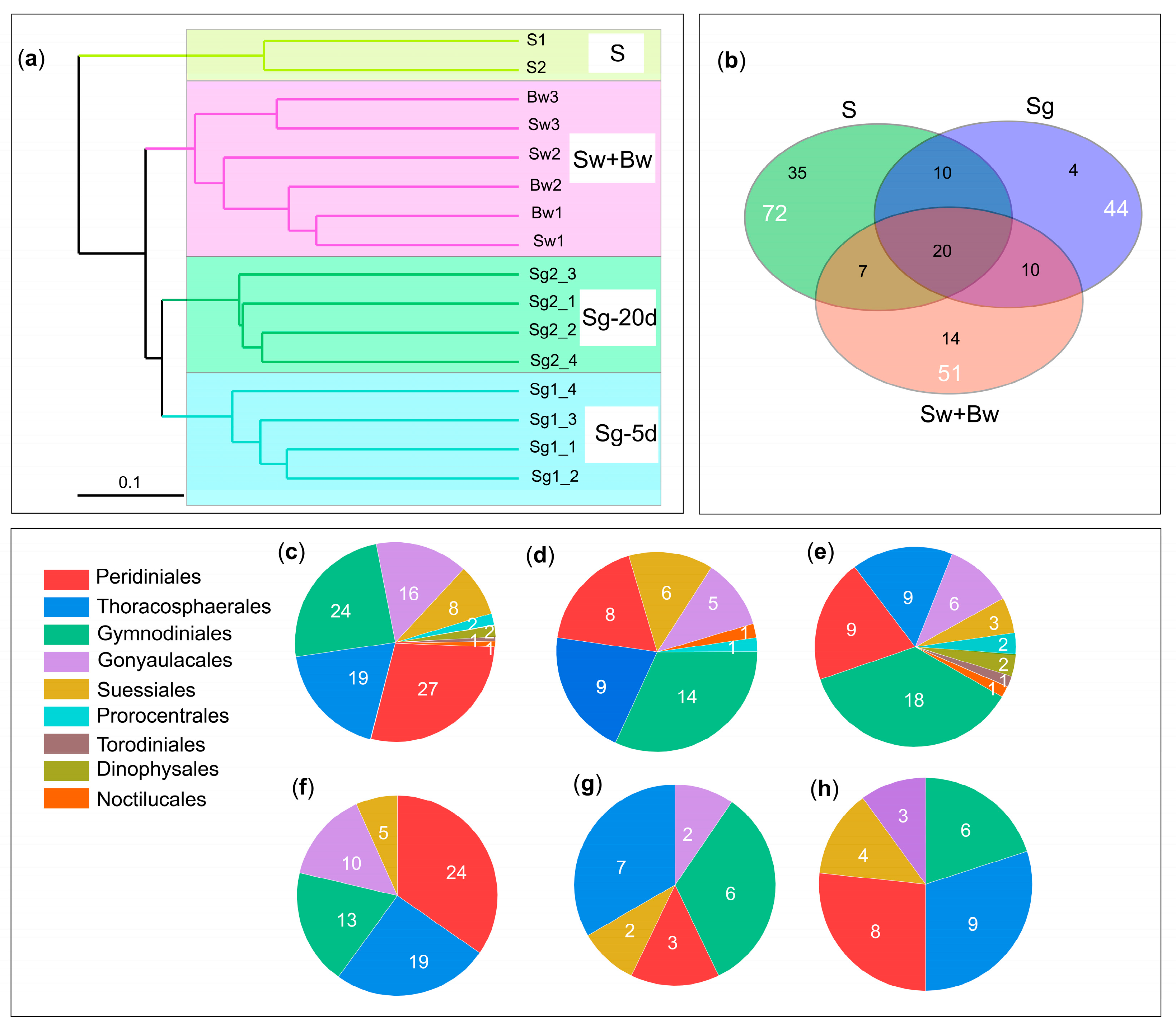
The UPGMA cluster analysis clearly grouped the 16 samples into three distinct clusters (Sg, Sw+Bw, and S) (Figure 3a). The three clusters revealed significant differences among samples from different sources (i.e., water samples within the GEHA, water samples surrounding the GEHA, and sediment samples) but minor differences within samples of the same cluster in species diversity, indicating a similar community structure within Sg, Sw+Bw, and S each but greater variability among the three groups. The differences in dinoflagellate species across Sg, Sw+Bw, and S are detailed as follows: (1) Across all samples (Sg, Sw+Bw, and S), 100 dinoflagellate species were identified, belonging to 54 genera, 26 families, and 9 orders (Figure 3c, Table S3). (2) In group Sg, 44 dinoflagellate species were identified, belonging to 31 genera, 18 families, and 7 orders (Figure 3d, Table S3). (3) In group Sw+Bw, 51 dinoflagellate species were identified, belonging to 33 genera, 24 families, and 9 orders (Figure 3e, Table S3). (4) In group S, 71 dinoflagellate species were identified, belonging to 37 genera, 15 families, and 5 orders (Figure 3f, Table S3). (5) In total, 20 species were shared among all three groups, belonging to 12 genera, 9 families, and 5 orders (Figure 3b,g, Table S3). (6) In total, 30 species were shared between Sg and S, belonging to 20 genera, 12 families, and 5 orders (Figure 3b,h, Table S3), accounting for 70% of the total germinated species in group Sg.
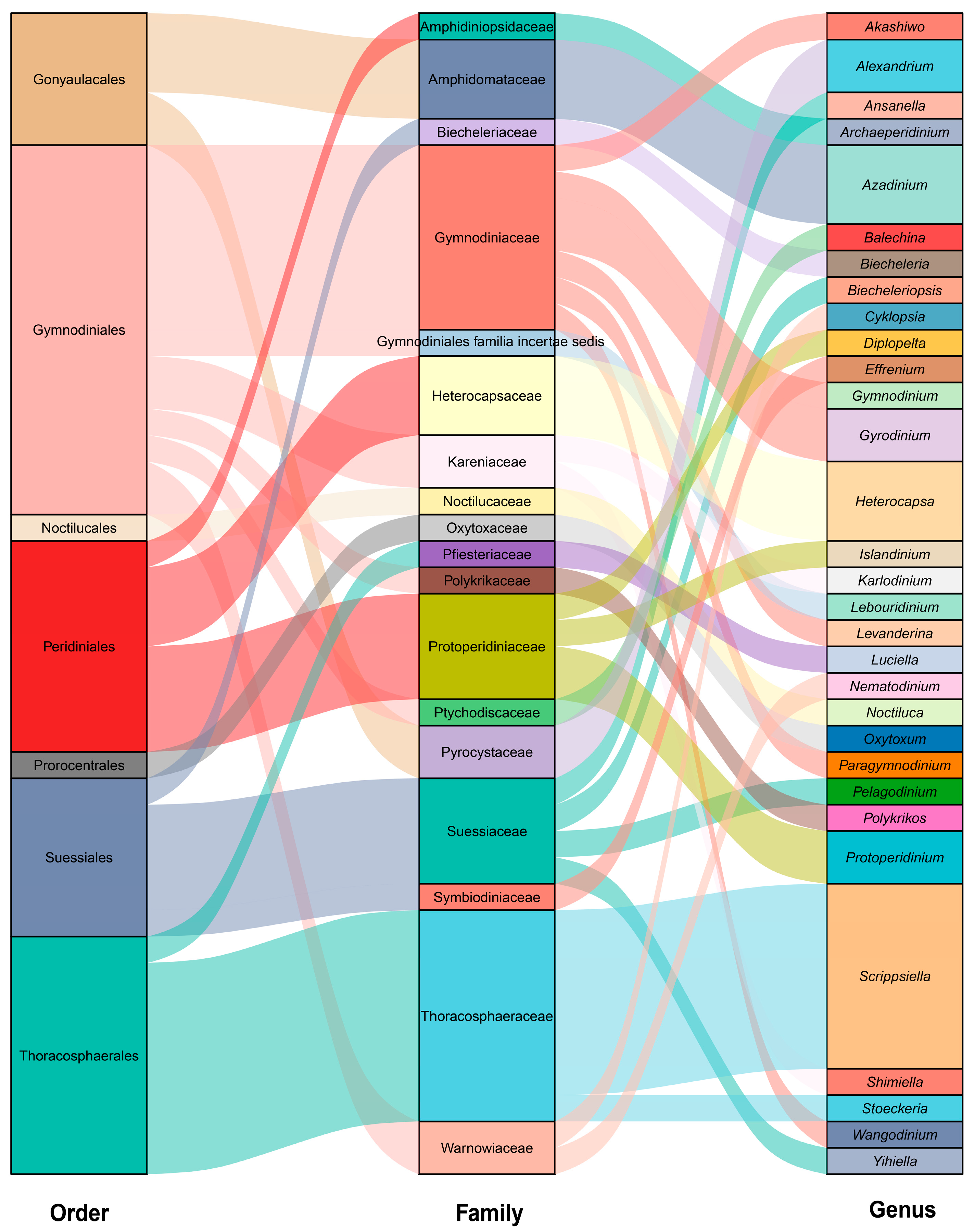
A total of 44 dinoflagellate species, belonging to 31 genera, 18 families, and 7 orders, were identified from in situ germination of cysts in the GEHA (Figure 4). The three most dominant orders were Gymnodiniales (14 species), Thoracosphaerales (9 species), and Peridiniales (8 species). The top four dominant families were Thoracosphaeraceae (eight species), Gymnodiniaceae (seven species), Suessiaceae (four species), and Protoperidiniaceae (four species). The three most dominant genera were Scrippsiella (seven species), Heterocapsa (three species), and Azadinium (three species) (Figure 4). A total of 19 species had cells larger than 15 μm, 28 species exceeded 10 μm, 42 species were larger than 5 μm, and 2 species were approximately 5 μm (Table S4). After 5 days of in situ germination (Sg-5 d), 23 dinoflagellate species were identified, spanning 19 genera, 15 families, and 6 orders. The order Gymnodiniales was the most diverse, with 10 species, followed by Peridiniales (4 species), Suessiales (3 species), Thoracosphaerales (3 species), Gonyaulacales (2 species), and Noctilucales (1 species) (Figure 5a, Table S3). Among these, 9 species were strictly heterotrophic, and 14 species were mixotrophic (Figure 5c, Table S5). After 20 days of in situ germination (Sg-20 d), 34 dinoflagellate species were identified, encompassing 25 genera, 14 families, and 6 orders. The order Gymnodiniales again exhibited the highest diversity with 11 species, followed by Thoracosphaerales (8 species), Peridiniales (7 species), Suessiales (4 species), Gonyaulacales (3 species), and Prorocentrales (1 species) (Figure 5a, Table S3). Of these, 12 species were strictly heterotrophic, and 22 species were mixotrophic (Figure 5c, Table S5). A total of 13 species, representing 12 genera, 9 families, and 4 orders, were shared between the 5-day (Sg-5d) and 20-day (Sg-20d) in situ germination samples (Figure 5a,b, Table S3).
During the 5-day in situ germination period (Sg-5 d), temperature increased from 11.17 °C to 11.99 °C, dissolved oxygen declined from 9.21 mg/L to 8.29 mg/L, and salinity rose from 31.79 ppt to 32.50 ppt (Figure 5c). During the in situ 20-day germinating period (Sg-20 d), temperature rose to 14.2 °C, dissolved oxygen dropped to 7.99 mg/L, and salinity was 32.50 ppt (Figure 5c). Principal coordinates analysis (PCoA) clearly grouped the eight in situ germinated samples into two distinct clusters, with the first principal coordinate axis accounting for more than 50% of the variance (Figure 5d). Both PCoA and UPGMA cluster analysis revealed significant differences in species diversity between those germinated in 5 days (Sg-5 d) and those in 20 days (Sg-20 d), which also indicated a highly similar community structure within the samples incubated for the same period of time (Figure 3a and Figure 5d). From Sg-5 d to Sg-20 d, the number of germinated dinoflagellate species increased by 11 species and six genera (Figure 5a,c). Specifically, the order Thoracosphaerales increased from three to eight species, with the genus Scrippsiella contributing four species; Peridiniales increased from four to seven species; and Gymnodiniales, Suessiales, and Prorocentrales each gained one species (Figure 5a). Additionally, the number of HAB-causing species increased from 7 to 10, while the number of corresponding orders changed from four to three (Table S3). The ratio of mixotrophic to heterotrophic species remained stable (approximately 2:1) across germination durations (Sg-5 d vs. Sg-20 d), closely mirroring the ratio in the community of sediment and seawater outside the GEHA (Figure 5c, Table S5).

3.3. Species of Particular Importance from In Situ Germinated Dinoflagellate Cysts

Among the 44 in situ germinated dinoflagellate species, 12 were HAB-causing species, including Azadinium polongum, Azadinium poporum, Alexandrium leei, Alexandrium pacificum, Levanderina fissa, Akashiwo sanguinea, Karlodinium veneficum, Lebouridinium glaucum, Noctiluca scintillans, Stoeckeria algicida, Scrippsiella acuminata, and Luciella masanensis (Table 1). Of these, six species (Azadinium poporum, Alexandrium leei, Alexandrium pacificum, Akashiwo sanguinea, Karlodinium veneficum, and Stoeckeria algicida) are known to be toxic (Table 1). In addition, five species were identified as novel cyst producers, including the widely distributed HAB-causing species Noctiluca scintillans, Balechina gracilis, Cyklopsia gemma, Gyrodinium cf. spirale, and Nematodinium parvum (Table 1). Among the 39 species previously reported to produce cysts, 5 were observed for the first time in this study to germinate from marine sediments: HAB-causing species Stoeckeria algicida, the symbiotic dinoflagellate Effrenium voratum, Scrippsiella lachrymose, Scrippsiella aff. acuminata, and Yihiella yeosuensis (Table 1).

4. Discussion

4.1. GEHA Combined with Metabarcoding Analysis Detected High Dinoflagellate Diversity from In Situ Germination

The challenges associated with in situ germination studies have historically limited research, with previous investigations relying primarily on morphological identification, resulting in the documentation of only nine dinoflagellate species from in situ germination: Scippsiella sp. [23], Ensiculifera carinata, Gonyaulax spinifera, Gonyaulax verior, Protoperidinium claudicans, Protoperidinium conicoides, Protoperidinium conicum [20], Alexandrium catenella [16,17], and Alexandrium pacificum [17]. Therefore, we developed the GEHA, a novel in situ germination device, for the in situ monitoring of the germination of resting-stage cells of plankton [37]. In this study, we employed the GEHA, combined with metabarcoding analysis targeting 28S rDNA-specific primers for dinoflagellates, to identify 44 dinoflagellate species from in situ germinated cysts, spanning 31 genera, 18 families, and 7 orders. Compared to 51 and 71 dinoflagellates identified in surface/bottom seawater surrounding the GEHA (Sw+Bw) and in situ sediments samples (S), respectively, the lower species richness in the GEHA samples (Sg) may be attributed to three factors: (1) Variable germination requirements, as different dinoflagellate cysts have distinct optimal germination temperatures [100,101], potentially leading to lower germination rates for species like Scrippsiella acuminata [102], which was abundant in sediments but less prevalent in GEHA. (2) “Dominance bias” in high-throughput sequencing, which may obscure low-abundance species [103,104]. Notably, dominant species, such as the winter bloom-causing dinoflagellate Heterocapsa rotundata [105] in the GEHA and the HAB-causing dinoflagellate Levanderina fissa in the surrounding seawater, contrasted with higher species evenness in sediment samples, mitigating the masking of rare species. (3) Due to the differences between sediment and seawater samples, we employed specific DNA extraction kits (as described in the Materials and Methods) to maximize DNA extraction efficiency. For sediment samples, the majority of cells obtained after SPT treatment were resting-stage cells, which generally have thick cell walls that are difficult to break using conventional kits. Therefore, we used a DNA extraction kit specifically designed for sediments. For water samples, since algal cells were filtered onto polycarbonate membranes, the sediment-specific kit could not fully dissolve the membranes. Hence, we adopted a DNA extraction kit containing organic solvents for membrane dissolution. Two specialized kits were employed to maximize the DNA extraction from all environmental samples, which may have influenced the observed species divergence. In addition to DNA extraction, for factors such as sequencing depth, limitations in genetic marker resolution, and bioinformatic parameters, we maintained consistency across all samples. Crucially, UPGMA clustering and PCoA of the Sg, Sw+Bw, and S groups, as well as the Sg-5d and Sg-20d samples, revealed distinct community structures inside and outside the GEHA, further validating its reliability for in situ germination monitoring.
Notably, the consistent ratio (approximately 2:1) of mixotrophic to heterotrophic dinoflagellates in the samples of in situ sediments and seawater outside GEHA, as well as across germination durations (Sg-5 d vs. Sg-20 d), further demonstrates that the germination condition within the GEHA does resemble the in situ scenario (i.e., the GEHA did not significantly alter the genuine field condition). As Barrie Dale noted in a recent interview [106], “Cysts indicate potential bloom initiation size but not bloom magnitude or fate, as water-column factors (e.g., currents, nutrients, predation, competition) dominate outcomes.” Therefore, the in situ cyst germination by the GEHA further solidifies our findings (i.e., many HAB-causing species can germinate in the field) and logically proves which species produce cysts and whether/when these cysts can germinate in a certain ecosystem setting; together, this proves the existence of a “seed source” for the vegetative cell population and possibility of HAB outbreaks under favorable growth conditions. However, as noted above, the scale or fate of HABs is influenced by a combination of environmental factors and species-specific traits (Figure 6). In this study, dinoflagellates were classified into mixotrophic and strictly heterotrophic categories. Theoretically, dinoflagellates exhibit three primary modes of nutrition, including autotrophy, heterotrophy, and mixotrophy [107,108]. According to previous studies, at least half of dinoflagellates are strictly heterotrophs [109,110]. In contrast, autotrophy may exist only theoretically; in practice, Lin et al. (2022) [111] have demonstrated that most dinoflagellates capable of autotrophy are mixotrophic because most (or all) dinoflagellate species lack functional metE genes with an N-terminal domain or do not have metE at all. Consequently, we classified dinoflagellate nutritional modes into mixotrophy and strict heterotrophy, without separating autotrophy as an independent category.
The use of 5 μm mesh nylon membranes in the GEHA introduces a potential limitation, as the two species with cell sizes of approximately 5 μm (Paragymnodinium asymmetricum [86] and Biecheleriopsis adriatica [63]) may have originated from external seawater rather than in situ sediment germination. However, given the double-layered 5 μm nylon mesh design and that B. adriatica is known to produce resting cysts [63,86], their presence in the GEHA most likely originated from in situ germination, though the external possibility cannot be completely ruled out. Furthermore, we conducted a blank control experiment for the GEHA by deploying it with only 0.22 μm of filtered seawater filled in it (i.e., no sediment introduced) on the seabed for 14 d. Subsequent microscopic examination of the water retrieved from the device revealed no cell debris or species larger than 5 μm. Therefore, among the 44 species detected from in situ germination in the GEHA, except for Paragymnodinium asymmetricum and Biecheleriopsis adriatica, the 5 μm mesh nylon membrane should prevent all the other 42 species (with sizes > 5 μm) from crossing the “baffle”, and hence, they were most likely germinated in situ within the GEHA.

4.2. Many Dinoflagellates of Particular Ecological Importance Germinated In Situ from Marine Sediments

Our results demonstrated that a few HAB-causing and toxic species that were proven as cyst producers in the laboratory after 2010 (e.g., Akashiwo sanguinea [50], Karlodinium veneficum [74], Azadinium polongum [60], and Azadinium poporum [61]) do produce resting cysts in the field and can also germinate in the field, which logically implies the possible role played by these cysts in seeding (or initiating) the HABs of these species. Notably, Karlodinium veneficum produces a suite of ichthyotoxic polyketides known as karlotoxins [75], which induce gill epithelial damage and osmoregulatory failure [112,113], and consequently, K. veneficum blooms are frequently associated with mass mortality events of aquatic organisms [76,114]. Azadinium poporum is known for the production potential of AZA (azaspiracid toxins) and is widely distributed in coastal waters in China [61]. Blooms of Akashiwo sanguinea have caused mass mortalities of invertebrates and fish [115,116], as well as seabirds [117], while concurrently incurring substantial economic losses to aquaculture via ecosystem disruption [118]. Additionally, Stoeckeria algicida has not been shown to have a specific toxin, though it has been confirmed to cause mortality in fish and other marine microalgae [97,119,120]. The cyst germination of S. algicida from sediments was reported for the first time in this study, which has only been observed in laboratory cultures previously [97]. It is noteworthy that the HAB-causing species Noctiluca scintillans, detected in the GEHA, has caused multiple HAB events in temperate, subtropical, and tropical coastal waters worldwide [84,121,122] and has also caused many serious HABs in Jiaozhou Bay [28]. Although Noctiluca scintillans have not been reported to produce toxins directly lethal to marine life or pose indirect threats to human health, the high biomass accumulation during its blooms causes significant physical damage to marine ecosystems, including gill clogging in fish and invertebrates [123], and light attenuation, leading to seagrass/coral photosynthesis suppression [124]. N. scintillans has not been reported to produce cysts but was detected from in situ germination within the GEHA, which provides direct evidence not only for the production of resting cysts but also for the capacity of the cysts to germinate from natural sediments. Furthermore, five species were identified as novel cyst producers, and five others were documented for the first time as germinating from marine sediment cysts, offering robust field evidence for their cyst-forming and germination capabilities.
The in situ germination of E. voratum, a symbiotic dinoflagellate, is particularly significant. Our previous study [66] first reported this species’ production of resting cysts through asexual reproduction and described cyst germination in clonal cultures, making E. voratum the first reported cyst-producing species in the family Symbiodiniaceae. However, the germination of E. voratum cysts has not been observed in marine sediments, and it remains doubtful whether the cysts can germinate in situ. This study provides the first evidence of in situ germination of E. voratum cysts from marine sediments, highlighting their ecological importance to coral reef ecosystems and their potential relevance to global climate change.

4.3. Species Diversity of In Situ Germinated Dinoflagellate Cysts with Germination Duration Extended

Over the 5-day (Sg-5 d) and 20-day (Sg-20 d) germination periods, significant differences in temperature and dissolved oxygen (t-tests, p < 0.05) were observed, while no significant difference was observed in salinity (t-tests, p > 0.05). Concurrently, germinated dinoflagellate species increased from 23 to 34, suggesting that temperature, dissolved oxygen, and germination duration are highly likely to be key drivers of cyst germination diversity. Previous studies have shown that cyst germination is regulated by both internal and external factors [7]. After the completion of mandatory dormancy, cyst germination occurs when environmental conditions become favorable [125]. Thus, environmental factors, such as temperature, light, dissolved oxygen, and nutrients, can influence cyst germination. The impact of these factors varies significantly across different cyst types: (1) For temperature, Anderson and Morel (1979) [14] combined field observations with laboratory experiments and found that increasing temperatures enhanced the germination rates of both field-collected cysts and laboratory-stored Alexandrium tamarense cysts after four months of storage. Accoroni et al. (2015) [126] found that the water temperature threshold of 25 °C played a key role in the germination of Ostreopsis cf. ovate cysts. (2) For light, Anderson et al. (1987) [127] found Gonyaulax polyedra cysts required light to germinate, while cysts of Alexandrium tamarense, Scrippsiella sp., and Gonyaulax verior exhibited 5 to 20 times higher germination rates under light conditions compared to darkness. In contrast, Gonyaulax rugosum showed high germination rates under both light and dark conditions. (3) For dissolved oxygen, cysts of Alexandrium tamarense and Alexandrium catenella required a certain level of dissolved oxygen for germination, and germination was completely inhibited when dissolved oxygen levels dropped to 0.01 mg/L [128]. (4) For nutrients, when dinoflagellate cysts in surface sediments were exposed to a culture environment containing organic phosphorus, their germination rates increased significantly [100]. However, Wang et al. (2010) [129] found that nutrient concentrations had no effect on the germination of Scrippsiella acuminata cysts. Beyond environmental factors, some studies have reported that phytohormones (e.g., abscisic acid, gibberellic acid, and melatonin) may also play vital roles in regulating cyst germination [130,131]. However, most research has been conducted in controlled laboratory settings, which struggle to replicate complex field conditions. There are only a few relevant studies, such as Natsuike et al. (2017) [17], on the germination dynamics of Alexandrium catenella and A. pacificum, and Ishikawa et al. (2022) [21,22], on the germination dynamics of Chattonella and centric diatoms, that focus on individual species germination fluxes and rely on microscopic examination. Our study, utilizing the GEHA with environmental monitoring and molecular identification, demonstrated that the temperature, dissolved oxygen, and germination duration within the observed ranges of variations may have all contributed to the elevated number of species detected in the GEHA, but other factors (e.g., nutrients, light, and hydrodynamic scenarios) might be also responsible for the observations, and all the abovementioned factors should be definitely measured in future investigations that use the GEHA to monitor, in situ, the seasonal dynamics of cyst germination in the field. Therefore, much more in situ field monitoring work is required in different environmental settings, which is exactly what our approach was developed for.
Additionally, we found the number of HAB-causing species increased from 7 to 10, while the number of corresponding orders changed from four to three with extended germination duration. These results indicated that the duration of cyst germination within GEHA and environmental factors appeared to have influenced the germination and possibly growth of HAB-causing species. However, due to the relatively short time period of this study, the germination of HAB-causing species also requires further investigation under different conditions, such as seasonal dynamics and fluctuations under diverse marine environments. Long-term continual monitoring of cyst germination is essential for establishing a framework of early warning and forecasting models of HABs, which will be the focus of our future research. While we did not observe that HAB-causing species are more likely to germinate after longer incubation, we analyzed whether the presence or absence of thecal plates was associated with the major germinating taxa. The analysis revealed that unarmored dinoflagellates accounted for 52% of the germinated taxa, whereas they accounted for only 32% of the taxa in the sediment. The number of unarmored dinoflagellate species in the germinated water within the GEHA was higher than that in the sediment, which suggests that unarmored dinoflagellates seem more likely to germinate under natural conditions, but this needs more extensive, convincing observations. Additionally, we found that cysts did not exhibit a clear tendency for which nutritional mode is more conducive to germination in the field, as the proportions of mixotrophic and heterotrophic dinoflagellates were similar in both the sediment and the germinated water within the GEHA. It is worth further investigating which types of cysts are more likely to germinate in the field, such as those with varying cyst wall thickness, different nutritional modes, and the presence or absence of thecal plates. We also noticed that those orders and families that produce thick-walled cysts and/or are the most speciose taxa were more detected from the GEHA (e.g., Gymnodiniales, Thoracosphaerales, and Peridiniales). Moreover, most of the species detected from the GEHA were also those commonly detected in marine sediments. These results demonstrate the reliability and authenticity of the GEHA for in situ cyst germination, laying a solid foundation for future long-term, continual research.

5. Conclusions

By integrating the Germlings Harvester (GEHA) with metabarcoding analysis and environmental monitoring, this study identified 44 dinoflagellate species that germinated in situ from marine sediments in Jiaozhou Bay, China. This included 12 species associated with HABs, 6 toxic species, 5 novel cyst producers, and 5 species with newly documented germination from sediment cysts. Additionally, the number of germinated species increased by 11 by extending the germination duration from 5 to 20 days, and the consistent ratio (approximately 2:1) of mixotrophic to heterotrophic dinoflagellates was observed in samples of in situ sediments and seawater outside the GEHA, as well as across germination durations (Sg-5 d vs. Sg-20 d). These findings establish a robust technical framework combining the GEHA, molecular identification, and in situ environmental monitoring (e.g., temperature and nutrients) in long-term in situ monitoring studies, demonstrating significant potential for predicting and providing early warnings of HAB dynamics. Furthermore, this approach shows promise for broader applications across diverse taxonomic groups in freshwater, estuarine, and marine ecosystems.

Supplementary Materials

The following supporting information can be downloaded at https://www.mdpi.com/article/10.3390/life15111670/s1: Figure S1: Rarefaction curve of metabarcoding sequencing. Table S1: The sampling name, sampling sites, and time of 16 samples. Table S2: The community diversity parameters in the samples. Table S3: All species identified from in situ sediments (S), in situ sediment germination of GEHA (Sg), bottom seawater (Bw), and surface seawater (Sw) surrounding GEHA. Table S4: Cell size of the 44 identified dinoflagellates from in situ cyst germination in GEHA. Table S5: Mixotrophic and heterotrophic species in the samples of in situ sediments (S) and surface and bottom seawater surrounding GEHA (Sw+Bw), as well as across the 5 d (Sg-5 d) and 20 d (Sg-5 d) in situ germination periods. References [52,55,57,58,60,61,62,63,64,65,69,70,73,80,83,84,86,87,89,95,96,97,98,99,132,133,134,135,136,137,138,139,140,141,142,143,144,145] are cited in the supplementary materials.

Author Contributions

Conceptualization, Y.-Z.T. and Z.C.; methodology, Y.-Z.T. and Z.C.; formal analysis, S.S., Z.T. and W.Y.; investigation, S.S., Z.T., W.Y., F.L., B.W., C.Y., Y.D. and L.S.; data curation, S.S.; writing—original draft preparation, S.S.; writing—review and editing, Y.-Z.T. and Z.C.; supervision, Y.-Z.T. and Z.C.; funding acquisition, Y.-Z.T. and Z.C. All authors have read and agreed to the published version of the manuscript.

Funding

This research was funded by the National Science Foundation of China (grant No. 42406210), the Key Research Infrastructures in the CAS Field Stations of the Chinese Academy of Science (grant No. KFJ-SWYW047), the National Key R&D Program of China (grant No. 2024YFF0506901), the National Science Foundation of China (grant No. U23A203), and the Shandong Provincial Natural Science Foundation (grant No. ZR2024MD109).

Institutional Review Board Statement

Not applicable.

Informed Consent Statement

Not applicable.

Data Availability Statement

The data are contained within the article and the Supplementary Materials.

Acknowledgments

We would like to express our gratitude to the anonymous reviewers for their constructive suggestions and comments. We acknowledge financial support from the National Science Foundation of China (grant No. 42406210), the Key Research Infrastructures in the CAS Field Stations of the Chinese Academy of Science (grant No. KFJ-SWYW047), the National Key R&D Program of China (grant No. 2024YFF0506901), the National Science Foundation of China (grant No. U23A203), and the Shandong Provincial Natural Science Foundation (grant No. ZR2024MD109). We are extremely grateful to the members of the ship anchored at the shore in Jiaozhou Bay used in the study. Data analysis was supported by the Oceanographic Data Center, Institute of Oceanology, Chinese Academy of Sciences.

Conflicts of Interest

The authors declare no conflicts of interest. The funders had no role in the design of the study; in the collection, analyses, or interpretation of the data; in the writing of the manuscript; or in the decision to publish the results.

References

  1. Smayda, T.J. Harmful algal blooms: Their ecophysiology and general relevance to phytoplankton blooms in the sea. Limnol. Oceanogr. 1997, 42, 1137–1153. [Google Scholar] [CrossRef]
  2. Jeong, H.J.; Kang, H.C.; Lim, A.S.; Jang, S.H.; Lee, K.; Lee, S.Y.; Ok, J.H.; You, J.H.; Kim, J.H.; Lee, K.H.; et al. Feeding diverse prey as an excellent strategy of mixotrophic dinoflagellates for global dominance. Sci. Adv. 2021, 7, eabe4214. [Google Scholar] [CrossRef] [PubMed]
  3. Yu, Z.; Tang, Y.; Gobler, C.J. Harmful algal blooms in China: History, recent expansion, current status, and future prospects. Harmful Algae 2023, 129, 102499. [Google Scholar] [CrossRef] [PubMed]
  4. Minstry of Natural Resources Bulletin of China Marine Disaster 1989–2022. Available online: https://www.mnr.gov.cn (accessed on 18 October 2024). (In Chinese)
  5. Bravo, I.; Figueroa, R.I. Towards an ecological understanding of dinoflagellate cyst functions. Microorganisms 2014, 2, 11–32. [Google Scholar] [CrossRef]
  6. Guiry, M.D. How many species of algae are there? A reprise. Four kingdoms, 14 phyla, 63 classes and still growing. J. Phycol. 2024, 60, 214–228. [Google Scholar] [CrossRef]
  7. Tang, Y.; Hu, Z.; Deng, Y. Characteristical life history (resting cyst) provides a mechanism for recurrence and geographic expansion of harmful algal blooms of dinoflagellates: A Review. Stud. Mar. Sin. 2016, 51, 132–154. [Google Scholar]
  8. Tang, Y.Z.; Gu, H.; Wang, Z.; Liu, D.; Wang, Y.; Lu, D.; Hu, Z.; Deng, Y.; Shang, L.; Qi, Y. Exploration of resting cysts (stages) and their relevance for possibly HABs-causing species in China. Harmful Algae 2021, 107, 102050. [Google Scholar] [CrossRef]
  9. Anderson, D.; Wall, D. Potential importance of benthic cysts of Gonyaulax tamarensis and Gonyaulax excavata in initiating toxic dinoflagellate blooms. J. Phycol. 1978, 14, 224–234. [Google Scholar] [CrossRef]
  10. Brosnahan, M.L.; Ralston, D.K.; Fischer, A.D.; Solow, A.R.; Anderson, D.M. Bloom termination of the toxic dinoflagellate Alexandrium catenella: Vertical migration behavior, sediment infiltration, and benthic cyst yield. Limnol. Oceanogr. 2017, 62, 2829–2849. [Google Scholar] [CrossRef]
  11. Hallegraeff, G.; Bolch, C. Transport of toxic dinoflagellate cysts via ships ballast water. Mar. Pollut. Bull. 1991, 22, 27–30. [Google Scholar] [CrossRef]
  12. Hallegraeff, G. A review of Harmful algal blooms and their apparent global increase. Phycologia 1993, 32, 79–99. [Google Scholar] [CrossRef]
  13. Shang, L.; Hu, Z.; Deng, Y.; Liu, Y.; Zhai, X.; Chai, Z.; Liu, X.; Zhan, Z.; Dobbs, F.C.; Tang, Y.Z. Metagenomic sequencing identifies highly diverse assemblages of dinoflagellate cysts in sediments from ships’ ballast tanks. Microorganisms 2019, 7, 250. [Google Scholar] [CrossRef] [PubMed]
  14. Anderson, D.; Morel, F. Seeding of 2 red tide blooms by the germination of benthic Gonyaulax tamarensis hypnocysts. Estuarine Coastal Mar. Sci. 1979, 8, 279–293. [Google Scholar] [CrossRef]
  15. Ishikawa, A.; Hattori, M.; Imai, I. Development of the “plankton emergence trap/chamber (PET Chamber)”, a new sampling device to collect in situ germinating cells from cysts of microalgae in surface sediments of coastal waters. Harmful Algae 2007, 6, 301–307. [Google Scholar] [CrossRef]
  16. Ishikawa, A.; Hattori, M.; Ishii, K.-I.; Kulis, D.M.; Anderson, D.M.; Imai, I. In situ dynamics of cyst and vegetative cell populations of the toxic dinoflagellate Alexandrium catenella in Ago Bay, central Japan. J. Plankton Res. 2014, 36, 1333–1343. [Google Scholar] [CrossRef]
  17. Natsuike, M.; Yokoyama, K.; Nishitani, G.; Yamada, Y.; Yoshinaga, I.; Ishikawa, A. Germination fluctuation of toxic Alexandrium fundyense and A. pacificum cysts and the relationship with bloom occurrences in Kesennuma Bay, Japan. Harmful Algae 2017, 62, 52–59. [Google Scholar] [CrossRef]
  18. Ishikawa, A.; Fujita, N.; Taniguchi, A. A sampling device to measure in situ germination rates of dinoflagellate cysts in surface sediments. J. Plankton Res. 1995, 17, 647–651. [Google Scholar] [CrossRef]
  19. Rengefors, K.; Gustafsson, S.; Ståhl-Delbanco, A. Factors regulating the recruitment of cyanobacterial and eukaryotic phytoplankton from littoral and profundal sediments. Aquat. Microb. Ecol. 2004, 36, 213–226. [Google Scholar] [CrossRef]
  20. Ishikawa, A.; Taniguchi, A. In situ germination patterns of cysts, and bloom formation of some armored dinoflagellates in Onagawa Bay, north-east Japan. J. Plankton Res. 1997, 19, 1783–1791. [Google Scholar] [CrossRef]
  21. Ishikawa, A.; Takei, Y.; Ishii, K.-I.; Yamaguchi, M. Population dynamics of Chattonella (Raphidophyceae), causative flagellates of noxious red tides in Ago Bay, central Japan, with an emphasis on cyst germination. Plankton Benthos Res. 2022, 17, 383–392. [Google Scholar] [CrossRef]
  22. Ishii, K.-I.; Matsuoka, K.; Imai, I.; Ishikawa, A. Life cycle strategies of the centric diatoms in a shallow embayment revealed by the plankton emergence trap/chamber (PET chamber) experiments. Front. Mar. Sci. 2022, 9, 889633. [Google Scholar] [CrossRef]
  23. Ishikawa, A.; Taniguchi, A. Contribution of benthic cysts to the population dynamics of Scrippsiella spp. (Dinophyceae) in Onagawa bay, northeast Japan. Mar. Ecol.-Prog. Ser. 1996, 140, 169–178. [Google Scholar] [CrossRef]
  24. Anglès, S.; Garcés, E.; Hattenrath-Lehmann, T.K.; Gobler, C.J. In situ life-cycle stages of Alexandrium fundyense during bloom development in Northport Harbor (New York, USA). Harmful Algae 2012, 16, 20–26. [Google Scholar] [CrossRef]
  25. Anglès, S.; Garcés, E.; Reñé, A.; Sampedro, N. Life-cycle alternations in Alexandrium minutum natural populations from the NW Mediterranean Sea. Harmful Algae 2012, 16, 1–11. [Google Scholar] [CrossRef]
  26. Liu, S.; Cui, Z.; Zhao, Y.; Chen, N. Composition and spatial-temporal dynamics of phytoplankton community shaped by environmental selection and interactions in the Jiaozhou bay. Water Res. 2022, 218, 118488. [Google Scholar] [CrossRef]
  27. Wang, Q.; Song, J.; Yuan, H. Evolution of marine environmental health in Jiaozhou Bay in recent 40 years based on the ‘DualCore’ assessment framework. Ocean Dev. Manag. 2023, 40, 29–38. [Google Scholar] [CrossRef]
  28. Liu, S.; Chen, N. Advances in biodiversity analysis of phytoplankton and harmful algal bloom species in the Jiaozhou Bay. Mar. Sci. 2021, 45, 170–188. [Google Scholar]
  29. Sun, S.; Zhang, Y.; Wu, Y.; Zhang, G.; Zhang, F.; Pu, X. Annual variation of primary productivity in Jiaozhou bay. Oceanol. Limnol. Sin. 2005, 36, 3–8. [Google Scholar]
  30. Hu, Z.; Deng, Y.; Li, Y.; Tang, Y.Z. The morphological and phylogenetic characterization for the dinoflagellate Margalefidinium fulvescens (=Cochlodinium fulvescens) isolated from the Jiaozhou Bay, China. Acta Oceanol. Sin. 2018, 37, 11–17. [Google Scholar] [CrossRef]
  31. Lin, S.; Hu, Z.; Deng, Y.; Shang, L.; Gobler, C.J.; Tang, Y.Z. An assessment on the intrapopulational and intraindividual genetic diversity in LSU rDNA in the harmful algal blooms-forming dinoflagellate Margalefidinium (=Cochlodinium) fulvescens based on clonal cultures and bloom samples from Jiaozhou Bay, China. Harmful Algae 2020, 96, 101821. [Google Scholar] [CrossRef]
  32. Liu, X.; Liu, Y.; Chai, Z.; Hu, Z.; Tang, Y.Z. A combined approach detected novel species diversity and distribution of dinoflagellate cysts in the Yellow Sea, China. Mar. Pollut. Bull. 2023, 187, 114567. [Google Scholar] [CrossRef] [PubMed]
  33. Wang, Z.; Zhang, Y.; Lei, M.; Ji, S.; Chen, J.; Zheng, H.; Tang, Y.; Hu, R. Distribution of dinoflagellate cysts in surface sediments from the Qingdao coast, the Yellow sea, China: The potential risk of harmful algal blooms. Front. Mar. Sci. 2022, 9, 910327. [Google Scholar] [CrossRef]
  34. Shen, P.; Tang, Y.; Li, Y.; Tian, Y.; Liu, M. Relationship between dinoflagellate cyst and environmental factors in surface sediment of the Jiaozhou Bay. J. Yantai Univ. Nat. Sci. Eng. Ed. 2021, 34, 392–399. [Google Scholar] [CrossRef]
  35. Li, Y.; Tang, Y.; Shen, P.; Gu, H.; Tan, Y. Distribution of dinoflagellate resting cysts in surface sediment of Jiaozhou bay, China. Oceanol. Limnol. Sin. 2017, 48, 760–766. [Google Scholar]
  36. Guo, S.; Zhu, M.; Zhao, Z.; Liang, J.; Zhao, Y.; Du, J.; Sun, X. Spatial-temporal variation of phytoplankton community structure in Jiaozhou Bay, China. J. Oceanol. Limnol. 2019, 37, 1611–1624. [Google Scholar] [CrossRef]
  37. Shi, S.; Tao, Z.; Yang, W.; Li, F.; Wei, B.; Yue, C.; Pan, S.; Deng, Y.; Shang, L.; Chai, Z.; et al. A newly developed germlings harvester (GEHA) in combination with metabarcoding analysis detected numerous plankton species, particularly HABs-causing species, from in-situ germination of resting stage cells. Harmful Algae 2025, 150, 103002. [Google Scholar] [CrossRef]
  38. Yang, S.; Wang, L.; Shi, X. Phytoplankton community of the Jiaozhou bay in spring 2009. Oceanol. Limnol. Sin. 2014, 45, 1234–1240. [Google Scholar]
  39. Li, Y.; Li, R.; Wang, Z.; Zhu, M.; Sun, P.; Xia, B. A preliminary study on phytoplankton community structure and its changes in the Jiaozhou Bay. Adv. Mar. Sci. 2005, 23, 328–334. [Google Scholar]
  40. Bolch, C.J.S. The use of sodium polytungstate for the separation and concentration of living dinoflagellate cysts from marine sediments. Phycologia 1997, 36, 472–478. [Google Scholar] [CrossRef]
  41. Maki, K.A.A.; Wolff, B.; Varuzza, L.; Green, S.J.J.; Barb, J.J.J. Multi-amplicon microbiome data analysis pipelines for mixed orientation sequences using QIIME2: Assessing reference database, variable region and pre-processing bias in classification of mock bacterial community samples. PLoS ONE 2023, 18, e0280293. [Google Scholar] [CrossRef]
  42. Magoc, T.; Salzberg, S.L. FLASH: Fast length adjustment of short reads to improve genome assemblies. Bioinformatics 2011, 27, 2957–2963. [Google Scholar] [CrossRef] [PubMed]
  43. Rognes, T.; Flouri, T.; Nichols, B.; Quince, C.; Mahe, F. VSEARCH: A versatile open source tool for metagenomics. PeerJ 2016, 4, e2584. [Google Scholar] [CrossRef] [PubMed]
  44. Callahan, B.J.; McMurdie, P.J.; Rosen, M.J.; Han, A.W.; Johnson, A.J.A.; Holmes, S.P. DADA2: High-resolution sample inference from illumina amplicon data. Nat. Methods 2016, 13, 581–583. [Google Scholar] [CrossRef] [PubMed]
  45. Chai, Z.; Wang, H.; Deng, Y.; Hu, Z.; Tang, Y.Z. Harmful algal blooms significantly reduce the resource use efficiency in a coastal plankton community. Sci. Total Environ. 2020, 704, 135381. [Google Scholar] [CrossRef]
  46. Bolyen, E.; Rideout, J.R.; Dillon, M.R.; Bokulich, N.A.; Abnet, C.C.; Al-Ghalith, G.A.; Alexander, H.; Alm, E.J.; Arumugam, M.; Asnicar, F.; et al. Reproducible, interactive, scalable and extensible microbiome data science using QIIME 2. Nat. Biotechnol. 2019, 37, 852–857. [Google Scholar] [CrossRef]
  47. Tindall, D.; Dickey, R.; Carlson, R.; Moreygaines, G. Ciguatoxigenic dinoflagellates from the Caribbean Sea. ACS Symp. Ser. 1984, 262, 225–240. [Google Scholar]
  48. Wu, X.; Yang, Y.; Yang, Y.; Zhong, P.; Xu, N. Toxic characteristics and action mode of the mixotrophic dinoflagellate Akashiwo sanguinea on co-occurring phytoplankton and zooplankton. Int. J. Environ. Res. Public Health 2022, 19, 404. [Google Scholar] [CrossRef]
  49. Luo, Z.; Yang, W.; Leaw, C.P.; Pospelova, V.; Bilien, G.; Liow, G.R.; Lim, P.T.; Gu, H. Cryptic diversity within the harmful dinoflagellate Akashiwo sanguinea in coastal Chinese waters is related to differentiated ecological niches. Harmful Algae 2017, 66, 88–96. [Google Scholar] [CrossRef]
  50. Tang, Y.Z.; Gobler, C.J. Sexual resting cyst production by the dinoflagellate Akashiwo sanguinea: A potential mechanism contributing toward the ubiquitous distribution of a harmful alga. J. Phycol. 2015, 51, 298–309. [Google Scholar] [CrossRef]
  51. Jeong, H.; Yoo, Y.; Park, J.; Song, J.; Kim, S.; Lee, S.; Kim, K.; Yih, W. Feeding by phototrophic red-tide dinoflagellates: Five species newly revealed and six species previously known to be mixotrophic. Aquat. Microb. Ecol. 2005, 40, 133–150. [Google Scholar] [CrossRef]
  52. Gu, H.; Zeng, N.; Liu, T.; Yang, W.; Mueller, A.; Krock, B. Morphology, toxicity, and phylogeny of Alexandrium (Dinophyceae) species along the coast of China. Harmful Algae 2013, 27, 68–81. [Google Scholar] [CrossRef]
  53. Shikata, T.; Taniguchi, E.; Sakamoto, S.; Kitatsuji, S.; Yamasaki, Y.; Yoshida, M.; Oikawa, H. Phylogeny, growth and toxicity of the noxious red-tide dinoflagellate Alexandrium leei in Japan. Reg. Stud. Mar. Sci. 2020, 36, 101265. [Google Scholar] [CrossRef]
  54. Dai, L.; Geng, H.-X.; Yu, R.-C.; Liu, Y.; Zhao, J.-Y.; Wang, J.-X.; Zhang, Q.-C.; Kong, F.-Z.; Zhou, M.-J. Distribution of Alexandrium pacificum cysts in the area adjacent to the Changjiang River estuary, China. Mar. Pollut. Bull. 2020, 156, 111206. [Google Scholar] [CrossRef] [PubMed]
  55. John, U.; Litaker, R.W.; Montresor, M.; Murray, S.; Brosnahan, M.L.; Anderson, D.M. Formal revision of the Alexandrium tamarense species complex (Dinophyceae) taxonomy: The introduction of five species with emphasis on molecular-based (rDNA) classification. Protist 2014, 165, 779–804. [Google Scholar] [CrossRef]
  56. Lee, S.K.; Jeong, H.J.; Jang, S.H.; Lee, K.H.; Kang, N.S.; Lee, M.J.; Potvin, E. Mixotrophy in the newly described dinoflagellate Ansanella granifera: Feeding mechanism, prey species, and effect of prey concentration. Algae 2014, 29, 137–152. [Google Scholar] [CrossRef]
  57. Mertens, K.N.; Yamaguchi, A.; Kawami, H.; Ribeiro, S.; Leander, B.S.; Price, A.M.; Pospelova, V.; Ellegaard, M.; Matsuoka, K. Archaeperidinium saanichi sp. nov.: A new species based on morphological variation of cyst and theca within the Archaeperidinium minutum jörgensen 1912 species complex. Mar. Micropaleontol. 2012, 96–97, 48–62. [Google Scholar] [CrossRef]
  58. Tillmann, U.; Gottschling, M.; Nezan, E.; Krock, B.; Bilien, G. Morphological and molecular characterization of three new Azadinium species (Amphidomataceae, Dinophyceae) from the Irminger Sea. Protist 2014, 165, 417–444. [Google Scholar] [CrossRef]
  59. Tillmann, U.; Sanchez-Ramirez, S.; Krock, B.; Bernales-Jimenez, A. A bloom of Azadinium polongum in coastal waters off Peru. Rev. Biol. Mar. Oceanogr. 2017, 52, 591–610. [Google Scholar] [CrossRef]
  60. Tillmann, U.; Soehner, S.; Nézan, E.; Krock, B. First record of the genus Azadinium (Dinophyceae) from the shetland islands, including the description of Azadinium polongum sp. nov. Harmful Algae 2012, 20, 142–155. [Google Scholar] [CrossRef]
  61. Gu, H.; Luo, Z.; Krock, B.; Witt, M.; Tillmann, U. Morphology, phylogeny and azaspiracid profile of Azadinium poporum (Dinophyceae) from the China sea. Harmful Algae 2013, 21–22, 64–75. [Google Scholar] [CrossRef]
  62. Gomez, F.; Artigas, L.F.; Gast, R.J. Molecular phylogeny and synonymy of Balechina gracilis comb. nov. (=Gymnodinium gracile), a widespread polymorphic unarmored dinoflagellate (Dinophyceae). J. Phycol. 2021, 57, 694–697. [Google Scholar] [CrossRef]
  63. Hu, Z.; Song, X.; Wang, J.; Tao, Z.; Sun, Y.; Li, Y.; Liu, Y.; Deng, Y.; Shang, L.; Chai, Z.; et al. Reviving and characterizing three species of dinoflagellate cysts dormant for about 70 years in the East China Sea: Biecheleria brevisulcata, Biecheleriopsis adriatica, and Scrippsiella donghaienis. J. Oceanol. Limnol. 2022, 40, 2292–2311. [Google Scholar] [CrossRef]
  64. Cooney, E.C.; Holt, C.C.; Jacko-Reynolds, V.K.L.; Leander, B.S.; Keeling, P.J. Photosystems in the eye-like organelles of heterotrophic warnowiid dinoflagellates. Curr. Biol. 2023, 33, 4252–4260.e3. [Google Scholar] [CrossRef] [PubMed]
  65. Liu, T.; Mertens, K.N.; Gu, H. Cyst-theca relationship and phylogenetic positions of the diplopsalioideans (Peridiniales, Dinophyceae), with description of Niea and Qia gen. nov. Phycologia 2015, 54, 210–232. [Google Scholar] [CrossRef]
  66. Li, R.; Deng, Y.; Shang, L.; Liu, Y.; Tao, Z.; Chai, Z.; Tang, Y.Z. Evidence for the production of asexual resting cysts in a free-living species of Symbiodiniaceae (Dinophyceae). Harmful Algae 2024, 137, 102658. [Google Scholar] [CrossRef]
  67. Lee, K.H.; Jeong, H.J.; Jang, T.Y.; Lim, A.S.; Kang, N.S.; Kim, J.-H.; Kim, K.Y.; Park, K.-T.; Lee, K. Feeding by the newly described mixotrophic dinoflagellate Gymnodinium smaydae: Feeding mechanism, prey species, and effect of prey concentration. J. Exp. Mar. Biol. Ecol. 2014, 459, 114–125. [Google Scholar] [CrossRef]
  68. Du Yoo, Y.; Jeong, H.J.; Lee, S.Y.; Yoon, E.Y.; Kang, N.S.; Lim, A.S.; Lee, K.H.; Jang, S.H.; Park, J.Y.; Kim, H.S. Feeding by heterotrophic protists on the toxic dinoflagellate Ostreopsis cf. ovata. Harmful Algae 2015, 49, 1–9. [Google Scholar] [CrossRef]
  69. Rene, A.; Camp, J.; Garces, E. Diversity and phylogeny of Gymnodiniales (Dinophyceae) from the NW Mediterranean Sea revealed by a morphological and molecular approach. Protist 2015, 166, 234–263. [Google Scholar] [CrossRef]
  70. Iwataki, M.; Hansen, G.; Sawaguchi, T.; Hiroishi, S.; Fukuyo, Y. Investigations of body scales in twelve Heterocapsa species (Peridiniales, Dinophyceae), including a new species H. pseudotriquetra sp. nov. Phycologia 2004, 43, 394–403. [Google Scholar] [CrossRef]
  71. Wang, J.; Liu, Q.; Huang, S.; Mertens, K.N.; Pospelova, V.; Shen, X.; Gu, H. High-resolution DNA metabarcoding of modern surface sediments uncovers a diverse assemblage of dinoflagellate cysts in the Pacific and Arctic oceans. Mar. Pollut. Bull. 2025, 215, 117899. [Google Scholar] [CrossRef]
  72. Millette, N.C.; Pierson, J.J.; Aceves, A.; Stoecker, D.K. Mixotrophy in Heterocapsa rotundata: A mechanism for dominating the winter phytoplankton. Limnol. Oceanogr. 2017, 62, 836–845. [Google Scholar] [CrossRef]
  73. Liu, T.; Gu, H.; Mertens, K.N.; Lan, D. New dinoflagellate species Protoperidinium haizhouense sp. nov. (Peridiniales, Dinophyceae), its cyst-theca relationship and phylogenetic position within the Monovela group. Phycol. Res. 2014, 62, 109–124. [Google Scholar] [CrossRef]
  74. Liu, Y.; Hu, Z.; Deng, Y.; Tang, Y.Z. Evidence for resting cyst production in the cosmopolitan toxic dinoflagellate Karlodinium veneficum and the cyst distribution in the China seas. Harmful Algae 2020, 93, 101788. [Google Scholar] [CrossRef] [PubMed]
  75. Bachvaroff, T.R.; Adolf, J.E.; Squier, A.H.; Harvey, H.R.; Place, A.R. Characterization and quantification of karlotoxins by liquid chromatography-mass spectrometry. Harmful Algae 2008, 7, 473–484. [Google Scholar] [CrossRef]
  76. Place, A.R.; Bowers, H.A.; Bachvaroff, T.R.; Adolf, J.E.; Deeds, J.R.; Sheng, J. Karlodinium veneficum—The little dinoflagellate with a big bite. Harmful Algae 2012, 14, 179–195. [Google Scholar] [CrossRef]
  77. Liu, K.; Wang, H.; Liu, F.; Chen, N. Molecular analysis of a polyphasic red tide in Rongcheng Offshore, Shandong Peninsula in 2021~2022. J. Oceanol. Limnol. 2023, 54, 1645–1655. [Google Scholar]
  78. Jimenez, R. Ecological Factors Related to Gyrodinium instriatum Bloom in the Inner Estuary of the Gulf of Guayaquil. In Toxic Phytoplankton Blooms in the Sea, Proceedings of the Fifth International Conference on Toxic Marine Phytoplankton, Newport, RI, USA, 28 October–1 November 1991; Smayda, T.J., Shimizu, Y., Eds.; Elsevier Science Publisher B.V.: Amsterdam, The Netherlands, 1993; Volume 3, pp. 257–262. [Google Scholar]
  79. Shikata, T.; Nagasoe, S.; Matsubara, T.; Yamasaki, Y.; Shimasaki, Y.; Oshima, Y.; Uchida, T.; Jenkinson, I.R.; Honjo, T. Encystment and excystment of Gyrodinium instriatum Freudenthal et Lee. J. Oceanogr. 2008, 64, 355–365. [Google Scholar] [CrossRef]
  80. Mason, P.L.; Litaker, R.W.; Jeong, H.J.; Ha, J.H.; Reece, K.S.; Stokes, N.A.; Park, J.Y.; Steidinger, K.A.; Vandersea, M.W.; Kibler, S.; et al. Description of a new genus of Pfesteria-like dinoflagellate, Luciella gen. nov (Dinophyceae), including two new species: Luciella masanensis sp. nov and Luciella atlantis sp. nov. J. Phycol. 2007, 43, 799–810. [Google Scholar] [CrossRef]
  81. Noga, E.J.; Khoo, L.; Stevens, J.B.; Fan, Z.; Burkholder, J.M. Novel toxic dinoflagellate causes epidemic disease in estuarine fish. Mar. Pollut. Bull. 1996, 32, 219–224. [Google Scholar] [CrossRef]
  82. Tao, Z.; Liu, X.; Song, X.; Deng, Y.; Shang, L.; Chai, Z.; Hu, Z.; Liu, Y.; Tang, Y.Z. Species and genetic diversity of notorious dinoflagellates Pfiesteria piscicida, Luciella masanensis, and relatives in marine sediments of China. Harmful Algae 2024, 140, 102746. [Google Scholar] [CrossRef]
  83. García-Portela, M.; Moestrup, Ø.; Daugbjerg, N.; Altenburger, A.; Lundholm, N. Studies on the complex Warnowiaceae (Dinophyceae) I. Lohmann’s Pouchetia parva refound and renamed Nematodinium parvum comb. nov. (=Warnowia parva). Phycologia 2023, 62, 421–435. [Google Scholar] [CrossRef]
  84. Li, X.; Lu, W.; Jiang, F.; Chen, Z.; Chang, Y.; Wang, Z.; Yan, T.; Li, J.; Wang, W.; Chen, X. The dinoflagellate Noctiluca scintillans in China: A review of its distribution and role in harmful algal blooms. Mar. Pollut. Bull. 2023, 194, 115415. [Google Scholar] [CrossRef] [PubMed]
  85. Gottschling, M.; Wietkamp, S.; Bantle, A.; Tillmann, U. Oxytoxaceae are prorocentralean rather than peridinialean dinophytes and taxonomic clarification of heterotrophic Oxytoxum lohmannii (≡“Amphidiniumcrassum) by epitypification. Sci. Rep. 2024, 14, 6689. [Google Scholar] [CrossRef] [PubMed]
  86. Yokouchi, K.; Takahashi, K.; Nguyen, V.N.; Iwataki, M.; Horiguchi, T. Ultrastructure and systematics of two new species of dinoflagellate, Paragymnodinium asymmetricum sp. nov. and Paragymnodinium inerme sp. nov. (Gymnodiniales, Dinophyceae). J. Phycol. 2020, 56, 730–746. [Google Scholar] [CrossRef]
  87. Siano, R.; Montresor, M.; Probert, I.; Not, F.; de Vargas, C. Pelagodinium gen. nov and P. beii comb. nov., a dinoflagellate symbiont of planktonic foraminifera. Protist 2010, 161, 385–399. [Google Scholar] [CrossRef]
  88. Matsuoka, K.; Kawami, H.; Nagai, S.; Iwataki, M.; Takayama, H. Re-examination of cyst-motile relationships of Polykrikos kofoidii Chatton and Polykrikos schwartzii Butschli (Gymnodiniales, Dinophyceae). Rev. Palaeobot. Palynol. 2009, 154, 79–90. [Google Scholar] [CrossRef]
  89. Gu, H.; Sun, J.; Kooistra, W.H.C.F.; Zeng, R. Phylogenetic position and morphology of thecae and cysts of Scrippsiella (Dinophyceae) species in the East China Sea. J. Phycol. 2008, 44, 478–494. [Google Scholar] [CrossRef]
  90. Xiao, Y.; Qi, Y.; Wang, Z.; Lü, S. The relationship between Scrippsiella trochoidea red tide and cysts in the Daya Bay. Mar. Sci. 2001, 25, 50–54. [Google Scholar]
  91. You, J.H.; Ok, J.H.; Kang, H.C.; Park, S.A.; Eom, S.H.; Jeong, H.J. Five phototrophic Scrippsiella species lacking mixotrophic ability and the extended prey spectrum of Scrippsiella acuminata (Thoracosphaerales, Dinophyceae). Algae 2023, 38, 111–126. [Google Scholar] [CrossRef]
  92. Tao, Z.; Liu, Y.; Liu, X.; Yue, C.; Song, X.; Hu, Z.; Shi, S.; Li, R.; Deng, Y.; Shang, L.; et al. Single-cyst morpho-molecular identification detected an unexpected high species diversity of dinoflagellate resting cysts from the coastal seas of China. Harmful Algae 2025, 149, 102941. [Google Scholar] [CrossRef]
  93. Zinssmeister, C.; Soehner, S.; Kirsch, M.; Facher, E.; Meier, K.J.S.; Keupp, H.; Gottschling, M. Same but different: Two novel bicarinate species of extant calcareous dinophytes (Thoracosphaeraceae, Peridiniales) from the Mediterranean Sea. J. Phycol. 2012, 48, 1107–1118. [Google Scholar] [CrossRef]
  94. Kretschmann, J.; Zinssmeister, C.; Gottschling, M. Taxonomic clarification of the dinophyte Rhabdosphaera erinaceus KAMPTNER, Scrippsiella erinaceus comb. nov (Thoracosphaeraceae, Peridiniales). Syst. Biodivers. 2014, 12, 393–404. [Google Scholar] [CrossRef]
  95. Lewis, J. Cyst-theca relationships in Scrippsiella (Dinophyceae) and related Orthoperidinioid genera. Bot. Mar. 1991, 34, 91–106. [Google Scholar] [CrossRef]
  96. Ok, J.H.; Jeong, H.J.; Lee, S.Y.; Park, S.A.; Noh, J.H. Shimiella gen. nov. and Shimiella gracilenta sp. nov. (Dinophyceae, Kareniaceae), a kleptoplastidic dinoflagellate from Korean waters and its survival under starvation. J. Phycol. 2021, 57, 70–91. [Google Scholar] [CrossRef] [PubMed]
  97. Jeong, H.J.; Kim, J.S.; Park, J.Y.; Kim, J.H.; Kim, S.; Lee, I.; Lee, S.H.; Ha, J.H.; Yih, W.H. Stoeckeria algicida n. gen., n. sp. (Dinophyceae) from the coastal waters off southern Korea: Morphology and small subunit ribosomal DNA gene sequence. J. Eukaryotic Microbiol. 2005, 52, 382–390. [Google Scholar] [CrossRef] [PubMed]
  98. Tao, Z.; Yue, C.; Liu, Y.; Shi, S.; Li, R.; Chai, Z.; Deng, Y.; Shang, L.; Hu, Z.; Gu, H.; et al. A new ribo-type of Wangodinium sinense from germination of resting cysts isolated from ballast tank sediments of incoming ships to China. J. Mar. Sci. Eng. 2025, 13, 942. [Google Scholar] [CrossRef]
  99. Jang, S.H.; Jeong, H.J.; Moestrup, O.; Kang, N.S.; Lee, S.Y.; Lee, K.H.; Seong, K.A. Yihiella yeosuensis gen. Et sp. nov. (Suessiaceae, Dinophyceae), a novel dinoflagellate isolated from the coastal waters of Korea. J. Phycol. 2017, 53, 131–145. [Google Scholar] [CrossRef]
  100. Su, Y.; Zhong, Y.; You, X.; Weng, Y.; Zhuang, H. Research on the germination and proliferation of dinoflagellate cysts in the sediments of Xipi Reservoir in Jiulongjiang River. Acta Sci. Circumstantiae 2015, 35, 2875–2882. [Google Scholar] [CrossRef]
  101. Zheng, X.; Lin, L.; Xue, M.; Wang, Q.; Wang, J.; Wu, H. Study on the diversity and germination of dinoflagellate cysts in the sediments of foreign ships in Shanghai port. Mar. Pollut. Bull. 2024, 205, 116566. [Google Scholar] [CrossRef]
  102. Wang, Z.; Qi, Y.; Gu, X. Seasonal fluctuation of Scrippsiella trochoidea cyst population in Daya Bay, South China Sea. J. Trop. Subtrop. Bot. 2007, 15, 9–15. [Google Scholar]
  103. Smith, B.; Wilson, J.B. A consumer’s guide to evenness indices. Oikos 1996, 76, 70–82. [Google Scholar] [CrossRef]
  104. Hillebrand, H.; Bennett, D.M.; Cadotte, M.W. Consequences of dominance: A review of evenness effects on local and regional ecosystem processes. Ecology 2008, 89, 1510–1520. [Google Scholar] [CrossRef]
  105. Millette, N.C.; Stoecker, D.K.; Pierson, J.J. Top-down control by micro- and mesozooplankton on winter dinoflagellate blooms of Heterocapsa rotundata. Aquat. Microb. Ecol. 2015, 76, 15–25. [Google Scholar] [CrossRef]
  106. Mertens, K. Interview with Barrie Dale. Harmful Algae News 2025, 80, 27–33. [Google Scholar]
  107. Burkholder, J.; Glasgow, H. Interactions of a toxic estuarine dinoflagellate with microbial predators and prey. Arch. Protistenkd. 1995, 145, 177–188. [Google Scholar] [CrossRef]
  108. Sherr, E.B.; Sherr, B.F. Significance of predation by protists in aquatic microbial food webs. Antonie Leeuwenhoek Int. J. Gen. Mol. Microbiol. 2002, 81, 293–308. [Google Scholar] [CrossRef]
  109. Jeong, H.J.; Yoo, Y.D.; Kang, N.S.; Rho, J.R.; Seong, K.A.; Park, J.W.; Nam, G.S.; Yih, W. Ecology of Gymnodinium aureolum. I. Feeding in western Korean waters. Aquat. Microb. Ecol. 2010, 59, 239–255. [Google Scholar] [CrossRef]
  110. Li, A.S.; Stoecker, D.K.; Adolf, J.E. Feeding, pigmentation, photosynthesis and growth of the mixotrophic dinoflagellate Gyrodinium galatheanum. Aquat. Microb. Ecol. 1999, 19, 163–176. [Google Scholar] [CrossRef]
  111. Lin, S.; Hu, Z.; Song, X.; Gobler, C.J.; Tang, Y.Z. Vitamin B12-auxotrophy in dinoflagellates caused by incomplete or absent cobalamin-independent methionine synthase genes (metE). Fundam. Res. 2022, 2, 727–737. [Google Scholar] [CrossRef]
  112. Fernandez-Tejedor, M.; Soubrier-Pedreno, M.A.; Furones, M.D. Mitigation of lethal effects of Karlodinium veneficum and K. armiger on Sparus aurata: Changes in haematocrit and plasma osmolality. Dis. Aquat. Org. 2007, 77, 53–59. [Google Scholar] [CrossRef]
  113. Llanos-Rivera, A.; Alvarez-Munoz, K.; Astuya-Villalon, A.; Lopez-Rosales, L.; Garcia-Camacho, F.; Sanchez-Miron, A.; Krock, B.; Jose Gallardo-Rodriguez, J. Sublethal effect of the toxic dinoflagellate Karlodinium veneficum on early life stages of zebrafish (Danio rerio). Environ. Sci. Pollut. Res. 2023, 30, 27113–27124. [Google Scholar] [CrossRef] [PubMed]
  114. Deeds, J.R.; Terlizzi, D.E.; Adolf, J.E.; Stoecker, D.K.; Place, A.R. Toxic activity from cultures of Karlodinium micrum (=Gyrodinium galatheanum) (Dinophyceae)—A dinoflagellate associated with fish mortalities in an estuarine aquaculture facility. Harmful Algae 2002, 1, 169–189. [Google Scholar] [CrossRef]
  115. Shumway, S.E. A review of the effects of algal blooms on shellfish and aquaculture. J. World Aquacult Soc. 1990, 21, 65–104. [Google Scholar] [CrossRef]
  116. Kahru, M.; Michell, B.G.; Diaz, A.; Miura, M. MODIS detects a devastating algal bloom in Paracas Bay, Peru. EOS Trans. Trans. Am. Geophys. Union 2004, 85, 465–472. [Google Scholar] [CrossRef]
  117. Jessup, D.A.; Miller, M.A.; Ryan, J.P.; Nevins, H.M.; Kerkering, H.A.; Mekebri, A.; Crane, D.B.; Johnson, T.A.; Kudela, R.M. Mass stranding of marine birds caused by a surfactant-producing red tide. PLoS ONE 2009, 4, e4550. [Google Scholar] [CrossRef]
  118. Wu, Y.; Zhou, C.; Zhang, Y.; Pu, X.; Li, W. Evolution and causes of formation of Gymnodinium sanguineum bloom in Yantai Sishili Bay. Oceanol. Limnol. Sin. 2001, 32, 159–167. [Google Scholar]
  119. Song, L.; Liu, W.; Wu, J.; Song, G.; Song, Y.; Sun, M.; Wang, N. Distribution of the toxic dinoflagellate Stoeckeria algicida in Liaodong Bay. Acta Ecol. Sin. 2017, 37, 1339–1345. [Google Scholar] [CrossRef]
  120. Jeong, H.J.; Kim, J.S.; Kim, J.H.; Kim, S.T.; Seong, K.A.; Kim, T.H.; Song, J.Y.; Kim, S.K. Feeding and grazing impact of the newly described heterotrophic dinoflagellate Stoeckeria algicida on the harmful alga Heterosigma akashiwo. Mar. Ecol. Prog. Ser. 2005, 295, 69–78. [Google Scholar] [CrossRef]
  121. Harrison, P.J.; Furuya, K.; Glibert, P.M.; Xu, J.; Liu, H.B.; Yin, K.; Lee, J.H.W.; Anderson, D.M.; Gowen, R.; Al-Azri, A.R.; et al. Geographical distribution of red and green Noctiluca scintillans. Chin. J. Oceanol. Limnol. 2011, 29, 807–831. [Google Scholar] [CrossRef]
  122. Wang, K.; Lin, H.; Peng, C.; Sun, L.; Gao, Y.; Chen, B. Long-term changes in Noctiluca scintillans blooms along the Chinese coast from 1933 to 2020. Global Change Biol. 2023, 29, 5099–5113. [Google Scholar] [CrossRef]
  123. Asefi, M.A.; Attaran-Fariman, G. Harmful blooming of Noctiluca scintillans in the southeast coastal waters of Iran, Oman sea. Iran. J. Fish. Sci. 2023, 22, 261–277. [Google Scholar] [CrossRef]
  124. Raj, K.D.; Mathews, G.; Obura, D.O.; Laju, R.L.; Bharath, M.S.; Kumar, P.D.; Arasamuthu, A.; Kumar, T.K.A.; Edward, J.K.P. Low oxygen levels caused by Noctiluca scintillans bloom kills corals in Gulf of Mannar, India. Sci. Rep. 2020, 10, 22133. [Google Scholar] [CrossRef] [PubMed]
  125. Kremp, A.; Anderson, D.M. Factors regulating germination of resting cysts of the spring bloom dinoflagellate Scrippsiella hangoei from the northern Baltic Sea. J. Plankton Res. 2000, 22, 1311–1327. [Google Scholar] [CrossRef]
  126. Accoroni, S.; Glibert, P.M.; Pichierri, S.; Romagnoli, T.; Marini, M.; Totti, C. A conceptual model of annual Ostreopsis cf. ovata blooms in the northern Adriatic Sea based on the synergic effects of hydrodynamics, temperature, and the N:P ratio of water column nutrients. Harmful Algae 2015, 45, 14–25. [Google Scholar] [CrossRef]
  127. Anderson, D.; Taylor, C.; Armbrust, E. The effects of darkness and anaerobiosis on dinoflagellate cyst germination. Limnol. Oceanogr. 1987, 32, 340–351. [Google Scholar] [CrossRef]
  128. Gu, H.; Lan, D.; Fang, Q.; Wang, Z.; Cai, F. Distribution and germination of Alexandrium sp. cysts in coastal areas of Southeast China Sea. Chin. J. Appl. Ecol. 2003, 14, 1147–1150. [Google Scholar]
  129. Wang, Z.; Zhang, Y.; Cao, Y. Laboratory studies on germination of Scrippsiella trochoidea resting cyst in different environmental conditions. J. Trop. Oceanogr. 2010, 29, 93–96. [Google Scholar]
  130. Delebecq, G.; Schmidt, S.; Ehrhold, A.; Latimier, M.; Siano, R. Revival of ancient marine dinoflagellates using molecular biostimulation. J. Phycol. 2020, 56, 1077–1089. [Google Scholar] [CrossRef]
  131. Deng, Y.; Hu, Z.; Shang, L.; Peng, Q.; Tang, Y.Z. Transcriptomic analyses of Scrippsiella trochoidea reveals processes regulating encystment and dormancy in the life cycle of a dinoflagellate, with a particular attention to the role of abscisic acid. Front. Microbiol. 2017, 8, 2450. [Google Scholar] [CrossRef]
  132. Koening, M.L.; Flores Montes, M.J.; Eskinazi Leca, E.; Tiburcio, A.S.X.S. New record of Akashiwo sanguinea (Dinophyta) in the tropical estuarine waters of Northeastern Brazil (Western Atlantic). Braz. J. Biol. 2014, 74, 191–198. [Google Scholar] [CrossRef]
  133. Larsen, J. Unarmoured dinoflagellates from Australian waters II. Genus Gyrodinium (Gymnodiniales, Dinophyceae). Phycologia 1996, 35, 342–349. [Google Scholar] [CrossRef]
  134. Nagai, S.; Matsuyama, Y.; Takayama, H.; Kotani, Y. Morphology of Polykrikos kofoidii and P. schwartzii (Dinophyceae, Polykrikaceae) cysts obtained in culture. Phycologia 2002, 41, 319–327. [Google Scholar] [CrossRef]
  135. Moestrup, O.; Hakanen, P.; Hansen, G.; Daugbjerg, N.; Ellegaard, M. On Levanderina fissa gen. & comb. nov. (Dinophyceae) (syn. Gymnodinium fissum, Gyrodinium instriatum, Gyr. uncatenum), a dinoflagellate with a very unusual sulcus. Phycologia 2014, 53, 265–292. [Google Scholar] [CrossRef]
  136. Cooney, E.C.; Jacobson, D.M.; Wolfe, G.V.; Bright, K.J.; Saldarriaga, J.F.; Keeling, P.J.; Leander, B.S.; Strom, S.L. Morphology, behavior, and phylogenomics of Oxytoxum lohmannii, dinoflagellata. J. Eukaryotic Microbiol. 2024, 71. [Google Scholar] [CrossRef] [PubMed]
  137. Kretschmann, J.; Elbraezchter, M.; Zinssmeister, C.; Soehner, S.; Kirsch, M.; Kusber, W.H.; Gottschling, M. Taxonomic clarification of the dinophyte Peridinium acuminatum Ehrenb., Scrippsiella acuminata, comb. nov. (Thoracosphaeraceae, Peridiniales). Phytotaxa 2015, 220, 239–256. [Google Scholar] [CrossRef]
  138. Gomez, F.; Takayama, H.; Moreira, D.; Lopez-Garcia, P. Unarmoured dinoflagellates with a small hyposome: Torodinium and Lebouridinium gen. nov for Katodinium glaucum (Gymnodiniales, Dinophyceae). Eur. J. Phycol. 2016, 51, 226–241. [Google Scholar] [CrossRef] [PubMed]
  139. Tillmann, U.; Hoppenrath, M.; Gottschling, M.; Kusber, W.H.; Elbraechter, M. Platepattern clarification of the marine dinophyte Heterocapsa triquetra sensu Stein (Dinophyceae) collected at the Kiel Fjord (Germany). J. Phycol. 2017, 53, 1305–1324. [Google Scholar] [CrossRef]
  140. Nunes, C.C.d.S.; da Silva, D.M.L.; Affe, H.M.d.J.; Nunes, J.M.d.C. Occurrence and distribution of Scrippsiella cf. acuminata (Dinophyta, Thoracospharaceae) in a tropical estuarine gradient. Rodriguésia 2022, 73, e02162020. [Google Scholar] [CrossRef]
  141. Hansen, G. Analysis of the thecal plate pattern in the dinoflagellate Heterocapsa rotundata (Lohmann) comb. nov. (=Katodinium rotundatum (Lohmann) Loeblich). Phycologia 1995, 34, 166–170. [Google Scholar] [CrossRef]
  142. Jeong, H.J.; Jang, S.H.; Moestrup, O.; Kang, N.S.; Lee, S.Y.; Potvin, E.; Noh, J.H. Ansanella granifera gen. et sp. nov (Dinophyceae), a new dinoflagellate from the coastal waters of Korea. Algae 2014, 29, 75–99. [Google Scholar] [CrossRef]
  143. Bergholtz, T.; Daugbjerg, N.; Moestrup, O.; Fernández-Tejedor, M. On the identity of Karlodinium veneficum and description of Karlodinium armiger sp. nov (Dinophyceae), based on light and electron microscopy, nuclear-encoded LSU rDNA, and pigment composition. J. Phycol. 2006, 42, 170–193. [Google Scholar] [CrossRef]
  144. Kang, N.S.; Kim, E.S.; Lee, J.A.; Kim, K.M.; Kwak, M.S.; Yoon, M.; Hong, J.W. First report of the dinoflagellate genus Effrenium in the east sea of Korea: Morphological, genetic, and fatty acid characteristics. Sustainability 2020, 12, 3928. [Google Scholar] [CrossRef]
  145. Kang, N.S.; Jeong, H.J.; Moestrup, O.; Lee, S.Y.; Lim, A.S.; Jang, T.Y.; Lee, K.H.; Lee, M.J.; Jang, S.H.; Potvin, E.; et al. Gymnodinium smaydae n. sp., a new planktonic phototrophic dinoflagellate from the coastal waters of western Korea: Morphology and molecular characterization. J. Eukaryotic Microbiol. 2014, 61, 182–203. [Google Scholar] [CrossRef]
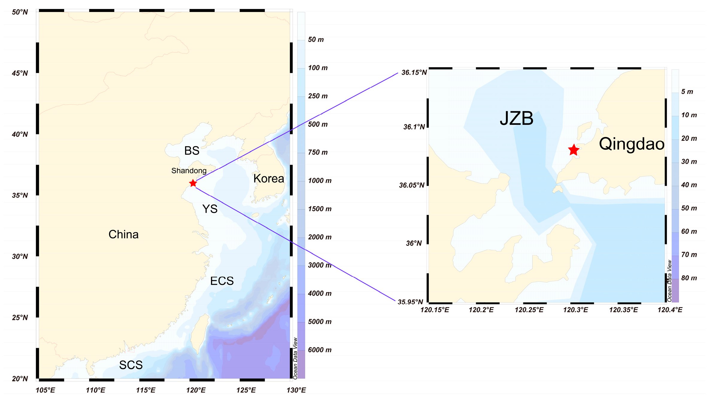
Figure 1. The sampling site (the closed red pentagram) in Jiaozhou Bay (120.314° E, 36.084° N). Notes: BS = Bohai Sea, YS = Yellow Sea, ECS = East China Sea, SCS = South China Sea, JZB = Jiaozhou Bay.
Life 15 01670 g001
Figure 2. The schematic diagram of Germlings Harvester (GEHA).
Life 15 01670 g002
Figure 3. Species diversity of dinoflagellates in Sg, S, Sw+Bw. (a) UPGMA cluster analysis of Sg, S, Sw+Bw; (b) Venn diagram of Sg, S, Sw+Bw; (c) number of orders and corresponding species from all three groups (Sg, S, Sw+Bw); (d) numbers of order and corresponding species from Sg; (e) number of orders and corresponding species from Sw+Bw; (f) number of orders and corresponding species from S; (g) number of common orders and corresponding species shared among Sg, S, Sw+Bw; (h) number of common orders and corresponding species shared between Sg and S. Notes: In situ sediment germination of GEHA = Sg; surface and bottom seawater surrounding GEHA = Sw+Bw; in situ sediments = S; in situ germination period of 5 d = Sg-5 d; in situ germination period of 20 d = Sg-20 d.
Life 15 01670 g003
Figure 4. Genera, families, and orders of in situ germinated dinoflagellate cysts.
Life 15 01670 g004
Figure 5. Shifts in dinoflagellate species within GEHA during in situ germination periods of 5 d (Sg-5 d) and 20 d (Sg-20 d). (a) Genus and corresponding species number of dinoflagellates within GEHA during in situ germination periods of 5 d and 20 d; (b) Venn diagram of species diversity from in situ germination periods of 5 d and 20 d; (c) schematic representation of dinoflagellate species and species number within GEHA and environmental conditions during in situ germination periods of 5 d and 20 d; (d) principal coordinates analysis (PCoA) of in situ germination periods of 5 d and 20 d. Notes: In situ sediment germination of GEHA = Sg; surface and bottom seawater surrounding GEHA = Sw+Bw; in situ sediments = S; in situ germination period of 5 d = Sg-5 d; in situ germination period of 20 d = Sg-20 d; H = heterotrophic; M = mixotrophic; TEMP = temperature; DO = dissolved oxygen; SAL = salinity; the red upward arrows indicate an increase in species number.
Life 15 01670 g005
Figure 6. A conceptual diagram showing the ecological processes and environmental drivers for outbreaks of dinoflagellate HABs that initiate from resting cyst germination.
Life 15 01670 g006
Table 1. The 44 identified dinoflagellate species that were germinated in situ from resting cysts within GEHA and the relevant information.
SpeciesSynonymsHAB EffectsWas It the Resting Cyst Formation Reported Before?Was Germination of Resting Cysts Collected from the Marine Sediment Reported Before?Is It Heterotrophic?References
Akashiwo sanguineaGymnodinium sanguineumB/TYY [47,48,49,50,51]
Alexandrium leeiProtogonyaulax leeiB/TYY [52,53]
Alexandrium pacificum B/TYY [17,54,55]
Ansanella granifera YY [32,37,56]
Archaeperidinium saanichi YYY[57]
Azadinium cuneatum YY [58]
Azadinium polongum BYY [37,59,60]
Azadinium poporum B/TYY [61]
Balechina gracilisGymnodinium gracile Y[62]
Biecheleria brevisulcata YY [63]
Biecheleriopsis adriatica YY [63]
Cyklopsia gemma Y[64]
Diplopelta pusillaLebouraia pusilla YYY[65]
Effrenium voratumSymbiodinium voratum Y [66]
Gymnodinium smaydae YY [37,67]
Gyrodinium cf. spirale Y[68,69]
Gyrodinium heterogrammum YYY[13,37,69]
Heterocapsa pseudotriquetra YY [37,70]
Heterocapsa rotundata YY [71,72]
Heterocapsa steinii YY [71]
Islandinium tricingulatumProtoperidinium tricingulatum YYY[73]
Karlodinium veneficum B/TYY [74,75,76]
Lebouridinium glaucumKatodinium glaucumBYYY[37,77]
Levanderina fissaGyrodinium instriatumBYY [78,79]
Luciella masanensis BYYY[37,80,81,82]
Nematodinium parvumWarnowia parva [83]
Noctiluca scintillansNoctiluca pacificaB Y[84]
Oxytoxum lohmanniiAmphidinium crissum;
Oxytoxum longum
YYY[37,85]
Paragymnodinium
asymmetricum
YY [37,86]
Pelagodinium beiiGymnodinium bei YY [37,87]
Polykrikos kofoidii YYY[68,88]
Protoperidinium americanumPeridinium americanum YYY[73]
Protoperidinium parthenopes YYY[73]
Scrippsiella acuminataScrippsiella trochoideaBYY [89,90,91]
Scrippsiella aff. acuminata Y [92]
Scrippsiella bicarinata YY [32,93]
Scrippsiella cf. acuminata YY [32]
Scrippsiella cf. erinaceus YY [94]
Scrippsiella donghaiensis YY [63]
Scrippsiella lachrymosa Y [95]
Shimiella gracilenta YYY[37,96]
Stoeckeria algicida B/TY Y[97]
Wangodinium sinense YY [98]
Yihiella yeosuensis Y [99]
Notes: B = harmful agal bloom-causing species; T = toxic species (all toxic species are harmful agal bloom-causing species); Y = yes.
Disclaimer/Publisher’s Note: The statements, opinions and data contained in all publications are solely those of the individual author(s) and contributor(s) and not of MDPI and/or the editor(s). MDPI and/or the editor(s) disclaim responsibility for any injury to people or property resulting from any ideas, methods, instructions or products referred to in the content.