Article

Advancing Duodenoscope Reprocessing with Alginate-Coated Calcium Peroxide Nanoparticles

by
Adrian Fifere
1,
Cristian-Dragos Varganici
1,
Elena-Laura Ursu
1,
Tudor Pinteala
1,2,
Vasile Sandru
3,4,
Ioana-Andreea Turin-Moleavin
1,*,
Irina Rosca
1,* and
Gheorghe G. Balan
2,5
1
Petru Poni Institute of Macromolecular Chemistry, Grigore Ghica Voda Alley 41A, 700487 Iasi, Romania
2
Faculty of Medicine, “Grigore T. Popa” University of Medicine and Pharmacy, 700115 Iasi, Romania
3
Department of Gastroenterology, Carol Davila University of Medicine and Pharmacy, 050474 Bucharest, Romania
4
Clinical Emergency Hospital of Bucharest, 105402 Bucharest, Romania
5
St. Spiridon Iasi County Emergency Clinical Hospital, 700111 Iasi, Romania
*
Authors to whom correspondence should be addressed.
Life 2025, 15(11), 1643; https://doi.org/10.3390/life15111643
Submission received: 26 September 2025 / Revised: 17 October 2025 / Accepted: 20 October 2025 / Published: 22 October 2025
(This article belongs to the Special Issue Emerging Applications of Nanobiotechnology in Medicine and Health)

Abstract

Background/Objectives: Although significant advances in duodenoscope reprocessing have been introduced since mid-2010s—including enhanced cleaning protocols, disposable distal endcaps, and the introduction of fully single-use duodenoscopes—residual contamination and infection risks remain unresolved. Moreover, repeated reprocessing may cause cumulative damage to the polymer surfaces, elevator mechanisms, and internal channels of the duodenoscopes, making them more susceptible to residual contamination. To minimize the duodenoscope polymer degradation caused by intensive use and reprocessing, new alternatives are urgently needed. In this context, calcium peroxide nanoparticles coated with sodium alginate (CaO2–Alg NPs), synthesized by our group, were tested for the first time as a disinfectant capable of combating nosocomial pathogens while reducing device deterioration associated with repeated investigations and reprocessing. Methods: The disinfectant properties of the CaO2–Alg NPs were evaluated under biomimetic conditions using reference bacterial strains commonly associated with nosocomial infections. In addition, the compatibility of the nanoparticles with the polymeric duodenoscope coatings was assessed after simulated intensive use. The external polymer coating was structurally and morphologically characterized by Fourier Transform Infrared Spectroscopy (FTIR), Differential Scanning Calorimetry (DSC), Atomic Force Microscopy (AFM), and Scanning Electron Microscopy (SEM). Results: The nanoparticles exhibited important antimicrobial activity against the reference bacterial strains Staphylococcus aureus, Escherichia coli, Enterococcus faecalis, and Klebsiella pneumoniae after only 20 min of incubation. Intensive exposure to the CaO2–Alg NPs did not cause additional structural or morphological damage to the duodenoscope’s external polymers and did not alter their anti-adhesive properties. Conclusions: The CaO2–Alg NPs appear to be a safe and effective disinfectant for the duodenoscope reprocessing, offering both antimicrobial efficacy and material compatibility.

1. Introduction

Duodenoscope-associated infections remain a critical patient-safety concern in 2025. Despite nearly a decade of intensified reprocessing protocols, mandatory US Food and Drug Administration (FDA) post-market surveillance, and the introduction of innovative technologies such as disposable distal endcaps and fully single-use duodenoscopes, the problem has not been eradicated [1]. Contamination rates of up to 15% have been documented in so-called “patient-ready” duodenoscopes [2], and even under enhanced surveillance methods, 5–9% still harbour high-concern pathogens [3,4]. While large-scale outbreaks have become less frequent, any transmission of multidrug-resistant organisms carries profound clinical and public health consequences, in line with Centers for Disease Control and Prevention (CDC) alerts on Carbapenem-resistant Enterobacteriaceae (CRE). This underscores the reality that engineering redesigns, rigorous quality systems, and broader adoption of single-use technologies are not optional, but essential, to move toward eliminating this preventable source of healthcare-associated infection [5].
Reprocessing of the duodenoscopes remains a cornerstone of infection prevention in gastrointestinal endoscopy, given the well-documented risk of patient-to-patient transmission of multidrug-resistant organisms through inadequately reprocessed instruments [6,7]. The complex design of the duodenoscopes—particularly the presence of the elevator mechanism and associated narrow recesses—creates significant challenges for complete decontamination and high-level disinfection (HLD), even when protocols are correctly followed [8,9,10].
Current international guidelines advocate a structured, multistep reprocessing workflow encompassing pre-cleaning, manual cleaning, high-level disinfection, drying, and continuous quality assurance [9,10]. Pre-cleaning should occur immediately after the procedure, with detergent solution aspirated through all channels to prevent drying of organic material and facilitate later cleaning [10,11]. Manual cleaning involves leak testing, disassembly of detachable components, and meticulous brushing and flushing of all internal channels and the elevator recess using enzymatic detergent, followed by thorough rinsing with clean water [9,12]. Subsequently, the duodenoscope undergoes HLD—either within an automated flexible endoscope reprocessor or by validated manual immersion—using chemical agents such as glutaraldehyde, ortho-phthalaldehyde, or peracetic acid, with strict adherence to manufacturer-specified contact times and temperatures [9,10,11]. However, it has been previously shown that HLD cycles and routine instrument/brush passage can produce cumulative surface defects (scratches, pits, residue) and channel roughening, creating niches for biofilm and microbial persistence despite guideline-concordant reprocessing [12,13].
Following HLD, channels must be flushed with sterile or bacteria-free water and purged with forced air. Active drying—often preceded by an alcohol flush—is indispensable, as residual moisture promotes microbial proliferation and biofilm formation [7,10]. The processed duodenoscope should then be stored in a ventilated drying cabinet with continuous filtered airflow to prevent recontamination [10,12]. Finally, guidelines emphasize quality assurance, surveillance, and documentation; reprocessing records should be maintained, and regular microbiological culturing—particularly of the elevator mechanism and distal tip—should be performed as part of ongoing surveillance [9,10,13].
Despite adherence to best practices, duodenoscope-associated infections continue to be reported, underscoring inherent limitations of current designs and reprocessing technologies [8,13]. Consequently, expert societies and regulatory agencies advocate supplementary mitigation strategies—such as double HLD cycles, ethylene oxide sterilization, or “culture-and-hold” protocols—and encourage progressive transition to the duodenoscopes with disposable distal caps or fully single-use designs [8,10,14,15].
Our earlier studies proved the effects of sterilisation cycles on the duodenoscope, emphasising changes to the coatings and working channel polymers due to intensive usage, mostly to the proximal part [16]. Additionally, our previous studies emphasized that there are more friendly alternatives to the duodenoscope reprocessing than those frequently used [17,18]. In the same framework, we approached calcium peroxide as a metastable compound that has been widely investigated for environmental and biomedical applications. Previously, calcium peroxide has been employed in wastewater treatment [19], and sludge treatment [20], for the removal of organic pollutants, due to its strong oxidative reactivity. In addition, it exhibits remarkable antimicrobial activity [21], and has been explored as an oxygen-releasing material in tissue engineering [22,23], highlighting its versatility in biomedical applications. Recently, we reported the synthesis of the CaO2−Alg NPs [24], with a high specific surface area. Importantly, these nanoparticles possess a relatively low oxidizing potential, allowing them to retain their antimicrobial efficacy while minimizing damage to disinfected surfaces.
When used as part of the HLD process, they would gradually release reactive oxygen species, primarily hydrogen peroxide and hydroxyl radicals, providing sustained antimicrobial activity while minimizing surface corrosion and toxic residue formation. The alginate matrix enhances stability and controlled release, allowing efficient biofilm disruption within narrow luminal channels. Preliminary experimental data suggest that the CaO2–Alg formulations could achieve broad-spectrum bactericidal and sporicidal efficacy comparable to peracetic acid or glutaraldehyde, but under milder conditions and with reduced environmental impact on medical devices [24].
In this context, the aim of our study was, for the first time, to evaluate an alternative disinfectant based on the CaO2–Alg NPs to improve the duodenoscope reprocessing and reduce the incidence of nosocomial infections associated with the use of this medical device. Additionally, we assessed the impact of NP treatment on the structure and properties of the polymer external coating after intensive use.

2. Materials and Methods

2.1. Duodenoscope Samples

Samples were collected from a reference duodenoscope, TJF-160F, Olympus Corporation, Tokyo, Japan, previously used in a gastroenterology center for approximately 500 endoscopic retrograde cholangiopancreatography (ERCP). The duodenoscope was quarantined for six months and was manually reprocessed according to the manufacturer’s revised instructions. HLD was performed using a peracetic acid solution. Subsequently, cultures were collected from the external surfaces of the elevator, working channels, water, and air. The culturing process involved a combination of channel flushing, channel brushing, and elevator swabbing, which was subsequently followed by inoculation in a liquid medium of tryptic soy broth at 37 °C for 48 h. All cultures were negative, which allowed the duodenoscope to be disassembled, and approximately 1 cm2 study samples of the outer coating polymers were taken from the area 10–20 cm from the distal end (as presented in Figure 1). The disassembly process was performed in a microbiologically controlled environment using a Class II microbiological safety cabinet (Bioquell, Andover, UK).

2.2. The CaO2–Alg NPs Synthesis

The CaO2–Alg NPs were prepared by chemical precipitation with H2O2 in the presence of sodium alginate as surfactant, following our previously reported protocol without any modifications [24]. The formation and physico-chemical characterization of synthesized NPs were investigated by X-ray diffraction, Fourier transform infrared spectroscopy, electronic microscopy, dynamic light scattering, electron spin resonance, and thermal analysis, as described in our earlier work.

2.3. Biological Investigation

For the microbiological assays there were four reference bacterial strains used, represented as follows: Staphylococcus aureus ATCC25923 (S. aureus), Escherichia coli ATCC25922 (E. coli), Enterococcus faecalis ATCC29212 (E. faecalis), and Klebsiella pneumoniae ATCC10031 (K. pneumoniae).

2.3.1. Evaluation of the Disinfectant Properties of the CaO2–Alg NPs Under Biomimetic Conditions

The study samples were contaminated with bacterial suspensions in biomimetic medium and subsequently treated with the CaO2–Alg NPs at successive time intervals. The control samples were isolated in sterile plates. The experiment was performed in three replicates at each sampling location. To obtain biomimetic conditions, 10 mL of bacterial suspension standardized at 108 CFU/mL in ultrapure sterilized water was mixed with 90 mL of standardized intestinal biomimetic fluid FeSSIF (Fed State Simulated Intestinal Fluid) (Biorelevant, London, UK), which replicates the environment after a high-fat meal, containing an increase in bile salts and lipids, to simulate a physiologically contaminated environment. The mixture was continuously maintained at a temperature of 37 °C. The duodenoscope samples were immersed in the bacterial solution under biomimetic conditions for 15 min at a temperature of 37 °C, with continuous mixing.
After this interval, the samples were taken from the medium and left on a sterile surface for 1 min to dry and subsequently put in contact with the CaO2–Alg NPs suspension in successive stages for 5, 10, 15, 20, and 30 min, respectively, with periodic shaking every 5 min. In parallel, the control samples were immersed in sterile distilled water. After treatment with the nanoparticles or distilled water, respectively, the samples were left to dry on a sterile surface for 1 min. After drying, they were transferred to containers containing 30 mL of sterile tryptone-soy broth medium, sealed, and incubated at 37 °C for 72 h. The presence of turbidity following incubation suggested bacterial growth, indicating the presence of viable microorganisms in the samples post-disinfection. Conversely, the absence of turbidity after incubation demonstrated a lack of viable microorganisms, confirming the disinfectant efficacy of the CaO2–Alg NPs for the specified contact duration. All incubated bottles, regardless of the appearance of the media, were subcultured on appropriate solid media to confirm the absence of viable bacteria. Negative subcultures and the lack of turbidity (clear medium) were considered indicative of HLD. All experiments were conducted in a Class II microbiological safety cabinet (Bioquell, Andover, UK) to prevent contamination with extraneous microbial species.

2.3.2. Evaluation of the Compatibility of the CaO2−Alg NPs with the Polymeric Coating of the Duodenoscope

To evaluate the effect of the repeated CaO2–Alg NP treatments on the duodenoscope polymer structure, a challenge test was conducted. Contaminated device samples were immersed in the CaO2–Alg NP suspensions for 30 min daily over a 45-day period. Control samples were treated under identical conditions, using distilled water instead of the CaO2–Alg NPs. All samples (both treated and untreated) were analyzed by scanning electron microscopy (SEM), atomic force microscopy (AFM), and energy-dispersive X-ray spectroscopy (EDAX).
  • Fourier Transform Infrared Spectroscopy (FT-IR)
FT-IR spectra were obtained using a Bruker Vertex 70 FT-IR spectrometer (Billerica, MA, USA), conducted at room temperature with a resolution of 2 cm−1 in the range of 500–4000 cm−1.
  • Differential Scanning Calorimetry (DSC)
DSC measurements were recorded on a DSC 200 F3 Maia device (Netzsch–Gerätebau GmbH, Selb, Germany). 8 mg of each sample was heated in aluminum crucibles sealed shut with pierced lids. Nitrogen atmosphere was used (flow rate of 50 mL min−1) and a heating rate of 10 °C min−1 was applied. Calibration was conducted with standard indium.
  • Atomic Force Microscopy (AFM)
Images from the study samples collected from the surface of the duodenoscope were recorded in air, in tapping mode using an NTEGRA Spectra (NT-MDT, Zelenograd, Russia) instrument with a 3.1–37.6 N/m force constant cantilever of silicon nitride cantilevers (NSC10, NT-MDT, Moscow, Russia).
  • Scanning Electron Microscopy (SEM)
The surface morphology of the study samples was observed using a Quanta200 scanning electron microscope (FEI Company, Hillsboro, OR, USA), working in a low vacuum mode, at 20 kV with an LFD detector. To obtain the elemental information, EDX analysis using a silicon drift detector was performed on both controls and samples.

2.3.3. Evaluation of Microbial Adhesion on the Duodenoscope Surface Following Prolonged Contact with Metal Oxide Nanoparticles

The anti-adhesion properties of polymer samples after treatment with the CaO2–Alg NPs were investigated in accordance with the Japanese Industrial Standard JIS Z2801:2000 [25]. The JIS Z 2801:2000 method is a standardized test to quantitatively measure the antibacterial activity of antimicrobial surfaces, such as plastics, ceramics, and metals, by inoculating them with bacteria and incubating for 24 h. After incubation, the number of surviving bacteria is counted to calculate the log reduction, which indicates the surface’s efficacy in inhibiting or killing microorganisms. The same bacterial strains were activated in liquid culture medium by incubation for 24 h at 37 °C. The preparation of the tested surfaces involved placing each sample in a sterile Petri dish. The bacterial inoculum was adjusted to standard values of 0.5 McFarland with dilutions to obtain a bacterial concentration of 2.4 × 10⁵ CFU/mL. Subsequently, 0.1 mL of the obtained inoculum was placed on the studied samples and these were subsequently incubated for 24 h at 37 °C. After incubation, the samples were rinsed repeatedly with sterile ultrapure water, and the resulting suspension underwent binary dilutions, which were cultured on PCA (plate count agar) medium and incubated for 24 h at 37 °C, and then the number of resulting colonies was recorded. The interpretation of the results was conducted according to the specifications of JIS Z2801:2000 Tokyo, Japan.

3. Results and Discussion

3.1. Evaluation of the Disinfectant Properties of the CaO2–Alg NPs Under Biomimetic Conditions

The impact of repeated exposure of polymer resins on the duodenoscope surface to the CaO2–Alg NPs was assessed using a previously presented case-control experimental study. The results of the antimicrobial activity of the CaO2–Alg NPs are presented in Table 1. Thus, after 5, 10, and 15 min of exposure, all four strains could still be identified on control cultures. After 20 min of treatment with the CaO2–Alg NPs, both liquid and solid cultures showed negative results, consistent with the absence of bacterial growth and, therefore, with HLD. The results were the same after 30 min of incubation. The total absence of all four bacterial strains after 20 min of sample exposure proves the high-grade disinfectant activity on polymer samples.
The CaO2 NPs have been employed extensively in different fields for the treatment of bacterial contamination, because they provide oxygen (O2) and hydrogen peroxide (H2O2) to the treatment site [21,26,27,28,29]. In dry settings, the CaO2 is quite stable, but in an aqueous environment, it slowly decomposes. According to the literature, the CaO2 has also been utilized to cure cancer because it decomposes to H2O2 more quickly in an acidic environment [30,31,32,33]. The bacterial infection site pH is substantially lower than that of the surrounding tissues, and it is very similar to the microenvironment of a tumour; it has been proven that the CaO2 NPs’ rapid release of the H2O2 may destroy the bacterial cells [34]. Previously, we have proved that the release of hydrogen peroxide increases significantly at low pH compared to neutral pH [24].
These processes could provide an advantage for the use of the CO2 as a disinfectant, because in the immediate vicinity of the acidic bacterial environment, there is an increase in the concentration of the Ca2+ and the H2O2, while in the rest of the dispersion medium, the oxidative and calcium ion levels are lower. H2O2–induced oxidative stress targets important bacterial components such as lipids, proteins, and nucleic acids, leading to cellular dysfunction and then cell death [35,36,37,38,39]. The calcium ions may also contribute to the antibacterial efficiency by disrupting cell membranes [21,40,41]. Moreover, these NP may inhibit the proliferation of bacterial biofilms, primarily by causing oxidative damage to the extracellular polymeric substances (EPS) from the biofilm structure [35,36,37]. Therefore, the CaO2 NPs may be excellent options for the duodenoscope reprocessing.

3.2. Evaluation of the Compatibility of the CaO2–Alg NPs with the Polymeric Duodenoscope Coating

Elemental composition analysis studies of the duodenoscope samples were carried out using the X-ray spectroscopy technique (EDAX), which highlighted the presence of the atoms that form the material covering the duodenoscope, namely: C, N, O, Na, Mg, P, S, and K, in different atomic percentage ratios, as presented in Table 2. Compared to the duodenoscope control sample, for the sample treated with the CaO2–Alg NPs, a slight increase in the mass percentages of C (from 66.59% in the control sample to 68.72% for the treated sample) and O (from 18.65% in the control sample to 21.7% for the treated sample) were observed, as well as the appearance of the Ca atom (0.79 atomic%), which is due to the presence of traces of the CaO2–Alg NPs on the surface of the material as a result of the material disinfection. The elemental composition of the samples, which contains: C, N, O, Na, Mg, Al, P, S, and K, is similar to the one revealed by our previous study [17]; the difference between the percentages is caused by the fact that this study is based on samples from a single area of the duodenoscope.
These traces of NPs are not significant because they can be easily removed by a final washing step of the disinfection process. Additionally, as previously studied by our group, in case of remanence on the duodenoscope surface after completing the reprocessing process, these NPs are biocompatible up to a maximum concentration of 25 μg/mL for the two tested cellular lines taken into study, namely, Normal Human Dermal Fibroblast (NHDF) and Human Gingival Fibroblast (HGF) [24].
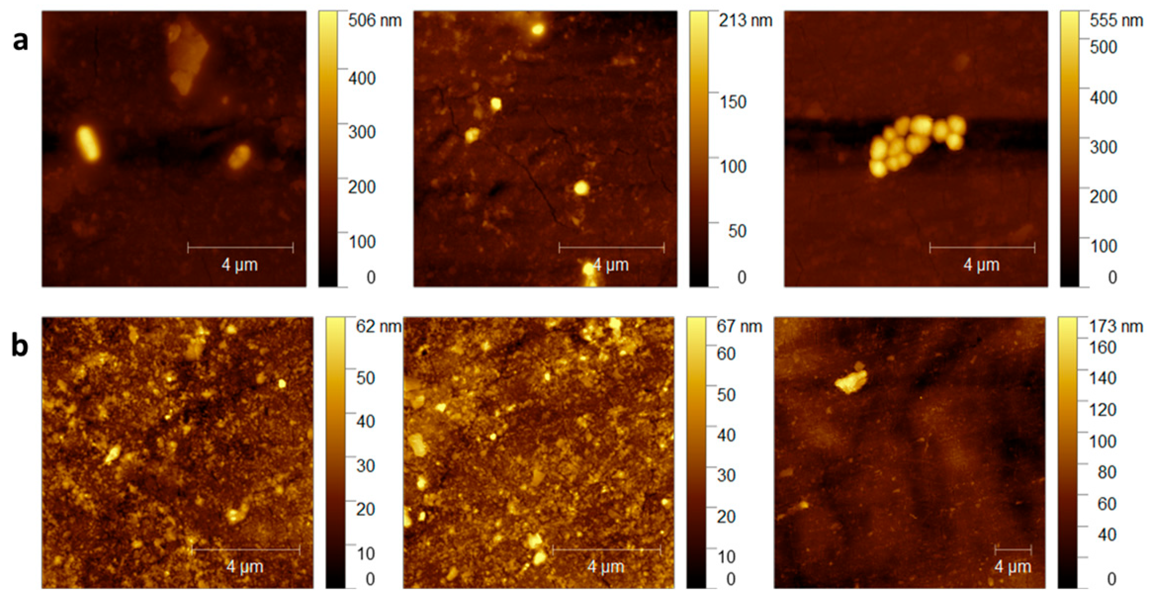
The AFM morphological analysis of the duodenoscope samples shown in (Figure 2) did not reveal any evident new lesions of the polymeric surface of the duodenoscope following exposure to the CaO2–Alg NPs when compared with the controls. These conclusions correlated with the previously presented chemical analysis of the coating samples, which may lead to the conclusion that the intensive use of the NPs does not bring additional damage to the external duodenoscope coating. However, in comparison, bacterial colonies and biofilm formation can be observed on the control samples shown in (Figure 2a), whereas these features are absent on the samples treated with the CaO2–Alg NPs (Figure 2b). On the treated and disinfected surfaces, traces of nanoparticles were still visible but could be easily removed during the final washing step of reprocessing. The morphology of the untreated samples was consistent with the topographic images obtained in our previous study, which revealed an inhomogeneous surface and the presence of microcracks resulting from the intensive use of the duodenoscope, leading over time to polymer degradation [16].
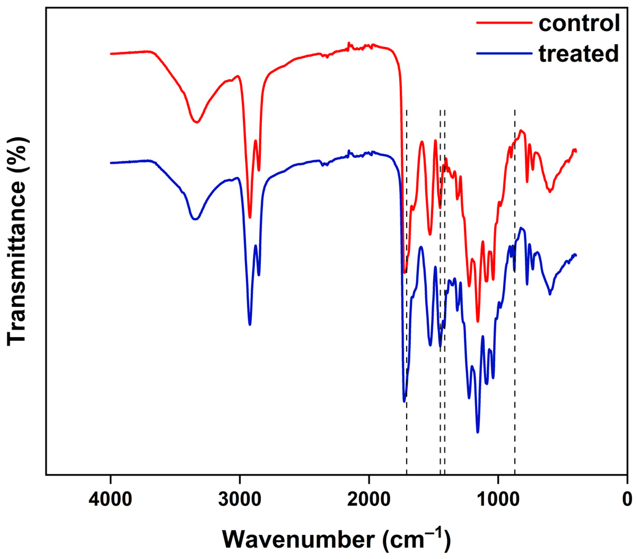
Structural characterization of the material used to cover the duodenoscope was evaluated using the FT-IR technique. For the analysis, two material samples were collected from the same area of the duodenoscope, one control sample and one sample treated with the CaO2–Alg NPs for 45 days. From the FT-IR spectra analysis of the two samples shown in (Figure 3), it may be observed that the absorption bands do not show major changes following treatment with the CaO2–Alg NPs disinfectant. The structural characterization of the control and treated samples is in accordance with the characterization made by our group in a previous study that emphasized structural changes of the external polymer due to intensive usage of the duodenoscope [16].
Both spectra display absorption bands at 2900 and 2853 cm−1, respectively, characteristic of the stretching vibrations of the CH and CH2 groups, the absorption bands present in the 1760–1600 cm−1 region are characteristic of the vibrations of the C=O and C=N groups, and those in the 1040-1200 cm−1 region are generally associated with the vibrations of the C–O, C–O–C, C–N groups. Compared to the control sample, the treated material sample shows traces of the CaO2–Alg NPs that can be observed in the FT-IR spectrum of the treated sample through slightly more intense vibrational bands at 1611 and 1418 cm−1, which were attributed to the symmetric and antisymmetric stretching vibrations of the COO group salt [35], through the intensification of the band at 1449 cm−1 attributed to the O–Ca–O deformation vibration as well as the appearance of the absorption band at 876 cm−1 corresponding to the stretching vibration of the peroxide group, O–O [24].
The present findings are consistent with our previous reports, which indicated that the CaO2–Alg NPs generate a moderately oxidative environment under neutral conditions, as suggested by the electron spin resonance (ESR) analysis of free radicals produced by the hydrogen peroxide released [24]. This suggests that by moderating the oxidizing capacity while maintaining effective disinfectant properties, these nanoparticles offer an advantage by minimizing surface damage to the duodenoscope during the disinfection process.
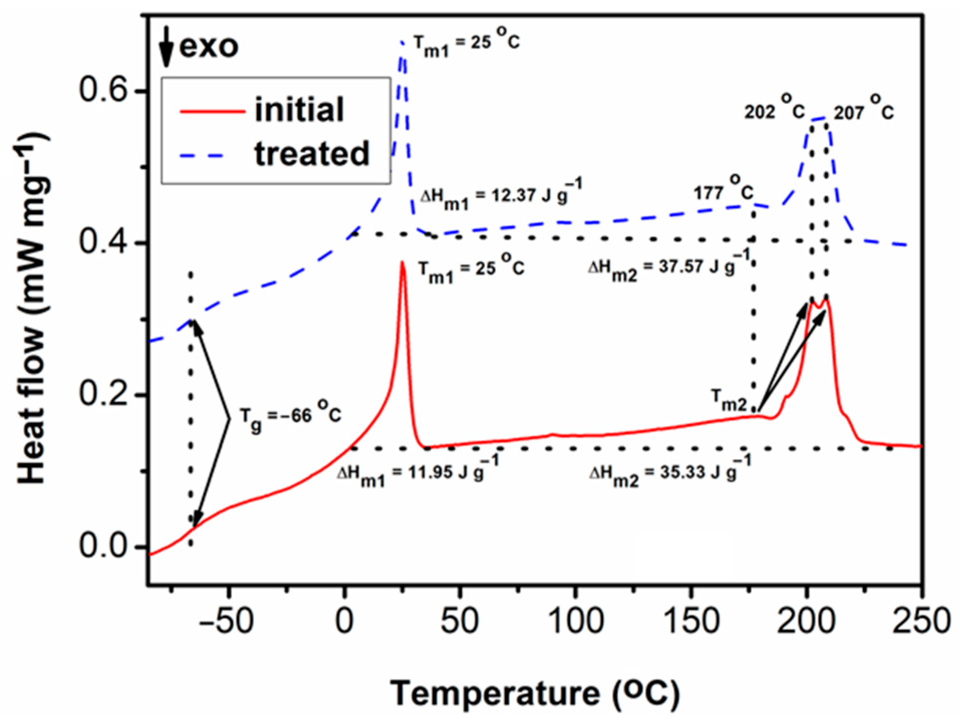
Figure 4 shows the second DSC heating curves of the studied materials. It may be observed that the initial and treated samples show no difference in the glass transition temperature (Tg = −66 °C). The same is in the case of the two melting profile temperatures [16], the one at a lower temperature (Tm1 = 25 °C) and the multiple peaked one at higher temperatures (Tm2 = 177 °C, 202 °C and 207 °C). By analysing the DSC data, it may also be observed that there is only a slight increase in the melting enthalpy (ΔHm) values. ΔHm1 increases from 11.95 J g−1 for the initial sample to 12.37 J g−1 for the treated sample, and ΔHm2 increases from 35.33 J g−1 for the initial sample to 37.57 J g−1 for the treated sample. All data indicate that the treatment process does not induce noteworthy modifications in the thermal behaviour of the material.

3.3. Evaluation of Microbial Adhesion on the Duodenoscope Surface Following Prolonged Contact with the CaO2–Alg NPs

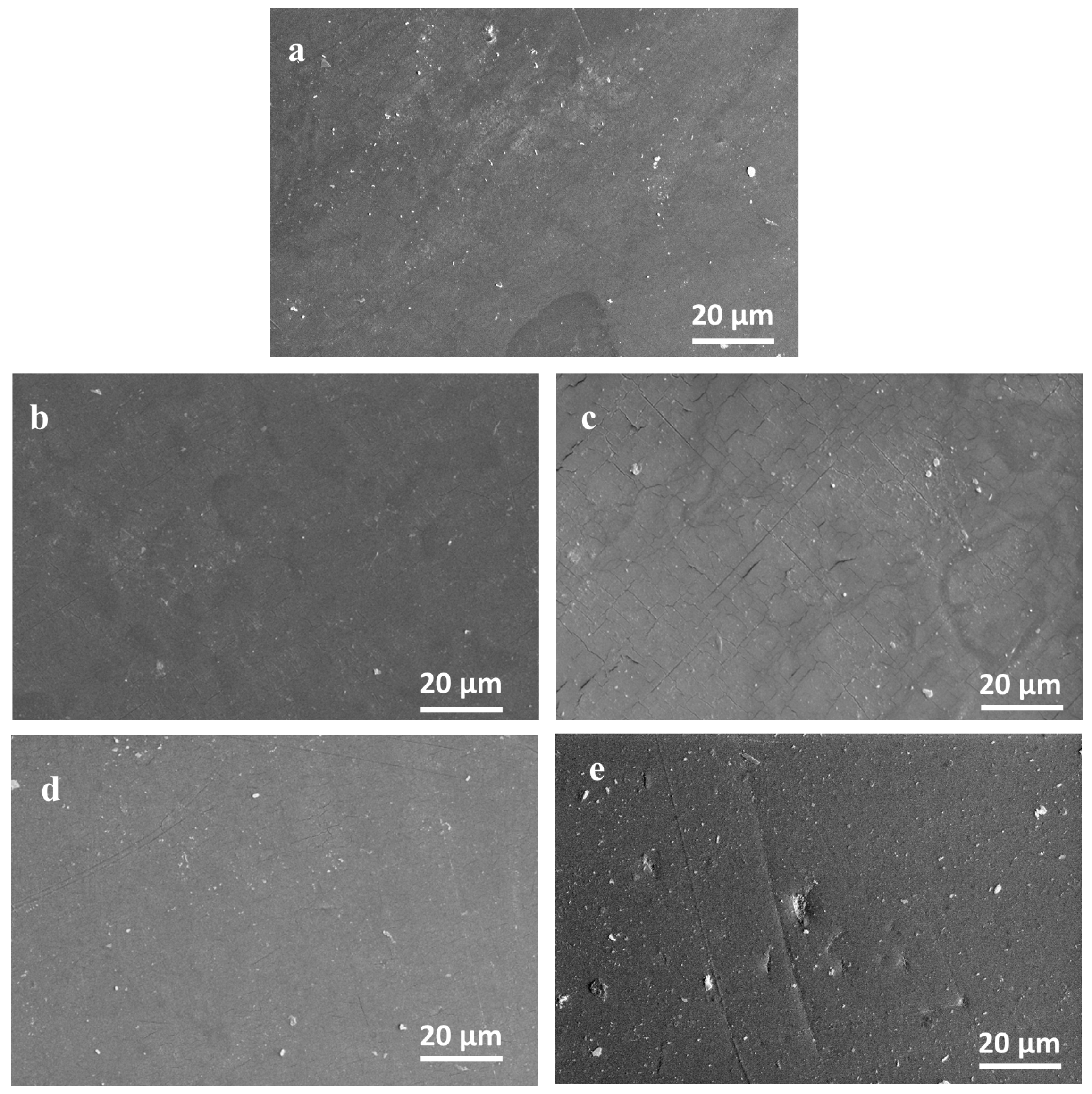
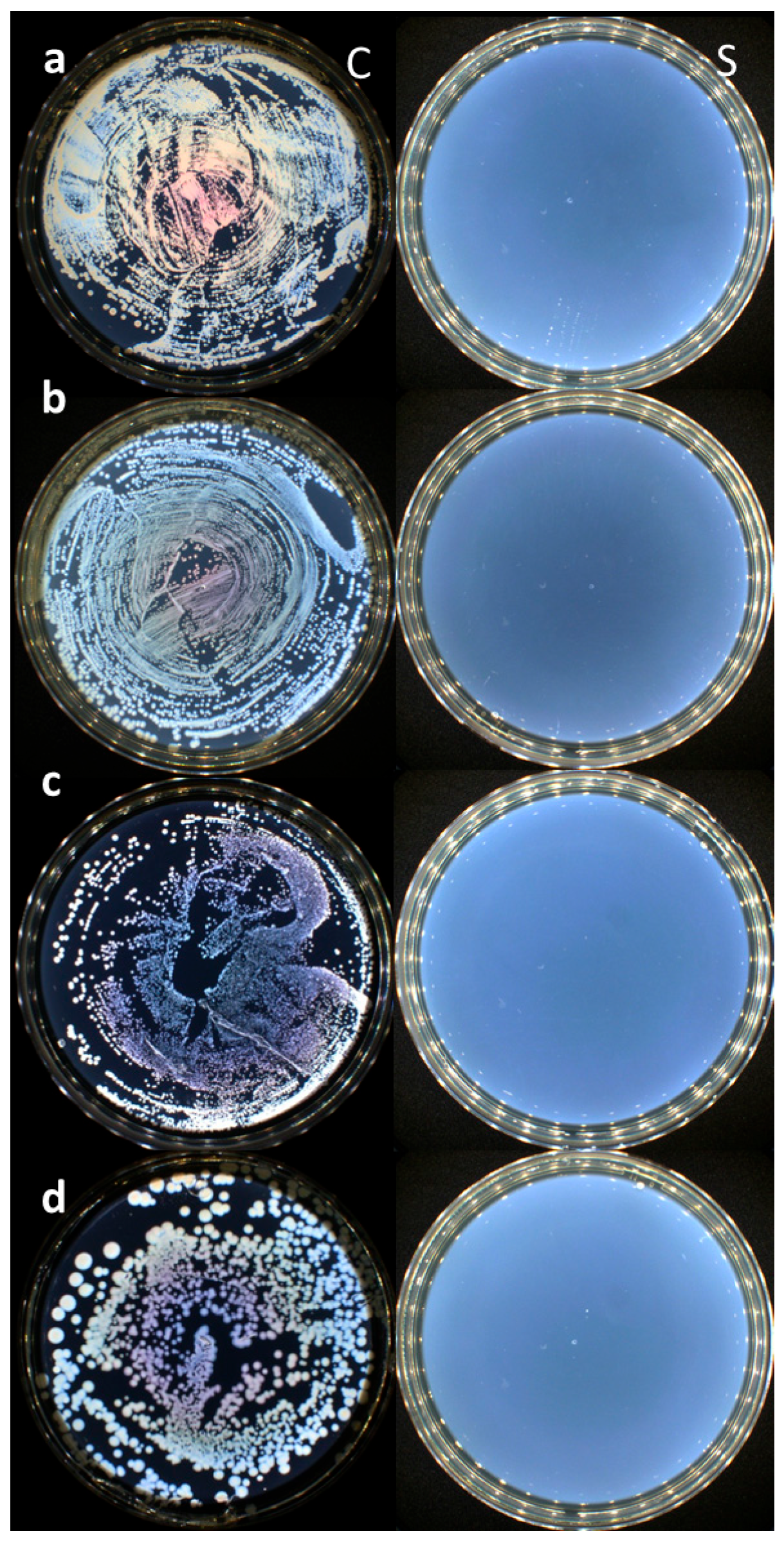
All the duodenoscope samples studied in contact with the CaO2–Alg preserved their anti-adherence properties according to the Japanese Industrial Standard JIS Z2801:2000. Thus, no adhesion and bacterial growth processes were evidenced for the Gram-positive (S. aureus, E. faecalis) and Gram-negative (E. coli, K. pneumoniae) bacterial strains tested as revealed by the SEM analysis of the treated samples and controls (see Figure 5). The duodenoscope surface seems to present the same erosion and deterioration induced by the intensive usage and repeated reprocessing as presented by our team in a previous study [16]. Moreover, no bacterial incubation was identified in the Petri plates after 24 h of incubation at 37 °C (Figure 6), which proves the disinfectant properties of the CaO2–Alg NPs.
These results are also in accordance with our previously made studies on an in-use duodenoscope [16], proving that the material is preserving its antibacterial properties after the prolonged and intensive utilization of the new proposed disinfectant represented by the CaO2–Alg NPs.

3.4. Evaluation of the Economic Burden and Subsequent Challenges of Single-Use Versus Current Multiple-Use Reprocessible Duodenoscopes

Although single-use duodenoscopes offer the theoretical advantage of eliminating cross-infection from reuse, their adoption in general practice has been slow and remains limited by several practical, environmental, and economic constraints. Recent analyses suggest that single-use duodenoscopes incur a higher per-procedure cost compared to well-maintained reusable systems, although the balance may shift in certain scenarios (e.g., low-volume centers or high-infection-risk). A systematic review of economic evaluations concluded that per-procedure cost has generally been higher using single-use endoscopes (versus reusable), although a subset of cost-utility analyses showed single-use designs could become cost-effective under favourable assumptions, especially regarding the high infection costs [42]. A recent European Societies’ position statement emphasizes concerns about the environmental and waste burden of routine single-use devices and suggests against their indiscriminate use, recommending instead a case-by-case assessment [43,44,45]. Meanwhile, when analysing the economic burden and cost trade-offs of all enhanced reprocessing practices currently recommended, one could easily conclude that widespread implementation of single-use duodenoscopes would impose substantial capital and operational costs on health care systems accustomed to high volumes of ERCP and reuse workflows [3,46,47]. Another analysis calculated break-even pricing for single-use devices; in the Dutch setting, single-use scopes would need to be priced between €140 and €250 to rival reusable scopes when used selectively in high–risk patients [48]. Even in such selected scenarios, their numbers are far from the current economic reality.
Thus, despite recognized infection risks from reusable scopes, the balance of sustainability, cost, infrastructure, and waste management continues to hinder the broad deployment of single-use duodenoscopes in favour of reprocessing multiple-use duodenoscopes. Hence, there is still a constant need for optimizing reprocessing cycles in both efficacy and long-term sustainability, both pleading for potential alternative environmental and scope-friendly measures. However, in low-volume or high-risk settings, single-use devices may approach financial viability under favourable pricing, reimbursement, or infection-cost assumptions.

4. Conclusions

Although this is clearly a preliminary study, due to the limited evaluation of the CaO2–Alg NP disinfectant properties and their effects on different regions of the duodenoscope with varying materials, the present research provides novel insights and demonstrates the potential utility of these nanoparticles as an alternative in the duodenoscope reprocessing. The goal is to optimize bacterial and biofilm removal while minimizing polymer resin degradation during reprocessing. Although still at an exploratory stage, integrating such biomaterials into reprocessing workflows may offer a pathway to reduce contamination rates and mitigate infection risk, thereby enhancing patient safety in endoscopic practice.

Author Contributions

Conceptualization, A.F., G.G.B. and I.R.; methodology, A.F., I.-A.T.-M., C.-D.V., T.P., E.-L.U. and I.R.; validation, A.F., V.S. and G.G.B.; resources, I.R. and G.G.B.; writing—original draft preparation, I.R. and I.-A.T.-M.; writing—review and editing, A.F., T.P. and G.G.B.; funding acquisition, I.R. All authors have read and agreed to the published version of the manuscript.

Funding

This research received no external funding.

Institutional Review Board Statement

Not applicable.

Informed Consent Statement

Not applicable.

Data Availability Statement

The original contributions presented in this study are included in the article. Further inquiries can be directed to the corresponding authors.

Acknowledgments

This work was supported by a grant of the Ministry of Research, Innovation and Digitization, CNCS—UEFISCDI, project number PN-III-P1-1.1-TE-2021-0739, within PNCDI III.

Conflicts of Interest

The authors declare no conflicts of interest.

References

  1. Center for Drug Evaluation and Research. Novel Drug Approvals for 2025; FDA: Silver Spring, MD, USA, 2025. [Google Scholar]
  2. Larsen, S.; Russell, R.V.; Ockert, L.K.; Spanos, S.; Travis, H.S.; Ehlers, L.H.; Mærkedahl, A. Rate and Impact of Duodenoscope Contamination: A Systematic Review and Meta-Analysis. eClinicalMedicine 2020, 25, 100451. [Google Scholar] [CrossRef]
  3. Bomman, S.; Klair, J.S.; Canakis, A.; Muthusamy, A.K.; Nagra, N.; Chandra, S.; Shanmugam, M.; Perisetti, A.; Aggarwal, A.; Gavini, H.K.; et al. Safety and Efficacy of Endoscopic Full Thickness Resection of Upper Gastrointestinal Lesions Using a Full Thickness Resection Device: A Systematic Review and Meta-Analysis. J. Clin. Gastroenterol. 2024, 58, 46–52. [Google Scholar] [CrossRef]
  4. Rutala, W.A.; Weber, D.J. Best Practices for Disinfection of Noncritical Environmental Surfaces and Equipment in Health Care Facilities: A Bundle Approach. Am. J. Infect. Control 2019, 47, A96–A105. [Google Scholar] [CrossRef]
  5. Heuvelmans, M.; Wunderink, H.F.; van der Mei, H.C.; Monkelbaan, J.F. A Narrative Review on Current Duodenoscope Reprocessing Techniques and Novel Developments. Antimicrob. Resist. Infect. Control 2021, 10, 171. [Google Scholar] [CrossRef]
  6. Günșahin, D.; Șandru, V.; Constantinescu, G.; Ilie, M.; Cabel, T.; Popescu, R.Ș.; Ungureanu, B.S.; Miron, V.D.; Balan, G.G.; Cotigă, D.; et al. A Comprehensive Review of Digestive Endoscopy-Associated Infections: Bacterial Pathogens, Host Susceptibility, and the Impact of Viral Hepatitis. Microorganisms 2025, 13, 2128. [Google Scholar] [CrossRef] [PubMed]
  7. Ofstead, C.L.; Buro, B.L.; Hopkins, K.M.; Eiland, J.E.; Wetzler, H.P.; Lichtenstein, D.R. Duodenoscope-Associated Infection Prevention: A Call for Evidence-Based Decision Making. Endosc. Int. Open 2020, 8, E1769–E1781. [Google Scholar] [CrossRef] [PubMed]
  8. Verfaillie, C.J.; Bruno, M.J.; Voor in ’t Holt, A.F.; Buijs, J.G.; Poley, J.-W.; Loeve, A.J.; Severin, J.A.; Abel, L.F.; Smit, B.J.; de Goeij, I.; et al. Withdrawal of a Novel-Design Duodenoscope Ends Outbreak of a VIM-2-Producing Pseudomonas aeruginosa. Endoscopy 2015, 47, 493–502. [Google Scholar] [CrossRef] [PubMed]
  9. Day, L.W.; Muthusamy, V.R.; Collins, J.; Kushnir, V.M.; Sawhney, M.S.; Thosani, N.C.; Wani, S. Multisociety Guideline on Reprocessing Flexible GI Endoscopes and Accessories. Gastrointest. Endosc. 2021, 93, 11–33.e6. [Google Scholar] [CrossRef]
  10. Speer, T.; Alfa, M.; Jones, D.; Vickery, K.; Griffiths, H.; Sáenz, R.; LeMair, A. WGO Guideline-Endoscope Disinfection Update. J. Clin. Gastroenterol. 2023, 57, 1–9. [Google Scholar] [CrossRef]
  11. Rutala, W.A.; Weber, D.J. Disinfection and Sterilization: An Overview. Am. J. Infect. Control 2013, 41, S2–S5. [Google Scholar] [CrossRef]
  12. Beilenhoff, U.; Biering, H.; Blum, R.; Brljak, J.; Cimbro, M.; Dumonceau, J.-M.; Hassan, C.; Jung, M.; Kampf, B.; Neumann, C.; et al. Reprocessing of Flexible Endoscopes and Endoscopic Accessories Used in Gastrointestinal Endoscopy: Position Statement of the European Society of Gastrointestinal Endoscopy (ESGE) and European Society of Gastroenterology Nurses and Associates (ESGENA)—Update 2018. Endoscopy 2018, 50, 1205–1234. [Google Scholar] [CrossRef] [PubMed]
  13. Epstein, L.; Hunter, J.C.; Arwady, M.A.; Tsai, V.; Stein, L.; Gribogiannis, M.; Frias, M.; Guh, A.Y.; Laufer, A.S.; Black, S.; et al. New Delhi Metallo-β-Lactamase–Producing Carbapenem-Resistant Escherichia coli Associated with Exposure to Duodenoscopes. JAMA 2014, 312, 1447–1455. [Google Scholar] [CrossRef] [PubMed]
  14. Gromski, M.A.; Sherman, S. Technological Review: Developments in Innovative Duodenoscopes. Gastrointest. Endosc. 2022, 95, 42–50. [Google Scholar] [CrossRef] [PubMed]
  15. van der Ploeg, K.; Haanappel, C.P.; Voor In ’t Holt, A.F.; de Groot, W.; Bulkmans, A.J.C.; Erler, N.S.; Mason-Slingerland, B.C.G.C.; Severin, J.A.; Vos, M.C.; Bruno, M.J. Unveiling 8 Years of Duodenoscope Contamination: Insights from a Retrospective Analysis in a Large Tertiary Care Hospital. Gut 2024, 73, 613–621. [Google Scholar] [CrossRef]
  16. Balan, G.G.; Rosca, I.; Ursu, E.-L.; Fifere, A.; Varganici, C.-D.; Doroftei, F.; Turin-Moleavin, I.-A.; Sandru, V.; Constantinescu, G.; Timofte, D.; et al. Duodenoscope-Associated Infections beyond the Elevator Channel: Alternative Causes for Difficult Reprocessing. Molecules 2019, 24, 2343. [Google Scholar] [CrossRef]
  17. Bălan, G.G.; Roșca, I.; Ursu, E.-L.; Doroftei, F.; Bostănaru, A.-C.; Hnatiuc, E.; Năstasă, V.; Șandru, V.; Ștefănescu, G.; Trifan, A.; et al. Plasma-Activated Water: A New and Effective Alternative for Duodenoscope Reprocessing. Infect. Drug Resist. 2018, 11, 727–733. [Google Scholar] [CrossRef]
  18. Ailincai, D.; Turin Moleavin, I.-A.; Sarghi, A.; Fifere, A.; Dumbrava, O.; Pinteala, M.; Balan, G.G.; Rosca, I. New Hydrogels Nanocomposites Based on Chitosan, 2-Formylphenylboronic Acid, and ZnO Nanoparticles as Promising Disinfectants for Duodenoscopes Reprocessing. Polymers 2023, 15, 2669. [Google Scholar] [CrossRef]
  19. Li, A.; Qian, C.; Wen, J.; Wang, T. Synchronous Removal of Organic Pollutants and Phosphorus from Emergency Wastewater in Chemical Industry Park by Plasma Catalysis System Based on Calcium Peroxide. Catalysts 2025, 15, 486. [Google Scholar] [CrossRef]
  20. Xu, Q.; Huang, Q.-S.; Wei, W.; Sun, J.; Dai, X.; Ni, B.-J. Improving the Treatment of Waste Activated Sludge Using Calcium Peroxide. Water Res. 2020, 187, 116440. [Google Scholar] [CrossRef]
  21. Shasha, Z.; Chuanchuan, H.; Yawen, Z. The Progress and Prospect of Calcium Peroxide Nanoparticles in Antibacterial Activity. Colloid Interface Sci. Commun. 2024, 61, 100793. [Google Scholar] [CrossRef]
  22. Mollajavadi, M.Y.; Saadatmand, M.; Ghobadi, F. Effect of Calcium Peroxide Particles as Oxygen-Releasing Materials on Cell Growth and Mechanical Properties of Scaffolds for Tissue Engineering. Iran. Polym. J. 2023, 32, 599–608. [Google Scholar] [CrossRef]
  23. Daneshmandi, L.; Laurencin, C.T. Regenerative Engineered Vascularized Bone Mediated by Calcium Peroxide. J. Biomed. Mater. Res. Part A 2020, 108, 1045–1057. [Google Scholar] [CrossRef] [PubMed]
  24. Turin Moleavin, I.A.; Sarghi, A.; Ursu, E.L.; Sandu, A.I.; Balan, G.G.; Rosca, I.; Fifere, A.; Pinteala, M. Alginate-Coated Calcium Peroxide Nanoparticles as a Disinfectant for Duodenoscope Reprocessing. ACS Appl. Nano Mater. 2023, 6, 23103–23113. [Google Scholar] [CrossRef]
  25. JIS Z 2801; Antibacterial Products—Test for Antibacterial Activity and Efficacy. Japan Standards Association (JSA): Tokyo, Japan, 2010.
  26. Khodaveisi, J.; Banejad, H.; Afkhami, A.; Olyaie, E.; Lashgari, S.; Dashti, R. Synthesis of Calcium Peroxide Nanoparticles as an Innovative Reagent for in Situ Chemical Oxidation. J. Hazard. Mater. 2011, 192, 1437–1440. [Google Scholar] [CrossRef] [PubMed]
  27. Lu, S.; Zhang, X.; Xue, Y. Application of Calcium Peroxide in Water and Soil Treatment: A Review. J. Hazard. Mater. 2017, 337, 163–177. [Google Scholar] [CrossRef]
  28. Wu, J.; Shen, P.; Qin, X.; Yang, Y.; Lin, C.; Li, X.; Geng, W.; Gao, P.; Chen, L.; Miao, L.; et al. Self-Supply of H2O2 and O2 by a Composite Nanogenerator for Chemodynamic Therapy/Hypoxia Improvement and Rapid Therapy of Biofilm-Infected Wounds. Chem. Eng. J. 2023, 459, 141507. [Google Scholar] [CrossRef]
  29. Wang, H.; Zhao, Y.; Li, T.; Chen, Z.; Wang, Y.; Qin, C. Properties of Calcium Peroxide for Release of Hydrogen Peroxide and Oxygen: A Kinetics Study. Chem. Eng. J. 2016, 303, 450–457. [Google Scholar] [CrossRef]
  30. Zhang, M.; Song, R.; Liu, Y.; Yi, Z.; Meng, X.; Zhang, J.; Tang, Z.; Yao, Z.; Liu, Y.; Liu, X.; et al. Calcium-Overload-Mediated Tumor Therapy by Calcium Peroxide Nanoparticles. Chem 2019, 5, 2171–2182. [Google Scholar] [CrossRef]
  31. Wu, D.; Zhu, Z.-Q.; Tang, H.-X.; Shi, Z.-E.; Kang, J.; Liu, Q.; Qi, J. Efficacy-Shaping Nanomedicine by Loading Calcium Peroxide into Tumor Microenvironment-Responsive Nanoparticles for the Antitumor Therapy of Prostate Cancer. Theranostics 2020, 10, 9808–9829. [Google Scholar] [CrossRef]
  32. Wang, Y.; He, X.; Zhou, C.; Bai, Y.; Li, T.; Liu, J.; Ju, S.; Wang, C.; Xiang, G.; Xiong, B. Nanoscale CaO2 Materials for Synergistic Transarterial Chemoembolization in a VX2 Orthotopic Rabbit Liver Cancer Model. Acta Biomater. 2022, 154, 536–548. [Google Scholar] [CrossRef]
  33. Ruan, S.; Yin, W.; Chang, J.; Yang, Y.; Sun, J.; Ma, X.; Liu, Y.; Zang, J.; Liu, Y.; Li, Y.; et al. Acidic and Hypoxic Tumor Microenvironment Regulation by CaO2-Loaded Polydopamine Nanoparticles. J. Nanobiotechnol. 2022, 20, 544. [Google Scholar] [CrossRef] [PubMed]
  34. Thi, P.L.; Lee, Y.; Tran, D.L.; Thi, T.T.H.; Park, K.M.; Park, K.D. Calcium Peroxide-Mediated in Situ Formation of Multifunctional Hydrogels with Enhanced Mesenchymal Stem Cell Behaviors and Antibacterial Properties. J. Mater. Chem. B 2020, 8, 11033–11043. [Google Scholar] [CrossRef] [PubMed]
  35. Chang, M.; Nguyen, T.T. Strategy for Treatment of Infected Diabetic Foot Ulcers. Acc. Chem. Res. 2021, 54, 1080–1093. [Google Scholar] [CrossRef] [PubMed]
  36. Mishra, A.; Pradhan, D.; Halder, J.; Biswasroy, P.; Rai, V.K.; Dubey, D.; Kar, B.; Ghosh, G.; Rath, G. Metal Nanoparticles against Multi-Drug-Resistance Bacteria. J. Inorg. Biochem. 2022, 237, 111938. [Google Scholar] [CrossRef]
  37. Seixas, A.F.; Quendera, A.P.; Sousa, J.P.; Silva, A.F.Q.; Arraiano, C.M.; Andrade, J.M. Bacterial Response to Oxidative Stress and RNA Oxidation. Front. Genet. 2022, 12, 821535. [Google Scholar] [CrossRef]
  38. Chaudhary, P.; Janmeda, P.; Docea, A.O.; Yeskaliyeva, B.; Abdull Razis, A.F.; Modu, B.; Calina, D.; Sharifi-Rad, J. Oxidative Stress, Free Radicals and Antioxidants: Potential Crosstalk in the Pathophysiology of Human Diseases. Front. Chem. 2023, 11, 1158198. [Google Scholar] [CrossRef]
  39. Asad, N.R.; Asad, L.M.B.O.; de Almeida, C.E.B.; Felzenszwalb, I.; Cabral-Neto, J.B.; Leitão, A.C. Several Pathways of Hydrogen Peroxide Action That Damage the E. coli Genome. Genet. Mol. Biol. 2004, 27, 291–303. [Google Scholar] [CrossRef]
  40. Gu, P.; Wang, Y.; Wu, H.; Chen, L.; Zhang, Z.; Yang, K.; Zhang, Z.; Ren, X.; Miao, H.; Zheng, Z. Efficient Control of Cyanobacterial Blooms with Calcium Peroxide: Threshold and Mechanism. Sci. Total Environ. 2023, 882, 163591. [Google Scholar] [CrossRef]
  41. Liu, L.; Pan, Y.; Zhi, X.; Chen, L.; Zhu, H. Bacterial Antioxidant Mechanism in Calcium Peroxide Aided Sludge Anaerobic Fermentation. Bioresour. Technol. 2023, 384, 129327. [Google Scholar] [CrossRef]
  42. Zanganeh, M.; Jiang, Y.; Waugh, N.; Brown, A.; Chen, Y.-F.; Arasaradnam, R.P.; Andronis, L.; “SUMU Endo” Project Group. Single-Use versus Reusable Endoscopes in Gastroenterology: Systematic Review of Full and Partial Economic Evaluations. Endosc. Int. Open 2025, 13, a26451463. [Google Scholar] [CrossRef]
  43. Rodríguez de Santiago, E.; Dinis-Ribeiro, M.; Pohl, H.; Agrawal, D.; Arvanitakis, M.; Baddeley, R.; Bak, E.; Bhandari, P.; Bretthauer, M.; Burga, P.; et al. Reducing the Environmental Footprint of Gastrointestinal Endoscopy: European Society of Gastrointestinal Endoscopy (ESGE) and European Society of Gastroenterology and Endoscopy Nurses and Associates (ESGENA) Position Statement. Endoscopy 2022, 54, 797–826. [Google Scholar] [CrossRef]
  44. Hassan, C.; Ponchon, T.; Bisschops, R.; van Hooft, J.E.; Messmann, H.; Gralnek, I.M.; Dinis-Ribeiro, M. European Society of Gastrointestinal Endoscopy (ESGE) Publications Policy—Update 2020. Endoscopy 2020, 52, 123–126. [Google Scholar] [CrossRef]
  45. Lenzen, M.; Malik, A.; Li, M.; Fry, J.; Weisz, H.; Pichler, P.-P.; Chaves, L.S.M.; Capon, A.; Pencheon, D. The Environmental Footprint of Health Care: A Global Assessment. Lancet Planet. Health 2020, 4, e271–e279. [Google Scholar] [CrossRef]
  46. Ma, G.K.; Pegues, D.A.; Kochman, M.L.; Alby, K.; Fishman, N.O.; Saunders, M.; Grous, C.; Dempsey, D.T.; Ginsberg, G.G. Implementation of a Systematic Culturing Program to Monitor the Efficacy of Endoscope Reprocessing: Outcomes and Costs. Gastrointest. Endosc. 2018, 87, 104–109.e3. [Google Scholar] [CrossRef]
  47. Barakat, M.; Ghosh, S.; Banerjee, S. 775 Cost Utility Analysis Comparing Duodenoscope Reprocessing/Sterilization, Novel Duodenoscopes with Disposable Endcaps and Fully Disposable Duodenoscopes. Gastrointest. Endosc. 2020, 91, AB67–AB68. [Google Scholar] [CrossRef]
  48. Kwakman, J.A.; Poley, M.J.; Vos, M.C.; Bruno, M.J. Single-Use Duodenoscopes Compared with Reusable Duodenoscopes in Patients Carrying Multidrug-Resistant Microorganisms: A Break-Even Cost Analysis. Endosc. Int. Open 2023, 11, E571–E580. [Google Scholar] [CrossRef]
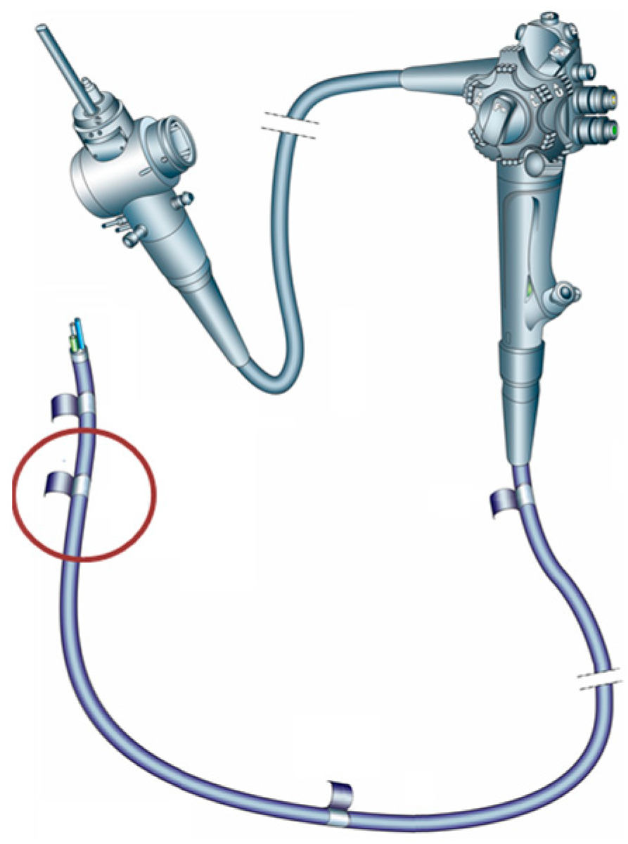
Figure 1. Duodenoscope scheme with the sampling site marked in red.
Life 15 01643 g001
Figure 2. AFM morphological evaluation of control (a), and disinfected samples (b).
Life 15 01643 g002
Figure 3. ATR-FT-IR spectra for control (red) and treated (blue) duodenoscope samples.
Life 15 01643 g003
Figure 4. Representative DSC curves of untreated (blue) and CaO2–Alg NP—treated (red) duodenoscope samples.
Life 15 01643 g004
Figure 5. Scanning electron microscopy (SEM) evaluation of duodenoscope samples: control (a) infected with S. aureus, (b) E. coli, (c) E. faecalis, (d) K. pneumoniae, (e) and disinfected with CaO2–Alg NPs.
Life 15 01643 g005
Figure 6. Antibacterial efficiency of CaO2−Alg NPs (S) compared with the controls (C) against the following: (a) S. aureus, (b) E. coli, (c) E. faecalis, and (d) K. pneumoniae.
Life 15 01643 g006
Table 1. Evaluation of the antimicrobial activity of CaO2–Alg NPs at different time intervals.
Tested Strain5 min10 min15 min20 min30 min
E. coli+++
K. pneumoniae+++
E. faecalis+++
S. aureus+++
Note: + indicates the presence of turbidity in the culture media (microbial growth); − indicates no turbidity (confirmed by complete absence of growth on subcultures, that is, HLD).
Table 2. Mass and atomic ratios obtained by the EDAX technique following the analysis of the controls and CaO2–Alg NPs treated samples.
ElementControlTreated Sample
Weight %Atomic %Weight %Atomic %
C59.1466.5959.6568.72
N11.9811.565.405.33
O22.0718.6525.0921.70
Na1.470.860.570.34
Mg0.500.280.400.23
P2.681.174.291.92
S1.710.721.280.55
K0.470.160.630.22
Ca--2.290.79
Disclaimer/Publisher’s Note: The statements, opinions and data contained in all publications are solely those of the individual author(s) and contributor(s) and not of MDPI and/or the editor(s). MDPI and/or the editor(s) disclaim responsibility for any injury to people or property resulting from any ideas, methods, instructions or products referred to in the content.