Article

The Brazilian Caatinga Biome as a Hotspot for the Isolation of Antibiotic-Producing Actinomycetota

by
Sayoane Pessoa Fernandes
1,†,
Luana Layse Câmara de Almeida
1,†,
Adrielly Silva Albuquerque de Andrade
2,
Lucas Silva Abreu
3,
Yuri Mangueira Nascimento
4,
Thalisson Amorim de Souza
4,
Evandro Ferreira da Silva
4,
Fabiana Caroline Zempulski Volpato
5,
Afonso Luis Barth
6,
Josean Fechine Tavares
4,
Demetrius Antonio Machado de Araújo
2,
Valnês da Silva Rodrigues-Junior
7,* and
Samuel Paulo Cibulski
8,*
1
Programa de Pós-Graduação em Ciências Farmacêuticas, Departamento de Farmácia, Universidade Estadual da Paraíba (UEPB), Campina Grande 58429-500, PB, Brazil
2
Laboratório de Biotecnologia Celular e Molecular, Centro de Biotecnologia, Universidade Federal da Paraíba (UFPB), João Pessoa 58051-900, PB, Brazil
3
Departamento de Química Orgânica, Instituto de Química, Universidade Federal Fluminense (UFF), Niterói 24020-007, RJ, Brazil
4
Laboratório Multiusuário de Caracterização e Análise (LMCA), Instituto de Pesquisa em Fármacos e Medicamentos, Universidade Federal da Paraíba (UFPB), João Pessoa 58051-900, PB, Brazil
5
Departamento de Biociências, Universidade Federal do Paraná (UFPR), Setor Palotina, Palotina 85950-000, PR, Brazil
6
Laboratório de Pesquisa Em Resistência Bacteriana (LABRESIS), Hospital de Clínicas de Porto Alegre, Porto Alegre 90035-007, RS, Brazil
7
Programa de Pós-Graduação em Produtos Naturais e Sintéticos Bioativos, Universidade Federal da Paraíba (UFPB), João Pessoa 58051-900, PB, Brazil
8
FACISA—Faculdade de Ciências da Saúde do Trairi, Universidade Federal do Rio Grande do Norte (UFRN), Santa Cruz 59200-000, RN, Brazil
*
Authors to whom correspondence should be addressed.
These authors contributed equally to this work.
Life 2025, 15(10), 1494; https://doi.org/10.3390/life15101494
Submission received: 29 August 2025 / Revised: 19 September 2025 / Accepted: 22 September 2025 / Published: 23 September 2025

Abstract

Antimicrobial resistance represents a critical global health challenge, intensifying the urgency of discovering novel antibiotics. Actinomycetota species, the most prolific source of clinical antibiotics, remain underexplored in unique ecosystems. In this study, we isolated 340 Actinomycetota strains from soils of the Brazilian semiarid Caatinga biome. Screening revealed that 122 isolates (35.9%) exhibited antimicrobial activity against clinically relevant pathogens (Escherichia coli, Pseudomonas aeruginosa, Staphylococcus aureus, and Candida albicans). Notably, 19 isolates showed activity against Mycobacterium tuberculosis H37Ra. MALDI-TOF MS analysis successfully provided genus-level identification for a subset of isolates, with approximately 32% assigned to the Streptomyces genus. However, the limited resolution of the database for the majority of the strains indicates high phylogenetic diversity and suggests the presence of potentially novel species. Metabolomic profiling via LC-MS/MS and GNPS molecular networking suggested the production of known antibiotics such as actinomycins, cyclomarins and anthracyclines and unveiled distinct molecular families putatively assigned to undescribed metabolites. Our work establishes the Caatinga biome as a valuable reservoir of bioactive Actinomycetota, encoding both known and potentially novel antimicrobial compounds. These results underscore the potential of underexplored and extreme environments in the quest to overcome antibiotic resistance.

Graphical Abstract

1. Introduction

Antimicrobial resistance (AMR) is recognized by the World Health Organization (WHO) as one of the top 10 global health threats. This silent pandemic of drug-resistant infections disproportionately affects low- and middle-income countries and vulnerable populations, including children and the elderly [1]. If unaddressed, AMR could compromise the treatment of common infections and make routine medical procedures increasingly risky. Research on antibiotics is therefore critical to counteract resistance and ensure preparedness for emerging infectious challenges.
Combating AMR demands coordinated interdisciplinary efforts. Microbiologists, molecular biologists, chemists, clinicians, epidemiologists, and economists, alongside governments and international organizations, must collaborate to discover, responsibly use, and preserve antibiotics. Such integrated action is essential to slow the rise of resistance and safeguard the continued efficacy of these life-saving drugs [2].
In this context, the Actinomycetota represent a critical resource, as this phylum of Gram-positive bacteria produces a milieu of bioactive metabolites, including antibiotics (e.g., streptomycin, rifamycins, and erythromycin), antiproliferative drugs (e.g., doxorubicin, mitomycin, and bleomycin), immunosuppressive drugs (e.g., rapamycin and tacrolimus), and other important medical molecules [3]. More than 25,000 bioactive secondary metabolites from microorganisms have been identified, and about 70% of these are produced by Actinomycetota species, including more than a hundred antibiotics used in human and animal therapy and agriculture [4]. However, the use of general and classical approaches to discover and develop new drugs from these microorganisms is becoming increasingly complex and demanding. To circumvent the challenges of drug development from Actinomycetota, new concepts based on the triad “genomics, chemical dereplication and new habitats” have been described [5]. In other words, new habitats harbor new species, which should contain new gene clusters synthesizing novel secondary metabolites. The alliance of simple, easy, and cheap classical microbiological methods with modern genomic and metabolomics techniques that focus on Actinomycetota species from new habitats, such as oceans, extreme environments including arid regions, plants, and feces of animals and lichens, could be a perfect marriage, offering hope for the development of novel drugs to combat old and new enemies.
According to the United Nations Environment Programme (UNEP), Brazil is recognized as the most biologically diverse country on Earth, hosting an estimated 15–20% of all known animal and plant species. While the country’s macrodiversity has been increasingly documented, its microbial diversity remains largely unexplored [6]. Microorganisms represent a largely untapped reservoir of bioactive compounds, offering immense potential for the discovery of novel drugs and other molecules of medical and biotechnological significance. Exploring this hidden microbial diversity is therefore critical to harnessing Brazil’s full biotechnological potential. Therefore, considering (i) the urgent need for novel, potent, and safe antibiotic therapies in the face of the rising burden of multidrug-resistant infections, (ii) the fact that many of the most effective antibiotics were originally discovered in Actinomycetota, and (iii) the largely unexplored and highly diverse Brazilian habitats that may harbor antibiotic-producing Actinomycetota, we applied classical microbiological methods to isolate such microorganisms from an “extreme” Brazilian semiarid biome, the Caatinga.
Thus, the aim of this study was to isolate and characterize Actinomycetota from the Caatinga biome, screen their antimicrobial and anti-tubercular potential, and employ metabolomic profiling to dereplicate known compounds and prioritize isolates for the discovery of potentially novel metabolites. By combining the strategy of targeting microorganisms from underexplored environments with modern dereplication approaches, this work aims to identify promising candidates for future development into new therapeutic agents against resistant infections.

2. Material and Methods

2.1. Collection of Soil Samples and Isolation of Actinomycetota

Six soil samples were collected aseptically from six different sites within the Caatinga biome in Paraíba State, Brazil (coordinates: #1: 7.31893° S, 36.39578° W; #2: 7.39248° S, 36.52796° W; #3: 7.40133° S, 36.52487° W; #4: 7.43368° S, 36.48676° W; #5: 7.49937° S, 36.34497° W; #6: 7.47623° S, 36.28489° W). Details from study area are described in Supplementary Figure S1A. Samples were retrieved from the upper layer of soil (~15 cm) using sterile plastic bags and immediately transported to the laboratory for further isolation steps. The soil collection and isolation procedures were performed in October 2018, in the dry season of Caatinga (Supplementary Figure S1B).
Actinomycetota isolation was performed using standard microbiological techniques. Briefly, 10 g of each soil sample was diluted with 90 mL of saline solution and homogenized in rotary shaker at 230 rpm for 1 h. Then, a 10 mL aliquot was heated to 65 °C during 5 min and serial dilutions up to 10−4 were carried out in saline solution. An amount of 100 μL from 10−2, 10−3, and 10−4 dilutions was spread on soil–agar (pH 7.0) and tap water–agar (pH 10.0) (HiMedia, Mumbai, India) and incubated at 30 °C for 7 days. Soil extracts were prepared from soil rich in organic matter, free of roots and debris. Briefly, 400 g of soil was mixed with 1 L of water, autoclaved at 121 °C for 60 min, cooled, filtered, and autoclaved again at 121 °C for 20 min. The sterile extract was stored at room temperature. Soil–agar was prepared by dissolving 15 g of bacteriological agar (HiMedia, Mumbai, India) in 1 L of the sterile soil extract and autoclaving at 121 °C for 15 min.
Colonies with a tough or powdery texture, dry or folded appearance and branching filaments with or without aerial pseudo mycelia were sub-cultured. A re-purification procedure was performed using the streak plate method and then the isolate was stored in 40% glycerol at −80 °C. Standard Gram staining was applied to verify the filamentous nature of the isolates. The microorganisms were registered in the National System for the Management of Genetic Heritage and Associated Traditional Knowledge (SisGen) under number A9E11C6.

2.2. Antimicrobial Activity Screening

The antimicrobial activity of the Actinomycetota isolates was evaluated against Staphylococcus aureus ATCC 25923, Escherichia coli ATCC 25922, Pseudomonas aeruginosa ATCC 27853, and Candida albicans ATCC 10231. Two complementary methods were employed.
For E. coli, P. aeruginosa, and C. albicans, the cross-streaking antibiosis assay was performed as described by Waksman [7]. Briefly, each Actinomycetota isolate was streaked as a single line onto M9 agar plates and incubated at 28 °C for 14 days. After incubation, indicator strains were streaked perpendicularly to the Actinomycetota growth line. Plates were incubated under optimal conditions (37 °C for bacteria, 30 °C for C. albicans) and inhibition of growth at the intersection was recorded. The M9 minimal salt solution was prepared as follows: 64 g/L Na2HPO4·7H2O, 15 g/L KH2PO4, 2.5 g/L NaCl, 5.0 g/L NH4Cl and 1.5% of bacteriological agar (HiMedia, India). After autoclaving, the following filter-sterilized solutions were added to the specified final concentrations: 2 mL/L of 1 M MgSO4, 0.1 mL/L of 1 M CaCl2, and 20 mL/L of 20% glucose (as a carbon source). The pH was adjusted to 7.0 prior to the addition of agar (15 g/L).
For S. aureus, the agar block/plug assay was performed on Mueller–Hinton agar, a nutrient-rich medium required due to its higher growth demands compared to the actinomycete indicator strains grown on M9 agar [8]. Actinomycetota isolates were cultured on M9 agar at 28 °C for 14 days, and 6 mm plugs of agar containing Actinomycetota growth were aseptically removed and placed on Müeller–Hinton agar plates previously seeded with S. aureus. Plates were incubated at 37 °C for 24 h, and antimicrobial activity was determined by measuring zones of inhibition around the plugs. All assays were performed in triplicate. Isolates were considered active when consistent inhibition was observed in at least two independent assays.

2.3. Anti-Tubercular Potential of Actinomycetota Extracts

Each Actinomycetota isolate was subcultured in soil–agar until sporulation (~7 days), and then spores were inoculated in deep-well plates containing 2 mL of M9 media and then incubated on rotary shaker incubator (200 rpm) at 37 °C for fourteen days. After, the culture was centrifuged at 3000× g for 1 h. The cell-free broth obtained was extracted three times with ethyl acetate (EtOAc, Dinâmica, Indaiatuba, SP, Brazil). The organic phase was concentrated to dryness, dissolved in 100 µL of dimethyl sulfoxide (DMSO) and used for anti-tubercular screening as further described.
Anti-mycobacterial activity was assessed using Mycobacterium tuberculosis H37Ra and Mycobacterium smegmatis mc2155 strains, determined using the resazurin reduction microplate assay (REMA) as a growth indicator, as previously reported [9]. For safety reasons, the anti-tubercular assays were performed using the attenuated M. tuberculosis H37Ra strain; while suitable for initial screening, this strain may not fully reflect the clinical behavior of virulent strains. Briefly, mycobacterial suspensions were cultivated and diluted in Middlebrook 7H9 medium (BD Difco, USA) plus ADC enrichment (albumin, dextrose, catalase; BD Difco, Franklin Lakes, NJ, USA) at an optical density (OD595 nm) of 0.001 for M. smegmatis and 0.006 for M. tuberculosis, and 200 μL was added to each well. Further, 5 μL of DMSO-diluted Actinomycetota extract was added to wells. Following incubation at 37 °C for 24 h for M. smegmatis, or 7 days for M. tuberculosis, 30 μL of a sterile resazurin solution (0.02%) was added to the plates and the results were evaluated after 24 h for M. smegmatis, or 48 h for M. tuberculosis [10]. Growth inhibition activity was considered when the Actinomycetota extract added prevented a color change from blue (resazurin) to pink (resorufin). The minimum inhibitory concentration (MIC) of the control drugs isoniazid and rifampicin (both purchased from Sigma-Aldrich, St. Louis, MO, USA) were determined in each independent test. Three independent experiments were performed.

2.4. Microbial Identification Using Matrix-Assisted Laser Desorption Ionization–Time of Flight Mass Spectrometry (MALDI-TOF MS)

For MALDI-TOF MS identification, microbial isolates (Table 1) were grown in Luria–Bertani (LB) broth for 24–48 h under agitation. The biomass was collected by centrifugation, transferred onto a MALDI target plate, and overlaid with 1 μL of sinapinic acid matrix solution. Mass spectra acquisition was performed using a Microflex LT mass spectrometer (Bruker Daltonics, Bremen, Germany) and the Flex Control software (version 3.4) with default parameter settings. Evaluation of the mass spectra was carried out using the Flex Analysis and MALDI Biotyper 3.1 software and library (version 3.1.66). The standard MALDI Biotyper® interpretative criteria were applied as follows: unreliable identification (score 0.000–1.699); probable genus identification (score 1.700–1.999); secure genus and probable species identification (score 2.000–2.299); and highly probable species identification (score 2.300–3.000).

2.5. LC-MS/MS Analyses and Molecular Networking

Nineteen isolates of Actinomycetota able to exhibit a broad inhibition spectrum (highlighted in Figure 1A) plus isolates that inhibited M. tuberculosis were selected to LC-MS/MS analysis. Actinomycetota isolates were initially cultured on soil–agar until sporulation (~7 days). Spores were then inoculated into 125 mL Erlenmeyer flasks containing 50 mL of M9 medium and incubated on a rotary shaker at 200 rpm and 37 °C for 14 days. Following incubation, cultures were centrifuged at 3000× g for 20 min. The resulting cell-free supernatant was extracted three times with EtOAc, and the combined organic phases were concentrated to dryness under reduced pressure.
A Shimadzu® (Kyoto, Japan) High-Performance Liquid Chromatography (HPLC) system coupled to an AmazonX (Bruker Daltonics, Billerica, MA, USA) equipped with an electrospray ionization (ESI) source was used for ESI-MSn analysis. The LC system comprised an LC-20AD solvent pump (flow rate 600 µL/min), DGU-20A5 online degasser, CBM-20A system controller, and an SPD-M20A diode array detector (190–800 nm) (Shimadzu®, Kyoto, Japan). Separation was performed on a Kromasil C18 column (5 µm, 100 Å, 250 × 4.6 mm, Kromasil, Uppsala, Sweden). Injections of 20 µL (1 mg/mL in acetonitrile) were performed using an autosampler (SIL-20A). The mobile phase consisted of 0.1% formic acid in water (solvent A) and acetonitrile (solvent B), using a linear gradient from 5–100% B over 60 min.
ESI-MSn parameters were as follows: capillary voltage 4.5 kV, positive ion mode, final plate offset 500 V, nebulizer 4.0 bar, dry nitrogen gas at 8 mL/min, and a temperature of 200 °C. Collision-induced dissociation (CID) fragmentation was performed in auto MS/MS mode using advanced resolution for both MS and MS/MS. Spectra were recorded every 2 s over m/z 50–1500. MS data were processed using Bruker Data Analysis software v4.2.
LC-MS/MS data were converted to mzXML using MSConvert and uploaded to the Global Natural Product Social (GNPS) molecular networking platform [11]. Molecular networking was generated with precursor and fragment ion mass tolerances of 0.8 and 0.2 Da, respectively. Advanced network parameters included minimum pair cosine 0.7, TopK 10, maximum connected component size 100, maximum matched fragment ions 4, and minimum cluster size 2. For library searches, a minimum of 6 matching peaks with a score threshold ≥ 0.7 was used. All other parameters were kept at default values. Networks were visualized in Cytoscape v3.6.1.
To enhance chemical structural information within the molecular network, information from in silico structure annotations from GNPS Library Search were incorporated into the network using the GNPS MolNetEnhancer workflow [12].

3. Results and Discussion

3.1. Recovery of Actinomycetota from the Caatinga Biome

Using simple, inexpensive, and classical methodologies for microbial isolation, a total of 340 Actinomycetota-like colonies were recovered from soil samples collected in the Brazilian semiarid region (Caatinga). On average, 56 isolates were obtained per soil sample (ranging from 43 to 78 colonies). Regarding the isolation media, 66.7% of the Actinomycetota were recovered from tap water–agar plates (pH 10.0), while the remaining isolates grew on soil–agar plates (Supplementary Figure S2). Most isolates exhibited typical Actinomycetota features, such as production of pseudo-hyphae, dry and powdery colonies, and pigment production, the latter often diffusing into the culture medium (Supplementary Figure S3A,B). Tap water–agar showed high selectivity, as >95% of the colonies displayed Actinomycetota-like morphology. Conversely, soil–agar plates also supported the growth of Bacillus-like colonies (Gram-positive rods) and Paenibacillus vortex-like colonies, indicating lower selectivity.
The high recovery of Actinomycetota under these conditions highlights the Caatinga biome as a potential reservoir of microorganisms well adapted to environmental stress. The selective performance of the tap water–agar (pH 10.0) is consistent with its oligotrophic composition, which limits the growth of fastidious bacteria and mimics the nutrient-poor conditions of dry soils. In contrast, soil–agar, a richer medium, allowed the proliferation of other spore-forming bacteria. These results reinforce the importance of tailoring cultivation strategies when targeting Actinomycetota in complex microbiomes.
The sampled region is located within the core of the Brazilian Caatinga, a seasonally dry tropical forest and an exclusive biome that represents one of the world’s most extensive and biodiverse semiarid ecosystems [13]. It is characterized by a hot semiarid climate (BSh, Köppen classification), with a mean annual precipitation of only 464.8 mm and average temperatures exceeding 26.5 °C (Supplementary Figure S1A). Soils were collected during an exceptionally dry period (October 2018), after approximately five months without rainfall (Supplementary Figure S1B). This ecosystem exhibits remarkable climatic seasonality, with a short, irregular rainy season followed by a prolonged and intense dry period that can last up to 11 months, imposing strong abiotic filters on the soil microbial community. Such conditions are known to favor drought-tolerant bacterial groups, particularly Actinomycetota, which are routinely enriched under drought due to traits such as spore formation and strong cell walls [13] and Bacillus spp., which are frequently selected in arid ecosystems and known to enhance plant drought resistance via hormonal and metabolic mechanisms [14].
While other underexplored and extreme biomes, such as deserts (e.g., Atacama), mangroves, and caves, are recognized reservoirs of microbial novelty, the Caatinga presents a unique combination of abiotic pressures. Unlike hyper-arid deserts, the Caatinga is a seasonally dry tropical forest characterized by intense, predictable interannual drought cycles punctuated by irregular, heavy rainfall events ecosystems [13]. This creates a powerful cycle of dehydration and rehydration stress. Furthermore, it experiences consistently high solar irradiation and elevated soil temperatures year-round. While mangroves exert strong osmotic pressure, the Caatinga’s stress is primarily thermohydric. We hypothesize that this specific combination of extreme UV exposure, cyclic drought, and high temperatures selects for a microbial community with specialized adaptations distinct from those found in other well-studied extreme environments. This is reflected in our initial finding that a significant portion of our isolates were not identifiable via MALDI-TOF MS, suggesting genetic and metabolic divergence (discussed in detail in a dedicated section).
From an ecological perspective, the abundance of pigmented and sporulating Actinomycetota suggests adaptive strategies against UV stress and desiccation. Similar patterns have been observed in polyextremophilic Actinomycetota isolated from high-altitude Andean lakes, where many isolates displayed resistance to multiple environmental stress factors, including intense UV-B radiation, highlighting pigmentation and spore formation as critical survival mechanisms in harsh, sun-exposed habitats [15]. Pigment production and powdery colony morphology are not only survival traits but are also often linked to secondary metabolite biosynthesis. Similar findings have been reported in desert environments, where Actinomycetota contribute to soil resilience and are recognized as prolific producers of bioactive compounds. Indeed, deserts are increasingly acknowledged as underexplored sources of diverse Actinomycetota taxa with high biotechnological potential, having already yielded over 50 novel compounds with antibacterial, antiviral, antitumor, anti-inflammatory, and other bioactivities [16]. More broadly, Actinomycetota isolated from extreme habitats such as deserts, caves, and mangroves have consistently been demonstrated as prolific producers of antibiotics, immunosuppressive agents, enzymes, and other secondary metabolites [17]. In particular, the Atacama Desert has already produced a notable diversity of natural products derived from its resident Actinomycetota communities [18,19,20]. Thus, the recovery of a large and morphologically diverse set of isolates from Caatinga soils indicates that this biome may represent a promising, yet underexplored, source of metabolically versatile Actinomycetota with potential for biotechnological applications.

3.2. Antimicrobial Activity Spectrum of Actinomycetota Isolates from Caatinga Biome

Among the 340 Actinomycetota isolates screened, 35.9% (122/340) exhibited antimicrobial activity against at least one medically relevant microorganism (Figure 1A,B). Approximately 19% of isolates produced metabolites capable of inhibiting Gram-negative bacteria (E. coli, 65 isolates; P. aeruginosa, 63 isolates). Among them, 25 isolates showed activity exclusively against Gram-negative bacteria, without inhibiting S. aureus or C. albicans, suggesting the production of selective bioactive compounds. For S. aureus, 61 isolates (17.9%) demonstrated inhibitory activity, with 33 isolates (52.3%) showing specificity for this Gram-positive bacterium. C. albicans growth was inhibited by 39 isolates (11.5%), of which 12 were selective for the yeast alone.
Amid the escalating global crisis of antimicrobial resistance, the discovery of new bioactive compounds is critical. The selective activity observed against S. aureus, Gram-negative bacteria (E. coli and P. aeruginosa), and C. albicans highlights the biomedical potential of Actinomycetota isolates from the Caatinga biome. These pathogens are listed among high-priority antibiotic-resistant organisms by the WHO, reflecting their widespread resistance and the urgent need for novel therapeutics [21]. Emerging fungal threats, including Candida auris and other multidrug-resistant species, further underscore the scarcity of effective antifungal options [22].
Overall, the high prevalence of cultivable, bioactive Actinomycetota highlights the Caatinga as a promising, yet underexplored, reservoir of microorganisms capable of producing novel antimicrobial compounds. These findings are in agreement with studies in other extreme habitats, where Actinomycetota have consistently yielded diverse metabolites with antibacterial, antifungal, and other bioactive properties [16,17]. While our findings highlight promising antimicrobial activities, it is important to acknowledge that translating crude extracts into clinically useful compounds is a complex and lengthy process, involving isolation, structural characterization, optimization, and rigorous preclinical and clinical evaluation.

3.3. Anti-Tubercular Activity of Actinomycetota Isolates from Caatinga Biome

The discovery of streptomycin as the first effective antibiotic against tuberculosis (TB) from Actinomycetota initiated extensive exploration of these filamentous microorganisms, leading to a wealth of bioactive compounds [23]. However, the discovery of novel natural products has declined, with roughly half of current antimicrobials originating from the “Golden Age” of the 1950s–1960s. Meanwhile, the emergence of drug-resistant M. tuberculosis strains remains a pressing global health challenge, highlighting the need to explore underinvestigated microbial sources. In this study, nineteen Actinomycetota isolates (19/340, ~6%) produced metabolites capable of inhibiting M. tuberculosis H37Ra (Figure 1C), demonstrating that these isolates represent a promising reservoir for anti-tubercular compounds and reinforcing the value of bioprospecting in extreme or understudied habitats. In addition, we observed that six isolates inhibited the growth of M. smegmatis mc2155 (6/340, ~1.8%). Notably, all extracts active against M. tuberculosis also inhibited M. smegmatis (Table 1). M. smegmatis is widely used as a surrogate model for M. tuberculosis due to its faster growth rate, non-pathogenic nature, and genetic similarity to pathogenic mycobacteria, making it a reliable tool for preliminary screening of anti-tubercular compounds, including those with activity against drug-resistant strains [24].
The antimicrobial screening of Caatinga Actinomycetota extracts revealed a selective inhibitory profile against M. tuberculosis H37Ra. While some isolates exhibited broad-spectrum activity, inhibiting both Gram-positive and Gram-negative bacteria, the majority of active isolates (14/19, ~74%) showed specificity towards M. tuberculosis, as indicated by the dark-shaded cells in Table 1. This selective activity suggests that these Actinomycetota produce metabolites with targeted antimycobacterial properties, highlighting their potential as sources of novel compounds for TB drug discovery, particularly for combating multidrug-resistant TB.
Table 1. Identification, antimicrobial activity, and GNPS dereplication of Actinomycetota isolates from the Caatinga biome.
Actinomycetota Isolate IDBiotyper Identification, Score and Best
Actinomycetota Matched Pattern
Gram (−)Gram (+)YeastMycobacteriaGNPS Dereplication
ECPASACAMtbMsmeg
I001NIP */<1.69/S. violaceoruber ----
I007S. nogalater/2.24/S. nogalater/ ----
I021NIP/<1.69/S. chartreusis ---
I022NIP/<1.69/Streptomyces sp. HKI D58 HKJ ---
I033NA ---
I052NIP/<1.69/S. lavendulae---- --
I068NIP/<1.69/S. phaeochromogenes---- Actinomycins
I070NIP/<1.69/S. nogalater---- Actinomycins
I072Streptomyces sp./1.70/S. nogalater --Rabelomycin
I082NA---- --
I087S. nogalater/2.32/S. nogalater ---
I136NIP/<1.69/S. violaceoruber - -
I139NIP/<1.69/S. violaceoruber - --
I144Streptomyces sp./1.76/S. nogalater---- --
I152NIP/<1.69/S. nogalater---- --
I159NA ---
I184NIP/<1.69/S. hirsutus---- -Actinomycins
I185NIP/<1.69/S. chartreusis---- -Actinomycins
I217NIP/<1.69/S. badius---- --
I227NIP/<1.69/S. chartreusis---- -Actinomycins
I243NIP/<1.69/S. badius ----
I248NIP/<1.69/S. violaceoruber - --
I251NIP/<1.69/S. violaceoruber---- -Cyclomarin A
I271Streptomyces sp./1.74/S. violaceoruber ---
I299NIP/<1.69/S. violaceoruber---- -Cyclomarin A
I300Streptomyces sp./1.86/S. violaceoruber ----
I301NA ----
I304Streptomyces sp./1.86/S. nogalater ---
I307NIP/<1.69/S. chartreusis---- Actinomycins
I310NIP/<1.69/S. hirsutus---- Actinomycins
I311S. nogalater/2.02/S. nogalater ----
I315Streptomyces sp./1.94/S. violaceoruber ----
I327S. violaceoruber/2.06/S. violaceoruber -Rabelomycin
I339NIP/<1.69/S. chartreusis Actinomycins
* NIP: No identification possible, according to the standard Biotyper Bruker interpretative criteria. Abbreviations: E. coli (EC), P. aeruginosa (PA), S. aureus (SA), C. albicans (CA), M. tuberculosis (Mtb), M. smegmatis (Msmeg). The colored area represents growth inhibition. “-”, no growth inhibition. NA, not available.

3.4. MALDI-TOF MS-Based Characterization of Actinomycetota Isolates

Actinomycetota isolates exhibiting broad-spectrum activity against Gram-positive and Gram-negative bacteria (n = 19), as well as those demonstrating inhibitory activity against M. tuberculosis (n = 19), were characterized by Gram staining. Microscopically, the highly filamentous pseudo-mycelium was stable and unfragmented, consistent with characteristics of probable Streptomyces species, as described in Bergey’s Manual of Systematic Bacteriology (Supplementary Figure S3A,B) [25]. In addition to classical morphological analyses, the isolates were characterized using mass spectra acquired with a MALDI-TOF mass spectrometer.
Three isolates were identified as Streptomyces nogalater (I007, I087 and I311) and one as Streptomyces violaceoruber (I327) with a score above 2.0, supporting secure genus-level and probable species-level identification (Table 1). All three Streptomyces nogalater isolates exhibited a comparable inhibition profile, active against Gram-negative and Gram-positive bacteria but inactive against M. tuberculosis and M. smegmatis. This concordance reinforces that the isolates are closely related, likely representing the same species, and suggests that they share a similar repertoire of bioactive metabolites. Six isolates (I072, I144, I271, I300, I304 and I315) yielded scores between 1700 and 1999, which corresponds to probable genus-level identification, and were classified as Streptomyces spp. (Table 1). The assignment at the genus level reflects the remarkable taxonomic diversity within Streptomyces, a genus known for harboring numerous closely related species with overlapping phenotypic and biochemical traits. This finding suggests that the isolates may represent putatively distinct species within the genus, potentially including strains with unexplored metabolic capabilities and the ability to produce novel bioactive compounds. However, the method was unable to determine the taxonomic identity of the remaining isolates (~70%), with scores below 1.699. Despite the low scores obtained, the software indicated the best-matched patterns with species of the genus Streptomyces, suggesting a likely affiliation even with limited or low-confidence data (Table 1).
MALDI-TOF MS has been widely adopted in clinical microbiology laboratories and is recognized as a rapid, reliable, and cost-effective method for bacterial species identification [26]. Moreover, numerous studies have validated the performance of MALDI-TOF MS for the identification of environmental bacteria through comparison with molecular techniques and have concluded that this technique is useful and applicable to environmental bacteria [27]. However, in this study, the MALDI-TOF MS-based methodology proved largely unsuccessful for the identification of the majority of isolates. This limitation is primarily attributable to the low number of reference Actinomycetota spectra in database libraries. An equally compelling explanation is that Actinomycetota from the Caatinga biome are highly divergent from previously characterized strains, likely as a direct adaptive response to extreme cyclical stresses. This divergence presumably drives the evolution of specialized biosynthetic pathways essential for survival and competition in this oligotrophic environment.
Therefore, our results highlight a critical gap in current microbiological resources. The low MALDI-TOF MS identification scores obtained here underscore the limitation of relying solely on this method for taxonomic identification of microbes from underexplored environments. Consequently, this finding highlights the need for genomic analyses (e.g., 16S rRNA or whole-genome sequencing) to accurately determine their phylogenetic placement and confirm the potential novelty suggested by our data. In parallel, multi-institutional initiatives aimed at creating and sharing specialized mass spectrometry datasets for Actinomycetota from extreme biomes are crucial. Such efforts will greatly enhance the high-throughput characterization of new strains and foster taxonomically guided discovery of novel natural products from these promising sources [28].

3.5. Metabolomic Fingerprint of Antibiotic-Producing Actinomycetota from Caatinga

To investigate whether the observed bioactivities could be attributed to known metabolites present in the extracts, or instead suggest the presence of novel natural products, we performed a dereplication analysis of 34 active extracts (Table 1). Over the last decade, molecular networking via the GNPS platform has become a routine and indispensable tool for the exploratory analysis of large extract libraries, fundamentally changing the workflow of natural product discovery [29,30]. For this purpose, the GNPS molecular networking platform was employed, enabling automated detection of MS/MS-based structural relatedness among features. The resulting molecular networks grouped spectra into families, linking unknown spectra to reference spectra of known compounds based on cosine similarity [11]. Currently, often allied with other computational metabolomics methods, this strategy has enabled the discovery of numerous novel bioactive metabolite classes from diverse sources [11,30]. This approach was applied both for dereplication and for the construction of molecular networks, allowing the identification of spectral families associated with known secondary metabolites.
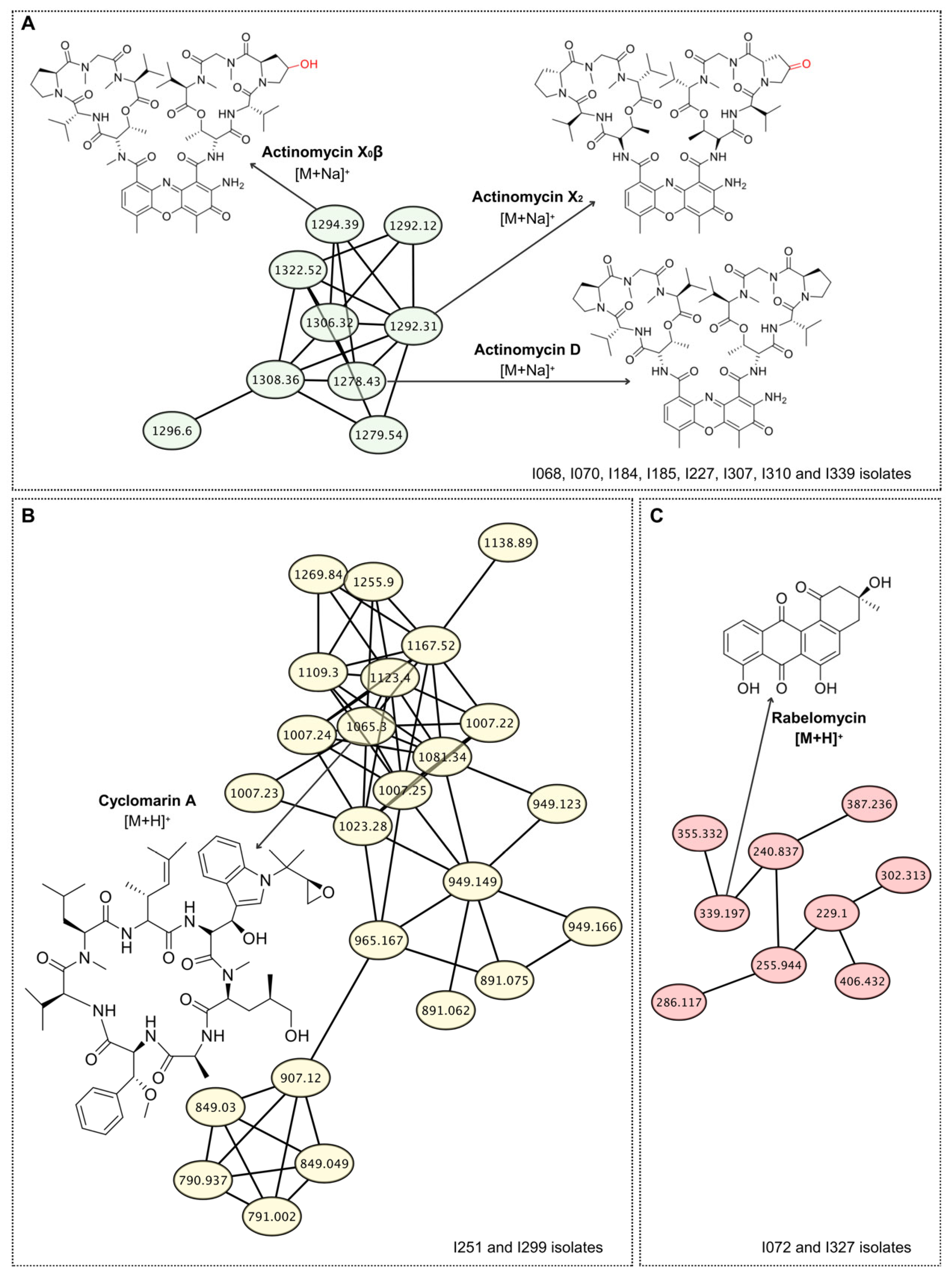
Using this approach, eight Actinomycetota isolates (I068, I070, I184, I185, I227, I307, I310 and I339) were found to produce one or more members of the actinomycin family (Table 1). Actinomycins are chromopeptide antibiotics belonging to the depsipeptide class, characterized by a phenoxazinone chromophore linked to a cyclic peptide lactone [31]. They represent a well-known group of secondary metabolites widely produced by diverse Streptomyces species and are recognized for their potent antibacterial and antitumor properties [32]. Analysis of the mass spectra of the eight isolates confirmed the presence of actinomycin D (m/z 1255.6 [M+H]+), actinomycin X2 (m/z 1269.6 [M+H]+) and actinomycin X0β (m/z 1271.6 [M+H]+) in all samples annotated by GNPS, demonstrating consistency of the dereplication data. Importantly, inspection of the GNPS molecular network revealed six additional clusters of related molecules beyond the three known actinomycins, strongly suggesting that the isolates also produce actinomycin D analogues (Figure 2A). This finding not only underscores the metabolic richness of the strains but also highlights their potential as a valuable source of structurally diverse and bioactive natural products with relevance for antibiotic discovery.
Although actinomycins are well known for their antibacterial activity against Gram-positive bacteria, such as S. aureus, only isolate I339 displayed inhibitory activity in the agar plug test (Table 1). This discrepancy may be related to the amount and specific type of actinomycin produced by different Streptomyces species. For example, actinomycin D exhibits approximately two-fold-lower potency against S. aureus compared to actinomycin X2, highlighting that structural variants can significantly influence antibacterial efficacy [33].
The eight Actinomycetota isolates producing actinomycins were active against M. tuberculosis, whereas only five of them exhibited activity against M. smegmatis. In this context, the identification of actinomycin-producing Streptomyces isolates presents a promising avenue for the discovery of new anti-TB agents. Nonetheless, to translate these findings into viable treatments, it is imperative to focus on the development of actinomycin analogs with reduced toxicity, thereby expanding their clinical applicability and contributing to the global effort against drug-resistant TB.
Extracts from isolates I251 and I299 displayed spectra consistent with cyclomarin A, and molecular networking analysis further grouped additional related spectra (Figure 2B), suggesting that these isolates may produce not only cyclomarin A but also novel structural analogues of this molecule. Moreover, both extracts showed selective inhibition of M. tuberculosis growth, reinforcing cyclomarin-type metabolites as highly promising scaffolds for the development of next-generation anti-tubercular drugs. Cyclomarin A is a cyclic heptapeptide originally isolated from a marine Streptomyces species and belongs to the family of nonribosomal peptides characterized by unusual amino acid residues and structural complexity [34]. It has attracted considerable attention due to its potent and selective antimycobacterial activity, particularly against M. tuberculosis [35]. Mechanistic studies revealed that cyclomarin A targets the essential protease ClpC1, a component of the caseinolytic protease complex, leading to dysregulation of protein homeostasis and bacterial death [36]. To our knowledge, this is the first report of cyclomarin A detection in terrestrial Actinomycetota, highlighting the Caatinga biome as a promising reservoir of novel bioactive scaffolds for anti-tubercular drug discovery.
Rabelomycin, a member of the aromatic polyketide angucycline family, is characterized by its benz[a]anthracene tetracyclic skeleton. It is generally considered an intermediate or shunt product in the biosynthesis of more complex angucyclines, such as urdamycins and landomycins [37,38]. Although not widely recognized for potent antibacterial activity on its own, rabelomycin exhibits significant cytotoxic and antiproliferative properties, making it a compound of potential interest in cancer research [39]. Its frequent detection in metabolomic studies also establishes it as a reliable chemotaxonomic marker for angucycline-producing Actinomycetota [38]. In this study, two isolates, I072 and I327, displayed mass spectra consistent with rabelomycin, as well as other related angucycline compounds, as evidenced by the GNPS molecular network (Figure 2C). This finding corroborates the conserved presence of angucycline-type metabolites in Streptomyces isolates and highlights the genus’s potential as a reservoir for structurally diverse bioactive molecules. Both isolates, I072 and I327, exhibited a broad-spectrum antimicrobial profile, inhibiting the growth of Gram-positive and Gram-negative bacteria, as well as C. albicans. This pattern aligns with the known activity of angucyclines, such as rabelomycin, which have demonstrated antibacterial effects against Gram-positive bacteria [40]. While rabelomycin’s antifungal activity is less documented, some studies have reported antifungal properties in related compounds [41].
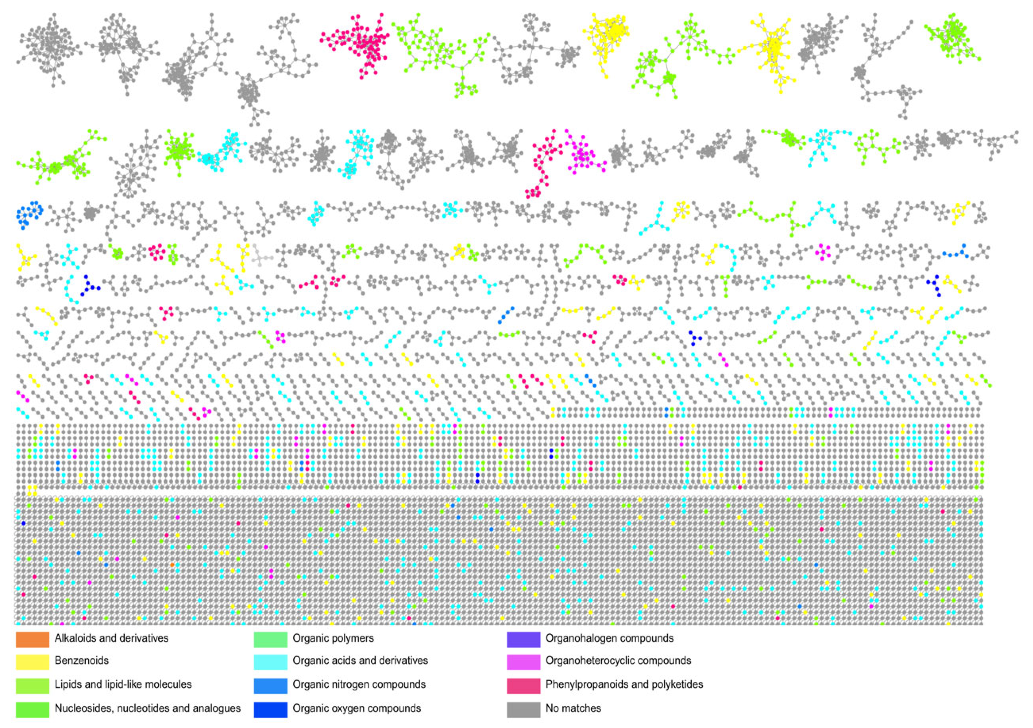
Notably, the majority of molecular families detected in the GNPS-generated network could not be assigned to known compounds (Figure 3). This pattern closely mirrors findings from Ribeiro et al. (2020), where, after dereplication of 26 Actinomycetota extracts exhibiting antimicrobial or cytotoxic activity, 19 showed no database matches; further molecular networking revealed 11 clusters unique to single extracts, many of which were considered prime candidates for novel natural products [42]. Likewise, in our study, most of the detected molecular clusters could not be annotated based on existing spectral libraries (dereplication-resistant compounds), underscoring the presence of chemically uncharted molecular families and reinforcing the potential of the Caatinga biome as a repository of potential and strong targets for the future isolation and elucidation of new natural products.

4. Conclusions

Our findings firmly establish the Brazilian Caatinga biome as an untapped reservoir of bioactive Actinomycetota. The high recovery rate of isolates exhibiting potent and selective activity against WHO priority pathogens, including M. tuberculosis, underscores the strategic value of bioprospecting in extreme environments. While taxonomic characterization revealed a likely dominance of Streptomyces, the limited database matches suggest a wealth of novel diversity awaiting description.
Critically, our integrated approach, which combines classical microbiology with modern metabolomics, proved highly effective. GNPS molecular networking not only suggested the production of potent known antibiotics like actinomycins and the anti-TB cyclomarin A but, more importantly, revealed a vast uncharted chemical landscape dominated by molecular families with no match to existing databases.
This work provides a robust foundation for future drug discovery efforts. The prioritized isolate collection represents a high-value starting point for the isolation and structural elucidation of novel anti-infective agents, directly contributing to the global arsenal against antimicrobial resistance.

Supplementary Materials

The following supporting information can be downloaded at https://www.mdpi.com/article/10.3390/life15101494/s1, Figure S1: Study area, climatic features, and Actinomycetota isolation from Caatinga soils.; Figure S2: Number of Actinomycetota colonies recovered from each soil sample (#1–#6).; Figure S3: Actinomycetota isolates.

Author Contributions

S.P.F., L.L.C.d.A., A.S.A.d.A., T.A.d.S., F.C.Z.V., V.d.S.R.-J. and S.P.C. contributed to the conceptualization and methodology. Formal analysis was performed by S.P.C., E.F.d.S., V.d.S.R.-J., L.S.A. and Y.M.N. Data curation and visualization were conducted by S.P.C., T.A.d.S. and V.d.S.R.-J. The original draft was written by S.P.F., L.L.C.d.A., S.P.C. and V.d.S.R.-J. Supervision was provided by J.F.T., A.L.B. and D.A.M.d.A., and project administration was carried out by S.P.C. and V.d.S.R.-J. All authors have read and agreed to the published version of the manuscript.

Funding

This project was supported by the National Institutes of Science and Technology Program (INCT-RENNOFITO, No. 465536/2014-0). DAMA and JFT are fellows of the National Council for Scientific and Technological Development (CNPq) (#310187/2022-2 and 306633/2021-3, respectively). This study was financed in part by the Coordenação de Aperfeiçoamento de Pessoal de Nível Superior—Brazil (CAPES)—Finance Code 001. None of the authors have any financial disclosures.

Institutional Review Board Statement

Not applicable.

Informed Consent Statement

Not applicable.

Acknowledgments

We especially acknowledge Tereza Cristina Grisi and Renata Almeida for their help in Actinomycetota isolation procedures. Special thanks to Ubiratan Ribeiro-Filho and Monalisa Mercês for maintaining pure cultures. We acknowledge Lucas Lima III and Matheus Fabião for helping in microbial isolation processes and Anauara Lima for helping in producing the precipitation data plot and microscopy images. We gratefully acknowledge the financial support provided by the Instituto Nacional de Pesquisa em Resistência Antimicrobiana (INPRA)—Brazil, through grants from CNPq (INCT 465718/2014-0) and FAPERGS (INCT 17/2551-0000514-7). We acknowledge our team’s diversity, including LGBTQIA+ individuals, women, and underrepresented groups, reflecting our commitment to equity and inclusion.

Conflicts of Interest

The authors declare no conflicts of interest.

References

  1. Murray, C.J.L.; Ikuta, K.S.; Sharara, F.; Swetschinski, L.; Robles Aguilar, G.; Gray, A.; Han, C.; Bisignano, C.; Rao, P.; Wool, E.; et al. Global Burden of Bacterial Antimicrobial Resistance in 2019: A Systematic Analysis. Lancet 2022, 399, 629–655. [Google Scholar] [CrossRef] [PubMed]
  2. Rodriguez, B.; Prinzi, A.M.; Hill, B.K.; Tibbetts, R.; Salazar, H.; McAdams, D.; Rivera-Acosta, A.M.; Wungwattana, M.; Silbert, S.; Niles, D.T. Uniting Disciplines against Antimicrobial Resistance (AMR): Highlights from a Multidisciplinary Inaugural AMR Summit. Antimicrob. Steward. Healthc. Epidemiol. 2025, 5, e149. [Google Scholar] [CrossRef] [PubMed]
  3. van Bergeijk, D.A.; Terlouw, B.R.; Medema, M.H.; van Wezel, G.P. Ecology and Genomics of Actinobacteria: New Concepts for Natural Product Discovery. Nat. Rev. Microbiol. 2020, 18, 546–558. [Google Scholar] [CrossRef]
  4. Bérdy, J. Thoughts and Facts about Antibiotics: Where We Are Now and Where We Are Heading. J. Antibiot. 2012, 65, 385–395. [Google Scholar] [CrossRef]
  5. Hug, J.; Bader, C.; Remškar, M.; Cirnski, K.; Müller, R. Concepts and Methods to Access Novel Antibiotics from Actinomycetes. Antibiotics 2018, 7, 44. [Google Scholar] [CrossRef]
  6. Pylro, V.S.; Roesch, L.F.W.; Ortega, J.M.; do Amaral, A.M.; Tótola, M.R.; Hirsch, P.R.; Rosado, A.S.; Góes-Neto, A.; da Costa da Silva, A.L.; Rosa, C.A.; et al. Brazilian Microbiome Project: Revealing the Unexplored Microbial Diversity—Challenges and Prospects. Microb. Ecol. 2014, 67, 237–241. [Google Scholar] [CrossRef]
  7. Waksman, S. The Actinomycetes; The Williams & Wilkins Company: Baltimore, MD, USA, 1961; Volume II. [Google Scholar]
  8. Balouiri, M.; Sadiki, M.; Ibnsouda, S.K. Methods for in Vitro Evaluating Antimicrobial Activity: A Review. J. Pharm. Anal. 2016, 6, 71–79. [Google Scholar] [CrossRef]
  9. Sidrônio, M.G.S.; Castelo Branco, A.P.O.T.; Abbadi, B.L.; Macchi, F.; Silveira, M.D.; Lock, G.d.A.; Costa, T.D.; de Araújo, D.M.; Cibulski, S.; Bizarro, C.V.; et al. Effects of Tafenoquine against Active, Dormant and Resistant Mycobacterium Tuberculosis. Tuberculosis 2021, 128, 102089. [Google Scholar] [CrossRef]
  10. Muradás, T.C.; Abbadi, B.L.; Villela, A.D.; Macchi, F.S.; Bergo, P.F.; de Freitas, T.F.; Sperotto, N.D.M.; Timmers, L.F.S.M.; Norberto de Souza, O.; Picada, J.N.; et al. Pre-Clinical Evaluation of Quinoxaline-Derived Chalcones in Tuberculosis. PLoS ONE 2018, 13, e0202568. [Google Scholar] [CrossRef]
  11. Wang, M.; Carver, J.J.; Phelan, V.V.; Sanchez, L.M.; Garg, N.; Peng, Y.; Nguyen, D.D.; Watrous, J.; Kapono, C.A.; Luzzatto-Knaan, T.; et al. Sharing and Community Curation of Mass Spectrometry Data with Global Natural Products Social Molecular Networking. Nat. Biotechnol. 2016, 34, 828–837. [Google Scholar] [CrossRef]
  12. Ernst, M.; Kang, K.B.; Caraballo-Rodríguez, A.M.; Nothias, L.-F.; Wandy, J.; Chen, C.; Wang, M.; Rogers, S.; Medema, M.H.; Dorrestein, P.C.; et al. MolNetEnhancer: Enhanced Molecular Networks by Integrating Metabolome Mining and Annotation Tools. Metabolites 2019, 9, 144. [Google Scholar] [CrossRef] [PubMed]
  13. Caatinga; Silva, J.M.C.d., Leal, I.R., Tabarelli, M., Eds.; Springer International Publishing: Cham, Switzerland, 2017; ISBN 978-3-319-68338-6. [Google Scholar]
  14. Naylor, D.; DeGraaf, S.; Purdom, E.; Coleman-Derr, D. Drought and Host Selection Influence Bacterial Community Dynamics in the Grass Root Microbiome. ISME J. 2017, 11, 2691–2704. [Google Scholar] [CrossRef] [PubMed]
  15. Marasco, R.; Fusi, M.; Rolli, E.; Ettoumi, B.; Tambone, F.; Borin, S.; Ouzari, H.; Boudabous, A.; Sorlini, C.; Cherif, A.; et al. Aridity Modulates Belowground Bacterial Community Dynamics in Olive Tree. Environ. Microbiol. 2021, 23, 6275–6291. [Google Scholar] [CrossRef] [PubMed]
  16. Rasuk, M.C.; Ferrer, G.M.; Kurth, D.; Portero, L.R.; Farías, M.E.; Albarracín, V.H. UV-Resistant Actinobacteria from High-Altitude Andean Lakes: Isolation, Characterization and Antagonistic Activities. Photochem. Photobiol. 2017, 93, 865–880. [Google Scholar] [CrossRef]
  17. Xie, F.; Pathom-aree, W. Actinobacteria from Desert: Diversity and Biotechnological Applications. Front. Microbiol. 2021, 12, 765531. [Google Scholar] [CrossRef]
  18. Mohammadipanah, F.; Wink, J. Actinobacteria from Arid and Desert Habitats: Diversity and Biological Activity. Front. Microbiol. 2015, 6, 1541. [Google Scholar] [CrossRef]
  19. Rateb, M.E.; Ebel, R.; Jaspars, M. Natural Product Diversity of Actinobacteria in the Atacama Desert. Antonie Leeuwenhoek 2018, 111, 1467–1477. [Google Scholar] [CrossRef]
  20. Vásquez-Dean, J.; Maza, F.; Morel, I.; Pulgar, R.; González, M. Microbial Communities from Arid Environments on a Global Scale. A Systematic Review. Biol. Res. 2020, 53, 29. [Google Scholar] [CrossRef]
  21. Bouskill, N.J.; Lim, H.C.; Borglin, S.; Salve, R.; Wood, T.E.; Silver, W.L.; Brodie, E.L. Pre-Exposure to Drought Increases the Resistance of Tropical Forest Soil Bacterial Communities to Extended Drought. ISME J. 2013, 7, 384–394. [Google Scholar] [CrossRef]
  22. Sati, H.; Carrara, E.; Savoldi, A.; Hansen, P.; Garlasco, J.; Campagnaro, E.; Boccia, S.; Castillo-Polo, J.A.; Magrini, E.; Garcia-Vello, P.; et al. The WHO Bacterial Priority Pathogens List 2024: A Prioritisation Study to Guide Research, Development, and Public Health Strategies against Antimicrobial Resistance. Lancet Infect. Dis. 2025, 25, 1033–1043. [Google Scholar] [CrossRef]
  23. Mishra, S.K.; Yasir, M.; Willcox, M. Candida auris: An Emerging Antimicrobial-Resistant Organism with the Highest Level of Concern. Lancet Microbe 2023, 4, e482–e483. [Google Scholar] [CrossRef]
  24. de Almeida, L.L.C.; Fernandes, S.P.; de Oliveira, G.D.; da Silveira Silva, M.; de Souza, T.A.; Rodrigues-Junior, V.S.; Cibulski, S.P. Harnessing Actinobacteria Secondary Metabolites for Tuberculosis Drug Discovery: Historical Trends, Current Status and Future Outlooks. Nat. Prod. Bioprospect. 2025, 15, 52. [Google Scholar] [CrossRef] [PubMed]
  25. Lelovic, N.; Mitachi, K.; Yang, J.; Lemieux, M.R.; Ji, Y.; Kurosu, M. Application of Mycobacterium Smegmatis as a Surrogate to Evaluate Drug Leads against Mycobacterium Tuberculosis. J. Antibiot. 2020, 73, 780–789. [Google Scholar] [CrossRef] [PubMed]
  26. Boone, D.R.; Castenholz, R.W.; Garrity, G.M. (Eds.) Bergey’s Manual of Systematic Bacteriology, 2nd ed.; Springer: New York, NY, USA, 2001; Volume 1, ISBN 0387987711. [Google Scholar]
  27. Tsuchida, S.; Umemura, H.; Nakayama, T. Current Status of Matrix-Assisted Laser Desorption/Ionization–Time-of-Flight Mass Spectrometry (MALDI-TOF MS) in Clinical Diagnostic Microbiology. Molecules 2020, 25, 4775. [Google Scholar] [CrossRef] [PubMed]
  28. Ashfaq, M.Y.; Da’na, D.A.; Al-Ghouti, M.A. Application of MALDI-TOF MS for Identification of Environmental Bacteria: A Review. J. Environ. Manag. 2022, 305, 114359. [Google Scholar] [CrossRef]
  29. Zuffa, S.; Schmid, R.; Bauermeister, A.; Gomes, P.W.P.; Caraballo-Rodriguez, A.M.; El Abiead, Y.; Aron, A.T.; Gentry, E.C.; Zemlin, J.; Meehan, M.J.; et al. MicrobeMASST: A Taxonomically Informed Mass Spectrometry Search Tool for Microbial Metabolomics Data. Nat. Microbiol. 2024, 9, 336–345. [Google Scholar] [CrossRef]
  30. Gaudry, A.; Marcourt, L.; Kaiser, M.; Flückiger, J.; David, B.; Grondin, A.; Ioset, J.-R.; Mäser, P.; Queiroz, E.F.; Allard, P.-M.; et al. Efficient Constitution of a Library of Rotenoid Analogs Active against Trypanosoma cruzi from a Digitalized Plant Extract Collection. RSC Adv. 2025, 15, 15240–15251. [Google Scholar] [CrossRef]
  31. Gaudry, A.; Pagni, M.; Mehl, F.; Moretti, S.; Quiros-Guerrero, L.-M.; Cappelletti, L.; Rutz, A.; Kaiser, M.; Marcourt, L.; Queiroz, E.F.; et al. A Sample-Centric and Knowledge-Driven Computational Framework for Natural Products Drug Discovery. ACS Cent. Sci. 2024, 10, 494–510. [Google Scholar] [CrossRef]
  32. Liu, M.; Jia, Y.; Xie, Y.; Zhang, C.; Ma, J.; Sun, C.; Ju, J. Identification of the Actinomycin D Biosynthetic Pathway from Marine-Derived Streptomyces costaricanus SCSIO ZS0073. Mar. Drugs 2019, 17, 240. [Google Scholar] [CrossRef]
  33. Lo, Y.-S.; Tseng, W.-H.; Chuang, C.-Y.; Hou, M.-H. The Structural Basis of Actinomycin D–Binding Induces Nucleotide Flipping out, a Sharp Bend and a Left-Handed Twist in CGG Triplet Repeats. Nucleic Acids Res. 2013, 41, 4284–4294. [Google Scholar] [CrossRef]
  34. Wang, D.; Wang, C.; Gui, P.; Liu, H.; Khalaf, S.M.H.; Elsayed, E.A.; Wadaan, M.A.M.; Hozzein, W.N.; Zhu, W. Identification, Bioactivity, and Productivity of Actinomycins from the Marine-Derived Streptomyces heliomycini. Front. Microbiol. 2017, 8, 1147. [Google Scholar] [CrossRef]
  35. Schultz, A.W.; Oh, D.-C.; Carney, J.R.; Williamson, R.T.; Udwary, D.W.; Jensen, P.R.; Gould, S.J.; Fenical, W.; Moore, B.S. Biosynthesis and Structures of Cyclomarins and Cyclomarazines, Prenylated Cyclic Peptides of Marine Actinobacterial Origin. J. Am. Chem. Soc. 2008, 130, 4507–4516. [Google Scholar] [CrossRef]
  36. Schmitt, E.K.; Riwanto, M.; Sambandamurthy, V.; Roggo, S.; Miault, C.; Zwingelstein, C.; Krastel, P.; Noble, C.; Beer, D.; Rao, S.P.S.; et al. The Natural Product Cyclomarin Kills Mycobacterium Tuberculosis by Targeting the ClpC1 Subunit of the Caseinolytic Protease. Angew. Chem. Int. Ed. 2011, 50, 5889–5891. [Google Scholar] [CrossRef] [PubMed]
  37. Vasudevan, D.; Rao, S.P.S.; Noble, C.G. Structural Basis of Mycobacterial Inhibition by Cyclomarin A. J. Biol. Chem. 2013, 288, 30883–30891. [Google Scholar] [CrossRef] [PubMed]
  38. Liu, W.-C.; Parker, W.L.; Slusarchyk, D.S.; Greenwood, G.L.; Graham, S.F.; Meyers, E. Isolation, characterization, and structure of rabelomycin, a new antibiotic. J. Antibiot. 1970, 23, 437–441. [Google Scholar] [CrossRef] [PubMed]
  39. Kharel, M.K.; Pahari, P.; Shepherd, M.D.; Tibrewal, N.; Nybo, S.E.; Shaaban, K.A.; Rohr, J. Angucyclines: Biosynthesis, Mode-of-Action, New Natural Products, and Synthesis. Nat. Prod. Rep. 2012, 29, 264–325. [Google Scholar] [CrossRef]
  40. Rohr, J.; Thiericke, R. Angucycline Group Antibiotics. Nat. Prod. Rep. 1992, 9, 103. [Google Scholar] [CrossRef]
  41. Liu, H.-S.; Chen, H.-R.; Huang, S.-S.; Li, Z.-H.; Wang, C.-Y.; Zhang, H. Bioactive Angucyclines/Angucyclinones Discovered from 1965 to 2023. Mar. Drugs 2025, 23, 25. [Google Scholar] [CrossRef]
  42. Ribeiro, I.; Girão, M.; Alexandrino, D.A.M.; Ribeiro, T.; Santos, C.; Pereira, F.; Mucha, A.P.; Urbatzka, R.; Leão, P.N.; Carvalho, M.F. Diversity and Bioactive Potential of Actinobacteria Isolated from a Coastal Marine Sediment in Northern Portugal. Microorganisms 2020, 8, 1691. [Google Scholar] [CrossRef]
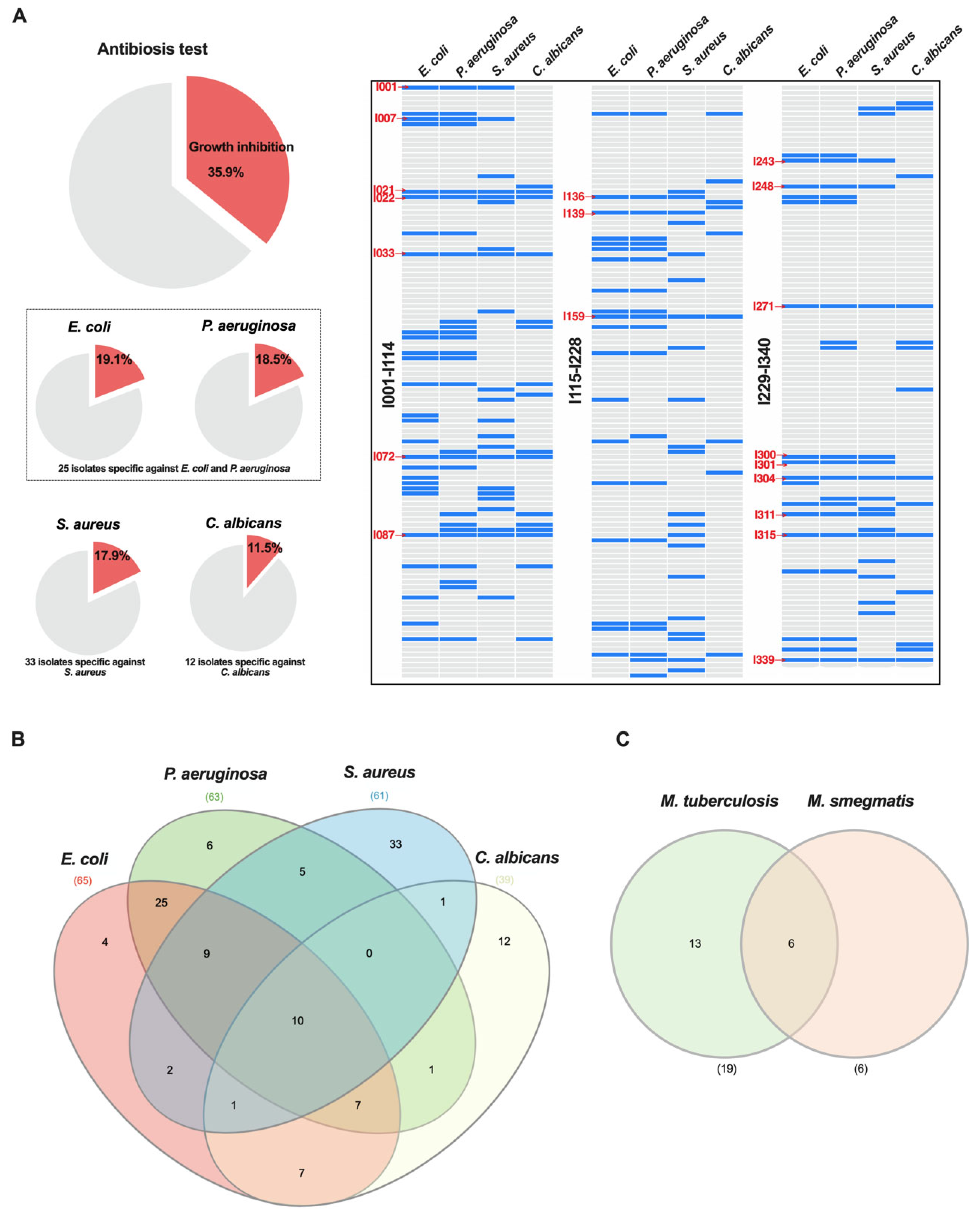
Figure 1. Antimicrobial activity and of Actinomycetota isolates from Caatinga soils. (A) Antimicrobial activity of 340 Actinomycetota isolates against E. coli, P. aeruginosa, S. aureus, and C. albicans. Pie charts indicate the percentage of isolates exhibiting inhibitory activity for each indicator microorganism. Heatmaps show the activity profile of the isolates (I001-340), with blue bars representing growth inhibition. Red labels indicate isolates with broad activity against both Gram-positive and -negative pathogens. (B) Venn diagram depicting the overlap of Actinomycetota isolates active against the four tested microorganisms. Numbers indicate the quantity of isolates inhibiting one or more pathogens. (C) Venn diagram showing isolates with antimycobacterial activity.
Life 15 01494 g001
Figure 2. Molecular networking unveils the specialized metabolome of Actinomycetota strains, highlighting known and variant antibiotics. LC-MS/MS data from Actinomycetota extracts were processed using GNPS to generate molecular families based on MS/MS spectral similarity. (A) A cluster annotated to the actinomycin family, with nodes corresponding to actinomycin D, X0β, and X2, identified by spectral matching (cosine score > 0.7). (B) A distinct molecular family led to the identification of the antimycobacterial cyclic peptide cyclomarin A. (C) Annotation of a cluster as the angucycline-class antibiotic rabelomycin, suggesting the potential for related derivatives. All annotations were confirmed by comparison to reference MS/MS spectra from public spectral libraries.
Life 15 01494 g002
Figure 3. Molecular networking analysis and chemical annotation of specialized metabolites from Actinomycetota extracts. Molecular networks were generated from LC-MS/MS data processed using GNPS. Nodes represent molecular families (clusters) of MS/MS spectra grouped by structural similarity. Chemical annotation was performed using the MolNetEnhancer workflow, which integrates: (i) spectral library matching, (ii) in silico molecular class prediction via CANOPUS (Class Assignment and Ontology Prediction Using Mass Spectrometry), and (iii) chemical substructure annotation via NAP (Network Annotation Propagation). Node colors represent the major chemical classes identified, highlighting a diverse metabolome that includes several clusters of specialized metabolites putatively annotated.
Life 15 01494 g003
Disclaimer/Publisher’s Note: The statements, opinions and data contained in all publications are solely those of the individual author(s) and contributor(s) and not of MDPI and/or the editor(s). MDPI and/or the editor(s) disclaim responsibility for any injury to people or property resulting from any ideas, methods, instructions or products referred to in the content.