

Longitudinal Changes of Cornea Volume Measured by Means of Anterior Segment-Optical Coherence Tomography in Patients with Stable and Progressive Keratoconus


 and
and 
Abstract
1. Introduction
2. Materials and Methods
Statistical Methods
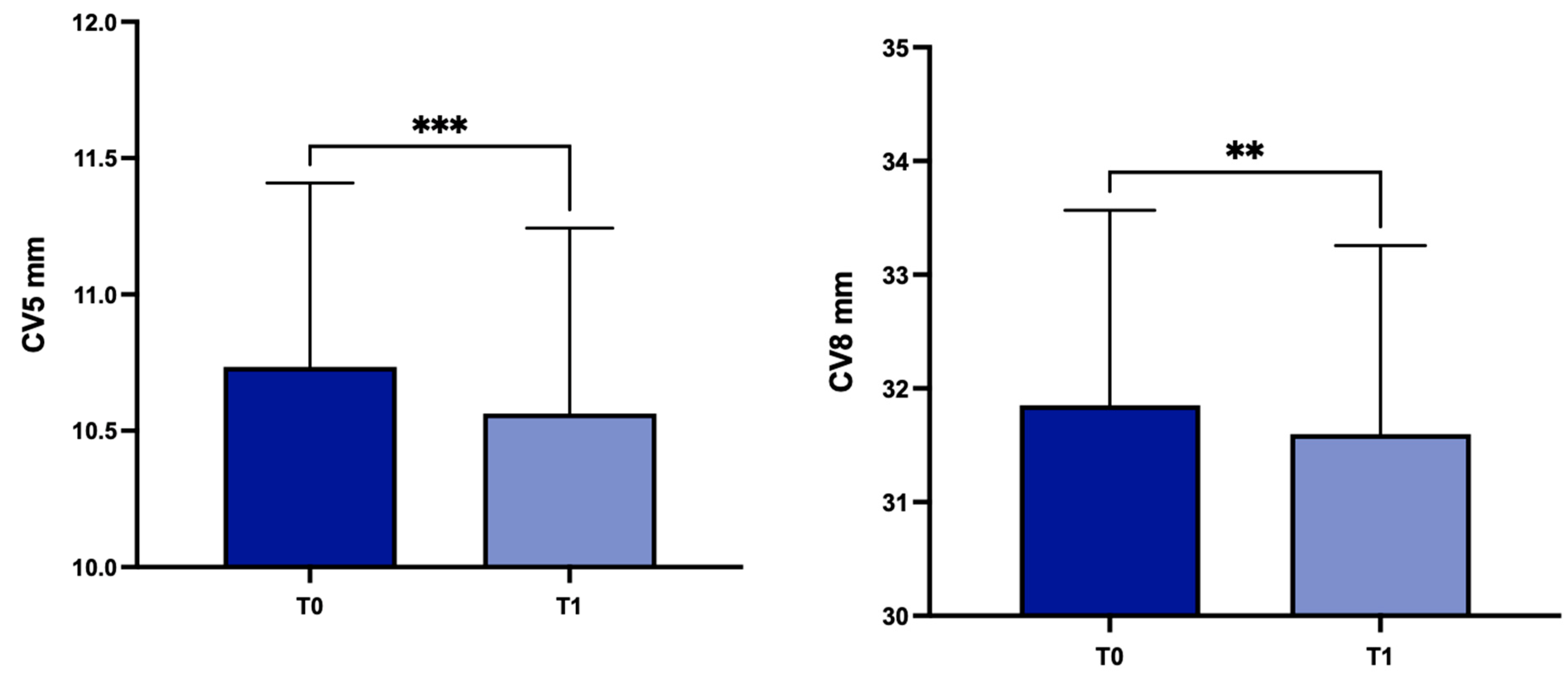
3. Results
4. Discussion
Author Contributions
Funding
Institutional Review Board Statement
Informed Consent Statement
Data Availability Statement
Conflicts of Interest
References
- Rabinowitz, Y.S.; Galvis, V.; Tello, A.; Rueda, D.; García, J.D. Genetics vs chronic corneal mechanical trauma in the etiology of keratoconus. Exp. Eye Res. 2021, 202, 108328. [Google Scholar] [CrossRef]
- Bui, A.D.; Truong, A.; Pasricha, N.D.; Indaram, M. Keratoconus Diagnosis and Treatment: Recent Advances and Future Directions. Clin. Ophthalmol. 2023, 17, 2705–2718. [Google Scholar] [CrossRef] [PubMed]
- Galvis, V.; Tello, A.; Barrera, R.; Niño, C.A. Inflammation in Keratoconus. Cornea 2015, 34, e22–e23. [Google Scholar] [CrossRef]
- Elbeyli, A.; Kurtul, B.E. Systemic immune-inflammation index, neutrophil-to-lymphocyte ratio, and platelet-to-lymphocyte ratio levels are associated with keratoconus. Indian J. Ophthalmol. 2021, 69, 1725–1729. [Google Scholar]
- Loh, I.P.; Sherwin, T. Is Keratoconus an Inflammatory Disease? The Implication of Inflammatory Pathways. Ocul. Immunol. Inflamm. 2022, 30, 246–255. [Google Scholar] [CrossRef] [PubMed]
- Wei, R.H.; Zhao, S.Z.; Lim, L.; Tan, D.T. Incidence and characteristics of unilateral keratoconus classified on corneal topography. J. Refract. Surg. 2011, 10, 745–751. [Google Scholar] [CrossRef]
- McMahon, T.T.; Szczotka-Flynn, L.; Barr, J.T.; Anderson, R.J.; Slaughter, M.E.; Lass, J.H.; Iyengar, S.K. CLEK Study Group. A new method for grading the severity of keratoconus: The Keratoconus Severity Score (KSS). Cornea 2006, 7, 794–800. [Google Scholar] [CrossRef]
- Krachmer, J.H.; Feder, R.S.; Belin, M.W. Keratoconus and related noninflammatory corneal thinning disorders. Surv. Ophthalmol. 1984, 28, 293–322. [Google Scholar] [CrossRef] [PubMed]
- Gomes, J.A.; Tan, D.; Rapuano, C.J.; Belin, M.W.; Ambrósio, R., Jr.; Guell, J.L.; Malecaze, F.; Nishida, K.; Sangwan, V.S. Group of Panelists for the Global Delphi Panel of Keratoconus and Ectatic Diseases. Global consensus on keratoconus and ectatic diseases. Cornea 2015, 34, 359–369. [Google Scholar] [CrossRef]
- Andreassen, T.T.; Simonsen, A.H.; Oxlund, H. Biomechanical properties of keratoconus and normal corneas. Exp. Eye Res. 1980, 31, 435–441. [Google Scholar] [CrossRef]
- Morishige, N.; Shin-Gyou-Uchi, R.; Azumi, H.; Ohta, H.; Morita, Y.; Yamada, N.; Kimura, K.; Takahara, A.; Sonoda, K.H. Quantitative analysis of collagen lamellae in the normal and keratoconic human cornea by second harmonic generation imaging microscopy. Investig. Ophthalmol. Vis. Sci. 2014, 55, 8377–8385. [Google Scholar] [CrossRef] [PubMed]
- Meek, K.M.; Tuft, S.J.; Huang, Y.; Gill, P.S.; Hayes, S.; Newton, R.H.; Bron, A.J. Changes in collagen orientation and distribution in keratoconus corneas. Investig. Ophthalmol. Vis. Sci. 2005, 46, 1948–1956. [Google Scholar] [CrossRef] [PubMed]
- Hayes, S.; Boote, C.; Tuft, S.J.; Quantock, A.J.; Meek, K.M. A study of corneal thickness, shape and collagen organisation in keratoconus using videokeratography and X-ray scattering techniques. Exp. Eye Res. 2007, 84, 423–434. [Google Scholar] [CrossRef] [PubMed]
- Balasubramanian, S.A.; Pye, D.C.; Willcox, M.D. Are proteinases the reason for keratoconus? Curr. Eye Res. 2010, 35, 185–191. [Google Scholar] [CrossRef]
- Foster, J.W.; Parikh, R.N.; Wang, J.; Bower, K.S.; Matthaei, M.; Chakravarti, S.; Jun, A.S.; Eberhart, C.G.; Soiberman, U.S. Transcriptomic and Immunohistochemical Analysis of Progressive Keratoconus Reveal Altered WNT10A in Epithelium and Bowman’s Layer. Investig. Ophthalmol. Vis. Sci. 2021, 62, 16. [Google Scholar] [CrossRef] [PubMed]
- Akoto, T.; Cai, J.; Nicholas, S.; McCord, H.; Estes, A.J.; Xu, H.; Karamichos, D.; Liu, Y. Unravelling the Impact of Cyclic Mechanical Stretch in Keratoconus-A Transcriptomic Profiling Study. Int. J. Mol. Sci. 2023, 24, 7437. [Google Scholar] [CrossRef] [PubMed]
- Giannaccare, G.; Murano, G.; Carnevali, A.; Yu, A.C.; Vaccaro, S.; Scuteri, G.; Maltese, L.; Scorcia, V. Comparison of Amsler–Krumeich and Sandali Classifications for Staging Eyes with Keratoconus. Appl. Sci. 2021, 11, 4007. [Google Scholar] [CrossRef]
- Piñero, D.P.; Nieto, J.C.; Lopez-Miguel, A. Characterization of corneal structure in keratoconus. J. Cataract. Refract. Surg. 2012, 38, 2167–2183. [Google Scholar] [CrossRef]
- Ahmadi Hosseini, S.M.; Mohidin, N.; Abolbashari, F.; Mohd-Ali, B.; Santhirathelagan, C.T. Corneal thickness and volume in subclinical and clinical keratoconus. Int. Ophthalmol. 2013, 33, 139–145. [Google Scholar] [CrossRef]
- Cerviño, A.; Gonzalez-Meijome, J.M.; Ferrer-Blasco, T.; Garcia-Resua, C.; Montes-Mico, R.; Parafita, M. Determination of corneal volume from anterior topography and topographic pachymetry: Application to healthy and keratoconic eyes. Ophthalmic Physiol. Opt. 2009, 29, 652–660. [Google Scholar] [CrossRef]
- Yadav, R.; Kottaiyan, R.; Ahmad, K.; Yoon, G. Epithelium and Bowman’s layer thickness and light scatter in keratoconic cornea evaluated using ultrahigh resolution optical coherence tomography. J. Biomed. Opt. 2012, 11, 116010. [Google Scholar] [CrossRef] [PubMed]
- Sandali, O.; El Sanharawi, M.; Temstet, C.; Hamiche, T.; Galan, A.; Ghouali, W.; Goemaere, I.; Basli, E.; Borderie, V.; Laroche, L. Fourier-domain optical coherence tomography imaging in keratoconus: A corneal structural classification. Ophthalmology 2013, 120, 2403–2412. [Google Scholar] [CrossRef]
- Fukuda, R.; Usui, T.; Miyai, T.; Mori, Y.; Miyata, K.; Amano, S. Corneal thickness and volume measurements by swept source anterior segment optical coherence tomography in normal subjects. Curr. Eye Res. 2013, 38, 531–536. [Google Scholar] [CrossRef] [PubMed]
- Ambrósio, R., Jr.; Alonso, R.S.; Luz, A.; Coca Velarde, L.G. Corneal-thickness spatial profile and corneal-volume distribution: Tomographic indices to detect keratoconus. J. Cataract. Refract. Surg. 2006, 32, 1851–1859. [Google Scholar] [CrossRef]
- Mannion, L.S.; Tromans, C.; O’Donnell, C. Reduction in corneal volume with severity of keratoconus. Curr. Eye Res. 2011, 36, 522–527. [Google Scholar] [CrossRef]
- Piñero, D.P.; Alió, J.L.; Alesón, A.; Escaf Vergara, M.; Miranda, M. Corneal volume, pachymetry, and correlation of anterior and posterior corneal shape in subclinical and different stages of clinical keratoconus. J. Cataract. Refract. Surg. 2010, 36, 814–825. [Google Scholar] [CrossRef]
- Fontes, B.M.; Ambrósio, R., Jr.; Jardim, D.; Velarde, G.C.; Nosé, W. Corneal biomechanical metrics and anterior segment parameters in mild keratoconus. Ophthalmology 2010, 117, 673–679. [Google Scholar] [CrossRef]
- Morishige, N.; Magome, K.; Ueno, A.; Matsui, T.A.; Nishida, T. Relations Among Corneal Curvature, Thickness, and Volume in Keratoconus as Evaluated by Anterior Segment-Optical Coherence Tomography. Investig. Ophthalmol. Vis. Sci. 2019, 60, 3794–3802. [Google Scholar] [CrossRef]
- Chatzis, N.; Hafezi, F. Progression of keratoconus and efficacy of pediatric [corrected] corneal collagen cross-linking in children and adolescents. J. Refract. Surg. 2012, 28, 753–758. [Google Scholar] [CrossRef]
- Gore, D.M.; Shortt, A.J.; Allan, B.D. New clinical pathways for keratoconus. Eye 2013, 27, 329–339. [Google Scholar] [CrossRef] [PubMed]
- Cui, J.; Zhang, X.; Hu, Q.; Zhou, W.Y.; Yang, F. Evaluation of Corneal Thickness and Volume Parameters of Subclinical Keratoconus Using a Pentacam Scheimflug System. Curr. Eye Res. 2016, 41, 923–926. [Google Scholar] [CrossRef]
- Cavas-Martínez, F.; Bataille, L.; Fernández-Pacheco, D.G.; Cañavate, F.J.F.; Alio, J.L. Keratoconus Detection Based on a New Corneal Volumetric Analysis. Sci. Rep. 2017, 7, 15837. [Google Scholar] [CrossRef]
- Lu, N.J.; Hafezi, F.; Koppen, C.; Alió Del Barrio, J.L.; Aslanides, I.M.; Awwad, S.T.; Ní Dhubhghaill, S.; Pineda, R., 2nd; Torres-Netto, E.A.; Wang, L.; et al. New keratoconus staging system based on OCT. J. Cataract. Refract. Surg. 2023, 49, 1098–1105. [Google Scholar] [CrossRef]
- Hu, P.; Lin, L.; Wu, Z.; Jin, X.; Ni, H. Kayser-Fleischer ring with keratoconus: A coincidence? A case report. BMC Ophthalmol. 2020, 20, 190. [Google Scholar] [CrossRef] [PubMed]
- Steinwender, G.; Kollenc, A.; Shajari, M.; Sommer, M.; Borenich, A.; Horwath-Winter, J.; Lindner, E.; Woltsche, N.; List, W.; Wedrich, A. Determining the center of a keratoconus: Comparison of different tomographic parameters and impact of disease severity. Front. Med. 2022, 9, 968318. [Google Scholar] [CrossRef] [PubMed]
- Heidari, Z.; Jafarzadehpour, E.; Mohammadpour, M.; Hashemi, H. Best indices of dual Scheimpflug/Placido tomographer for keratoconus detection. Int. Ophthalmol. 2023, 43, 1353–1362. [Google Scholar] [CrossRef] [PubMed]


| All (n = 116) | Group 1 (n = 72) | Group 2 (n = 34) | Group 3 (n = 10) | |
|---|---|---|---|---|
| Corneal volume 3 mm Ø (mm3) mean ± SD | 3.67 ± 0.29 | 3.77 ± 0.25 | 3.53 ± 0.27 | 3.24 ± 0.28 |
| Corneal volume 5 mm Ø (mm3) mean ± SD | 10.79 ± 0.75 | 11.01 ± 0.70 | 10.55 ± 0.73 | 9.92 ± 0.90 |
| Corneal volume 8 mm Ø (mm3) mean ± SD | 32.05 ± 1.90 | 32.38 ± 1.89 | 31.70 ± 2.01 | 30.68 ± 2.31 |
| Correlation Matrix | |||||
|---|---|---|---|---|---|
| TP | CV 3 mm | CV 5 mm | CV 8 mm | ||
| TP | Pearson’s r | - | |||
| df | - | ||||
| p-value | - | ||||
| CV 3 mm | Pearson’s r | 0.918 | - | ||
| df | 114 | - | |||
| p-value | <0.001 | - | |||
| CV 5 mm | Pearson’s r | 0.891 | 0.971 | - | |
| df | 114 | 114 | - | ||
| p-value | <0.001 | <0.001 | - | ||
| CV 8 mm | Pearson’s r | 0.788 | 0.888 | 0.960 | - |
| df | 114 | 114 | 114 | - | |
| p-value | <0.001 | <0.001 | <0.001 | - | |
Disclaimer/Publisher’s Note: The statements, opinions and data contained in all publications are solely those of the individual author(s) and contributor(s) and not of MDPI and/or the editor(s). MDPI and/or the editor(s) disclaim responsibility for any injury to people or property resulting from any ideas, methods, instructions or products referred to in the content. |
© 2024 by the authors. Licensee MDPI, Basel, Switzerland. This article is an open access article distributed under the terms and conditions of the Creative Commons Attribution (CC BY) license (https://creativecommons.org/licenses/by/4.0/).
Share and Cite
Vaccaro, S.; Vivarelli, C.; Yu, A.C.; Pecora, N.; Lionetti, G.; Gioia, R.; Scorcia, V.; Giannaccare, G. Longitudinal Changes of Cornea Volume Measured by Means of Anterior Segment-Optical Coherence Tomography in Patients with Stable and Progressive Keratoconus. Life 2024, 14, 176. https://doi.org/10.3390/life14020176
Vaccaro S, Vivarelli C, Yu AC, Pecora N, Lionetti G, Gioia R, Scorcia V, Giannaccare G. Longitudinal Changes of Cornea Volume Measured by Means of Anterior Segment-Optical Coherence Tomography in Patients with Stable and Progressive Keratoconus. Life. 2024; 14(2):176. https://doi.org/10.3390/life14020176
Chicago/Turabian StyleVaccaro, Sabrina, Chiara Vivarelli, Angeli Christy Yu, Nicolò Pecora, Giovanna Lionetti, Raffaella Gioia, Vincenzo Scorcia, and Giuseppe Giannaccare. 2024. "Longitudinal Changes of Cornea Volume Measured by Means of Anterior Segment-Optical Coherence Tomography in Patients with Stable and Progressive Keratoconus" Life 14, no. 2: 176. https://doi.org/10.3390/life14020176
APA StyleVaccaro, S., Vivarelli, C., Yu, A. C., Pecora, N., Lionetti, G., Gioia, R., Scorcia, V., & Giannaccare, G. (2024). Longitudinal Changes of Cornea Volume Measured by Means of Anterior Segment-Optical Coherence Tomography in Patients with Stable and Progressive Keratoconus. Life, 14(2), 176. https://doi.org/10.3390/life14020176