Correction: Ulman-Macón et al. Morphological Changes of the Suboccipital Musculature in Women with Myofascial Temporomandibular Pain: A Case-Control Study. Life 2023, 13, 1159


 , , and
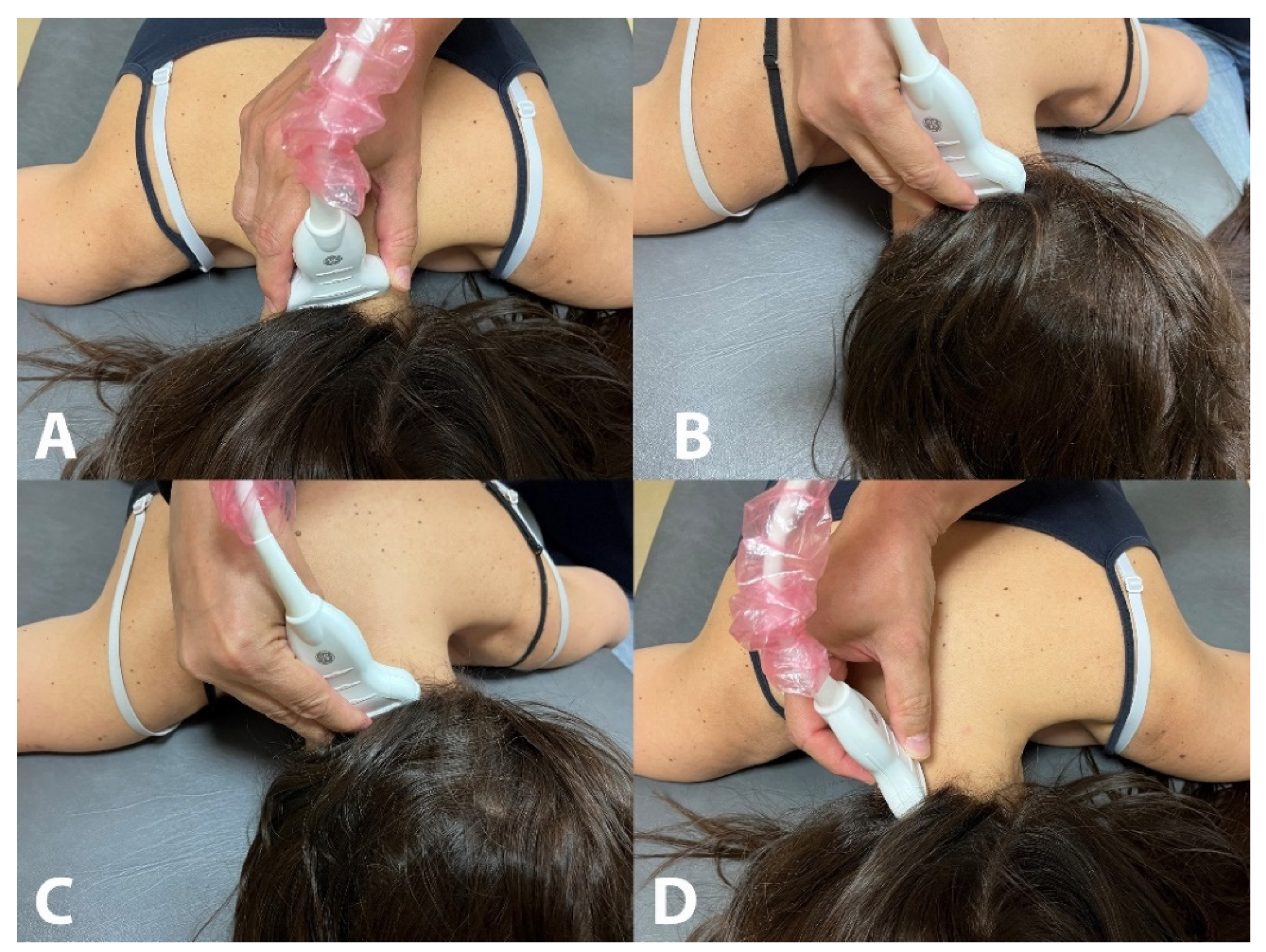
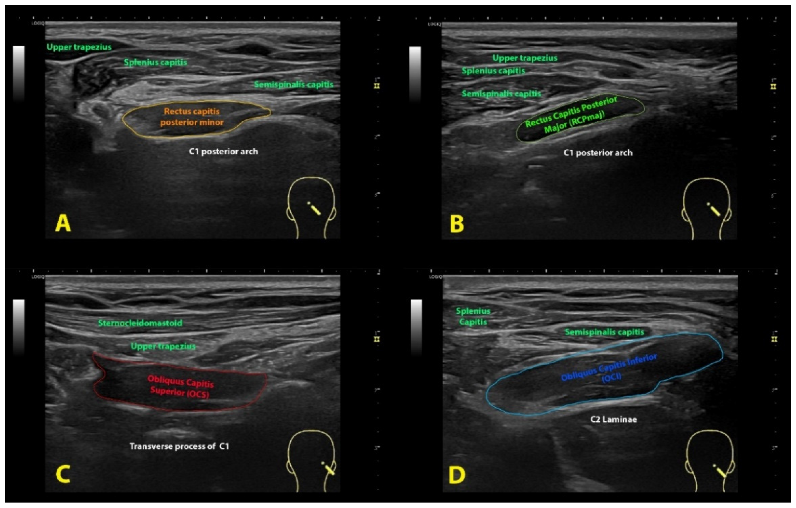
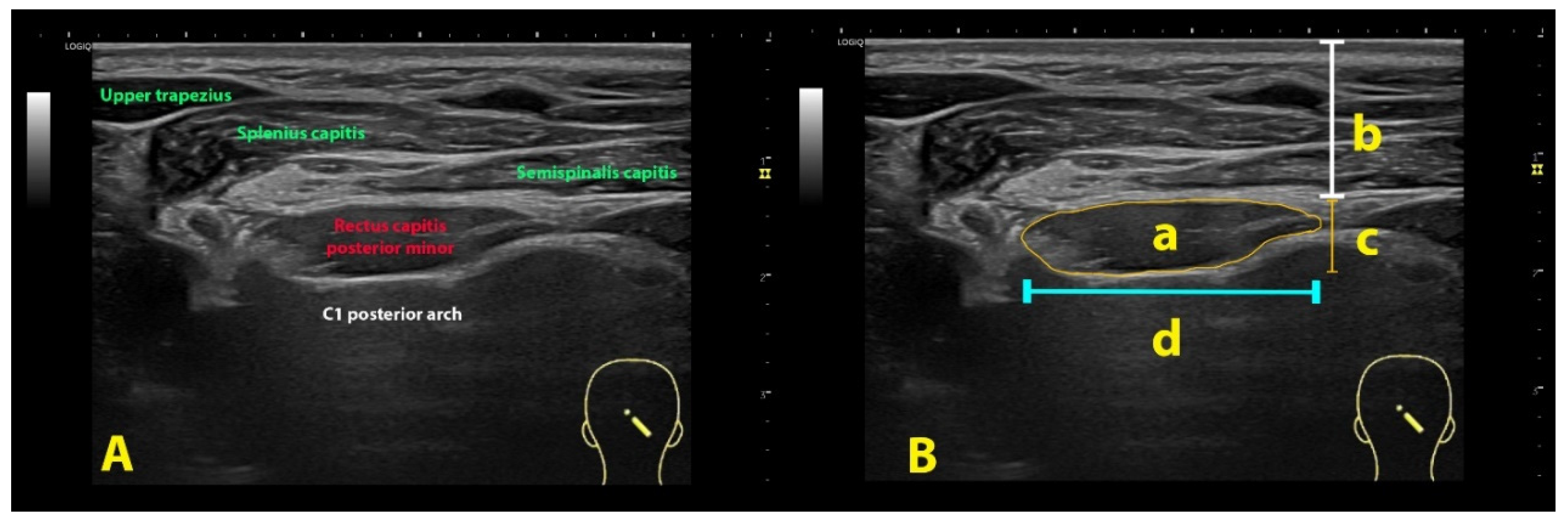
- Error in Figures
- Text Correction
Reference
- Ulman-Macón, D.; Fernández-de-las-Peñas, C.; Angulo-Díaz-Parreño, S.; Arias-Buría, J.L.; Mesa-Jiménez, J.A. Morphological Changes of the Suboccipital Musculature in Women with Myofascial Temporomandibular Pain: A Case-Control Study. Life 2023, 13, 1159. [Google Scholar] [CrossRef] [PubMed]
Disclaimer/Publisher’s Note: The statements, opinions and data contained in all publications are solely those of the individual author(s) and contributor(s) and not of MDPI and/or the editor(s). MDPI and/or the editor(s) disclaim responsibility for any injury to people or property resulting from any ideas, methods, instructions or products referred to in the content. |
© 2024 by the authors. Licensee MDPI, Basel, Switzerland. This article is an open access article distributed under the terms and conditions of the Creative Commons Attribution (CC BY) license (https://creativecommons.org/licenses/by/4.0/).
Share and Cite
Ulman-Macón, D.; Fernández-de-las-Peñas, C.; Angulo-Díaz-Parreño, S.; Arias-Buría, J.L.; Mesa-Jiménez, J.A. Correction: Ulman-Macón et al. Morphological Changes of the Suboccipital Musculature in Women with Myofascial Temporomandibular Pain: A Case-Control Study. Life 2023, 13, 1159. Life 2024, 14, 1387. https://doi.org/10.3390/life14111387
Ulman-Macón D, Fernández-de-las-Peñas C, Angulo-Díaz-Parreño S, Arias-Buría JL, Mesa-Jiménez JA. Correction: Ulman-Macón et al. Morphological Changes of the Suboccipital Musculature in Women with Myofascial Temporomandibular Pain: A Case-Control Study. Life 2023, 13, 1159. Life. 2024; 14(11):1387. https://doi.org/10.3390/life14111387
Chicago/Turabian StyleUlman-Macón, Daniel, César Fernández-de-las-Peñas, Santiago Angulo-Díaz-Parreño, José L. Arias-Buría, and Juan A. Mesa-Jiménez. 2024. "Correction: Ulman-Macón et al. Morphological Changes of the Suboccipital Musculature in Women with Myofascial Temporomandibular Pain: A Case-Control Study. Life 2023, 13, 1159" Life 14, no. 11: 1387. https://doi.org/10.3390/life14111387
APA StyleUlman-Macón, D., Fernández-de-las-Peñas, C., Angulo-Díaz-Parreño, S., Arias-Buría, J. L., & Mesa-Jiménez, J. A. (2024). Correction: Ulman-Macón et al. Morphological Changes of the Suboccipital Musculature in Women with Myofascial Temporomandibular Pain: A Case-Control Study. Life 2023, 13, 1159. Life, 14(11), 1387. https://doi.org/10.3390/life14111387