
Inflammatory Markers Used as Predictors of Subclinical Atherosclerosis in Patients with Diabetic Polyneuropathy
 , and
, and
Abstract
:1. Introduction
2. Materials and Methods
2.1. Study Design
2.2. Data Collection
2.3. Study Outcomes
2.4. Statistical Analysis
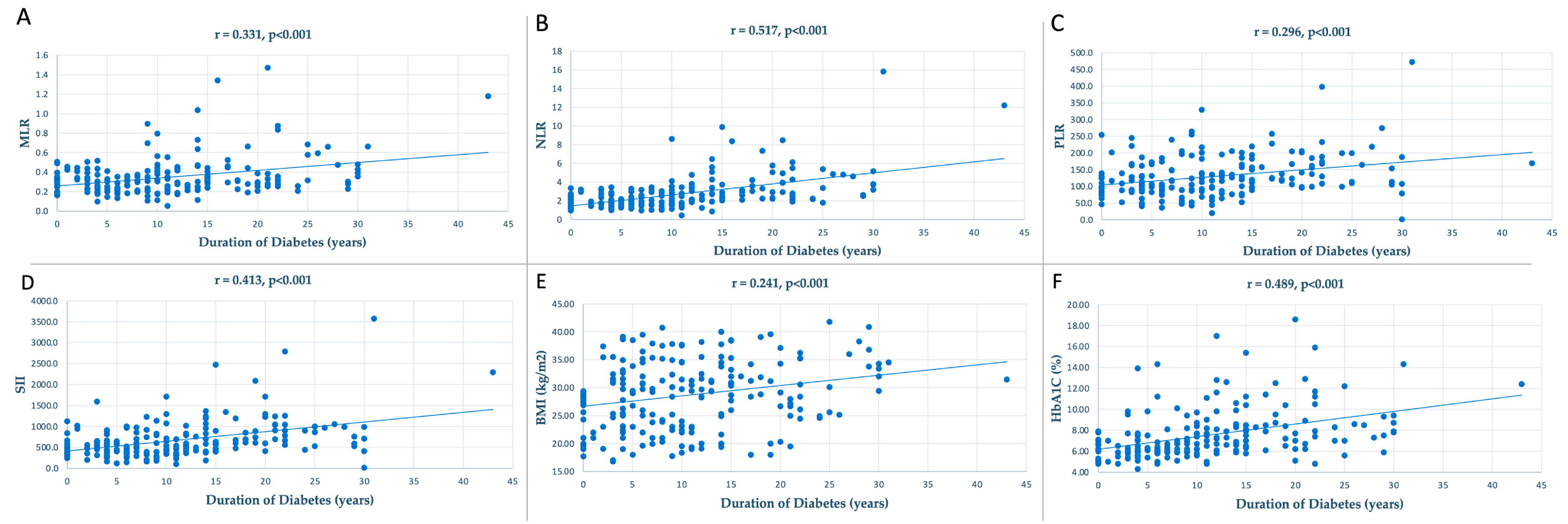
3. Results
4. Discussion
5. Conclusions
Author Contributions
Funding
Institutional Review Board Statement
Informed Consent Statement
Data Availability Statement
Acknowledgments
Conflicts of Interest
References
- Zheng, Y.; Ley, S.H.; Hu, F.B. Global Aetiology and Epidemiology of Type 2 Diabetes Mellitus and Its Complications. Nat. Rev. Endocrinol. 2018, 14, 88–98. [Google Scholar] [CrossRef] [PubMed]
- Saeedi, P.; Petersohn, I.; Salpea, P.; Malanda, B.; Karuranga, S.; Unwin, N.; Colagiuri, S.; Guariguata, L.; Motala, A.A.; Ogurtsova, K.; et al. Global and Regional Diabetes Prevalence Estimates for 2019 and Projections for 2030 and 2045: Results from the International Diabetes Federation Diabetes Atlas, 9th Edition. Diabetes Res. Clin. Pract. 2019, 157, 107843. [Google Scholar] [CrossRef] [PubMed]
- Fowkes, F.G.R.; Rudan, D.; Rudan, I.; Aboyans, V.; Denenberg, J.O.; McDermott, M.M.; Norman, P.E.; Sampson, U.K.A.; Williams, L.J.; Mensah, G.A.; et al. Comparison of Global Estimates of Prevalence and Risk Factors for Peripheral Artery Disease in 2000 and 2010: A Systematic Review and Analysis. Lancet Lond. Engl. 2013, 382, 1329–1340. [Google Scholar] [CrossRef] [PubMed]
- Malyar, N.M.; Freisinger, E.; Meyborg, M.; Lüders, F.; Gebauer, K.; Reinecke, H.; Lawall, H. Amputations and Mortality in In-Hospital Treated Patients with Peripheral Artery Disease and Diabetic Foot Syndrome. J. Diabetes Complicat. 2016, 30, 1117–1122. [Google Scholar] [CrossRef]
- Humphries, M.D.; Brunson, A.; Hedayati, N.; Romano, P.; Melnkow, J. Amputation Risk in Patients with Diabetes Mellitus and Peripheral Artery Disease Using Statewide Data. Ann. Vasc. Surg. 2016, 30, 123–131. [Google Scholar] [CrossRef]
- Freisinger, E.; Malyar, N.M.; Reinecke, H.; Lawall, H. Impact of Diabetes on Outcome in Critical Limb Ischemia with Tissue Loss: A Large-Scaled Routine Data Analysis. Cardiovasc. Diabetol. 2017, 16, 41. [Google Scholar] [CrossRef]
- Murabito, J.M.; D’Agostino, R.B.; Silbershatz, H.; Wilson, W.F. Intermittent Claudication. A Risk Profile from The Framingham Heart Study. Circulation 1997, 96, 44–49. [Google Scholar] [CrossRef]
- American Diabetes Association. Peripheral Arterial Disease in People with Diabetes. Diabetes Care 2003, 26, 3333–3341. [Google Scholar] [CrossRef]
- Marso, S.P.; Hiatt, W.R. Peripheral Arterial Disease in Patients with Diabetes. J. Am. Coll. Cardiol. 2006, 47, 921–929. [Google Scholar] [CrossRef]
- Armstrong, E.J.; Rutledge, J.C.; Rogers, J.H. Coronary Artery Revascularization in Patients with Diabetes. Circulation 2013, 128, 1675–1685. [Google Scholar] [CrossRef]
- Li, Y.; Woo, V.; Bose, R. Platelet Hyperactivity and Abnormal Ca2+ Homeostasis in Diabetes Mellitus. Am. J. Physiol. Heart Circ. Physiol. 2001, 280, H1480–H1489. [Google Scholar] [CrossRef]
- Fowler, M.J. Microvascular and Macrovascular Complications of Diabetes. Clin. Diabetes 2008, 26, 77–82. [Google Scholar] [CrossRef]
- Emerging Risk Factors Collaboration; Di Angelantonio, E.; Kaptoge, S.; Wormser, D.; Willeit, P.; Butterworth, A.S.; Bansal, N.; O’Keeffe, L.M.; Gao, P.; Wood, A.M.; et al. Association of Cardiometabolic Multimorbidity with Mortality. JAMA 2015, 314, 52–60. [Google Scholar] [CrossRef]
- Tesfaye, S.; Boulton, A.J.M.; Dickenson, A.H. Mechanisms and Management of Diabetic Painful Distal Symmetrical Polyneuropathy. Diabetes Care 2013, 36, 2456–2465. [Google Scholar] [CrossRef] [PubMed]
- Yuan, Q.; Wang, J.; Peng, Z.; Zhou, Q.; Xiao, X.; Xie, Y.; Wang, W.; Huang, L.; Tang, W.; Sun, D.; et al. Neutrophil-to-Lymphocyte Ratio and Incident End-Stage Renal Disease in Chinese Patients with Chronic Kidney Disease: Results from the Chinese Cohort Study of Chronic Kidney Disease (C-STRIDE). J. Transl. Med. 2019, 17, 86. [Google Scholar] [CrossRef]
- Azab, B.; Zaher, M.; Weiserbs, K.F.; Torbey, E.; Lacossiere, K.; Gaddam, S.; Gobunsuy, R.; Jadonath, S.; Baldari, D.; McCord, D.; et al. Usefulness of Neutrophil to Lymphocyte Ratio in Predicting Short- and Long-Term Mortality after Non-ST-Elevation Myocardial Infarction. Am. J. Cardiol. 2010, 106, 470–476. [Google Scholar] [CrossRef] [PubMed]
- Mertoglu, C.; Gunay, M. Neutrophil-Lymphocyte Ratio and Platelet-Lymphocyte Ratio as Useful Predictive Markers of Prediabetes and Diabetes Mellitus. Diabetes Metab. Syndr. 2017, 11 (Suppl. S1), S127–S131. [Google Scholar] [CrossRef] [PubMed]
- Liu, J.; Liu, X.; Li, Y.; Quan, J.; Wei, S.; An, S.; Yang, R.; Liu, J. The Association of Neutrophil to Lymphocyte Ratio, Mean Platelet Volume, and Platelet Distribution Width with Diabetic Retinopathy and Nephropathy: A Meta-Analysis. Biosci. Rep. 2018, 38, BSR20180172. [Google Scholar] [CrossRef]
- Liu, N.; Sheng, J.; Pan, T.; Wang, Y. Neutrophil to Lymphocyte Ratio and Platelet to Lymphocyte Ratio Are Associated with Lower Extremity Vascular Lesions in Chinese Patients with Type 2 Diabetes. Clin. Lab. 2019, 65, 180804. [Google Scholar] [CrossRef]
- Akbas, E.M.; Demirtas, L.; Ozcicek, A.; Timuroglu, A.; Bakirci, E.M.; Hamur, H.; Ozcicek, F.; Turkmen, K. Association of Epicardial Adipose Tissue, Neutrophil-to-Lymphocyte Ratio and Platelet-to-Lymphocyte Ratio with Diabetic Nephropathy. Int. J. Clin. Exp. Med. 2014, 7, 1794–1801. [Google Scholar]
- Ciray, H.; Aksoy, A.H.; Ulu, N.; Çizmecioğlu, A.; Gaipov, A.; Solak, Y. Nephropathy, but Not Angiographically Proven Retinopathy, Is Associated with Neutrophil to Lymphocyte Ratio in Patients with Type 2 Diabetes. Exp. Clin. Endocrinol. Diabetes 2015, 123, 267–271. [Google Scholar] [CrossRef] [PubMed]
- Wang, R.; Zhang, J.; Li, Y.; Liu, T.; Yu, K. Neutrophil-Lymphocyte Ratio Is Associated with Arterial Stiffness in Diabetic Retinopathy in Type 2 Diabetes. J. Diabetes Complicat. 2015, 29, 245–249. [Google Scholar] [CrossRef] [PubMed]
- Ulu, S.M.; Dogan, M.; Ahsen, A.; Altug, A.; Demir, K.; Acartürk, G.; Inan, S. Neutrophil-to-Lymphocyte Ratio as a Quick and Reliable Predictive Marker to Diagnose the Severity of Diabetic Retinopathy. Diabetes Technol. Ther. 2013, 15, 942–947. [Google Scholar] [CrossRef]
- Intensive Blood-Glucose Control with Sulphonylureas or Insulin Compared with Conventional Treatment and Risk of Complications in Patients with Type 2 Diabetes (UKPDS 33). Lancet 1998, 352, 837–853. [CrossRef]
- Diabetes Control and Complications Trial Research Group; Nathan, D.M.; Genuth, S.; Lachin, J.; Cleary, P.; Crofford, O.; Davis, M.; Rand, L.; Siebert, C. The Effect of Intensive Treatment of Diabetes on the Development and Progression of Long-Term Complications in Insulin-Dependent Diabetes Mellitus. N. Engl. J. Med. 1993, 329, 977–986. [Google Scholar] [CrossRef] [PubMed]
- Brownlee, M. Biochemistry and Molecular Cell Biology of Diabetic Complications. Nature 2001, 414, 813–820. [Google Scholar] [CrossRef]
- Ridker, P.M.; Cushman, M.; Stampfer, M.J.; Tracy, R.P.; Hennekens, C.H. Plasma Concentration of C-Reactive Protein and Risk of Developing Peripheral Vascular Disease. Circulation 1998, 97, 425–428. [Google Scholar] [CrossRef]
- Schaper, N.C.; Nabuurs-Franssen, M.H.; Huijberts, M.S. Peripheral Vascular Disease and Type 2 Diabetes Mellitus. Diabetes Metab. Res. Rev. 2000, 16 (Suppl. S1), S11–S15. [Google Scholar] [CrossRef]
- Pradhan, A.D.; Manson, J.E.; Rifai, N.; Buring, J.E.; Ridker, P.M. C-Reactive Protein, Interleukin 6, and Risk of Developing Type 2 Diabetes Mellitus. JAMA 2001, 286, 327–334. [Google Scholar] [CrossRef]
- Pickup, J.C.; Chusney, G.D.; Thomas, S.M.; Burt, D. Plasma Interleukin-6, Tumour Necrosis Factor Alpha and Blood Cytokine Production in Type 2 Diabetes. Life Sci. 2000, 67, 291–300. [Google Scholar] [CrossRef]
- Ross, R. Atherosclerosis Is an Inflammatory Disease. Am. Heart J. 1999, 138, S419–S420. [Google Scholar] [CrossRef]
- Yuuki, T.; Kanda, T.; Kimura, Y.; Kotajima, N.; Tamura, J.; Kobayashi, I.; Kishi, S. Inflammatory Cytokines in Vitreous Fluid and Serum of Patients with Diabetic Vitreoretinopathy. J. Diabetes Complicat. 2001, 15, 257–259. [Google Scholar] [CrossRef]
- Schröder, S.; Palinski, W.; Schmid-Schönbein, G.W. Activated Monocytes and Granulocytes, Capillary Nonperfusion, and Neovascularization in Diabetic Retinopathy. Am. J. Pathol. 1991, 139, 81–100. [Google Scholar] [PubMed]
- Chibber, R.; Ben-Mahmud, B.; Coppini, D.; Christ, E.; Kohner, E. Activity of the Glycosylating Enzyme, Core 2 GlcNAc (Β1,6) Transferase, Is Higher in Polymorphonuclear Leukocytes from Diabetic Patients Compared with Age-Matched Control Subjects: Relevance to Capillary Occlusion in Diabetic Retinopathy. Diabetes 2000, 49, 1724–1730. [Google Scholar] [CrossRef] [PubMed]
- Kasza, M.; Meleg, J.; Vardai, J.; Nagy, B.; Szalai, E.; Damjanovich, J.; Csutak, A.; Ujhelyi, B.; Nagy, V. Plasma E-Selectin Levels Can Play a Role in the Development of Diabetic Retinopathy. Graefes Arch. Clin. Exp. Ophthalmol. Albrecht Von Graefes Arch. Klin. Exp. Ophthalmol. 2017, 255, 25–30. [Google Scholar] [CrossRef] [PubMed]
- Limb, G.A.; Hickman-Casey, J.; Hollifield, R.D.; Chignell, A.H. Vascular Adhesion Molecules in Vitreous from Eyes with Proliferative Diabetic Retinopathy. Investig. Ophthalmol. Vis. Sci. 1999, 40, 2453–2457. [Google Scholar]
- Miyamoto, K.; Khosrof, S.; Bursell, S.E.; Rohan, R.; Murata, T.; Clermont, A.C.; Aiello, L.P.; Ogura, Y.; Adamis, A.P. Prevention of Leukostasis and Vascular Leakage in Streptozotocin-Induced Diabetic Retinopathy via Intercellular Adhesion Molecule-1 Inhibition. Proc. Natl. Acad. Sci. USA 1999, 96, 10836–10841. [Google Scholar] [CrossRef]
- Suzuki, Y.; Nakazawa, M.; Suzuki, K.; Yamazaki, H.; Miyagawa, Y. Expression Profiles of Cytokines and Chemokines in Vitreous Fluid in Diabetic Retinopathy and Central Retinal Vein Occlusion. Jpn. J. Ophthalmol. 2011, 55, 256–263. [Google Scholar] [CrossRef]
- Koleva-Georgieva, D.N.; Sivkova, N.P.; Terzieva, D. Serum Inflammatory Cytokines IL-1beta, IL-6, TNF-Alpha and VEGF Have Influence on the Development of Diabetic Retinopathy. Folia Med. 2011, 53, 44–50. [Google Scholar] [CrossRef]
- Boss, J.D.; Singh, P.K.; Pandya, H.K.; Tosi, J.; Kim, C.; Tewari, A.; Juzych, M.S.; Abrams, G.W.; Kumar, A. Assessment of Neurotrophins and Inflammatory Mediators in Vitreous of Patients with Diabetic Retinopathy. Investig. Ophthalmol. Vis. Sci. 2017, 58, 5594–5603. [Google Scholar] [CrossRef]
- Williams, G.; Pickup, J.C. Handbook of Diabetes; Wiley: Hoboken, NJ, USA, 2004; ISBN 978-1-4051-2052-4. [Google Scholar]
- Gheith, O.; Farouk, N.; Nampoory, N.; Halim, M.A.; Al-Otaibi, T. Diabetic Kidney Disease: World Wide Difference of Prevalence and Risk Factors. J. Nephropharmacology 2015, 5, 49–56. [Google Scholar] [CrossRef]
- Stenvinkel, P. Chronic Kidney Disease: A Public Health Priority and Harbinger of Premature Cardiovascular Disease. J. Intern. Med. 2010, 268, 456–467. [Google Scholar] [CrossRef]
- Tavafi, M. Diabetic Nephropathy and Antioxidants. J. Nephropathol. 2013, 2, 20–27. [Google Scholar] [CrossRef]
- Navarro, J.F.; Mora, C. Role of Inflammation in Diabetic Complications. Nephrol. Dial. Transplant. 2005, 20, 2601–2604. [Google Scholar] [CrossRef]
- Suzuki, D.; Miyazaki, M.; Naka, R.; Koji, T.; Yagame, M.; Jinde, K.; Endoh, M.; Nomoto, Y.; Sakai, H. In Situ Hybridization of Interleukin 6 in Diabetic Nephropathy. Diabetes 1995, 44, 1233–1238. [Google Scholar] [CrossRef]
- Wong, C.K.; Ho, A.W.Y.; Tong, P.C.Y.; Yeung, C.Y.; Kong, A.P.S.; Lun, S.W.M.; Chan, J.C.N.; Lam, C.W.K. Aberrant Activation Profile of Cytokines and Mitogen-Activated Protein Kinases in Type 2 Diabetic Patients with Nephropathy. Clin. Exp. Immunol. 2007, 149, 123–131. [Google Scholar] [CrossRef]
- Lim, A.K.H.; Tesch, G.H. Inflammation in Diabetic Nephropathy. Mediat. Inflamm. 2012, 2012, 146154. [Google Scholar] [CrossRef]
- Shang, J.; Wang, L.; Zhang, Y.; Zhang, S.; Ning, L.; Zhao, J.; Cheng, G.; Liu, D.; Xiao, J.; Zhao, Z. Chemerin/ChemR23 Axis Promotes Inflammation of Glomerular Endothelial Cells in Diabetic Nephropathy. J. Cell. Mol. Med. 2019, 23, 3417–3428. [Google Scholar] [CrossRef]
- Awad, A.S.; Kinsey, G.R.; Khutsishvili, K.; Gao, T.; Bolton, W.K.; Okusa, M.D. Monocyte/Macrophage Chemokine Receptor CCR2 Mediates Diabetic Renal Injury. Am. J. Physiol.-Ren. Physiol. 2011, 301, F1358–F1366. [Google Scholar] [CrossRef]
- Doupis, J.; Lyons, T.E.; Wu, S.; Gnardellis, C.; Dinh, T.; Veves, A. Microvascular Reactivity and Inflammatory Cytokines in Painful and Painless Peripheral Diabetic Neuropathy. J. Clin. Endocrinol. Metab. 2009, 94, 2157–2163. [Google Scholar] [CrossRef]
- Magrinelli, F.; Briani, C.; Romano, M.; Ruggero, S.; Toffanin, E.; Triolo, G.; Peter, G.C.; Praitano, M.; Lauriola, M.F.; Zanette, G.; et al. The Association between Serum Cytokines and Damage to Large and Small Nerve Fibers in Diabetic Peripheral Neuropathy. J. Diabetes Res. 2015, 2015, 547834. [Google Scholar] [CrossRef]
- Jin, H.Y.; Park, T.S. Role of Inflammatory Biomarkers in Diabetic Peripheral Neuropathy. J. Diabetes Investig. 2018, 9, 1016–1018. [Google Scholar] [CrossRef]
- Herder, C.; Kannenberg, J.M.; Huth, C.; Carstensen-Kirberg, M.; Rathmann, W.; Koenig, W.; Heier, M.; Püttgen, S.; Thorand, B.; Peters, A.; et al. Proinflammatory Cytokines Predict the Incidence and Progression of Distal Sensorimotor Polyneuropathy: KORA F4/FF4 Study. Diabetes Care 2017, 40, 569–576. [Google Scholar] [CrossRef]
- Eshcol, J.; Jebarani, S.; Anjana, R.M.; Mohan, V.; Pradeepa, R. Prevalence, Incidence and Progression of Peripheral Arterial Disease in Asian Indian Type 2 Diabetic Patients. J. Diabetes Complicat. 2014, 28, 627–631. [Google Scholar] [CrossRef]
- Lekshmi Narayanan, R.M.; Koh, W.P.; Phang, J.; Subramaniam, T. Peripheral Arterial Disease in Community-Based Patients with Diabetes in Singapore: Results from a Primary Healthcare Study. Ann. Acad. Med. Singapore 2010, 39, 525–527. [Google Scholar] [CrossRef]
- Rhee, S.Y.; Guan, H.; Liu, Z.M.; Cheng, S.W.-K.; Waspadji, S.; Palmes, P.; Tai, T.Y.; Suwanwalaikorn, S.; Kim, Y.S.; PAD-SEARCH Study Group. Multi-Country Study on the Prevalence and Clinical Features of Peripheral Arterial Disease in Asian Type 2 Diabetes Patients at High Risk of Atherosclerosis. Diabetes Res. Clin. Pract. 2007, 76, 82–92. [Google Scholar] [CrossRef]
- Merino, J.; Planas, A.; Elosua, R.; de Moner, A.; Gasol, A.; Contreras, C.; Vidal-Barraquer, F.; Clarà, A. Incidence and Risk Factors of Peripheral Arterial Occlusive Disease in a Prospective Cohort of 700 Adult Elderly Men Followed for 5 Years. World J. Surg. 2010, 34, 1975–1979. [Google Scholar] [CrossRef]
- Velescu, A.; Clara, A.; Peñafiel, J.; Grau, M.; Degano, I.R.; Martí, R.; Ramos, R.; Marrugat, J.; Elosua, R. Peripheral Arterial Disease Incidence and Associated Risk Factors in a Mediterranean Population-Based Cohort. The REGICOR Study. Eur. J. Vasc. Endovasc. Surg. Off. J. Eur. Soc. Vasc. Surg. 2016, 51, 696–705. [Google Scholar] [CrossRef]
- Arbănași, E.M.; Halmaciu, I.; Kaller, R.; Mureșan, A.V.; Arbănași, E.M.; Suciu, B.A.; Coșarcă, C.M.; Cojocaru, I.I.; Melinte, R.M.; Russu, E. Systemic Inflammatory Biomarkers and Chest CT Findings as Predictors of Acute Limb Ischemia Risk, Intensive Care Unit Admission, and Mortality in COVID-19 Patients. Diagnostics 2022, 12, 2379. [Google Scholar] [CrossRef]
- Halmaciu, I.; Arbănași, E.M.; Kaller, R.; Mureșan, A.V.; Arbănași, E.M.; Bacalbasa, N.; Suciu, B.A.; Cojocaru, I.I.; Runcan, A.I.; Grosu, F.; et al. Chest CT Severity Score and Systemic Inflammatory Biomarkers as Predictors of the Need for Invasive Mechanical Ventilation and of COVID-19 Patients’ Mortality. Diagnostics 2022, 12, 2089. [Google Scholar] [CrossRef]
- Kaller, R.; Arbănași, E.M.; Mureșan, A.V.; Voidăzan, S.; Arbănași, E.M.; Horváth, E.; Suciu, B.A.; Hosu, I.; Halmaciu, I.; Brinzaniuc, K.; et al. The Predictive Value of Systemic Inflammatory Markers, the Prognostic Nutritional Index, and Measured Vessels’ Diameters in Arteriovenous Fistula Maturation Failure. Life 2022, 12, 1447. [Google Scholar] [CrossRef] [PubMed]
- Mureșan, A.V.; Hălmaciu, I.; Arbănași, E.M.; Kaller, R.; Arbănași, E.M.; Budișcă, O.A.; Melinte, R.M.; Vunvulea, V.; Filep, R.C.; Mărginean, L.; et al. Prognostic Nutritional Index, Controlling Nutritional Status (CONUT) Score, and Inflammatory Biomarkers as Predictors of Deep Vein Thrombosis, Acute Pulmonary Embolism, and Mortality in COVID-19 Patients. Diagnostics 2022, 12, 2757. [Google Scholar] [CrossRef] [PubMed]
- Arbănași, E.M.; Mureșan, A.V.; Coșarcă, C.M.; Kaller, R.; Bud, T.I.; Hosu, I.; Voidăzan, S.T.; Arbănași, E.M.; Russu, E. Neutrophil-to-Lymphocyte Ratio and Platelet-to-Lymphocyte Ratio Impact on Predicting Outcomes in Patients with Acute Limb Ischemia. Life 2022, 12, 822. [Google Scholar] [CrossRef] [PubMed]
- Melinte, R.M.; Arbănași, E.M.; Blesneac, A.; Zolog, D.N.; Kaller, R.; Mureșan, A.V.; Arbănași, E.M.; Melinte, I.M.; Niculescu, R.; Russu, E. Inflammatory Biomarkers as Prognostic Factors of Acute Deep Vein Thrombosis Following the Total Knee Arthroplasty. Medicina 2022, 58, 1502. [Google Scholar] [CrossRef] [PubMed]
- Russu, E.; Mureșan, A.V.; Arbănași, E.M.; Kaller, R.; Hosu, I.; Voidăzan, S.; Arbănași, E.M.; Coșarcă, C.M. The Predictive Role of NLR and PLR in Outcome and Patency of Lower Limb Revascularization in Patients with Femoropopliteal Disease. J. Clin. Med. 2022, 11, 2620. [Google Scholar] [CrossRef] [PubMed]
- Arbănași, E.M.; Mureșan, A.V.; Arbănași, E.M.; Kaller, R.; Cojocaru, I.I.; Coșarcă, C.M.; Russu, E. The Neutrophil-to-Lymphocyte Ratio’s Predictive Utility in Acute Pulmonary Embolism: Systematic Review. J. Cardiovasc. Emergencies 2022, 8, 25–30. [Google Scholar] [CrossRef]
- Niculescu, R.; Russu, E.; Arbănași, E.M.; Kaller, R.; Arbănași, E.M.; Melinte, R.M.; Coșarcă, C.M.; Cocuz, I.G.; Sabău, A.H.; Tinca, A.C.; et al. Carotid Plaque Features and Inflammatory Biomarkers as Predictors of Restenosis and Mortality Following Carotid Endarterectomy. Int. J. Environ. Res. Public. Health 2022, 19, 13934. [Google Scholar] [CrossRef]
- Mureșan, A.V.; Russu, E.; Arbănași, E.M.; Kaller, R.; Hosu, I.; Arbănași, E.M.; Voidăzan, S.T. The Predictive Value of NLR, MLR, and PLR in the Outcome of End-Stage Kidney Disease Patients. Biomedicines 2022, 10, 1272. [Google Scholar] [CrossRef]
- Roumeliotis, S.; Neofytou, I.E.; Maassen, C.; Lux, P.; Kantartzi, K.; Papachristou, E.; Schurgers, L.J.; Liakopoulos, V. Association of Red Blood Cell Distribution Width and Neutrophil-to-Lymphocyte Ratio with Calcification and Cardiovascular Markers in Chronic Kidney Disease. Metabolites 2023, 13, 303. [Google Scholar] [CrossRef]
- Caicedo, D.; Alvarez, C.V.; Perez-Romero, S.; Devesa, J. The Inflammatory Pattern of Chronic Limb-Threatening Ischemia in Muscles: The TNF-α Hypothesis. Biomedicines 2022, 10, 489. [Google Scholar] [CrossRef]
- Taurino, M.; Aloisi, F.; Del Porto, F.; Nespola, M.; Dezi, T.; Pranteda, C.; Rizzo, L.; Sirignano, P. Neutrophil-to-Lymphocyte Ratio Could Predict Outcome in Patients Presenting with Acute Limb Ischemia. J. Clin. Med. 2021, 10, 4343. [Google Scholar] [CrossRef] [PubMed]
- Serra, R.; Ielapi, N.; Licastro, N.; Provenzano, M.; Andreucci, M.; Bracale, U.M.; Jiritano, F.; de Franciscis, S.; Mastroroberto, P.; Serraino, G.F. Neutrophil-to-Lymphocyte Ratio and Platelet-to-Lymphocyte Ratio as Biomarkers for Cardiovascular Surgery Procedures: A Literature Review. Rev. Recent Clin. Trials 2021, 16, 173–179. [Google Scholar] [CrossRef] [PubMed]
- Wachsmann-Maga, A.; Kaszuba, M.; Maga, M.; Włodarczyk, A.; Krężel, J.; Kaczmarczyk, P.; Bogucka, K.; Maga, P. Leukotrienes in the Atherosclerotic Cardiovascular Diseases—A Systematic Review. Acta Angiol. 2022, 28, 147–153. [Google Scholar] [CrossRef]
- Wachsmann, A.; Maga, M.; Cebeńko, M.; Schönborn, M.; Olszewska, M.; Blukacz, M.; Maga, P.; Trynkiewicz, A. Impact of Pre-Operative Glycated Haemoglobin A1C Level on 1-Year Outcomes of Endovascular Treatment in Patients with Critical Limb Ischemia in the Course of Diabetes Mellitus. Folia Med. Cracoviensia 2019, 59, 49–60. [Google Scholar]


| Variables | All Patients n = 198 | No-SA n = 126 | SA n = 72 | p-Value |
|---|---|---|---|---|
| Age mean ± SD (min–max) | 64.36 ± 10.18 (35–87) | 62.96 ± 9.99 (35–86) | 66.80 ± 10.13 (42–87) | 0.01 |
| Male/Female gender no. (%) | 93 (46.97%) 105 (53.03%) | 53 (42.06%) 73 (57.94%) | 40 (55.56%) 32 (44.44%) | 0.06 |
| Comorbidities and Risk factors, no. (%) | ||||
| Arterial Hypertension | 183 (92.42%) | 116 (92.06%) | 67 (93.05%) | 0.79 |
| Ischemic Heart Disease | 107 (54.04%) | 61 (48.41%) | 46 (63.88%) | 0.03 |
| Chronic Venous Insufficiency | 89 (44.94%) | 61 (48.41%) | 28 (38.88%) | 0.19 |
| Malignancy | 28 (14.14%) | 20 (15.87%) | 8 (11.11%) | 0.35 |
| Active Smoking | 81 (40.9%) | 25 (34.72%) | 56 (44.44%) | 0.18 |
| History of Stroke | 11 (5.55%) | 7 (5.55%) | 4 (5.55%) | NS |
| History of Myocardial Infraction | 18 (9.09%) | 7 (5.55%) | 11 (15.27%) | 0.02 |
| End Stage Kidney Disease | 28 (14.14%) | 15 (11.9%) | 13 (18.05%) | 0.23 |
| Diabetic Retinopathy | 54 (27.27%) | 30 (23.80%) | 24 (33.33%) | 0.14 |
| Diabetic Nephropathy | 40 (20.2%) | 19 (15.07%) | 21 (29.16%) | 0.01 |
| Anthropometric Characteristics, median [Q1–Q3] | ||||
| BMI (kg/m2) | 29.32 [23.92–33.68] | 25.81 [21.16–30.45] | 32 [30.1–35.36] | <0.0001 |
| Abdominal circumferential (cm) | 110 [100–120] | 109 [100–118.25] | 112 [101–121] | 0.052 |
| Duration of Diabetes (years) | 10 [5–15] | 7 [4–11] | 15 [11.75–22] | <0.0001 |
| Laboratory Findings, median [Q1–Q3] | ||||
| HbA1C (%) | 6.83 [6–8.3] | 6.2 [5.8–7] | 8.55 [7.3–11.2] | <0.0001 |
| Admission Glucose (mg/dL) | 141 [114–199.75] | 122 [101.25–143] | 218 [174–271.25] | <0.0001 |
| Cholesterol (mg/dL) | 167.3 [132–203.47] | 154.15 [122.1–190.85] | 189.4 [157.3–209.32] | <0.0001 |
| Triglyceride (mg/dL) | 162.1 [118.32–241.72] | 152 [116.15–237.9] | 177.6 [120.55–248.9] | 0.12 |
| AST (IU/L) | 21 [15.9–30.8] | 22.45 [16.15–31.97] | 19.2 [15.7–27.17] | 0.04 |
| ALT (IU/L) | 21.95 [15.87–35.52] | 23.15 [18–41] | 19.7 [14.2–30.92] | 0.007 |
| GGT (IU/L) | 34 [23–72] | 37 [21–75] | 31.5 [23–62] | 0.26 |
| BUN (mg/dL) | 41.55 [32.42–56.67] | 39.45 [30.45–56.35] | 43 [35.57–56.77] | 0.09 |
| Creatinine (mg/dL) | 0.95 [0.77–1.29] | 0.91 [0.73–1.23] | 1.02 [0.84–1.38] | 0.02 |
| Hemoglobin (g/dL) | 13.5 [12.3–14.6] | 13.3 [12.3–14.47] | 13.5 [12.47–14.87] | 0.18 |
| Hematocrit % | 40.65 [37.22–43.65] | 40.55 [37.1–43.5] | 40.7 [37.27–43.72] | 0.35 |
| WBC | 8.06 [6.63–9.78] | 8.18 [6.64–9.67] | 7.69 [6.32–9.84] | 0.21 |
| Neutrophil | 4.8 [3.83–6.12] | 4.66 [3.82–5.79] | 5.22 [3.92–7.01] | 0.02 |
| Monocyte | 0.61 [0.51–0.76] | 0.63 [0.53–0.77] | 0.59 [0.48–0.71] | 0.03 |
| Lymphocyte | 2.04 [1.53–2.65] | 2.34 [1.93–2.97] | 1.67 [1.31–1.98] | <0.0001 |
| PLT | 237 [200.25–290.75] | 246.5 [205.25–290.75] | 231.5 [198.5–201] | 0.20 |
| NLR | 2.24 [1.77–3.15] | 1.95 [1.60–2.50] | 3.16 [2.41–4.66] | <0.0001 |
| MLR | 0.30 [0.22–0.40] | 0.27 [0.21–0.35] | 0.35 [0.27–0.46] | <0.0001 |
| PLR | 116.52 [91.2–162.7] | 103.26 [84.4–128.89] | 157.64 [113.28–194.59] | <0.0001 |
| SII | 575.5 [399.9–838.7] | 468.6 [363.2–631.1] | 802.9 [577.1–1004.9] | <0.0001 |
| Variables | Cut-Off | AUC | Std. Error | 95% CI | Sensitivity | Specificity | p-Value |
|---|---|---|---|---|---|---|---|
| Subclinical Atherosclerosis of Lower Limb | |||||||
| MLR | 0.33 | 0.689 | 0.039 | 0.613–0.765 | 59.7% | 71.4% | <0.0001 |
| NLR | 2.53 | 0.820 | 0.031 | 0.759–0.881 | 73.6% | 77% | <0.0001 |
| PLR | 137.21 | 0.751 | 0.036 | 0.680–0.821 | 62.5% | 82.5% | <0.0001 |
| SII | 615.91 | 0.759 | 0.036 | 0.688–0.829 | 68.1% | 73.8% | <0.0001 |
| BMI | 30.87 | 0.793 | 0.031 | 0.732–0.854 | 68.1% | 79.4% | <0.0001 |
| HbA1C | 7.45 | 0.857 | 0.028 | 0.802–0.911 | 73.6% | 82.5% | <0.0001 |
| Duration of Diabetes | 9.5 | 0.812 | 0.032 | 0.749–0.875 | 83.3% | 65.1% | <0.0001 |
| Admission Glucose level | 161.5 | 0.899 | 0.025 | 0.850–0.948 | 80.6% | 85.7% | <0.0001 |
| Subclinical Atherosclerosis | |||
|---|---|---|---|
| OR | 95% CI | p-Value | |
| Demographic Characteristics | |||
| Age | 2.58 | 1.79–5.93 | <0.001 |
| Male | 2.30 | 1.26–4.19 | 0.006 |
| Comorbidities and Risk factors | |||
| Ischemic Heart Disease | 1.57 | 0.85–2.18 | 0.11 |
| History of Myocardial Infraction | 1.26 | 0.76–2.87 | 0.20 |
| Diabetic Nephropathy | 1.95 | 0.93–4.55 | 0.052 |
| Tobacco | 1.33 | 0.70–3.46 | 0.18 |
| Anthropometric Characteristics | |||
| BMI (kg/m2) | 7.71 | 3.57–14.90 | <0.001 |
| Abdominal circumferential (cm) | 1.86 | 0.84–3.78 | 0.06 |
| Duration of Diabetes (years) | 8.65 | 4.35–16.78 | <0.001 |
| Inflammatory Markers | |||
| NLR | 7.46 | 3.38–13.58 | <0.001 |
| MLR | 4.63 | 1.99–9.12 | <0.001 |
| PLR | 5.89 | 2.62–11.88 | <0.001 |
| SII | 6.09 | 2.68–12.16 | <0.001 |
| Diabetes Controlling Status | |||
| HbA1C | 10.4 | 8.27–28.74 | <0.001 |
| Admission Glucose | 10.78 | 8.69–32.45 | <0.001 |
Disclaimer/Publisher’s Note: The statements, opinions and data contained in all publications are solely those of the individual author(s) and contributor(s) and not of MDPI and/or the editor(s). MDPI and/or the editor(s) disclaim responsibility for any injury to people or property resulting from any ideas, methods, instructions or products referred to in the content. |
© 2023 by the authors. Licensee MDPI, Basel, Switzerland. This article is an open access article distributed under the terms and conditions of the Creative Commons Attribution (CC BY) license (https://creativecommons.org/licenses/by/4.0/).
Share and Cite
Mureșan, A.V.; Tomac, A.; Opriș, D.R.; Bandici, B.C.; Coșarcă, C.M.; Covalcic, D.C.; Hălmaciu, I.; Akácsos-Szász, O.-Z.; Rădulescu, F.; Lázár, K.; et al. Inflammatory Markers Used as Predictors of Subclinical Atherosclerosis in Patients with Diabetic Polyneuropathy. Life 2023, 13, 1861. https://doi.org/10.3390/life13091861
Mureșan AV, Tomac A, Opriș DR, Bandici BC, Coșarcă CM, Covalcic DC, Hălmaciu I, Akácsos-Szász O-Z, Rădulescu F, Lázár K, et al. Inflammatory Markers Used as Predictors of Subclinical Atherosclerosis in Patients with Diabetic Polyneuropathy. Life. 2023; 13(9):1861. https://doi.org/10.3390/life13091861
Chicago/Turabian StyleMureșan, Adrian Vasile, Alexandru Tomac, Diana Roxana Opriș, Bogdan Corneliu Bandici, Cătălin Mircea Coșarcă, Diana Carina Covalcic, Ioana Hălmaciu, Orsolya-Zsuzsa Akácsos-Szász, Flavia Rădulescu, Krisztina Lázár, and et al. 2023. "Inflammatory Markers Used as Predictors of Subclinical Atherosclerosis in Patients with Diabetic Polyneuropathy" Life 13, no. 9: 1861. https://doi.org/10.3390/life13091861
APA StyleMureșan, A. V., Tomac, A., Opriș, D. R., Bandici, B. C., Coșarcă, C. M., Covalcic, D. C., Hălmaciu, I., Akácsos-Szász, O.-Z., Rădulescu, F., Lázár, K., Stoian, A., & Tilinca, M. C. (2023). Inflammatory Markers Used as Predictors of Subclinical Atherosclerosis in Patients with Diabetic Polyneuropathy. Life, 13(9), 1861. https://doi.org/10.3390/life13091861