Article

Genetic Basis of Acinetobacter sp. K1 Adaptation Mechanisms to Extreme Environmental Conditions

1
Department of Microbiology, Institute of Biology and Ecology, Faculty of Sciences, Pavol Jozef Safarik University in Kosice, Srobarova 2, 041 54 Kosice, Slovakia
2
Centre of Biosciences, Institute of Animal Physiology, Slovak Academy of Sciences, Soltesovej 4-6, 040 01 Kosice, Slovakia
*
Author to whom correspondence should be addressed.
Life 2023, 13(8), 1728; https://doi.org/10.3390/life13081728
Submission received: 24 June 2023 / Revised: 27 July 2023 / Accepted: 8 August 2023 / Published: 11 August 2023
(This article belongs to the Special Issue Microbial Composition and Function in Soil and Groundwater Systems)

Abstract

:
Anthropogenic pollution often leads to the generation of technosols, technogenic soils with inhospitable conditions for all living organisms including microbiota. Aluminum production near Ziar nad Hronom (Slovakia) resulted in the creation of a highly alkaline and heavy-metal-rich brown mud landfill, from which a bacterial strain of a likely new species of the genus Acinetobacter, Acinetobacter sp. K1, was isolated. The whole-genome sequence analysis of this strain confirmed the presence of operon units enabling tolerance to the heavy metals copper, zinc, cobalt, cadmium, chromium, and metalloid arsenic, which are functionally active. Despite the predominance of plasmid-related sequences in the K1 genome, the results indicate that most of the resistance genes are chromosomally encoded. No significant alkali tolerance of Acinetobacter sp. K1 was observed in vitro, suggesting that community level mechanisms are responsible for the survival of this strain in the highly alkaline, brown mud bacterial community.

1. Introduction

Bacteria belonging to the genus Acinetobacter are characterized by the ability to inhabit a wide range of environments and to adapt to the prevailing extreme conditions. Many strains of Acinetobacter spp. can tolerate increased concentrations of heavy metals and different pH values of the environment and resist the action of various types of antibiotics [1,2]. The high tolerance is possible due to the activity of several protein groups encoded by genes located mainly on mobile genetic elements and thus can be transmitted horizontally within the bacterial population. In addition, bacteria of the Acinetobacter genus are able to form capsules, increasing their resistance to environmental impacts even more [3]. Environmental Acinetobacter spp. species, including A. lwoffii strains, are generally considered non-pathogenic compared to clinical A. baumannii strains. On the other hand, in recent years, the number of infections caused by this bacterial species has noticeably increased [4,5,6,7,8].
Various industrial activities cause environmental pollution with a subsequent negative impact on human and environmental health. The generation of large quantities of technosols or technogenic soils with diverse toxic properties is one of the emerging phenomena of these activities [9,10,11]. Such waste sludges present a challenge in finding effective methods for their removal. In recent years, research has shown the high potential of Acinetobacter spp. in biotechnologies and bioremediation of industrial waste sludges [12]. These approaches are being intensively studied because of the advantages offered by bacterial biomass, such as the short generation time of bacteria or the ability of their eventual genetic modification [13]. In recent decades, whole-genome sequencing has become a widely used method to better understand the organization of genes responsible for bacterial adaptation to polluted industrial environments.
The bacterial isolate Acinetobacter sp. K1 was isolated from a highly alkaline, brown mud landfill generated during the years of aluminum production near Ziar nad Hronom (Slovakia); the technosol was contaminated by high concentrations of heavy metals, including mercury [14,15]. The population in the brown mud was dominated by Gram-positive bacteria (Bacillus, Micrococcus, Isoptericola and Arthrobacter) and acinetobacters were the only representative of Gram-negative bacteria [14]. As acinetobacters are reported to prefer a slightly acid pH as the optimum for growth [16], we focused on the determination of genes responsible for the adaptation of the K1 isolate to extreme conditions of the brown mud and the putative role of horizontal gene transfer in the adaptation as the presence of multiple DNA plasmids (with a size of 3 kbp–25 kbp) were detected in the K1 genome [17].

2. Materials and Methods

2.1. Origin of the Bacterial Isolate Acinetobacter sp. K1 and Growth Conditions

The bacterial strain Acinetobacter sp. K1 was isolated from a brown mud created during aluminum production of the Slovalco Co. near Ziar nad Hronom (48°35′3″ N, 18°51′39″ E) in our previous research. To a 0.5 g solid brown mud sample, 10 mL of a sterile phosphate-buffered saline solution was added and the mixture was intensively mixed for 20 min. Subsequently, the aliquots were spread on tryptone soya agar (TSA) (Oxoid, Columbia, MD, USA) and cultivated for 24–72 h at 22 °C [14]. After the cell and colony morphology examination, subculturing and the purity check, the K1 strain was preliminary identified using matrix-assisted laser desorption ionization time of flight mass spectroscopy (MALDI-TOF MS) and phylogenetic analysis based on the 16S rRNA gene sequence. The phenotypic pattern was analyzed using a GEN III MicroPlate system (Biolog, Hayward, CA, USA) [14]. The isolate was routinely cultivated on TSA (BD Difco, Sparks, MD, USA) or liquid Luria–Bertani (LB) medium (BD Difco, Sparks, MD, USA) at 25 °C for 12–16 h in all experiments.

2.2. DNA Extraction and Purification

The genomic DNA was extracted from an overnight LB bacterial culture using an E.Z.N.A.® Bacterial DNA Kit (Omega Bio-tek, Norcross, GA, USA) according to the manufacturer’s instructions. Subsequently, the genomic DNA was purified by ethanol precipitation. We added 0.1 volumes of 3 mM sodium acetate (pH = 6) and 2.5 volumes of 96% ethanol to the DNA, mixed by inverting several times and incubated for 10 min at −70 °C. After 30 min of centrifugation at 4 °C and 13,000× g, we removed the supernatant and added 500 µL of 70% ethanol to the DNA sediment. The DNA was centrifuged for 15 min at 25 °C, the supernatant was removed, and the dried DNA was dissolved in a 10 mM TRIS solution (pH = 8) (Applichem, Germany). The electrophoresis was performed in a 1% agarose gel stained with ethidium bromide (0.5 μg/L) under the following conditions: 5 V/cm at 25° C for 30 min. The DNA was visualized under UV light using a Gel Logic 212 PRO Imaging System (Carestream, Health Inc., Rochester, NY, USA). The optimal purity and concentration were determined spectrophotometrically using a NanoDrop 2000c Spectrophotometer (Thermo Scientific, Carlsbad, CA, USA).

2.3. The Whole-Genome Sequence Analysis

Whole-genome sequencing of the isolate Acinetobacter sp. K1 was carried out using an Illumina HiSeq 2000 platform and the paired-end strategy (2 × 150 bp) in the NovaSeq 6000 mode using an S2 PE150 XP kit (Eurofins Genomics Europe Sequencing GmbH, Köln, Germany). The obtained sequences were processed using the tools implemented in the Unipro UGENE v35.0 bioinformatics software [18,19]. The quality of the raw data was evaluated by the program FastQC v0.11.8. Reads with low quality scores (<20) were removed using Trimmomatic v0.38 software. The resulting sequences were assembled de novo with the program SPAdes 3.12.0. Subsequently, the sequences shorter than 200 bp were excluded from further analyses. The contigs obtained were checked against the GenBank database available at https://blast.ncbi.nlm.nih.gov/Blast.cgi (accessed on 1 April 2022) using the Blastn algorithm [20] and Rapid Annotation using Subsystem Technology (RAST) was used to determine the gene contents of the Acinetobacter sp. K1 genome based on functional subsystem classifications [21,22,23].

2.4. Taxogenomics

We used the Type (Strain) Genome Server (TYGS, https://tygs.dsmz.de/) (accessed on 25 July 2023) for the taxogenomic comparisons of the K1 genome based on the Genome BLAST Distance Phylogeny (GBDP) method and comparison of the 16S rRNA gene [24]. The K1 genome relatedness to the reference genomes of A. lwoffii strains (accession numbers are listed in Table S2) was calculated using The Kostas Lab Average Nucleotide Identity calculator (ANI, http://enve-omics.ce.gatech.edu/ani/index) (accessed on 16 March 2023) with a cut-off value of 95% indicating the same species [25,26]. The similarity matrix created from the ANI values was used to build the phylogenetic tree using the Unweighted Pair Group Method with Arithmetic Mean (UPGMA) and visualized using the Iroki online tool available at https://www.iroki.net/viewer (accessed on 21 March 2023) [27].

2.5. Identification of the Plasmid-Related Sequences and Localization of Determinants Important for the Adaptation Mechanisms of the K1 Isolate

The putative plasmid-related sequences identified using the RAST server were verified using the Conserved Domain Database (CDD) [28] and ORF finder (https://www.ncbi.nlm.nih.gov/orffinder/) (accessed on 5 December 2022). We focused on the presence of genes involved in resistance to extreme environmental conditions and especially on the genes located in plasmid DNA. In addition, we compared the contig sequences with the genomes in the GenBank database using the Blastn and Blastx algorithms [20]. Plasmid-related sequences were predicted using the mlplasmids v2.1.0 online server (https://sarredondo.shinyapps.io/mlplasmids/) (accessed on 9 May 2023) [29]. The average GC content of contigs was calculated using the Genomics %G~C Content Calculator available at https://www.sciencebuddies.org/science-fair-projects/references/genomics-g-c-content-calculator (accessed on 27 March 2023).

3. Results

3.1. The Whole-Genome Sequence Analysis and Taxogenomics of the K1 Isolate

The electrophoretic analysis of the total DNA of Acinetobacter sp. K1 showed the presence of multiple extrachromosomal DNA bands [17]. To evaluate the potential role of plasmid-encoded genes in adaptation of the isolate to extreme alkalic conditions, whole-genome sequencing of the isolate Acinetobacter sp. K1 was performed. The sequencing provided 6,158,720 reads of 1,847,616,000 nucleotides with 543× coverage of the K1 genome. The raw reads were assembled into a draft genome of 3,357,632 bp, organized into 94 contigs with N50 = 93,093 bp and L50 = 11. The genome was deposited into the GenBank database under accession number NZ_JALGQY000000000.1. Annotation by the RAST server led to the identification of 3413 protein-coding genes and RNA genes. No CRISPR-cas9 system gene(s) were detected in the K1 genome. A quality check by the RAST server did not identify any warnings or fatal problems. The general characteristics of the K1 genome were compared to those of twelve genomes of A. lwoffii strains, one A. pseudolwoffii strain and three A. idrijaensis strains available in the GenBank database using the RAST server (Table S1). The average GC content of the K1 genome (42.8%) was similar to all Acinetobacter spp. strains included in this analysis. The A. idrijaensis MII strain showed the highest number of protein-coding sequences. The number of protein-coding sequences in the K1 genome was the fourth highest among all analyzed strains.
The K1 isolate showed the closest relationship to the A. idrijaensis MII according to the GBDP phylogenetic analysis (Figure 1) with a dDDH value below 70%. This finding was also confirmed by the phylogenetic analysis of the 16S rRNA gene sequences (Figure S1). However, the bootstrap value was too low to reliably assign the K1 isolate to the A. idrijaensis species and the TYGS server predicted that the K1 strain is likely to be a new species of the Acinetobacter genus.
The ANI value between the K1 genome and the type strain of A. lwoffii was 95.84%. The highest ANI value of 96.26% was observed between the genomes of Acinetobacter sp. K1 and A. lwoffii NIPH478. A similarity of 96% or more was also shown with two other strains of A. lwoffii (SH145 and UBA2051). An ANI value higher than 95% was observed with A. idrijaensis MII, A. mesopotamicus DSM 26953, and Prolinoborus fasciculus CIP 103579. An ANI index lower than 90% was shown with the strains A. lwoffii CIP64.10, A. lwoffii F78 and A. lwoffii WJ10621 (Table S3). The dendrogram constructed based on the ANI similarity matrix (Table S3) grouped the A. lwoffii strains in several separate branches, indicating non-homogeneity (resp. high genetic variability) of the A. lwoffii species (Figure S2). The K1 isolate formed a separate group with A. lwoffii NIPH478 that was distant from the A. idrijaensis MII strain.

3.2. Identification of the Plasmid-Related Sequences in the K1 Genome

Thirty-seven contigs (with a total size of 476,260 bp), representing 13.13% of the whole K1 genome, were predicted to be plasmid-related according to the mlplasmids v2.1.0 online server (Table S4). Three complete plasmid sequences were identified, including the sequence of the cryptic pALK1 plasmid (NZ_JALGQY010000067.1), whose sequence was obtained using the recombinant DNA techniques in our previous study (data not shown) and two other small plasmids unnamed1 (NZ_JALGQY010000057.1) and unnamed2 (NZ_JALGQY010000058.1) (Table S5). Plasmid unnamed1 carries the gene for a yet undescribed replication protein rep_pAB02_ORF2. This gene is often located near the repM gene; however, in plasmid unnamed 1, the presence of the genes encoding the Replicase superfamily and PriCT_1 protein of the Primase superfamily were detected behind the rep_pAB02_ORF2 gene. This plasmid also encodes a protein of the Serine recombinase family and the transcriptional regulator HipB. Plasmid unnamed2 encodes the replication protein Rep_3/RepM, the mobilization protein MobA_MobL and the relaxase Tra_Ti. In addition, the PhdYeFM superfamily of antitoxins and the YoeB-like toxin of the type II toxin–antitoxin system are encoded by the plasmid unnamed2 sequence. Several other contig sequences showed high similarities to Acinetobacter spp. plasmid sequences using the Blastn algorithm. However, some of these contigs (e.g., NODE 34, 50, 56 and 261) were proposed as being of chromosomal origin according to the mlplasmids v2.1.0 online server. The average GC content showed that majority of plasmid-related sequences clearly differed from the chromosomal DNA of the K1 isolate, indicating that plasmids were probably obtained by horizontal gene transfer events (Tables S4 and S5).
The most frequently occurring genes encoding replication proteins of putative plasmid-related sequences (such as NODE 38, 43, 47, 55, 66, 223 and plasmids pALK1, unnamed1 and unnamed 2) are rep_3, repM and rep_pAB02_ORF2 (Table S5). The mobilization protein MobA_MobL is encoded by a gene located on plasmid unnamed2 and on contigs NODE 55 and 2715, while genes coding proteins MobM and MobC were detected only on plasmid pALK1 and on contig NODE 43, respectively. The sequences of plasmid unnamed2 and contigs NODE 55 and 2568 encode the Tra_Ti protein. A considerable number of the analyzed contigs contain conserved domain sequences of the IS3, IS5_2, IS5_3, IS21, IS481 or ISNCY_2 Transposase families. Conserved domain sequences of integrases, for example, XerC, were also found in the K1 genome. The genes coding the ComFC protein superfamily were detected within contig NODE 47. Conserved “competence domain” sequence of ComEC proteins was located on contig NODE 97. In addition, contig NODE 97 also encodes transposases and, according to all predictions, it is probably a plasmid-related sequence.

3.3. Genetic Determinants Important to Adaptation Mechanisms of the K1 Isolate

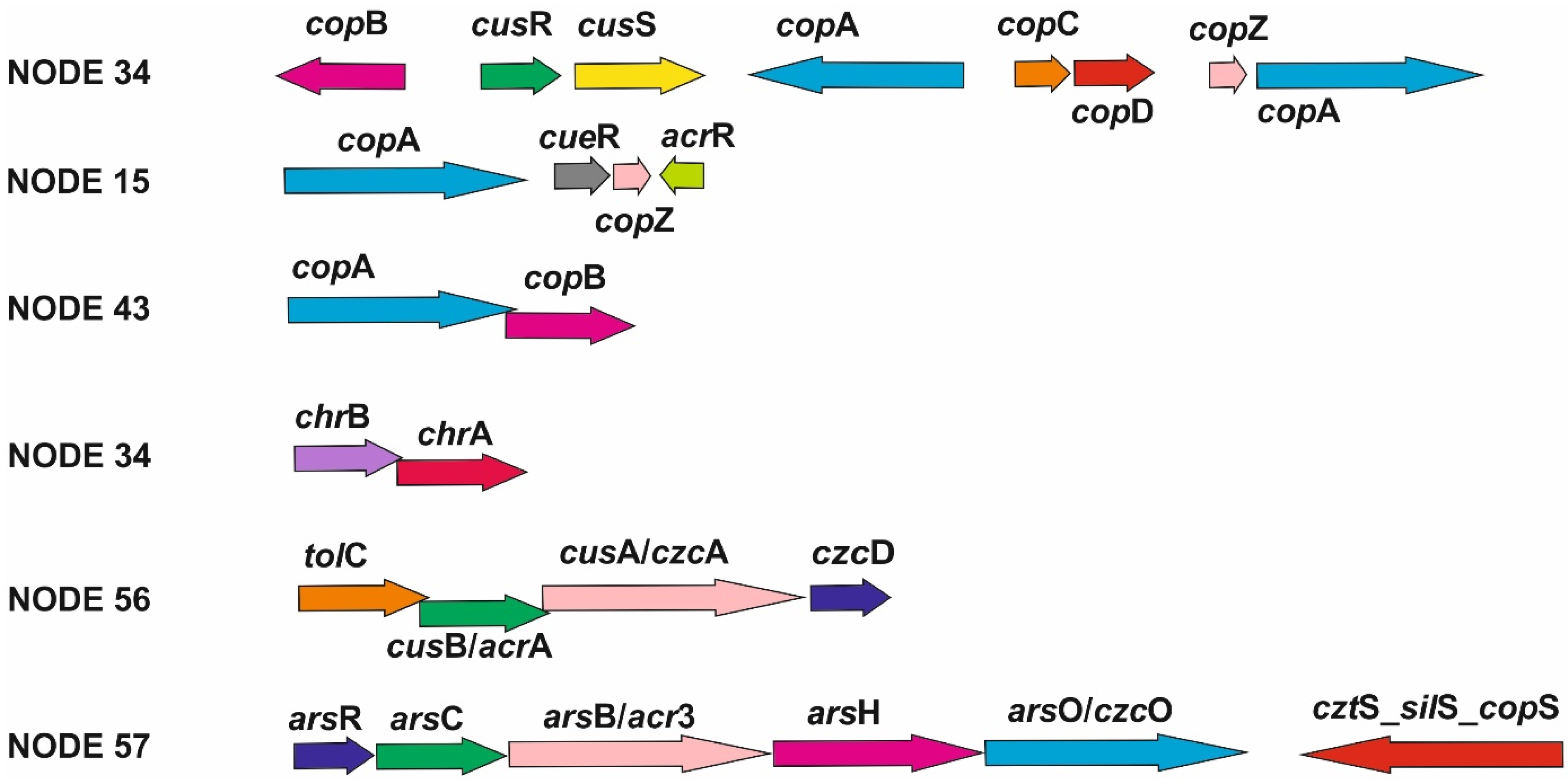
Multiple genes involved in heavy-metal-resistance mechanisms were identified on contig NODE 34, e.g., genes of the cop operon (copABCDZ, cusSR and pcoB) that are responsible for copper resistance; czcD, chrA and chrB which are involved in resistance mechanisms to high concentrations of cadmium, zinc, cobalt and chromium; zntA encoding a P-type ATPase that participates in the transport of zinc, cadmium and lead; and genes encoding the DmeF protein of the CDF protein family of efflux transporters. We have not been able to reliably determine the origin of the DNA due to the different results of the analyses (Figure 2, Tables S4 and S5). The mlplasmids v2.1.0 server determined this contig as chromosomal DNA; on the other hand, it showed a low average GC content and consists of the genes coding the IS6 family of transposases and a serine recombinase. The genes encoding the CopA and CopB proteins were also found on contig NODE 43, which was identified as plasmid DNA with a high probability. It carries genes encoding the replication protein RepM, the mobilization protein MobC and proteins of the IS48 and IS3 transposase families.
Heavy-metal resistance or efflux pump genes (czcA, czcD, tolC, arcA, cadR-pbrR, zntA and zitB) were also detected on other contigs, e.g., NODE 56 (Figure 2, Table S5), NODE 130, NODE 1947 (Table 1) and NODE 261 (Table S5). The mlplasmids server determined NODE 130 and NODE 1947 as plasmid-related DNA (Table S4). However, replication-protein-encoding genes have not been identified on any of these contigs. The conserved domain sequences of the IS6 and IS240 transposase families were found only on contig NODE 130. Contig NODE 261 showed a 99.37% similarity (100% coverage) to A. lwoffii FDAARGOS_557 plasmid DNA (CP054804.1) according to the Blastn analysis; however, the mlplasmids v2.1.0 server evaluated this sequence as being of chromosomal origin (Tables S4 and S5). In addition, this contig encodes FeoA and FeoB proteins involved in the transport of iron ions into the interior of bacterial cells [33].
Arsenic-resistance determinants of ars and smt operon were found on several contigs of putative chromosomal (NODE 15 and NODE 50) and plasmid origin (NODE 29, 48 and 57). However, only in the case of contig NODE 15 have we been able to reliably determine the chromosomal origin of the DNA. Contig NODE 50 showed significant similarities with plasmid pALWED3.1 (CP083572.1) using Blastn analysis and a lower average GC content. On the other hand, the mlplasmids v2.1.0 server determined this contig to be a sequence of chromosomal origin. The sequence of contig NODE 57 encodes genes for ArsH, ArsC, ArsO, ArsR and Acr3 proteins (Figure 2). Even though this sequence showed the highest similarity to Acinetobacter chromosomal DNA, the mlplasmids v2.1.0 server classified it as a plasmid-related sequence (Table 1).
Several other plasmid-encoded genes in the K1 genome were detected, e.g., crcB (NODE 1680) encoding a protein important for the maintaining fluoride concentrations inside bacterial cells [34]; telA involved in the resistance to tellurite (NODE 2568); a conserved domain sequence of terC encoding the integral membrane protein superfamily important for tellurium efflux from bacterial cells; and a gene encoding the Phenol_MetA_deg superfamily (NODE 211) involved in the MetA pathway of phenol degradation (Table 1).
The bacterial strain Acinetobacter sp. K1 carries the genes that are important for adaptation to alkaline pH, e.g., Na+/K+ antiporter genes, genes for cytochrome bd and genes for stress responses and production of cell wall components and bacterial capsules (Figure 3).
Acinetobacter sp. K1 also harbors genes important for the intracellular regulation of osmotic pressure. First, we detected the genes kup, mscL and kefA (mscS) encoding transport proteins that play a role in transport and subsequent accumulation of potassium cations [35] and/or organic solutes [36] in the bacterial cytoplasm and a conserved domain of ion transport protein family pfam00520. The K1 genome also encodes genes responsible for the synthesis of the compatible organic solutes betaine (betA, betB), proline (proA, proB, proC), glutamate (gdhA, glnA, gltB, gltD) and trehalose (otsA, otsB). Only genes encoding Kup, pfam00520, OtsA and OtsB proteins are plasmid-encoded (Table 1).
In the K1 genome, we found chromosomally encoded genes that are important for dealing with oxidative stress caused by various negative environmental conditions (such as high concentrations of some heavy metals [37]): sodB and sodC encoding superoxide dismutases, ahpC encoding alkylhydroperoxidase, osmC encoding organic hydroperoxide reductase, gshA encoding glutamate-cysteine ligase and gshB encoding glutathione synthetase.

4. Discussion

4.1. Taxogenomic Placement of the K1 Isolate

The bacterial isolate Acinetobacter sp. K1 was sampled from an environment characterized by extreme alkaline pH (11.6) and contamination with heavy metals (Hg: 10 mg/kg, Cu: 220 mg/kg, Cr: 400 mg/kg, V: 700 mg/kg, Pb: 150 mg/kg and As: 800 mg/kg) [14,15]. The initial identification of the K1 strain was carried out using MALDI-TOF MS by Kopčáková et al. [14]. The strain was classified as A. lwoffii based on the protein profile with a MALDI score of 1.961, which is below the value of 2 that is considered reliable for identification at the species level. The 16S rRNA gene sequence analysis also confirmed the highest similarity (99% identity, 96% coverage) of the K1 isolate to A. lwoffii DSM 2403 (NR 026209.1) [14].
In 1986, Bouvet and Grimont established the basics for the species classification of bacteria belonging to the genus Acinetobacter using DNA–DNA hybridization and comprehensive phenotypic testing [38]. In this study, we tried to reevaluate the classification of the K1 isolate as the A. lwoffii species using different phylogenetic approaches with different results. The K1 strain was identified as a potential new species based on the TYGS server analysis; however, it showed the highest similarity to the A. idrijaensis MII strain. A phylogenetic analysis based on the ANI similarity matrix showed that the closest relationship of the K1 strain was to the A. lwoffii NIPH478. However, Campos-Guillén et al. [39] demonstrated that the 16S rRNA gene of the A. idrijaensis MII strain is 99% identical to the A. lwoffii species, while the same being true for the K1 isolate. In addition, the taxonomic name idrijaensis was not published according to the rules of the International Code of Bacterial Nomenclature (Bacteriological Code) [40]. Similarly, Nemec [41] refuted the classification of the Acinetobacter strain GC2 as the species Acinetobacter mesopotamicus, which was proposed by Acer et al. [42]. Finally, Nemec [41] assigned this strain to the species A. lwoffii based on the ANI values and DNA–DNA hybridization. Because of these discrepancies, further analyses would be necessary for more precise species identification of these strains.

4.2. Genetic Determinants Important for Adaptation of the K1 Strain to Extreme Environmental Conditions

Various studies demonstrated the tolerance of bacterial strains belonging to the Acinetobacter spp. to heavy metals and their ability to survive in diverse environments [1,2,43,44,45]. The ability of the strain Acinetobacter sp. K1 to tolerate higher concentrations of heavy metals and an alkaline pH was confirmed experimentally in our previous work [17]. Our preliminary analyses also identified the complex plasmid population in the K1 isolate [17]. It has already been shown that plasmids could play a key role in the adaptability of Acinetobacter to their living conditions, including heavy metal resistance [46]. Moreover, some plasmids in Acinetobacter potentiate the accumulation and horizontal transfer of diverse accessory genes [47].
Bacterial genes involved in heavy-metal-resistance mechanisms are often organized into characteristic operon units. The cop operon is the best-studied operon unit, conferring bacterial tolerance to high copper concentrations [48,49]. This operon was detected on contig NODE 34 that we could not reliably identify as plasmid or chromosomal DNA using the various bioinformatic analyses. The discrepancy could be due to the mlplasmids v2.1.2 server being optimized for searching plasmid sequences in the A. baumannii species complex. Nevertheless, this contig encodes all the genes necessary to exclude copper ions from the cell (Figure 2). The presence of cusRS regulatory genes, as well as prediction by the mlplasmids v2.1.2 server, point to a chromosomal origin of this sequence [49]. In addition, the chr operon was found on this contig. The ChrA protein enables chromate efflux from the cell and the ChrB protein is a regulatory protein that responds to the concentration of chromium in the intracellular environment [50]. Such an arrangement of genes is characteristic for Acinetobacter spp. [50], but in various Acinetobacter species, we can also observe the presence of other genes that complement this operon [51]. Mindlin et al. [50] identified a recombination site XerC/XerD near this operon. We detected the conserved domain of the IS6 family of transposases in the contig sequence. Despite the likely chromosomal origin of this sequence, the presence of this enzyme suggests the possibility of horizontal gene transfer through transposition. Contig NODE 43 encoding the genes copA and copB is probably part of the plasmid DNA of the K1 genome. Genes involved in the resistance to copper are also present on probably chromosomal contig NODE 15. We can assume that the copper tolerance of the K1 strain determined in our previous study [17] is probably mediated by the cop operon genes distributed mainly within the chromosomal DNA of the K1 isolate.
Bacterial resistance to cobalt, zinc and cadmium is provided by the czc operon encoding the CzcABC proteins [52] that form an efflux pump. The genes coding CusB/AcrA and TolC protein were detected on contig NODE 56. Protein CusB/AcrA could function as a fusion periplasmic protein CzcB; TolC protein is an outer membrane protein like CzcC. These components, together with the inner membrane protein CzcA, can form a complex efflux pump of the CzcABC type and thus ensure the exclusion of Co2+, Zn2+ and Ca2+ ions from the cell, supported by the efflux pump CzcD, which is encoded by several contigs of chromosomal and plasmid origin. The tolerance of the K1 strain to cobalt and zinc (MIC 1 mM for cobalt and 2 mM for zinc) [17] is probably the result of cooperation of these determinants. However, the determinants responsible for HGT were only detected within contig NODE 34. Similar operons with an identical arrangement of czcABC genes (Figure 2) occur in various species of bacteria [53,54,55].
Operon ars (the components encoded by NODE 50 and 57) ensures the resistance to arsenic in various bacterial species [56]. Contig NODE 57 encodes the ArsC protein that catalyzes the reduction of arsenate to arsenite and the ArsH protein that reduces organoarsenical compounds. Arsenite can be excluded from the bacterial cell by the Acr3 protein performing the function of the typical ars operon protein, ArsB. Regulation of the operon unit is provided by the ArsR protein. The activity of the operon is supported by the protein ArsO, whose function is not yet precisely defined [57]. The cztS_silS_copS protein family contains a domain characteristic for bacterial signal proteins and is associated with the efflux systems of heavy metals such as copper, silver, cadmium and zinc [58,59,60]. Contig NODE 50 contains genes coding the three primary components that are necessary for the detoxification and elimination of arsenite in the bacterial cells. It is not clear whether the NODE 50 and 57 contigs are of plasmid origin; however, they carry genes encoding transposases. The tolerance of Acinetobacter sp. K1 to arsenic was examined in our previous study and the MIC of arsenic was determined as a value of 2.6 mM [61].
In the K1 genome, we failed to detect the complete mer operon, which plays an important role in mercury resistance. The genome encodes only the regulatory protein of this operon, MerR. According to our previous research, the MIC of mercury of the K1 strain was established as 0.025 mM [61]. Despite the absence of important genes of the mer operon, this isolate survived in the sewage sludge contaminated with mercury, in addition to other heavy metals. Our hypothesis for this phenomenon is that the K1 isolate survived thanks to the detoxification mechanisms of the bacterial community presented in the brown mud, enabling the survival of sensitive strains. Further studies will be necessary to better understand these findings.
Several studies documented that various Acinetobacter strains are able to successfully thrive in acidic or alkaline environments [62,63,64]. According to our previous study, the bacterial strain Acinetobacter sp. K1 was able to grow at a maximum pH of 9 in vitro [17]. Bacteria use various mechanisms to maintain the pH homeostasis of their intracellular environment. These mechanisms include, for example, increased expression of cation/proton antiporters [65,66]. In the K1 genome, Na+ and K+ ion antiporters were detected, such as the pfam07885 and pfam00520 protein families, located on contig NODE 57. In addition, the subunits A, B, C, D, E, F and G of the Na+/H+ antiporter were detected in the K1 genome. Some bacteria (E. coli) use an increased expression of cytochrome bd (Cyd) as a protective mechanism [67]. The gene coding cytochrome bd is encoded by contig NODE 34 in the K1 genome. Additionally, we compared the number of genes for stress responses among the strains of A. lwoffii, A. pseudolwoffii and A. idrijaensis (Figure 3a). The highest similarity was observed with environmental strains A. lwoffii ZS207, A. lwoffii UBA 2051, A. lwoffii UBA1649 and A. idrijaensis WKH.25 which have different origins (Table S1), but these strains are characterized by a lower number of genes of this group. Modification of the bacterial cell wall and the production of secondary polymers of the cell wall (teichuronopeptide and SlpA protein) are other possible mechanisms of the protection against high alkalinity [65,66]. Acinetobacter spp. synthesize bacterial capsules, which are composed of tightly arranged repeating units of polysaccharides. Production of this protective cover is primarily a defense mechanism of clinically significant types of bacteria, creating a barrier around the cell wall, thus providing protection against adverse environmental conditions (disinfection, drying, host immune response, etc.). They even confer resistance to various clinically relevant antimicrobial agents [3]. We observed a significantly lower number of genes involved in the formation of the cell wall components and capsules in the K1 genome compared to other A. lwoffii strains (Figure 3b). The highest similarity between gene numbers was observed between the same strains as in the case of genes involved in for stress responses. In addition, combinations of available protective mechanisms enable the survival of this strain up to a pH of 9 in vitro. One of the explanations for this observation could be that a highly alkaline environment of brown mud is probably inhabited by other alkali-tolerant microorganisms, providing protection to more sensitive bacterial strains, including the isolate Acinetobacter sp. K1. Bacterial survival based on their cooperation is also supported by biochemical tests performed using GEN III MicroPlate™ (Biolog, Hayward, CA, USA). The bacterial strain K1 is a non-fermenting strain that uses simple carboxylic acids as a carbon source instead of simple carbohydrates [14].
High osmotic pressure environments cause lethal turgor reduction and cytoplasm dehydration in bacteria. However, in microorganisms, two main mechanisms to manage osmotic pressure are observed: K+ accumulation in the cytoplasm and the biosynthesis and subsequent accumulation of various compatible organic solutes [68]. In a comparative genomic analysis of 312 Acinetobacter spp. strains [69], genes encoding the Kup system of potassium transport with low affinity [35] were found in all analyzed strains. While most of the analyzed strains have 2-4 potassium transport systems such as Kup, Ktr, Trk, Kdp and Ktr [69], the bacterial strain Acinetobacter sp. K1 harbors mechanosensitive channels MscL and MscS [70,71,72] and a conserved domain of the ion transport protein family pfam00520 in addition to the Kup system. The K1 strain can produce the compatible organic solutes betaine, trehalose, proline and glutamate which are not only involved in maintaining the bacterial cell osmotic balance but also in the stabilization of the cellular components including proteins during the high ionic stress [73].
In some cases, extreme environmental conditions lead to the production of reactive oxygen species, and massive accumulation in the bacterial cells is lethal [74]. Acinetobacter spp. were found to encode superoxide dismutases SodB or SodC [69,75] that catalyze the dismutation of O2 to O2 and H2O2 [76], which we also detected in the K1 strain. Bacterial strains of the genus Acinetobacter often encode enzymes catalyzing the conversion of H2O2 (catalases KatE and KatG, and alkylhydroperoxidase AhpC) and the repair of oxidized cysteine and methionine residues in bacterial proteins (disulfide isomerase DsbC and methionine sulfoxide reductase Msr) [69]. Bioinformatic analyses of the K1 genome confirmed only presence of the ahpC gene supplemented by the genes for organic hydroperoxide reductase (osmC/ohr) [77], glutamate-cysteine ligase (gshA) and glutathione synthetase (gshB) which can also protect bacteria against oxidative stress [78].
In addition to the proteins important for plasmid DNA replication, mobilization and transposition, we also detected genes of the competence system (pilA, pilQ, pilC, pilB, pilD, pilDm, pilT, pilM, pilN, pilO, pilP, pilY1, pilV, pilW, pilR, fimT and fimB) in the K1 genome, located in several contigs, which are important in the formation of the type IV pilus (T4P) [79]. We also detected the genes encoding proteins ComEC and ComFC, which are important for transport of single-stranded DNA across the inner membrane of the bacteria during the natural transformation process [79]. In conclusion, genetic determinants found in the K1 genome provide various possibilities for HGT, including conjugation, transposition and transformation. On the other hand, despite the frequent presence of plasmid DNA in the K1 genome, the genes important for various tolerance mechanisms to heavy metals tend to be encoded by the chromosomal DNA of this strain.

5. Conclusions

The bacterial strain Acinetobacter sp. K1 was isolated from alkaline and heavy=metal-contaminated technogenic soil produced by industrial activities near Ziar nad Hronom (Slovakia). The phylogenetic analyses suggest that this strain is potentially a new species of the Acinetobacter genus. A whole-genome analysis was used to decipher the molecular basis of the K1 strain’s survival in its extreme environment. The analysis confirmed the presence of the complete operon units for cop, czc, chr and ars, providing resistance to copper, zinc, cobalt, cadmium, chromium and arsenic. The lack of mercury resistance determinants in the K1 genome is probably the result of the presence of the complex bacterial community in the brown mud, in which the resistance mechanisms of other microorganisms provide protection to more sensitive bacterial strains. According to our analyses, despite the presence of multiple plasmids in the K1 genome, the genes responsible for the adaptation to extreme environmental conditions are mainly chromosomally encoded. Nevertheless, we detected other important genes potentially mediating horizontal gene transfer of resistance genes between the bacterial populations of the brown mud.

Supplementary Materials

The following supporting information can be downloaded at: https://www.mdpi.com/article/10.3390/life13081728/s1, Table S1: Comparison of the general genome characteristics of various Acinetobacter strains deposited in the GenBank database using the RAST server; Figure S1: Phylogenetic analysis of the 16S rRNA gene of the isolate Acinetobacter sp. K1 using the TYGS server; Table S2: The list of bacterial strains that were used to create the ANI similarity matrix and subsequent phylogenetic analysis; Table S3: ANI similarity matrix of the analyzed strains; Figure S2: UPGMA phylogenetic tree of different strains of A. lwoffii, A. idrijaensis MII, A. mesopotamicus DSM 26953, A. pecorum, Prolinoborus fasciculus CIP 103579 and Acinetobacter sp. K1 constructed based on the similarity matrix constructed from the ANI values between the analyzed strains; Table S4: Prediction of the inclusion of Acinetobacter sp. K1 genome contigs into chromosomal or plasmid DNA using the server mlplasmids v2.1.0 in correlation with the average GC content of individual contigs; Table S5: Genetic analysis of individual contig sequences of the Acinetobacter sp. K1 genome, which at the nucleotide level show similarities with Acinetobacter spp. plasmid DNA or encode genes for plasmid replication proteins, proteins involved in HGT processes or genes involved in various mechanisms of resistance and adaptation. The summary analysis was created by a combination of analyses using the servers, databases and online tools blastn, blastx, RAST, CD-Search (Conserved Domain Database), ORF finder, mlplasmids v2.1.0, Genomics %G~C Content Calculator.

Author Contributions

Conceptualization, P.P.; methodology, P.P.; formal analysis, N.P. and J.K.; investigation, N.P.; resources, M.K.; writing—original draft preparation, N.P.; writing—review and editing, N.P., P.P. and J.K.; visualization, N.P.; supervision, P.P.; project administration, P.P.; funding acquisition, P.P. All authors have read and agreed to the published version of the manuscript.

Funding

This work was supported by the Grant Agency of the Ministry of Education, Science, Research and Sport, Slovak Republic VEGA under Grant No. 1/0779/21.

Institutional Review Board Statement

Not applicable.

Informed Consent Statement

Not applicable.

Data Availability Statement

The data presented in this study are available on request from the corresponding author.

Conflicts of Interest

The authors declare no conflict of interest. The funders had no role in the design of the study; in the collection, analyses, or interpretation of data; in the writing of the manuscript; or in the decision to publish the results.

References

  1. Doughari, H.J.; Ndakidemi, P.A.; Human, I.S.; Benade, S. The Ecology, Biology and Pathogenesis of Acinetobacter spp.: An Overview. Microbes Environ. 2011, 26, 101–112. [Google Scholar] [CrossRef] [PubMed] [Green Version]
  2. Veress, A.; Nagy, T.; Wilk, T.; Kömüves, J.; Olasz, F.; Kiss, J. Abundance of mobile genetic elements in an Acinetobacter lwoffii strain isolated from Transylvanian honey sample. Sci. Rep. 2020, 10, 2969. [Google Scholar] [CrossRef] [PubMed] [Green Version]
  3. Singh, J.K.; Adams, F.G.; Brown, M.H. Diversity and function of capsular polysaccharide in Acinetobacter baumannii. Front. Microbiol. 2018, 9, 3301. [Google Scholar] [CrossRef]
  4. Robino, P.; Bert, E.; Tramuta, C.; Cerruti Sola, S.; Nebbia, P. Isolation of Acinetobacter lwoffii from a Lovebird (Agapornis roseicollis) with severe respiratory symptoms. Schweiz. Arch. Tierheilkd 2005, 147, 267–269. [Google Scholar] [CrossRef]
  5. Regalado, N.G.; Martin, G.; Antony, A.J. Acinetobacter lwoffii: Bacteremia associated with acute gastroenteritis. Travel Med. Infect. Dis. 2009, 7, 316–317. [Google Scholar] [CrossRef]
  6. Turton, J.F.; Shah, J.; Ozongwu, C.; Pike, R. Incidence of Acinetobacter species other than A. baumannii among clinical isolates of Acinetobacter: Evidence for emerging species. J. Clin. Microbiol. 2010, 48, 1445–1449. [Google Scholar] [CrossRef] [Green Version]
  7. Nakwan, N.; Wannaro, J.; Nakwan, N. Multidrug-resistant Acinetobacter lwoffii infection in neonatal intensive care units. Res. Rep. Neonatol. 2011, 1, 1–4. [Google Scholar] [CrossRef] [Green Version]
  8. Kulkarni, G.; Challa, J. The first Indian viridescent Acinetobacter lwoffii. Indian J. Med. Microbiol. 2021, 39, 130–132. [Google Scholar] [CrossRef]
  9. Kabala, C.; Galka, B.; Jezierski, P. Assessment and monitoring of soil and plant contamination with trace elements around Europe’s largest copper ore tailings impoundment. Sci. Total Environ. 2020, 738, 139918. [Google Scholar] [CrossRef]
  10. Uzarowicz, Ł.; Charzyński, P.; Greinert, A.; Hulisz, P.; Kabała, C.; Kusza, G.; Kwasowski, W.; Pędziwiatr, A. Studies of technogenic soils in Poland: Past, present, and future perspectives. Soil Sci. Ann. 2020, 71, 281–299. [Google Scholar] [CrossRef]
  11. Cimermanova, M.; Pristas, P.; Piknova, M. Biodiversity of Actinomycetes from Heavy Metal Contaminated Technosols. Microorganisms 2021, 9, 1635. [Google Scholar] [CrossRef] [PubMed]
  12. Abdel-El-Haleem, D. Minireview Acinetobacter: Environmental and biotechnological applications. Afr. J. Biotechnol. 2003, 2, 71–74. [Google Scholar] [CrossRef] [Green Version]
  13. Chojnacka, K. Biosorption and bioaccumulation—The prospects for practical applications. Environ. Int. 2010, 36, 299–307. [Google Scholar] [CrossRef] [PubMed]
  14. Kopčáková, A.; Stramová, Z.; Kvasnová, S.; Godany, A.; Perhacova, Z.; Pristas, P. Need for database extension for reliable identification of bacteria from extreme environments using MALDI TOF mass spectrometry. Chem. Pap. 2014, 68, 1435–1442. [Google Scholar] [CrossRef]
  15. Rasulov, O.; Schwarz, M.; Horváth, A.; Zoirov, F.; Fayz, N. Analysis of soil contamination with heavy metals in (the three) highly contaminated industrial zones. SN Appl. Sci. 2020, 2, 2013. [Google Scholar] [CrossRef]
  16. Towner, K.J. Biology of Acinetobacter spp. In Acinetobacter, Microbiology, Epidemiology, Infections, Management; Bergogne-Bérézin, E., Joly-Guillou, M.-L., Towner, K.J., Eds.; CRC Press: Boca Raton, FL, USA, 1996; pp. 13–36. [Google Scholar]
  17. Šipošová, N.Š.; Liptáková, V.; Kvasnová, S.; Kosorínová, P.; Pristaš, P. Genetic diversity of Acinetobacter spp. adapted to heavy metal polluted environments. Nova Biotechnol. Chim. 2017, 16, 42–47. [Google Scholar] [CrossRef]
  18. Okonechnikov, K.; Golosova, O.; Fursov, M.; UGENE Team. Unipro UGENE: A unified bioinformatics toolkit. Bioinformatics 2012, 28, 1166–1167. [Google Scholar] [CrossRef] [Green Version]
  19. Golosova, O.; Henderson, R.; Vaskin, Y.; Gabrielian, A.; Grekhov, G.; Nagarajan, V.; Oler, A.J.; Quiñones, M.; Hurt, D.; Fursov, M.; et al. Unipro UGENE NGS pipelines and components for variant calling, RNA-seq and ChIP-seq data analyses. PeerJ 2014, 2, e644. [Google Scholar] [CrossRef]
  20. Altschul, S.F.; Gish, W.; Miller, W.; Myers, E.W.; Lipman, D.J. Basic local alignment search tool. J. Mol. Biol. 1990, 215, 403–410. [Google Scholar] [CrossRef]
  21. Aziz, R.K.; Bartels, D.; Best, A.A.; DeJongh, M.; Disz, T.; Edwards, R.; Formsma, K.; Gerdes, S.; Glass, E.; Kubal, M.; et al. The RAST Server: Rapid annotations using subsystems technology. BMC Genom. 2008, 9, 75. [Google Scholar] [CrossRef] [Green Version]
  22. Overbeek, R.; Olson, R.; Pusch, G.D.; Olsen, G.J.; Davis, J.J.; Disz, T.; Edwards, R.A.; Gerdes, S.; Parrello, B.; Shukla, M.; et al. The SEED and the Rapid Annotation of microbial genomes using Subsystems Technology (RAST). Nucleic. Acids. Res. 2014, 42, 206–214. [Google Scholar] [CrossRef]
  23. Brettin, T.; Davis, J.J.; Disz, T.; Edwards, R.A.; Gerdes, S.; Olsen, G.J.; Olson, R.; Overbeek, R.; Parrello, B.; Pusch, G.D.; et al. RASTtk: A modular and extensible implementation of the RAST algorithm for building custom annotation pipelines and annotating batches of genomes. Sci. Rep. 2015, 5, 8365. [Google Scholar] [CrossRef] [PubMed] [Green Version]
  24. Meier-Kolthoff, J.P.; Göker, M. TYGS is an automated high-throughput platform for state-of-the-art genome-based taxonomy. Nat. Commun. 2019, 10, 2182. [Google Scholar] [CrossRef] [PubMed] [Green Version]
  25. Goris, J.; Konstantinidis, K.T.; Klappenbach, J.A.; Coenye, T.; Vandamme, P.; Tiedje, J.M. DNA–DNA hybridization values and their relationship to whole-genome sequence similarities. Int. J. Syst. Evol. Microbiol. 2007, 57, 81–91. [Google Scholar] [CrossRef] [PubMed] [Green Version]
  26. Rodriguez-R, L.M.; Konstantinidis, K.T. Estimating coverage in metagenomic data sets and why it matters. ISME J. 2014, 8, 2349–2351. [Google Scholar] [CrossRef] [Green Version]
  27. Moore, R.M.; Harrison, A.O.; McAllister, S.M.; Polson, S.W.; Wommack, K.E. Iroki: Automatic customization and visualization of phylogenetic trees. PeerJ 2020, 8, e8584. [Google Scholar] [CrossRef] [PubMed]
  28. Marchler-Bauer, A.; Lu, S.; Anderson, J.B.; Chitsaz, F.; Derbyshire, M.K.; DeWeese-Scott, C.; Fong, J.H.; Geer, L.Y.; Geer, R.C.; Gonzales, N.R.; et al. CDD: A Conserved Domain Database for the functional annotation of proteins. Nucleic Acids Res. 2011, 39, 225–229. [Google Scholar] [CrossRef] [Green Version]
  29. Arredondo-Alonso, S.; Rogers, M.R.C.; Braat, J.C.; Verschuuren, T.D.; Top, J.; Corander, J.; Willems, R.J.L.; Schürch, A.C. mlplasmids: A user-friendly tool to predict plasmid- and chromosome-derived sequences for single species. Microb. Genom. 2018, 4, e000224. [Google Scholar] [CrossRef] [Green Version]
  30. Lefort, V.; Desper, R.; Gascuel, O. FastME 2.0: A comprehensive, accurate, and fast distance-based phylogeny inference program. Mol. Biol. Evol. 2015, 32, 2798–2800. [Google Scholar] [CrossRef] [PubMed] [Green Version]
  31. Farris, J.S. Estimating phylogenetic trees from distance matrices. Am. Nat. 1972, 106, 645–667. Available online: https://www.jstor.org/stable/2459725 (accessed on 25 July 2023). [CrossRef]
  32. Holland, B.R.; Huber, K.T.; Dress, A.; Moulton, V. Delta plots: A tool for analyzing phylogenetic distance data. Mol. Biol. Evol. 2002, 19, 2051–2059. [Google Scholar] [CrossRef] [PubMed] [Green Version]
  33. Kim, H.; Lee, H.; Shin, D. The FeoA protein is necessary for the FeoB transporter to import ferrous iron. Biochem. Biophys. Res. Commun. 2012, 423, 733–738. [Google Scholar] [CrossRef] [PubMed]
  34. Baker, J.L.; Sudarsan, N.; Weinberg, Z.; Roth, A.; Stockbridge, R.B.; Breaker, R.R. Widespread Genetic Switches and Toxicity Resistance Proteins for Fluoride. Science 2012, 335, 233–235. [Google Scholar] [CrossRef] [PubMed] [Green Version]
  35. Epstein, W. The KdpD sensor kinase of Escherichia coli responds to several distinct signals to turn on expression of the Kdp transport system. J. Bacteriol. 2016, 198, 212–220. [Google Scholar] [CrossRef] [Green Version]
  36. Levina, N.; Tötemeyer, S.; Stokes, N.R.; Louis, P.; Jones, M.A.; Booth, I.R. Protection of Escherichia coli cells against extreme turgor by activation of MscS and MscL mechanosensitive channels: Identification of genes required for MscS activity. EMBO J. 1999, 18, 1730–1737. [Google Scholar] [CrossRef]
  37. Khalid, M.; Hassani, S.; Abdollahi, M. Metal-induced oxidative stress: An evidence-based update of advantages and disadvantages. Curr. Opin. Toxicol. 2020, 20–21, 55–68. [Google Scholar] [CrossRef]
  38. Bouvet, P.J.M.; Grimont, P.A.D. Taxonomy of the genus Acinetobacter with the recognition of Acinetobacter baumannii sp. nov., Acinetobacter haemolyticus sp. nov., Acinetobacter johnsonii sp. nov. and Acinetobacter junii sp. nov. And emended descriptions of Acinetobacter calcoaceticus and Acinetobacter lwoffii. Int. J. Syst. Evol. Microbiol. 1986, 36, 228–240. [Google Scholar] [CrossRef] [Green Version]
  39. Campos-Guillén, J.; Pérez, J.C.; Medina, J.A.C.; Vera, C.M.; Rosas, L.M.S.; Gutiérrez, C.L.; Salinas, I.G.; Ramírez, M.R.H.; Alonso, G.S.; Hernández, A.C.; et al. Draft Genome Sequence of the Mercury-Resistant Bacterium Acinetobacter idrijaensis Strain MII, Isolated from a Mine-Impacted Area, Idrija, Slovenia. Genome Announc. 2014, 2, e01177-14. [Google Scholar] [CrossRef] [Green Version]
  40. Lapage, S.P.; Sneath, P.H.A.; Lessel, E.F.; Skerman, V.B.D.; Seelinger, H.P.R.; Clark, W.A. International Code of Nomenclature of Bacteria (1990 Revision); American Society for Microbiology: Washington, DC, USA, 1992; ISBN 1-55581-039-X. [Google Scholar]
  41. Nemec, A. Strain “Acinetobacter mesopotamicus” GC2 does not represent a novel species, but belongs to the species Acinetobacter lwoffii as revealed by whole-genome sequence-based analysis. Curr. Microbiol. 2021, 78, 369–370. [Google Scholar] [CrossRef]
  42. Acer, Ö.; Güven, K.; Poli, A.; Di Donato, P.; Leone, L.; Buono, L.; Güven, R.G.; Nicolaus, B.; Finore, I. Acinetobacter mesopotamicus sp. nov., Petroleum-degrading Bacterium, Isolated from Petroleum-Contaminated Soil in Diyarbakir, in the Southeast of Turkey. Curr. Microbiol. 2020, 77, 3192–3200. [Google Scholar] [CrossRef]
  43. Rajbanshi, A. Study on heavy metal resistant bacteria in Guheswori sewage treatment plant. Our Nat. 2008, 6, 52–57. [Google Scholar] [CrossRef]
  44. Raja, C.E.; Elvam, G.S.; Omine, K. Isolation, identification and characterization of heavy metal resistent bacteria from sewage. In International Joint Symposium on Geodisaster Prevention and Geoenvironment in Asia; JS: Fukuoka, Japan, 2009; pp. 205–211. [Google Scholar]
  45. El-Sayed, M.H. Multiple heavy metal and antibiotic resistance of Acinetobacter baumannii strain HAF—13 isolated from industrial effluents. Am. J. Microbiol. Res. 2016, 4, 26–36. [Google Scholar]
  46. Maslova, O.; Mindlin, S.; Beletsky, A.; Mardanov, A.; Petrova, M. Plasmids as Key Players in Acinetobacter Adaptation. Int. J. Mol. Sci. 2022, 23, 10893. [Google Scholar] [CrossRef]
  47. Moran, R.A.; Liu, H.; Doughty, E.L.; Hua, X.; Cummins, E.A.; Liveikis, T.; McNally, A.; Zhou, Z.; van Schaik, W.; Yu, Y. GR13-type plasmids in Acinetobacter potentiate the accumulation and horizontal transfer of diverse accessory genes. Microb. Genom. 2022, 8, mgen000840. [Google Scholar] [CrossRef]
  48. Brown, N.L.; Barrett, S.R.; Camakaris, J.; Lee, B.T.; Rouch, D.A. Molecular genetics and transport analysis of the copper-resistance determinant (pco) from Escherichia coli plasmid pRJ1004. Mol. Microbiol. 1995, 17, 1153–1166. [Google Scholar] [CrossRef]
  49. Williams, C.L.; Neu, H.M.; Alamneh, Y.A.; Reddinger, R.M.; Jacobs, A.C.; Singh, S.; Abu-Taleb, R.; Michel, S.L.J.; Zurawski, D.V.; Merrell, D.S. Characterization of Acinetobacter baumannii Copper Resistance Reveals a Role in Virulence. Front. Microbiol. 2020, 11, 16. [Google Scholar] [CrossRef]
  50. Mindlin, S.; Petrenko, A.; Petrova, M. Chromium resistance genetic element flanked by XerC/XerD recombination sites and its distribution in environmental and clinical Acinetobacter strains. FEMS Microbiol. Lett. 2018, 365, fny047. [Google Scholar] [CrossRef] [Green Version]
  51. Viti, C.; Marchi, E.; Decorosi, F.; Giovannetti, L. Molecular mechanisms of Cr(VI) resistance in bacteria and fungi. FEMS Microbiol. Rev. 2013, 38, 633–659. [Google Scholar] [CrossRef] [Green Version]
  52. Hussain, S.; Khan, M.; Sheikh, T.M.M.; Mumtaz, M.Z.; Chohan, T.A.; Shamim, S.; Liu, Y. Zinc Essentiality, Toxicity, and Its Bacterial Bioremediation: A Comprehensive Insight. Front. Microbiol. 2022, 13, 900740. [Google Scholar] [CrossRef]
  53. Gutiérrez-Barranquero, J.A.; de Vicente, A.; Carrión, V.J.; Sundin, G.W.; Cazorla, F.M. Recruitment and Rearrangement of Three Different Genetic Determinants into a Conjugative Plasmid Increase Copper Resistance in Pseudomonas syringae. Appl. Environ. Microbiol. 2013, 79, 1028–1033. [Google Scholar] [CrossRef] [Green Version]
  54. Ou, H.-Y.; Kuang, S.N.; He, X.; Molgora, B.M.; Ewing, P.J.; Deng, Z.; Osby, M.; Chen, W.; Xu, H.H. Complete genome sequence of hypervirulent and outbreak-associated Acinetobacter baumannii strain LAC-4: Epidemiology, resistance genetic determinants and potential virulence factors. Sci. Rep. 2015, 5, 8643. [Google Scholar] [CrossRef] [PubMed] [Green Version]
  55. Mindlin, S.; Petrenko, A.; Kurakov, A.; Beletsky, A.; Mardanov, A.; Petrova, M. Resistance of permafrost and modern Acinetobacter lwoffii strains to heavy metals and arsenic revealed by genome analysis. BioMed Res. Int. 2016, 2016, 3970831. [Google Scholar] [CrossRef] [PubMed] [Green Version]
  56. Suhadolnik, M.L.S.; Salgado, A.P.C.; Scholte, L.L.S.; Bleicher, L.; Costa, P.S.; Reis, M.P.; Dias, M.F.; Ávila, M.P.; Barbosa, F.A.R.; Chartone-Souza, E.; et al. Novel arsenic-transforming bacteria and the diversity of their arsenic-related genes and enzymes arising from arsenic-polluted freshwater sediment. Front. Microbiol. 2017, 7, 11231. [Google Scholar] [CrossRef] [PubMed]
  57. Hao, X.; Zhu, J.; Rensing, C.; Liu, Y.; Gao, S.; Chen, W.; Huang, Q.; Liu, Y.-R. Recent advances in exploring the heavy metal(loid) resistant microbiome. Comput. Struct. Biotechnol. 2021, 19, 94–109. [Google Scholar] [CrossRef] [PubMed]
  58. Urano, H.; Yoshida, M.; Ogawa, A.; Yamamoto, K.; Ishihama, A.; Ogasawara, H. Cross-regulation between two common ancestral response regulators, HprR and CusR, in Escherichia coli. Microbiology 2017, 163, 243–252. [Google Scholar] [CrossRef]
  59. Urano, H.; Yamamoto, K.; Ogasawara, H.; Ishihama, A.; Umezawa, Y. Cooperative regulation of the common target genes between H2O2-sensing YedVW and Cu2+-sensing CusSR in Escherichia coli. Microbiology 2015, 161, 729–738. [Google Scholar] [CrossRef]
  60. Gudipaty, S.A.; Larsen, A.S.; Rensing, C.; McEvoy, M.M. Regulation of Cu(I)/Ag(I) efflux genes in Escherichia coli by the sensor kinase CusS. FEMS Microbiol. Lett. 2012, 130, 30–37. [Google Scholar] [CrossRef] [Green Version]
  61. Pristas, P.; Kvasnova, S.; Judova, J.; Perhacova, Z.; Vidova, B.; Sramkova, Z.; Godany, A. Non-ferrous metal industry waste disposal sites as a source of poly-extremotolerant bacteria. Nova Biotechnol. Chim. 2015, 14, 62–68. [Google Scholar] [CrossRef] [Green Version]
  62. Yavankar, S.P.; Pardesi, K.R.; Chopade, B.A. Species distribution and physiological characterization of Acinetobacter genospecies from healthy human skin of tribal population in India. Indian J. Med. Microbiol. 2007, 25, 336–345. [Google Scholar] [CrossRef]
  63. Li, T.; Luo, D.; Ning, N.; Liu, X.; Chen, F.; Zhang, L.; Bao, C.; Li, Z.; Li, D.; Gu, H.; et al. Acinetobacter baumannii adaptation to the host pH microenvironment is mediated by allelic variation in a single residue of BauA protein. PNAS Nexus 2023, 2, pgad079. [Google Scholar] [CrossRef]
  64. Jung, J.; Park, W. Acinetobacter species as model microorganisms in environmental microbiology: Current state and perspectives. Appl. Microbiol. Biotechnol. 2015, 99, 2533–2548. [Google Scholar] [CrossRef]
  65. Padan, E.; Bibi, E.; Ito, M.; Krulwich, T.A. Alkaline pH homeostasis in bacteria: New insights. Biochim. Biophys. Acta Bioenerg. 2005, 1717, 67–88. [Google Scholar] [CrossRef] [PubMed] [Green Version]
  66. Guo, J.; Ma, Z.; Gao, J.; Zhao, J.; Wei, L.; Liu, J.; Xu, N. Recent advances of pH homeostasis mechanisms in Corynebacterium glutamicum. World J. Microbiol. Biotechnol. 2019, 35, 192. [Google Scholar] [CrossRef] [PubMed]
  67. Krulwich, T.A.; Sachs, G.; Padan, E. Molecular aspects of bacterial pH sensing and homeostasis. Nat. Rev. Microbiol. 2011, 9, 330–343. [Google Scholar] [CrossRef] [PubMed] [Green Version]
  68. Gunde-Cimerman, N.; Plemenitaš, A.; Oren, A. Strategies of adaptation of microorganisms of the three domains of life to high salt concentrations. FEMS Microbiol. Rev. 2018, 42, 353–375. [Google Scholar] [CrossRef] [Green Version]
  69. Zhao, Y.; Wei, H.-M.; Yuan, J.-L.; Xu, L.; Sun, J.-Q. A comprehensive genomic analysis provides insights on the high environmental adaptability of Acinetobacter strains. Front. Microbiol. 2023, 14, 1177951. [Google Scholar] [CrossRef]
  70. Moe, P.C.; Blount, P.; Kung, C. Functional and structural conservation in the mechanosensitive channel MscL implicates elements crucial for mechanosensation. Mol. Microbiol. 1998, 28, 583–592. [Google Scholar] [CrossRef] [PubMed]
  71. Pivetti, C.D.; Yen, M.-R.; Miller, S.; Busch, W.; Tseng, Y.-H.; Booth, I.R.; Saier, M.H. Two families of mechanosensitive channel proteins. Microbiol. Mol. Biol. Rev. 2003, 67, 66–85. [Google Scholar] [CrossRef] [Green Version]
  72. Naismith, J.H.; Booth, I.R. Bacterial mechanosensitive channels—MscS: Evolution’s solution to creating sensitivity in function. Annu. Rev. Biophys. 2012, 41, 157–177. [Google Scholar] [CrossRef] [Green Version]
  73. Kempf, B.; Bremer, E. Uptake and synthesis of compatible solutes as microbial stress responses to high-osmolality environments. Arch. Microbiol. 1998, 170, 319–330. [Google Scholar] [CrossRef]
  74. Fasnacht, M.; Polacek, N. Oxidative stress in bacteria and the central dogma of molecular biology. Front. Mol. Biosci. 2021, 8, 671037. [Google Scholar] [CrossRef]
  75. Steimbrüch, B.A.; Sartorio, M.G.; Cortez, N.; Albanesi, D.; Lisa, M.-N.; Repizo, G.D. The distinctive roles played by the superoxide dismutases of the extremophile Acinetobacter sp. Ver3. Sci. Rep. 2022, 12, 4321. [Google Scholar] [CrossRef] [PubMed]
  76. Fridovich, I. Superoxide radical and superoxide dismutases. Annu. Rev. Biochem. 1995, 64, 97–112. [Google Scholar] [CrossRef] [PubMed]
  77. Cussiol, J.R.R.; Alegria, T.G.P.; Szweda, L.I.; Netto, L.E.S. Ohr (organic hydroperoxide resistance protein) possesses a previously undescribed activity, lipoyl-dependent peroxidase. J. Biol. Chem. 2010, 285, 21943–21950. [Google Scholar] [CrossRef] [PubMed] [Green Version]
  78. Masip, L.; Veeravalli, K.; Georgiou, G. The many faces of glutathione in bacteria. Antioxid. Redox Signal. 2006, 8, 753–762. [Google Scholar] [CrossRef]
  79. Hu, Y.; Zheng, J.; Zhang, J. Natural transformation in Acinetobacter baumannii W068: A genetic analysis reveals the involvements of the CRP, XcpV, XcpW, TsaP, and TonB2. Front. Microbiol. 2022, 12, 738034. [Google Scholar] [CrossRef]
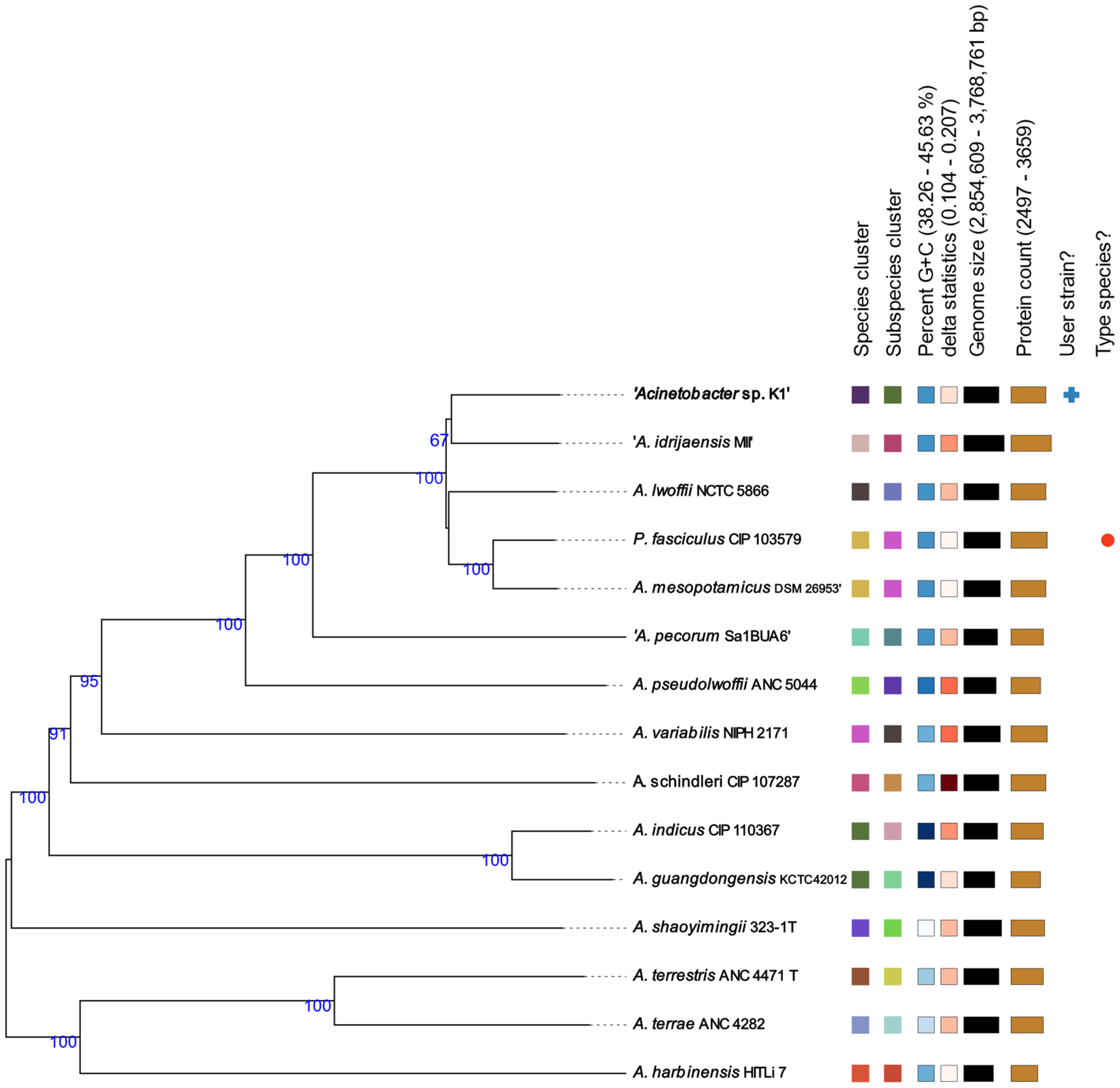
Figure 1. The GBDP phylogenetic analysis based on the whole-genome sequence comparison of different species of the genus Acinetobacter using the TYGS server [24]. Tree inferred with FastME 2.1.6.1 [30] from GBDP distances calculated from genome sequences. The branch lengths are scaled in terms of GBDP distance formula d5. The numbers above branches are GBDP pseudo-bootstrap support values > 60% from 100 replications, with an average branch support of 91.9%. The tree was rooted at the midpoint [31]. Leaf labels are annotated by affiliation to dDDH (digital DNA:DNA hybridization) species and subspecies clusters, genomic G+C content, delta values, overall genome sequence length and number of proteins. Delta statistics permit assessment of accuracy in terms of tree-likeness; the lower the delta value, the greater the accuracy [32].
Life 13 01728 g001
Figure 2. Organization of identified operon units mediating tolerance to heavy metals and metalloids in the strain Acinetobacter sp. K1.
Life 13 01728 g002
Figure 3. Comparison of the number of stress response genes (a) and genes involved in cell wall and capsule biosynthesis (b) in A. lwoffii strains using the RAST server [21,22,23].
Life 13 01728 g003
Table 1. Plasmid-related sequences of the K1 genome encoding genes important for adaptation of Acinetibacter sp. K1 to high concentrations and/or degradation of heavy metals, metalloids, phenol and antibiotics according to the mlplasmids v2.1.2 server [29].
ContigSize
(bp)
GenBank
Accession No.
ReplicationHGTResistance
NODE 4-234,994NZ_JALGQY010000005-IS5_3 transposase superfamilyAcrA, AcrB;
RND_mfp superfamily
NODE 1378,976NZ_JALGQY010000014--CzcD
NODE 1762,178NZ_JALGQY010000018-IS3, IS5, IS21, IS481, ISNCY transposase superfamilies;
XerC integrase
TauE
NODE 2944,217NZ_JALGQY010000029-LGT_TIGR03299 phage/plasmid-like protein;
COG5377 phage protein superfamily;
AlpA superfamily;
Inovirus_Gp2 superfamily;
IS66 transposase family;
XerC integrase
Fur; SmtA
NODE 3827,781NZ_JALGQY010000038.1rep_pAB02_ORF2IS3, IS481, IS6 transposase families; XerC integraseRcnR-FrmR-like_DUF156;
MefA protein family; AcrR;
GST_N_GTT1/GST_C superfamily
NODE 3926,463NZ_JALGQY010000039-IS5_2 transposase familyRND_mfp superfamily
OtsA, OtsB
NODE 4318,602NZ_JALGQY010000042.1RepMIS481 and IS3 transposase families
Relaxase; MobC
CopA, CopB
NODE 4814,852NZ_JALGQY010000047-IS3 transposase familyArsR/SmtB family;
TauE
NODE 558994NZ_JALGQY010000052.1Rep_3; RepM;
rep_pAB02_ORF2;
MobA_MobL;
TraA_Ti
TerC superfamily;
SUL1 superfamily
NODE 578544NZ_JALGQY010000054-IS5_3 transposase familyArsH, ArsC, ArsO, ArsR, Acr3; cztS_silS_copS
Pfam00520
NODE 666322NZ_JALGQY010000055.1Rep_3; RepM;
rep_pAB02_ORF2;
-RamA
NODE 1303934NZ_JALGQY010000063-IS6, IS240 transposase familyCadR-PbrR
Kup
NODE 2113202NZ_JALGQY010000065-Phage_antiter_QBeta-lactamase;
Phenol_MetA_deg
NODE 3752618NZ_JALGQY010000069-Serine recombinase
PinE recombinase
ABC_trans_N
NODE 16801426NZ_JALGQY010000076--CrcB
NODE 19471329NZ_JALGQY010000077--CzcD; MerR; CadR-PbrR; ZntA; ZitB
NODE 25681144NZ_JALGQY010000083-MobA_MobL;
TraA_Ti
TelA
Disclaimer/Publisher’s Note: The statements, opinions and data contained in all publications are solely those of the individual author(s) and contributor(s) and not of MDPI and/or the editor(s). MDPI and/or the editor(s) disclaim responsibility for any injury to people or property resulting from any ideas, methods, instructions or products referred to in the content.