Review

Possible Mechanisms Linking Obesity, Steroidogenesis, and Skeletal Muscle Dysfunction

by
Anna F. Sheptulina
1,2,
Karina Yu Antyukh
3,
Anton R. Kiselev
4,*,
Natalia P. Mitkovskaya
3,5 and
Oxana M. Drapkina
1,2
1
Department of Fundamental and Applied Aspects of Obesity, National Medical Research Center for Therapy and Preventive Medicine, 101990 Moscow, Russia
2
Department of Therapy and Preventive Medicine, A.I. Evdokimov Moscow State University of Medicine and Dentistry, 127473 Moscow, Russia
3
Republican Scientific and Practical Center of Cardiology, 220036 Minsk, Belarus
4
National Medical Research Center for Therapy and Preventive Medicine, 101990 Moscow, Russia
5
Department of Cardiology and Internal Diseases, Belarusian State Medical University, 220116 Minsk, Belarus
*
Author to whom correspondence should be addressed.
Life 2023, 13(6), 1415; https://doi.org/10.3390/life13061415
Submission received: 28 May 2023 / Revised: 12 June 2023 / Accepted: 16 June 2023 / Published: 19 June 2023

Abstract

:
Increasing evidence suggests that skeletal muscles may play a role in the pathogenesis of obesity and associated conditions due to their impact on insulin resistance and systemic inflammation. Skeletal muscles, as well as adipose tissue, are largely recognized as endocrine organs, producing biologically active substances, such as myokines and adipokines. They may have either beneficial or harmful effects on the organism and its functions, acting through the endocrine, paracrine, and autocrine pathways. Moreover, the collocation of adipose tissue and skeletal muscles, i.e., the amount of intramuscular, intermuscular, and visceral adipose depots, may be of major importance for metabolic health. Traditionally, the generalized and progressive loss of skeletal muscle mass and strength or physical function, named sarcopenia, has been thought to be associated with age. That is why most recently published papers are focused on the investigation of the effect of obesity on skeletal muscle function in older adults. However, accumulated data indicate that sarcopenia may arise in individuals with obesity at any age, so it seems important to clarify the possible mechanisms linking obesity and skeletal muscle dysfunction regardless of age. Since steroids, namely, glucocorticoids (GCs) and sex steroids, have a major impact on the amount and function of both adipose tissue and skeletal muscles, and are involved in the pathogenesis of obesity, in this review, we will also discuss the role of steroids in the interaction of these two metabolically active tissues in the course of obesity.

1. Introduction

The global prevalence of obesity has now reached epidemic proportions. This condition currently affects >2 billion people worldwide, and more than 1.9 billion adults globally are overweight [1]. According to the experts’ estimates, if the current incidence trend continues, by 2030, 60% of the world’s population (that is, 3.3 billion people) may be overweight or obese [2].
Obesity is a multifactorial condition, resulting mainly from the imbalance between energy intake and expenditure and leading to the excess accumulation of fat in the body. This, in turn, causes various metabolic and neurohumoral disturbances maintaining the vicious circle of excess fat accumulation and increasing the likelihood of a variety of conditions accompanied by high incidence of morbidity and mortality. They include, in particular, cardiovascular diseases, type 2 diabetes mellitus (T2DM) [3,4], chronic kidney disease [5], non-alcoholic fatty liver disease [6], diseases of the musculoskeletal system [7], depression [8], and different types of cancer [9]. The pathophysiology of obesity is considered to be complex and to include the interaction between genetic, environmental, neurohormonal, microbial, and psychosocial factors [2,4,10]. For a long period of time, it was believed that all of the negative health effects associated with obesity are due to the excess accumulation of adipose tissue in the body. However, increasing evidence suggests that a decrease in skeletal muscle mass and quality, as well as skeletal muscle function, may also be relevant. Muscles play an important role in energy homeostasis, and they may be involved in the pathogenesis of obesity due to their impact on insulin resistance, energy expenditure, and systemic inflammation [10].
Generally, the loss of muscle mass and function, i.e., sarcopenia, was considered a feature of aging. However, according to the ESPEN and EASO Consensus Statement on the Definition and Diagnostic Criteria for Sarcopenic Obesity [11], sarcopenia may arise in individuals with obesity at any age. Indeed, T.L. Silva et al. [12] showed that the prevalence of sarcopenia among young and middle-aged adults (n = 108, mean age: 43 ± 11.7 years) of both sexes ranged between 11.1% and 13.9% depending on the criteria being used for the diagnosis of sarcopenia (low muscle mass or low muscle strength, correspondingly). Additionally, in the study conducted by E. Poggiogalle et al. [13], the prevalence of sarcopenia in a cohort of 727 participants with obesity (141 males, mean age: 45.6 ± 13.5 years; 586 females, mean age 45.8 ± 13.6 years) was 34.8% in males and 50.1% in females. This condition, characterized by the simultaneous presence of sarcopenia and obesity, was named sarcopenic obesity, and is believed to be associated with worse health outcomes than obesity and sarcopenia on their own, including the higher incidence of osteoporosis [11,14].
It is well known that serum glucocorticoid (GC) levels are elevated in individuals with obesity, and this contributes to the development of insulin resistance, hyperglycemia, and dyslipidemia [15]. The accompanied increase in circulating free fatty acids may result in the lipid infiltration of the liver, pancreas, and skeletal muscles, leading to the deterioration of their function. Obesity is also associated with disturbances in sex steroid synthesis and signaling, negatively affecting fertility [16,17]. On the other hand, owing to the well-described effects of androgens and estrogens on muscle mass, structure, and function, the impaired sex steroid balance in obesity may result in the deterioration of muscle health [18]. The fatty infiltration of skeletal muscles is defined as myosteatosis, and is characterized by the decrease in muscle strength, regeneration capacity, and the profile of biologically active substances produced by myocytes, i.e., myokines. It was shown that myokines acting together with other organokines, such as adipokines and hepatokines, may take part in the pathogenesis of obesity and associated conditions [19,20]. Currently, they are extensively studied as potential prognostic markers and therapeutic targets [20,21]. We provide a comprehensive review on the structural and functional changes of skeletal muscle in the course of obesity, as well as the impact of steroids on this process. To the best of our knowledge, this is the first multi-faceted review focused on the interactions among adipose tissue, skeletal muscle, and steroids in patients with obesity.

2. Skeletal Muscles in Obesity

There are three main adipose tissue depots in the body: subcutaneous, visceral, and ectopic. Ectopic corresponds to the accumulation of adipose tissue in internal organs or other compartments of the body, including skeletal muscles, which are originally unappropriated for the storage of fat [14]. Depending on the localization, fat depots in skeletal muscles may be classified as intermuscular (adipocytes localized between muscle groups), intramuscular (adipocytes located between the muscle fibers), and intramyocellular (IMCL, lipids stored within the myocytes) (Figure 1) [22]. Usually, adipocytes localized between the muscle fibers (intramuscular fat) and muscle groups (literally intermuscular fat) are combined by the term “intermuscular” (INTM) fat as both of them represent the fat depots situated underneath the deep fascia of the muscle, are formed by adipocytes of the same origin, and have similar significance for the muscle metabolic state [23]. In this review, we will also use the term “intermuscular fat” to denote both depots.

2.1. Metabolic Properties of INTM Fat

Currently, INTM fat is most often considered to be related to metabolic disturbances. Indeed, several studies have established that the increase in the amount of INTM fat is significantly associated with a decrease in muscle mass and strength, as well as insulin sensitivity [24,25]. Several studies indicate that INTM fat is functionally quite similar to visceral adipose tissue (VAT), particularly with regard to its ability to induce and maintain inflammation and influence insulin sensitivity [24,26]. For instance, both INTM fat and VAT are characterized by the increased expression of interleukin (IL)-6, tumor necrosis factor (TNF), and plasminogen activator inhibitor 1 (PAI1) [27]. At the same time, the results of direct comparisons of the qualitive and quantitative composition of inflammatory proteins secreted by INTM fat, VAT, or subcutaneous adipose tissue (SAT) in individuals with obesity clearly indicate that INTM depots secrete substantially greater amounts of inflammatory cytokines (such as interferon-(IFN) γ, IL-2, IL-5, and IL-10) and chemokines compared to VAT or SAT [28]. Moreover, it was shown that the lipolytic capacity of INTM fat was significantly greater than the rate of lipolysis in subcutaneous adipose tissue and was similar to that in VAT [29,30]. Thus, INTM fat may increase the concentration of free fatty acids in the interstitial space, negatively influencing the local muscle microenvironment and promoting muscle insulin resistance. In an in vitro study, it was shown that proteins secreted by INTM fat were able to increase the concentration of 1,2-diacylglycerols within the myotubes, intramyocellular lipids associated with impaired insulin sensitivity [29]. Sachs et al. [26] demonstrated that the metabolic properties of INTM fat can change and become more unfavorable with the aggravation of metabolic health. Further, taking into account the role of insulin signaling in muscle protein synthesis, the impaired insulin sensitivity of skeletal muscles resulting from myosteatosis may directly contribute to the decrease in muscle strength and mass [31].
It is suggested that INTM fat may originate from several cell sources, in particular fibro-adipogenic progenitors, muscle satellite stem cells, adipose-derived stem cells, and bone-marrow-derived mesenchymal stem cells.

2.1.1. Fibro-Adipogenic Progenitors

It is believed that adipocytes constituting the INTM depots originate mainly from fibro-adipogenic progenitors (FAPs) [31]. These cells received their name because of their ability to differentiate either into fibroblasts or adipocytes. In in vitro studies, it was shown that the fibrogenic differentiation of FAPs can be induced by transforming growth factor-β (TGF-β), whereas their differentiation into adipocytes can be stimulated by insulin, 3-isobutyl-1-methylxanthine, and dexamethasone [32,33,34]. Except for being involved in muscle tissue regeneration under physiological conditions [35], these cells take part in the processes of muscle atrophy during disease and aging [36,37]. Moreover, animal studies showed that FAPs are essential for the maintenance of the amount and function of muscle stem cells, which are responsible for muscle regeneration and growth [38,39].
It is suggested that FAP differentiation into fibroblasts or adipocytes in the settings of insulin resistance and impaired metabolism can contribute to the muscle remodeling, degeneration, and fibrosis taking place in obesity [40]. For instance, it was shown that thrombospondin 1 (THBS1), an adipokine secreted by adipocytes of white adipose tissue, is capable of promoting the proliferation of FAPs in obese mice [41]. In addition, chronic inflammation, being an important pathogenic mechanism in the development of obesity and associated conditions, may sustain the activated state of FAP, thus contributing to the development and progression of the so-called fibrofatty degeneration of skeletal muscles in obesity. It is worth noting that this condition is accompanied not only by the decrease in muscle function, but also by the increase in the insulin resistance of the muscle tissue. Such effects may be due to the cytokines expressed either by M1 (IL-1β polarized) or M2 (IL-4 polarized) macrophages infiltrating muscle tissue, and their influence on the processes of FAP differentiation and adipogenesis [42]. Finally, it was shown that adipocytes derived from FAPs were characterized by the impaired phosphorylation of insulin receptors, which contributed to the increase in peripheral insulin resistance [43].

2.1.2. Other Cell Types as Possible Source of INTM Fat

B.H. Goodpaster et al. [25] suggested that, along with FAPs, several other cell types may participate in the generation of INTM fat depots, including muscle satellite stem cells (MSCs) and adipose-derived stem cells (ASCs), which migrate into the muscles from other adipose tissue depots. Although MSCs represent a major source of skeletal muscle cells and are responsible for muscle tissue regeneration and growth in the postnatal period, in vitro studies have shown that MSCs do differentiate into adipocytes in the presence of adipogenic factors [44,45]. However, the exact factors capable of stimulating the MSC differentiation into adipocytes in vivo remain unknown. For instance, L. Guo et al. [46] showed that intramuscular preadipocytes might hamper the differentiation of MSCs via C-Jun N-terminal kinases (JNK)/mitogen-activated protein kinase (MAPK) pathway and simultaneously facilitate their lipid deposition through peroxisome proliferator–activated receptor (PPAR) signaling in chickens.
Finally, ASCs and bone-marrow-derived mesenchymal stem cells constitute the third and even fourth possible sources of INTM fat depots, though the exact mechanisms, as well as factors contributing to their migration into the muscle tissue bed, require further investigation [47,48].

2.2. Intramyocellular Fat Depots

Intramyocellular (IMCL) fat depots refer to the accumulation of fat within the myofibers themselves. Inside the muscle fibers, lipids are stored mainly in the form of triacylglycerols (TAG), being localized within the lipid droplets. Other components of such lipid droplets include diacylglycerols (DAG), sphingolipids, and phospholipids. These depots are thought to be important for muscle contraction during physical exercises and to be associated with the impaired insulin sensitivity in patients with obesity and/or T2DM, as well as in elderly and sedentary individuals. Indeed, in their study, J.J. Dubé et al. [49] showed that the completion of a 16-week moderate exercise training program was accompanied by the increase in IMCL fat content by 21% in 25 older obese individuals. Additionally, the authors noticed that physical activity contributed to the increase in the content of triacylglycerols in skeletal muscles and to the decrease in the content of both diacylglycerols and ceramides within the IMCL depots. This, in turn, resulted in the improvement of insulin sensitivity. It is worth noting that the above-mentioned effect was independent of weight reduction, thus suggesting that physical activity itself may have a beneficial effect on the metabolic health of obese adults.
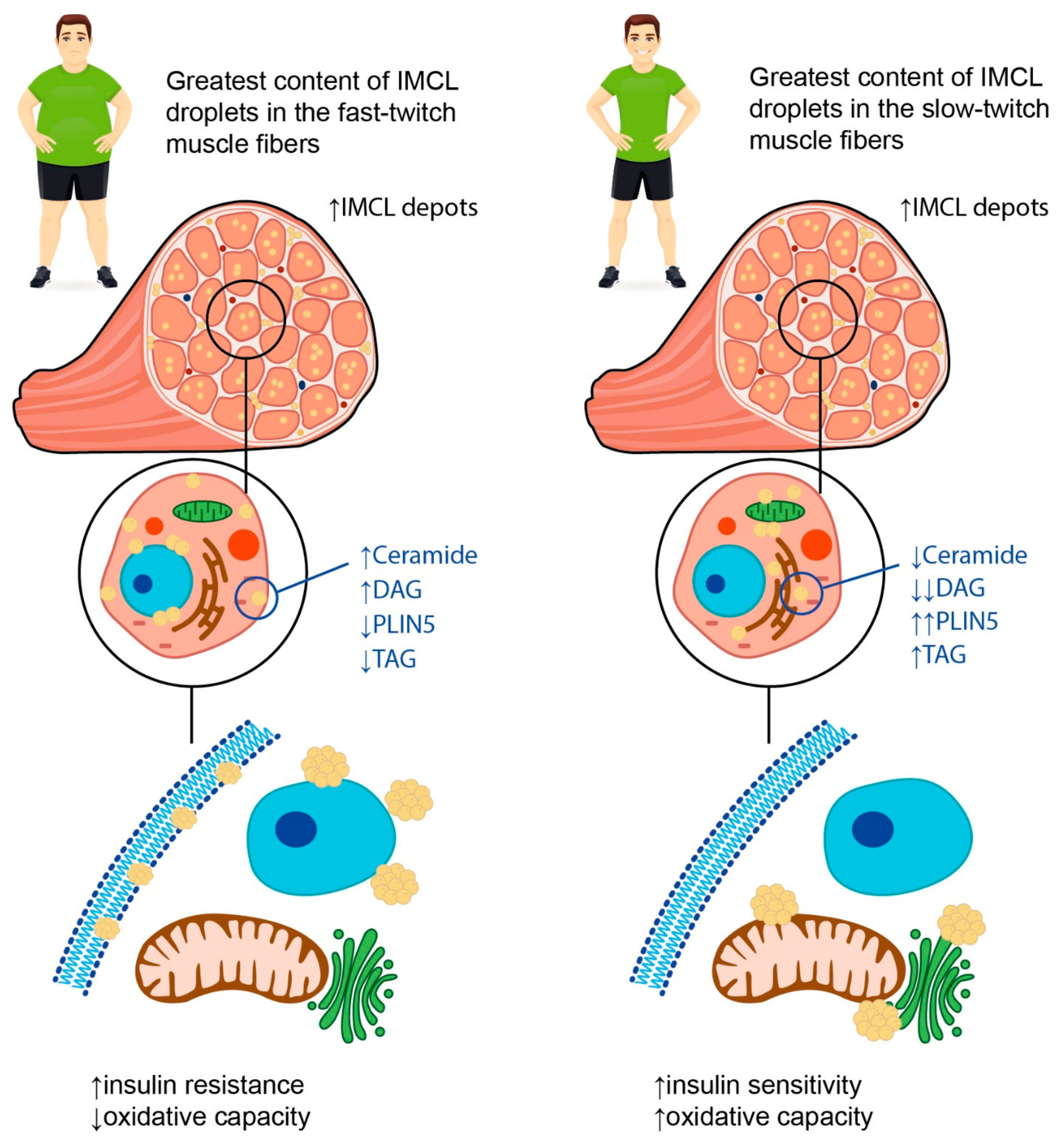
These described findings are in line with the so called “athlete’s paradox”; that is, the higher amounts of IMCL fat in the muscles are observed not only in patients with obesity and/or T2DM, but also in endurance-trained athletes (Figure 2) [49,50,51]. There are several possible explanations for this phenomenon. First, the increase in the IMCL fat amount in trained athletes is necessary to provide enough substrates for energy metabolism during regular physical exercise, whereas in obese patients and patients with T2DM, the IMCL depots serve as storage for excess fat [52]. Second, the content of IMCL depots does differ between the endurance-trained athletes and obese adults. In particular, the latter have greater amounts of DAGs and ceramides within the IMCL depots. These substances are regarded as lipid metabolites, and the increase in their content within the myofibers is suggested to be associated with impaired insulin sensitivity, possibly due to the decrease in muscle oxidative capacity [49]. Accordingly, B.C. Bergman et al. [51] demonstrated that physical exercise promoted the decrease in di-saturated DAG concentration both within the membrane and cytosol, and this resulted in the insulin sensitization of skeletal muscles. Moreover, A Gemmink et al. [53] showed that trained athletes had higher concentrations of perilipin 5 (PLIN5), a lipid droplet coating protein, compared to patients with T2DM. Although the content of PLIN5 was not significantly associated with the size and number of lipid droplets, it was positively correlated with the muscle oxidative capacity.
Third, the rate of TAG synthesis within the myofibers differs between the endurance-trained athletes, insulin resistant obese patients, and lean sedentary individuals, being the highest in the former. These data suggest that high rates of IMCL fat synthesis, which influences intramuscular lipid structuring and localization, can help to prevent the formation of insulin resistance [50,51]. In relation to lipid droplet partitioning, it was established that the accumulation of triglycerides in the sarcolemma and nucleus was negatively corelated with insulin sensitivity, whereas the localization of IMCL fat depots in close relation to mitochondria and the endoplasmic reticulum was not associated with the development of insulin resistance and appeared to facilitate the oxidation of TAGs [51,54]. Similar results were obtained in the study by M.C. Devries et al. [55] that included 11 obese and 12 lean sedentary women who underwent the 12-week endurance training program. Though the endurance training did not result in weight loss, it led to the increase in muscle oxidative capacity. Additionally, after the completion of the endurance training program, the localization of the IMCL fat depots changed in both groups: the content of IMCL lipid droplets in the subsarcolemmal region reduced, whereas the content of IMCL fat depots in the intermyofibrillar compartment increased. Finally, the saturated vs. unsaturated fatty acid ratio within the IMCL fat depots may also be relevant. Indeed, D. Kahn et al. [54] showed that patients with T2DM had the highest content of saturated fatty acids within the myofibers, and this appeared to be significantly associated with insulin resistance.
Interestingly, the degree of lipid accumulation within IMCL depots in obesity may depend on the type of muscle fibers. In their study, N. Umek et al. [56] demonstrated that the fast-twitch muscles of obese mice (i.e., gastrocnemius and intermediate plantaris muscles) were characterized by the greatest content of IMCL lipid droplets, while in slow-twitch muscles (soleus muscle), there was no significant lipid accumulation. Furthermore, the authors described an increase in the expression of fast-type myosin heavy chain in the slow-twitch soleus muscles of the obese mice.
In summary, one can conclude that not only the amount, but also the composition and localization of IMCL fat depots within the myofibers may be crucial for the development of insulin resistance and the deterioration of metabolic health in obese patients. Additionally, the capacity of skeletal muscles for lipid oxidation may be the factor determining the association between IMCL fat content and insulin resistance and may depend on muscle composition as well as metabolic flexibility.

2.3. Skeletal Muscle Composition and Metabolic Flexibility in Obesity

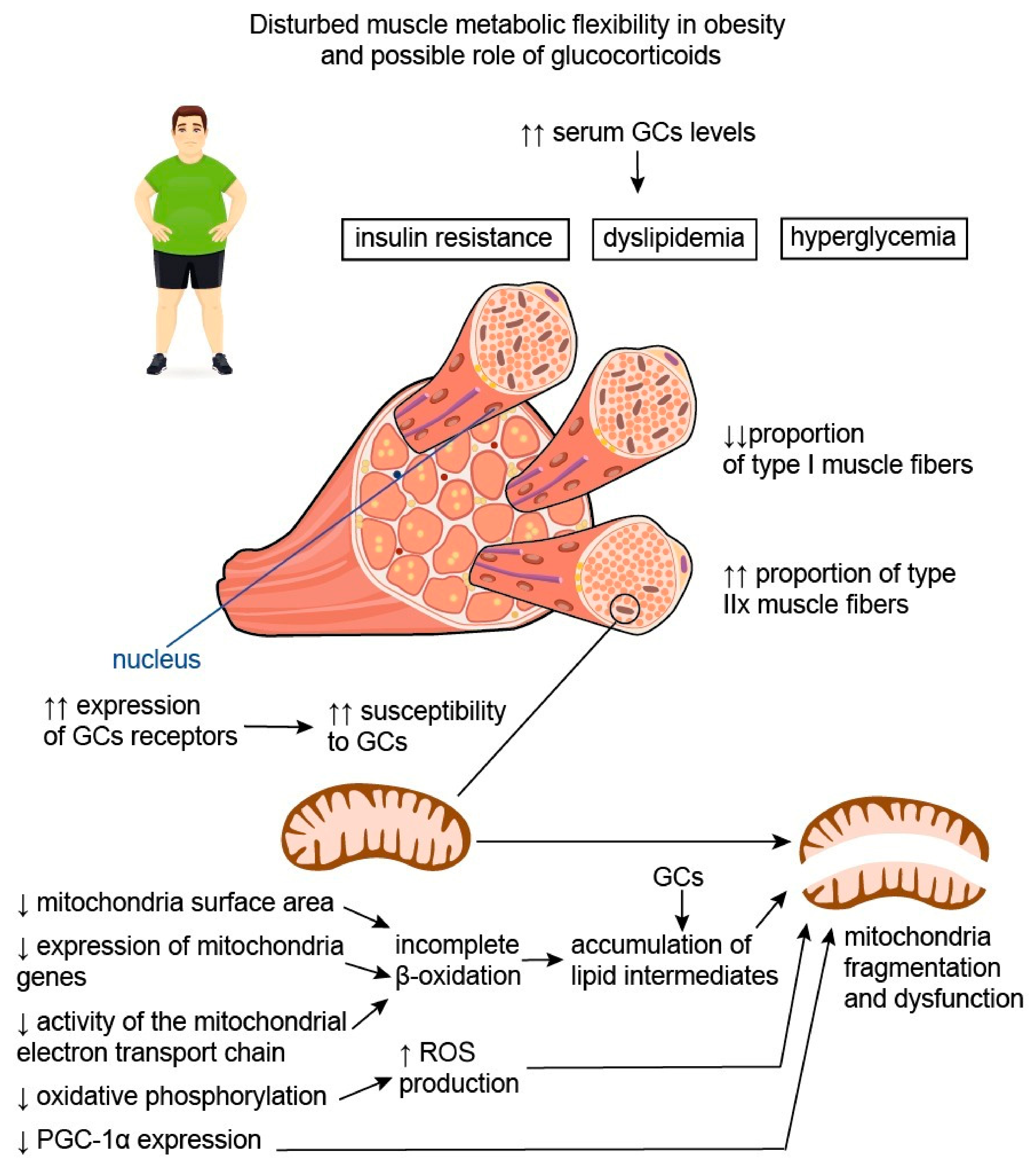
Skeletal muscle metabolism is crucial for total daily energy expenditure; along with the brain, liver, heart, and gastrointestinal tract, skeletal muscles are regarded as major contributors to overall metabolic rate [57]. This is due to their participation in resting energy expenditure and ability to increase energy expenditure with physical exercise [58]. Depending on metabolic activity, fatigability, mitochondrial content, and movement rates, muscle fibers may be classified into two different types: type I (slow-twitch or oxidative) and type II (fast-twitch or glycolytic) muscle fibers [59,60]. Type II fibers can be further classified as type IIa fibers, also called fast-oxidative glycolytic fibers or intermediate fibers, and IIx fibers, also termed fast glycolytic fibers. The latter contain high amounts of glycogen and use primarily anaerobic metabolism to generate adenosine triphosphate (ATP), so they are characterized by relatively small numbers of mitochondria [59,61]. It is believed that the distribution of muscle fiber types within muscle groups varies substantially among individuals and may be influenced by both environmental (nutrition, physical exercises, etc.) and biological factors (age, gender, genetics, etc.), thus determining the individual differences in daily energy expenditure, as well as the individual risk for sustaining a positive energy balance, and, as a consequence, weight gain [58,61]. Indeed, it was established that patients with obesity have lower proportions of type I muscle fibers characterized by an increased number of mitochondria and, at the same time, greater proportions of type IIx fibers compared with lean individuals [61,62,63]. These data are confirmed by the results of D.A. Reiter et al., suggesting that obesity is associated with reduced muscle energetic efficiency with a predominance of glycolysis over oxidation processes [64].
In addition to disturbed proportions of different muscle fibers, mitochondria content and muscle tissue metabolism are also altered in obesity. To define the ability of muscle fibers to use either glucose or fatty acids as a fuel depending on the availability of the substrates, physical activity, and conditional changes in metabolic or energy demand, the term “muscle metabolic flexibility” was proposed [65]. This concept, named the Randle Cycle, was first suggested by P.J. Randle et al. in 1963 [66] to describe the fuel selection in the transition from fasting to fed states and to characterize its role in insulin sensitivity and metabolic health. Nowadays, the term “metabolic flexibility” is used with respect to physiological adaptability and encompasses several other tissues and metabolic circumstances.
Increasing evidence suggests that an inability to adapt to metabolic stimuli, such as insulin signaling or fatty acid exposure, may lead to the decrease in muscle oxidative capacity characteristic of obesity [67,68,69]. In their study including 28 juvenile Iberian pigs fed either a control or a high-fructose, high-fat (HFF) diet for 10 weeks, H.C. Spooner et al. [70] demonstrated that the HFF diet resulted in decreased IMCL fat content and in the formation of less oxidative skeletal muscle phenotype, reflecting the disturbed ability of skeletal muscles to use lipids as a fuel. These changes were similar to the effects of detraining or muscle atrophy, indicating the reduced capacity of muscles to perform endurance-type exercises [71]. These effects may be followed by a long-term decrease in muscle mass and strength. Indeed, G.V. Hernandez et al. [72] showed that there was a reduction in plasma creatinine levels in juvenile Iberian pigs fed an HFF diet during 10 weeks, suggesting a tendency towards deterioration in muscle growth in this animal model.
Disturbed muscle oxidative capacity is believed to be associated with both changes in the proportions of muscle fiber types and mitochondrial dysfunction (Figure 3). It was shown that obesity is accompanied by a decrease in mitochondrial surface area by, on average, 20–60%, as well as by the substantially lower expression of mitochondrial genes and metabolites [67,68,73]. Accumulating data suggest that obesity has an unfavorable impact on the activity of the mitochondrial electron transport chain and oxidative phosphorylation, as well as stimulating the production of reactive oxygen species (ROS), facilitating mitochondrial fragmentation and mutations in mitochondrial DNA [74]. This may be due to mitochondria lipid overload, which causes incomplete β-oxidation and, consequently, the accumulation of lipid intermediates, such as ceramides. It was shown that ceramides could affect membrane potential, electron transport, and mitochondrial morphology, resulting in mitochondrial dysfunction [74]. In these settings, the expression of peroxisome proliferator-activated receptor–gamma coactivator-1alpha (PGC-1α), known to be involved in mitochondrial biogenesis, and to exert antioxidant action is being downregulated in order to prevent further mitochondrial function deterioration due to the accumulation of damaged mitochondrial DNA [75]. PGC-1α is also known to regulate lipid oxidation, energy homeostasis, and insulin sensitivity. In particular, it was demonstrated that the inactivation of PGC-1α expression in skeletal muscles in mice led to a shift from oxidative type I and IIa muscle fibers toward type IIx and IIb glycolytic muscle fibers [76]. On the contrary, elevated levels of PGC-1α in the skeletal muscles of transgenic mice were shown to protect against age-related obesity and T2DM [77]. The results of these studies reflect the important role of PGC-1α in promoting effective muscle energy metabolism and regulating energy balance in the body.
Interestingly, weight loss does not necessary lead to an increase in mitochondrial number and the recovery of mitochondrial function. In their study, E.V. Menshikova et al. [78] assessed the impact of weight loss through calorie restriction (n = 7) or moderate-intensity exercise (n = 10) on skeletal muscle mitochondrial content, mitochondrial enzyme activities, and insulin resistance in 17 overweight or obese individuals aged 60–75 years. Interestingly, although both interventions resulted in weight loss and improved insulin sensitivity, only physical activity was associated with an increase in mitochondria content and the activity of enzymes involved in mitochondrial electron transport chain and fatty acid oxidation within the skeletal muscle.

2.4. Myokines in Obesity

Recently it was established that skeletal muscles produce some biologically active substances called myokines, which facilitate the crosstalk between muscles and some other organs and tissues, such as adipose tissue, brain, liver, bone, and kidney. The production and secretion of nearly all such molecules, namely, irisin/fibronectin type III domain-containing protein 5 (FNDC5), IL-15, IL-6, brain-derived neurotrophic factor (BDNF), and myonectin, are upregulated during muscle contraction and physical exercise [75]. Here, we will discuss the role of the two most studied PGC-1α-dependent myokines, irisin and myostatin, in the deterioration of muscle structure and function in obesity.

2.4.1. Irisin

A recent meta-analysis concluded that circulating irisin levels were higher in obese individuals compared to healthy controls, though they appeared to be affected by ethnicity and age [79]. Irisin is produced by the proteolytical cleavage of FNDC5 during muscle contraction and physical exercise. It is a PGC-1α-dependent myokine, able to increase the browning of white adipose tissue by increasing the expression of uncoupling protein 1 (UCP-1). Adipose tissue and liver are also able to secret this molecule in small amounts [80]. The elevation in circulating irisin levels in patients with obesity without T2DM may be explained by the existence of obesity-induced metabolic dysfunction, particularly insulin resistance. In this case elevated irisin levels may represent an attempt to maximize glucose uptake by skeletal muscle and prevent hyperglycemia [81,82]. Interestingly, it was shown that after the onset of T2DM, the expression of FNDC5 in the muscle of treatment-naïve patients in vivo was reduced by ~15%. However, in in vitro experiments, myotubes isolated from these patients were capable of expressing FNDC5 in greater amounts than myotubes taken from lean individuals [81]. In contrast, it was shown that the irisin secretion from adipocytes in patients with obesity is lower than in lean controls [81,83]. However, as the body fat mass is substantially increased in patients with obesity, the overall levels of irisin secreted by adipose tissue in patients with obesity may be quite similar or even superior to those in lean individuals.
It is worth noting that plasma irisin levels may be significantly decreased following weight loss due to bariatric surgery. This effect may be explained by the decrease in fat-free mass during weight loss and, as a consequence, lower FNDC5 mRNA expression in skeletal muscle. In support of this suggestion, the irisin concentration returned to baseline levels in patients who regained the original weight [84,85]. However, the contribution of adipose tissue to the increase in total irisin levels upon weight regain cannot be excluded. These data also confirm the notion that an increase in irisin levels in patients with obesity may mirror metabolic disturbances characteristic of these patients, and may be directed at the compensation of these abnormalities [20].
According to J. Jia et al. [79], irisin levels were higher in young participants compared to older ones, possibly due to the age-related decline in muscle function. These authors further found out that irisin levels were higher in obese individuals compared to controls when they were from Africa, while no significant differences were described in European, Asian, or American populations. These inconsistencies may result from genetic factors capable of influencing the levels of irisin, as well as from the differences in body mass index (BMI) criteria used to diagnose overweight and obesity in the Asian population.
Moreover, it was shown that irisin expression may be associated with some anti-inflammatory markers [86]. In particular, irisin was able to suppress the expression of pro-inflammatory cytokines, nuclear factor-kappa B (NF-κB), TNF-α, and IL-6. In addition, irisin reduced the monocyte chemoattractant protein 1 (MCP-1) expression in cultured adipocytes with the subsequent attenuation of macrophage migration in the presence of irisin. Irisin was also able to induce the transition from the M1 (pro-inflammatory) macrophage phenotype to the M2 (anti-inflammatory) phenotype [86,87].
Therefore, taking into account the above-mentioned information, irisin seems to play a protective role in the development of metabolic dysfunction in obesity directed at the attenuation of insulin resistance and inflammation, as well as at the improvement of energy metabolism. However, its concentration may be decreased in case of sarcopenic obesity.

2.4.2. Myostatin

Myostatin, also known as growth and differentiation factor 8 (GDF8), is a member of the TGF-β superfamily. It is expressed predominantly in skeletal muscle, and its secretion and production are inhibited during muscle contraction and exercise [88]. Except for being involved in muscle atrophy [89], myostatin is regarded as a potential modulator of metabolic homeostasis acting through the regulation of adipose tissue function [90,91]. With regard to its effects, myostatin may be considered an irisin antagonist. For instance, it was demonstrated that the inhibition of myostatin was able to decelerate the development of insulin resistance and obesity in mice fed a high-fat diet, possibly due to the intensification of lipolysis and mitochondrial lipid oxidation in liver and adipose tissue. Moreover, it was shown that the inactivation of myostatin resulted in the formation of brown adipose tissue in the white adipose tissue of myostatin knockout mice [92]. This mouse line was also characterized by the upregulation of the expression and phosphorylation of adenosine monophosphate (AMP)-activated protein kinase (AMPK) in muscle, leading to the activation of PGC-1α and FNDC5 [93]. A study by M. Amor et al. reported an increase in serum myostatin concentrations in patients with obesity compared to lean individuals with no differences in myostatin expression in adipose tissue [90]. The authors showed that circulating myostatin levels were positively correlated with insulin resistance, while muscle myostatin gene expression was strongly associated with the expression of metabolic genes, particularly the insulin receptor substrate-1 (IRS-1) gene and sterol regulatory element binding transcription factor 1 (SREBTF1) gene [90]. Interestingly, the inhibition of myostatin expression in mice was accompanied by an improvement in insulin sensitivity: myostatin knockout mice were even more insulin sensitive than wild-type mice, suggesting that myostatin’s effect on insulin signaling may be independent of muscle mass [94]. Accordingly, physical exercises, bariatric surgery [95], and a calorie-restricted Dietary Approaches to Stop Hypertension (DASH) diet [96], due to their well-known beneficial effects on insulin sensitivity, were demonstrated to suppress myostatin production in skeletal muscles.
Accumulating data suggest that myostatin is expressed in the human reproductive system and may have different functions. For instance, myostatin is involved in the regulation of steroidogenesis, cell proliferation, the formation of extracellular matrix, and gonadotrophin responsiveness, thus influencing the process of cell differentiation in emerging follicles [97]. In addition, it is suggested that myostatin may be involved in the pathogenesis of some ovarian diseases, particularly polycystic ovary syndrome and ovarian hyperstimulation syndrome [98,99].
In view of the important role of myostatin in muscle atrophy, sarcopenia, and fat accumulation, this substance represents a promising direction for future research with respect to aging and obesity being associated with the growing incidence of chronic non-communicable diseases.

3. Steroids, Obesity, and Skeletal Muscles

3.1. Glucocorticoids

Glucocorticoids are considered to be major regulators of metabolism in the whole body. Skeletal muscle represents the key GC target tissue, where GCs primarily regulate protein and glucose metabolism [100]. The main metabolic consequence of GCs’ action in skeletal muscle is the increase in blood glucose concentration aiming to provide enough glucose to fuel the brain during stress. This may be achieved by the inhibition of glucose uptake by muscle cells, the decrease in protein synthesis and increase in proteolysis, and the suppression of glycogen synthesis in skeletal muscles [15,100].
A recent study A.Z. Shirif et al. [101] showed that the combination of a high-fructose diet and chronic stress resulted in the increased PPAR-α and PPAR-δ expression in the skeletal muscle of male rats after 9 weeks of exposure. This, in turn, led to the activation of β-oxidation and promoted the use of fatty acids instead of glucose as a fuel in skeletal muscles, concomitant accumulation of lipid intermediates, and induction of insulin resistance via the activation of inflammation [102]. Additionally, A.Z. Shirif et al. described a decrease in GC signaling in the skeletal muscles of fructose-fed stressed rats, which, in the author’s opinion, may predispose to the initiation and maintenance of lipid-induced inflammation in the muscle tissue, further affecting its metabolism [101].
Nowadays, GCs represent the first-line treatment for Duchenne muscular dystrophy (DMD), as they are able to slow the deterioration of muscle function and physical performance by reducing the inflammation [102]. Additionally, recent studies indicate that glucocorticoids may have either pro- or anti-adipogenic effects on FAPs, and this depends on the type of glucocorticoids used and on the culture conditions. For example, according to the results of in vitro studies, budesonide exerts anti-adipogenic properties when added to actively proliferating FAPs in the presence of adipogenic induction medium, in contrast with halcinonide and clobetasol, which do not influence adipogenesis [103].
It is considered that the effect of GCs on skeletal muscles may depend on the duration of administration and dosing regimen. For instance, it was established that short-term administration of GCs might be beneficial for physical performance and for muscle recovery from injury due to the anti-inflammatory properties of GCs [104,105]. Additionally, M. Quattrocelli et al. [106] showed that although both daily and pulsatile dosing regimens of GCs enhanced muscle repair after acute injury in dystrophic mice, only the pulsatile dosing regimen (once-weekly administration of GS over 4 weeks) was able to enhance muscle function and physical performance, while daily dosing of GCs resulted in muscle wasting. The authors documented that the negative effect of daily dosing was associated with the activation of atrophic pathways, particularly F-box protein 32 (Fbxo32) in skeletal muscles, while once-weekly dosing was accompanied by a decrease in Fbxo32 expression [106].
Chronic GC use is associated with negative consequences, namely, obesity and metabolic disturbances, as well as muscle atrophy [107,108]. The regulation of muscle atrophy induced by chronic GC exposure appears to be associated with the activity of enzyme 11-beta hydroxysteroid dehydrogenase 1 (11β-HSD1), which converts cortisone to cortisol. This enzyme is also involved in the development of GC-induced insulin resistance, a factor possibly contributing to muscle atrophy [109,110]. The effect of GCs on fast- and slow-twitch skeletal muscles is not similar: fast-twitch skeletal muscles are more susceptible to the negative impact of GCs (Figure 3). This phenomenon may have several explanations. First, slow-twitch skeletal muscles may have greater importance in life support, as they participate in the maintenance of posture and respiration. Second, the expression of the GC receptors differs between the fiber types, i.e., fast-twitch muscles are characterized by the abundant expression of GC receptors [111,112]. Third, the differences may be due to the expression of PGC-1α in the slow-twitch fibers, which is lacking in the fast-twitch fibers. This substance is believed to prevent the development of muscle atrophy induced by fasting [113] and to promote muscle tissue remodeling to a fiber-type composition characterized by greater oxidative (and, consequently, less glycolytic) capacity [114].
Taking into account the known sexual dimorphism of skeletal muscles, I. Salamone et al. [115] analyzed whether there are any differences in the muscle response to steroids between male and female mice. The authors showed that once-weekly GC exposure resulted in the increased expression of genes participating in the insulin growth factor 1 (IGF-1)/phosphoinositide 3-kinase (PI3K) pathway, which is known to be strongly implicated in muscle growth [116], as well as genes involved in calcium handling in male mice. In contrast, there was an upregulation of genes implicated in lipid metabolism in the skeletal muscles of female mice receiving GCs once-weekly during one month. Interestingly, the upregulated genes also have sexually dimorphic expression in skeletal muscles [117,118], and this may be due to the possible differences in fiber type, as female muscles appear to have a greater proportion of oxidative fibers compared to male muscles [119]. Moreover, weekly treated female mice exhibited a decrease in adipocyte size, whole-body percentage fat mass, and visceral fat pad size, while these changes were not described in treated male mice [115].
Other sex differences in muscle phenotypes between males and females are presented in Table 1.

3.2. Sex Steroid Hormones

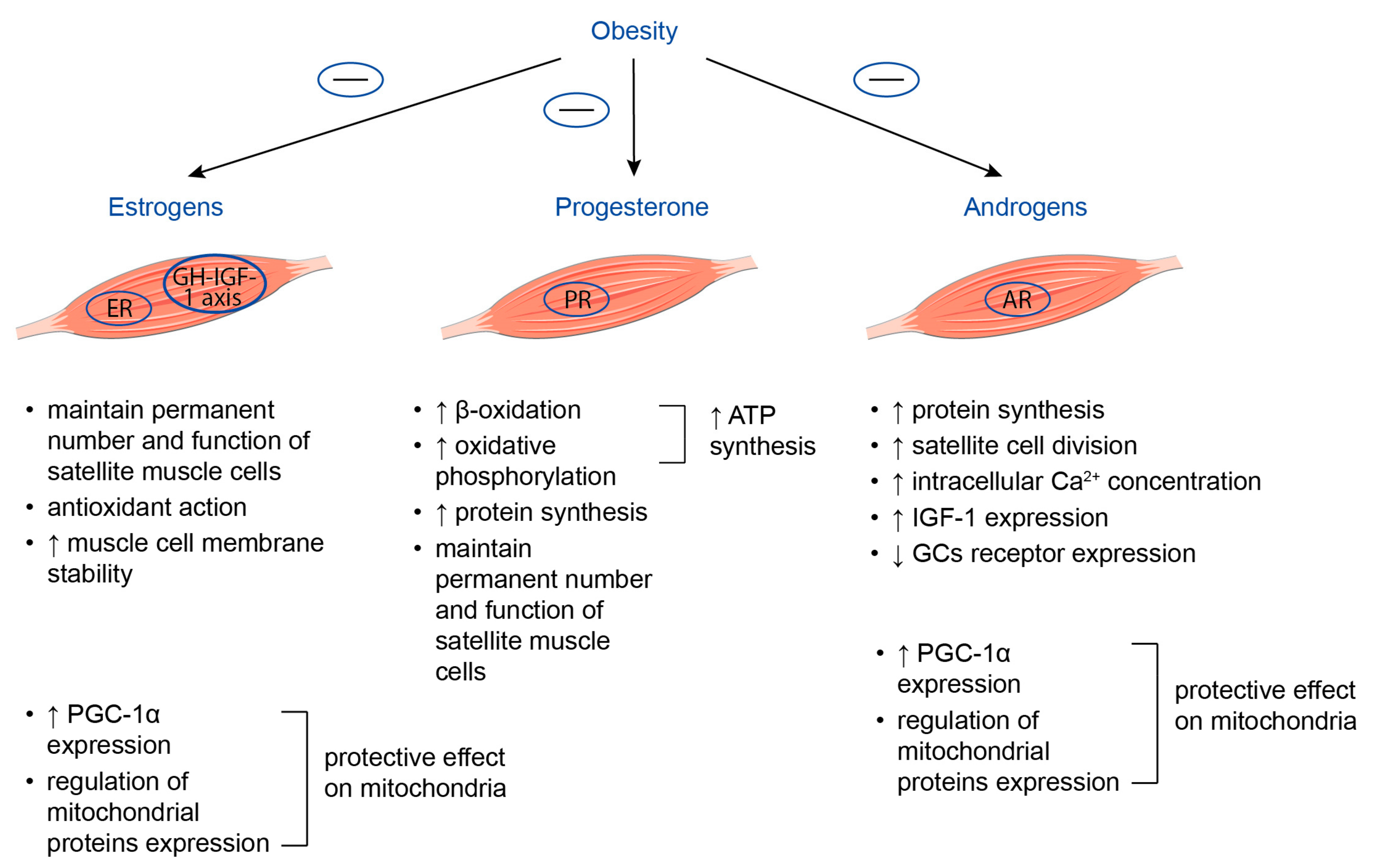
Sex steroid hormones, namely, testosterone, estrogen, and progesterone, act as anabolic hormones and are involved in the maintenance of skeletal muscle mass and function (Figure 4) [128]. Estrogen and androgen receptors are expressed by muscle satellite cells, myoblasts, and myocytes, while progesterone receptors may be found at myocytes [129].

3.2.1. Estrogens

It is considered that estrogen-mediated beneficial effects on skeletal muscle function, namely, the mitigation of muscle injury and enhancement of muscle repair after damage, may be associated with their ability to maintain a permanent number and function of satellite muscle cells, exert antioxidant action, particularly in the case of mitochondrial stress, and to increase muscle cell membrane stability. Additionally, estrogens are believed to participate in several signaling pathways, such as the modification of contractile proteins and apoptosis, involved in the preservation of muscle mass and strength [130]. Estrogens’ effects on skeletal muscle may be mediated by direct binding to their receptors situated at the level of muscle fibers, and indirectly through the regulation of the growth hormone–IGF-1 axis [131,132].
The decline in estradiol levels, characteristic of menopausal transition, is accompanied by the increased visceral adiposity as well as decreased muscle mass and strength and diminished bone mineral density [133]. Chronic low-intensive inflammation represents the pathophysiological mechanism common for all of the above-mentioned conditions. It was demonstrated that 17β-estradiol is capable of suppressing the production of several pro-inflammatory cytokines, namely, TNF-α, thus protecting skeletal muscle from wasting and facilitating recovery from injury [134,135]. It is worth noting that hormone replacement therapy containing estradiol was shown to be able to reverse both menopause-related obesity and loss of lean mass in healthy post-menopausal women, possibly due to the anabolic effect on skeletal muscle. Interestingly the overall body weight remained unchanged [136].

3.2.2. Progesterone

Progesterone is another female sex hormone, whose concentration increases dramatically during the luteal phase of the menstrual cycle. Studies concerning the impact of endogenous progesterone on skeletal muscle structure and function are limited. Most of the data concerning the influence of progesterone on skeletal muscles come from studies in women taking hormone replacement therapy or oral contraceptives. Increasing evidence suggests that the binding of progesterone to its receptors found on the outer membrane of mitochondria results in the enhancement of beta-oxidation and oxidative phosphorylation, which leads to increased ATP synthesis, thus providing muscles with the energy necessary for the maintenance of their mass and function [137,138]. Moreover, progesterone may have a beneficial effect on muscle protein turnover in postmenopausal women. It was demonstrated that 100 mg/day progesterone therapy for 14 days was accompanied by an increase in muscle protein synthesis by 50% in post-menopausal women [139]. Finally, progesterone, as estrogen, may preserve the regenerative capacity of skeletal muscles by maintaining the number and function of muscle satellite cells [140].
With regard to obesity, in a study by R.M. Whynott et al. [141], it was shown that both BMI and body weight correlated negatively with serum progesterone concentrations. These data were confirmed by the findings from J. Bellver et al. [142] indicating that serum progesterone concentrations decreased with increasing BMI; therefore, underweight or normal weight women had higher progesterone concentrations compared to overweight or obese women. Among others, decreased serum progesterone concentrations in obesity may participate in the negative impact of excess body fat on the overall body composition, particularly skeletal muscle mass.

3.2.3. Androgens

Androgens have anabolic effects on skeletal muscle, which may decline with age. Furthermore, it is well established that obesity is associated with the decreased production and secretion of androgens in men, providing an additional mechanism for the depletion of skeletal muscle mass and deterioration of their function in obesity. On the contrary, high levels of bioavailable serum testosterone may be the factor predisposing to the development of obesity in women [18]. These data are confirmed by the study in 1765 postmenopausal women indicating that high BMI at the moment of the inclusion was associated with elevated serum concentrations of androgen metabolites [143].
Although the precise mechanisms by which androgens affect muscle mass and strength remain unclear, several hypotheses are discussed. First, testosterone may increase muscle protein synthesis via the acceleration of intracellular amino acid reutilization. This, in turn, leads to the enlargement of muscle fibers and muscle hypertrophy [144]. At least in part, such a mechanism of testosterone action may be explained by its ability to upregulate the expression of myogenin and the mammalian target of rapamycin (mTOR) and to inhibit the production of myostatin, a myokine with a negative impact on muscle structure and function [145,146]. Second, the results of in vivo studies also demonstrate that testosterone may stimulate satellite cell division [147]. Third, it is suggested that testosterone is able to increase intracellular concentrations of Ca2+ in myoblasts via G-protein cupelled receptor, thus promoting myoblast growth [148]. Finally, androgens were reported to increase IGF-1 expression [149].
Importantly, androgen receptors and glucocorticoid receptors have 79% and 50% homology in the DNA- and ligand-binding domains; therefore, the anti-catabolic activity of testosterone may be explained by interfering with cortisol binding to its receptor or by competition with cortisol–glucocorticoid receptor complexes for DNA-binding sites. Moreover, testosterone is capable of decreasing glucocorticoid receptor expression in muscle tissue [149].
Both estrogen and testosterone have protective effects on mitochondria in muscle tissue, which may be attributed to their direct action on these organelles via binding with either estrogen or androgen receptors localized on them, as well as to indirect action through the regulation of mitochondrial protein expression at the level of nuclear and mitochondrial DNA. Estrogen and testosterone are also able to activate PGC-1α expression in the muscle and to inhibit mitophagy [150,151,152,153].

4. Conclusions

In conclusion, considering the high prevalence of obesity worldwide, as well as the increasingly recognized problem of sarcopenic obesity associated with negative health outcomes, clarifying the underlining pathophysiological mechanisms is of great importance. Obesity is characterized by ectopic fat accumulation in skeletal muscles, leading to increased intermuscular fat deposition and the enhancement of intramyocellular lipid droplet generation. Both these processes are accompanied by the deterioration of muscle structure and function, the disturbance of muscle regeneration capacity, and the impairment of energy metabolism. In turn, the above-mentioned negative changes may lead to the shift in myokine profile and, thus, to the formation of the vicious circle facilitating excess fat accumulation, decreasing muscle mass, and aggravating muscle function. Elevated serum GCs levels, as well as decreased serum concentrations of sex steroids associated with obesity, may represent the additional mechanism contributing to the disturbance in muscle metabolic flexibility, oxidative capacity, and decreased muscle energetic efficiency, further promoting the deterioration of muscle function, growth, and regeneration capacity. Taking into account the known sexual dimorphism of skeletal muscles and variations in the response to steroids depending on gender, the mechanisms of skeletal muscle dysfunction in obesity may differ between men and women. The provided data on the complex interaction between skeletal muscle, adipose tissue, and steroids in obesity indicate the possible need for differentiated and individualized approaches to obesity treatment and the correction of the accompanied decrease in muscle mass and function.

Author Contributions

Conceptualization, O.M.D. and N.P.M.; methodology, O.M.D.; investigation, A.F.S. and K.Y.A.; data curation, A.R.K.; writing—original draft preparation, A.F.S., K.Y.A. and O.M.D.; writing—review and editing, A.F.S. and A.R.K.; supervision, O.M.D.; funding acquisition, O.M.D. and N.P.M. All authors have read and agreed to the published version of the manuscript.

Funding

This work was supported by the Russian Science Foundation (Project No. 23-45-10030) as part of the scientific work “Prevalence and factors, associated with the musculoskeletal disorders in young and middle-aged patients with arterial hypertension and non-alcoholic fatty liver disease in Russian and Belarusian populations” performed at the National Medical Research Center for Therapy and Preventive Medicine in 2023–2025.

Institutional Review Board Statement

Not applicable.

Informed Consent Statement

Not applicable.

Data Availability Statement

Not applicable.

Conflicts of Interest

The authors declare no conflict of interest.

References

  1. World Health Organization. Obesity and Overweight Factsheets. 2020. Available online: https://www.who.int/news-room/fact-sheets/detail/obesity-and-overweight (accessed on 29 April 2023).
  2. Frühbeck, G.; Yumuk, V. Obesity: A Gateway Disease with a Rising Prevalence. Obes. Facts 2014, 7, 33–36. [Google Scholar] [CrossRef] [PubMed]
  3. Clinton, S.K.; Giovannucci, E.L.; Hursting, S.D. The World Cancer Research Fund/American Institute for Cancer Research Third Expert Report on Diet, Nutrition, Physical Activity, and Cancer: Impact and Future Directions. J. Nutr. 2020, 150, 663–671. [Google Scholar] [CrossRef] [PubMed]
  4. Tremmel, M.; Gerdtham, U.-G.; Nilsson, P.M.; Saha, S. Economic Burden of Obesity: A Systematic Literature Review. Int. J. Environ. Res. Public Health 2017, 14, 435. [Google Scholar] [CrossRef] [PubMed] [Green Version]
  5. Kovesdy, C.P.; Furth, S.L.; Zoccali, C.; World Kidney Day Steering Committee. Obesity and Kidney Disease: Hidden Consequences of the Epidemic. Can. J. Kidney Health Dis. 2017, 4, 2054358117698669. [Google Scholar] [CrossRef] [PubMed] [Green Version]
  6. Sarwar, R.; Pierce, N.; Koppe, S. Obesity and nonalcoholic fatty liver disease: Current perspectives. Diabetes Metab. Syndr. Obes. Targets Ther. 2018, 11, 533–542. [Google Scholar] [CrossRef] [Green Version]
  7. Onyemaechi, N.; Anyanwu, G.; Obikili, E.; Onwuasoigwe, O.; Nwankwo, O. Impact of overweight and obesity on the musculoskeletal system using lumbosacral angles. Patient Prefer. Adherence 2016, 10, 291–296. [Google Scholar] [CrossRef] [Green Version]
  8. Blasco, B.V.; García-Jiménez, J.; Bodoano, I.; Gutiérrez-Rojas, L. Obesity and Depression: Its Prevalence and Influence as a Prognostic Factor: A Systematic Review. Psychiatry Investig. 2020, 17, 715–724. [Google Scholar] [CrossRef]
  9. Pati, S.; Irfan, W.; Jameel, A.; Ahmed, S.; Shahid, R.K. Obesity and Cancer: A Current Overview of Epidemiology, Pathogenesis, Outcomes, and Management. Cancers 2023, 15, 485. [Google Scholar] [CrossRef]
  10. Lin, X.; Li, H. Obesity: Epidemiology, Pathophysiology, and Therapeutics. Front. Endocrinol. 2021, 12, 706978. [Google Scholar] [CrossRef]
  11. Donini, L.M.; Busetto, L.; Bischoff, S.C.; Cederholm, T.; Ballesteros-Pomar, M.D.; Batsis, J.A.; Bauer, J.M.; Boirie, Y.; Cruz-Jentoft, A.J.; Dicker, D.; et al. Definition and Diagnostic Criteria for Sarcopenic Obesity: ESPEN and EASO Consensus Statement. Obes. Facts 2022, 15, 321–335. [Google Scholar] [CrossRef]
  12. da Silva, T.L.; Nogueira, V.d.S.C.S.; Mulder, A.P. Sarcopenia and poor muscle quality associated with severe obesity in young adults and middle-aged adults. Clin. Nutr. ESPEN 2021, 45, 299–305. [Google Scholar] [CrossRef]
  13. Poggiogalle, E.; Lubrano, C.; Sergi, G.; Coin, A.; Gnessi, L.; Mariani, S.; Lenzi, A.; Donini, L.M. Sarcopenic obesity and metabolic syndrome in adult Caucasian subjects. J. Nutr. Health Aging 2015, 20, 958–963. [Google Scholar] [CrossRef]
  14. Bauer, J.M.; Cruz-Jentoft, A.J.; Fielding, R.A.; Kanis, J.A.; Reginster, J.-Y.; Bruyère, O.; Cesari, M.; Chapurlat, R.; Al-Daghri, N.; Dennison, E.; et al. Is There Enough Evidence for Osteosarcopenic Obesity as a Distinct Entity? A Critical Literature Review. Calcif. Tissue Int. 2019, 105, 109–124, 125–126. [Google Scholar] [CrossRef] [Green Version]
  15. Beaupere, C.; Liboz, A.; Fève, B.; Blondeau, B.; Guillemain, G. Molecular Mechanisms of Glucocorticoid-Induced Insulin Resistance. Int. J. Mol. Sci. 2021, 22, 623. [Google Scholar] [CrossRef]
  16. Sayın, S.; Kutlu, R.; Kulaksızoğlu, M. The relationship between sex steroids, insulin resistance and body compositions in obese women: A case-control study. J. Med. Biochem. 2020, 39, 25–31. [Google Scholar] [CrossRef] [Green Version]
  17. Zhou, Y.-H.; Guo, Y.; Wang, F.; Zhou, C.-L.; Tang, C.-Y.; Tang, H.-N.; Yan, D.-W.; Zhou, H.-D. Association of Sex Hormones and Fat Distribution in Men with Different Obese and Metabolic Statuses. Int. J. Gen. Med. 2022, 15, 1225–1238. [Google Scholar] [CrossRef]
  18. Loh, N.Y.; Humphreys, E.; Karpe, F.; Tomlinson, J.W.; Noordam, R.; Christodoulides, C. Sex hormones, adiposity, and metabolic traits in men and women: A Mendelian randomisation study. Eur. J. Endocrinol. 2022, 186, 407–416. [Google Scholar] [CrossRef]
  19. Chung, H.S.; Choi, K.M. Organokines in disease. Adv. Clin. Chem. 2020, 94, 261–321. [Google Scholar] [CrossRef]
  20. Lőrincz, H.; Somodi, S.; Ratku, B.; Harangi, M.; Paragh, G. Crucial Regulatory Role of Organokines in Relation to Metabolic Changes in Non-Diabetic Obesity. Metabolites 2023, 13, 270. [Google Scholar] [CrossRef]
  21. Guo, A.; Li, K.; Xiao, Q. Sarcopenic obesity: Myokines as potential diagnostic biomarkers and therapeutic targets? Exp. Gerontol. 2020, 139, 111022. [Google Scholar] [CrossRef]
  22. Straight, C.R.; Toth, M.J.; Miller, M.S. Current perspectives on obesity and skeletal muscle contractile function in older adults. J. Appl. Physiol. 2021, 130, 10–16. [Google Scholar] [CrossRef] [PubMed]
  23. Addison, O.; Marcus, R.L.; LaStayo, P.C.; Ryan, A.S. Intermuscular Fat: A Review of the Consequences and Causes. Int. J. Endocrinol. 2014, 2014, 309570. [Google Scholar] [CrossRef] [PubMed] [Green Version]
  24. Visser, M.; Goodpaster, B.H.; Kritchevsky, S.; Newman, A.B.; Nevitt, M.; Rubin, S.M.; Simonsick, E.M.; Harris, T.B. Muscle mass, muscle strength, and muscle fat infiltration as predictors of incident mobility limitations in well-functioning older persons. J. Gerontol. Ser. A 2005, 60, 324–333. [Google Scholar] [CrossRef] [PubMed]
  25. Goodpaster, B.H.; Bergman, B.C.; Brennan, A.M.; Sparks, L.M. Intermuscular adipose tissue in metabolic disease. Nat. Rev. Endocrinol. 2023, 19, 285–298. [Google Scholar] [CrossRef] [PubMed]
  26. Sachs, S.; Zarini, S.; Kahn, D.E.; Harrison, K.A.; Perreault, L.; Phang, T.; Newsom, S.; Strauss, A.; Kerege, A.; Schoen, J.A.; et al. Intermuscular adipose tissue directly modulates skeletal muscle insulin sensitivity in humans. Am. J. Physiol.-Endocrinol. Metab. 2019, 316, E866–E879. [Google Scholar] [CrossRef] [PubMed] [Green Version]
  27. Li, M.; Wu, H.; Wang, T.; Xia, Y.; Jin, L.; Jiang, A.; Zhu, L.; Chen, L.; Li, R.; Li, X. Co-methylated Genes in Different Adipose Depots of Pig are Associated with Metabolic, Inflammatory and Immune Processes. Int. J. Biol. Sci. 2012, 8, 831–837. [Google Scholar] [CrossRef]
  28. Kahn, D.E.; Bergman, B.C. Keeping It Local in Metabolic Disease: Adipose Tissue Paracrine Signaling and Insulin Resistance. Diabetes 2022, 71, 599–609. [Google Scholar] [CrossRef]
  29. Lyu, K.; Zhang, D.; Song, J.D.; Li, X.; Perry, R.J.; Samuel, V.T.; Shulman, G.I. Short-term overnutrition induces white adipose tissue insulin resistance through sn-1,2-diacylglycerol—PKCε—Insulin receptor T1160 phosphorylation. J. Clin. Investig. 2021, 6, e139946. [Google Scholar] [CrossRef]
  30. Perreault, L.; Newsom, S.A.; Strauss, A.; Kerege, A.; Kahn, D.E.; Harrison, K.A.; Snell-Bergeon, J.K.; Nemkov, T.; D’alessandro, A.; Jackman, M.R.; et al. Intracellular localization of diacylglycerols and sphingolipids influences insulin sensitivity and mitochondrial function in human skeletal muscle. J. Clin. Investig. 2018, 3, e96805. [Google Scholar] [CrossRef] [Green Version]
  31. Hamrick, M.W.; McGee-Lawrence, M.E.; Frechette, D.M. Fatty Infiltration of Skeletal Muscle: Mechanisms and Comparisons with Bone Marrow Adiposity. Front. Endocrinol. 2016, 7, 69. [Google Scholar] [CrossRef] [Green Version]
  32. Giuliani, G.; Vumbaca, S.; Fuoco, C.; Gargioli, C.; Giorda, E.; Massacci, G.; Palma, A.; Reggio, A.; Riccio, F.; Rosina, M.; et al. SCA-1 micro-heterogeneity in the fate decision of dystrophic fibro/adipogenic progenitors. Cell Death Dis. 2021, 12, 122. [Google Scholar] [CrossRef]
  33. Marinkovic, M.; Fuoco, C.; Sacco, F.; Perpetuini, A.C.; Giuliani, G.; Micarelli, E.; Pavlidou, T.; Petrilli, L.L.; Reggio, A.; Riccio, F.; et al. Fibro-adipogenic progenitors of dystrophic mice are insensitive to NOTCH regulation of adipogenesis. Life Sci. Alliance 2019, 2, e201900437. [Google Scholar] [CrossRef] [Green Version]
  34. Uezumi, A.; Ito, T.; Morikawa, D.; Shimizu, N.; Yoneda, T.; Segawa, M.; Yamaguchi, M.; Ogawa, R.; Matev, M.M.; Miyagoe-Suzuki, Y.; et al. Fibrosis and adipogenesis originate from a common mesenchymal progenitor in skeletal muscle. J. Cell Sci. 2011, 124, 3654–3664. [Google Scholar] [CrossRef] [Green Version]
  35. Malecova, B.; Gatto, S.; Etxaniz, U.; Passafaro, M.; Cortez, A.; Nicoletti, C.; Giordani, L.; Torcinaro, A.; De Bardi, M.; Bicciato, S.; et al. Dynamics of cellular states of fibro-adipogenic progenitors during myogenesis and muscular dystrophy. Nat. Commun. 2018, 9, 3670. [Google Scholar] [CrossRef] [Green Version]
  36. Dong, J.; Dong, Y.; Chen, Z.; Mitch, W.E.; Zhang, L. The pathway to muscle fibrosis depends on myostatin stimulating the differentiation of fibro/adipogenic progenitor cells in chronic kidney disease. Kidney Int. 2017, 91, 119–128. [Google Scholar] [CrossRef] [Green Version]
  37. Nachit, M.; Leclercq, I.A. Emerging awareness on the importance of skeletal muscle in liver diseases: Time to dig deeper into mechanisms! Clin. Sci. 2019, 133, 465–481. [Google Scholar] [CrossRef]
  38. Roberts, E.W.; Deonarine, A.; Jones, J.O.; Denton, A.E.; Feig, C.; Lyons, S.K.; Espeli, M.; Kraman, M.; McKenna, B.; Wells, R.J.; et al. Depletion of stromal cells expressing fibroblast activation protein-α from skeletal muscle and bone marrow results in cachexia and anemia. J. Exp. Med. 2013, 210, 1137–1151. [Google Scholar] [CrossRef] [Green Version]
  39. Uezumi, A.; Ikemoto-Uezumi, M.; Zhou, H.; Kurosawa, T.; Yoshimoto, Y.; Nakatani, M.; Hitachi, K.; Yamaguchi, H.; Wakatsuki, S.; Araki, T.; et al. Mesenchymal Bmp3b expression maintains skeletal muscle integrity and decreases in age-related sarcopenia. J. Clin. Investig. 2021, 131, e139617. [Google Scholar] [CrossRef]
  40. Collao, N.; Farup, J.; De Lisio, M. Role of Metabolic Stress and Exercise in Regulating Fibro/Adipogenic Progenitors. Front. Cell Dev. Biol. 2020, 8, 9. [Google Scholar] [CrossRef]
  41. Kaur, G.; Davies, M.R.; Liu, X.; Feeley, B.T. The Role of Fibro-Adipogenic Progenitors in Musculoskeletal Disease. Muscles Ligaments Tendons J. 2021, 11, 201–214. [Google Scholar] [CrossRef]
  42. Moratal, C.; Raffort, J.; Arrighi, N.; Rekima, S.; Schaub, S.; Dechesne, C.A.; Chinetti, G.; Dani, C. IL-1β- and IL-4-polarized macrophages have opposite effects on adipogenesis of intramuscular fibro-adipogenic progenitors in humans. Sci. Rep. 2018, 8, 17005. [Google Scholar] [CrossRef] [PubMed]
  43. Arrighi, N.; Moratal, C.; Clément, N.; Giorgetti-Peraldi, S.; Peraldi, P.; Loubat, A.; Kurzenne, J.-Y.; Dani, C.; Chopard, A.; Dechesne, A.C. Characterization of adipocytes derived from fibro/adipogenic progenitors resident in human skeletal muscle. Cell Death Dis. 2015, 6, e1733. [Google Scholar] [CrossRef] [PubMed] [Green Version]
  44. Asakura, A.; Rudnicki, M.A.; Komaki, M. Muscle satellite cells are multipotential stem cells that exhibit myogenic, osteogenic, and adipogenic differentiation. Differentiation 2001, 68, 245–253. [Google Scholar] [CrossRef]
  45. Wada, M.R.; Inagawa-Ogashiwa, M.; Shimizu, S.; Yasumoto, S.; Hashimoto, N. Generation of different fates from multipotent muscle stem cells. Development 2002, 129, 2987–2995. [Google Scholar] [CrossRef]
  46. Guo, L.; Cui, H.; Zhao, G.; Liu, R.; Li, Q.; Zheng, M.; Guo, Y.; Wen, J. Intramuscular preadipocytes impede differentiation and promote lipid deposition of muscle satellite cells in chickens. BMC Genom. 2018, 19, 838. [Google Scholar] [CrossRef]
  47. Corvera, S. Cellular Heterogeneity in Adipose Tissues. Annu. Rev. Physiol. 2021, 83, 257–278. [Google Scholar] [CrossRef]
  48. Majka, S.M.; Miller, H.L.; Sullivan, T.; Erickson, P.F.; Kong, R.; Weiser-Evans, M.; Nemenoff, R.; Moldovan, R.; Morandi, S.A.; Davis, J.A.; et al. Adipose lineage specification of bone marrow-derived myeloid cells. Adipocyte 2012, 1, 215–229. [Google Scholar] [CrossRef] [Green Version]
  49. Dubé, J.J.; Amati, F.; Stefanovic-Racic, M.; Toledo, F.G.S.; Sauers, S.E.; Goodpaster, B.H. Exercise-induced alterations in intramyocellular lipids and insulin resistance: The athlete’s paradox revisited. Am. J. Physiol.-Endocrinol. Metab. 2008, 294, E882–E888. [Google Scholar] [CrossRef] [Green Version]
  50. Goodpaster, B.H.; He, J.; Watkins, S.; Kelley, D.E. Skeletal Muscle Lipid Content and Insulin Resistance: Evidence for a Paradox in Endurance-Trained Athletes. J. Clin. Endocrinol. Metab. 2001, 86, 5755–5761. [Google Scholar] [CrossRef]
  51. Bergman, B.C.; Perreault, L.; Strauss, A.; Bacon, S.; Kerege, A.; Harrison, K.; Brozinick, J.T.; Hunerdosse, D.M.; Playdon, M.C.; Holmes, W.; et al. Intramuscular triglyceride synthesis: Importance in muscle lipid partitioning in humans. Am. J. Physiol.-Endocrinol. Metab. 2018, 314, E152–E164. [Google Scholar] [CrossRef]
  52. Belzunce, M.A.; Henckel, J.; Di Laura, A.; Hart, A. Intramuscular fat in gluteus maximus for different levels of physical activity. Sci. Rep. 2021, 11, 21401. [Google Scholar] [CrossRef] [PubMed]
  53. Gemmink, A.; Daemen, S.; Brouwers, B.; Huntjens, P.R.; Schaart, G.; Moonen-Kornips, E.; Jörgensen, J.; Hoeks, J.; Schrauwen, P.; Hesselink, M.K.C. Dissociation of intramyocellular lipid storage and insulin resistance in trained athletes and type 2 diabetes patients; involvement of perilipin 5? J. Physiol. 2018, 596, 857–868. [Google Scholar] [CrossRef] [PubMed] [Green Version]
  54. Kahn, D.; Perreault, L.; Macias, E.; Zarini, S.; Newsom, S.A.; Strauss, A.; Kerege, A.; Harrison, K.; Snell-Bergeon, J.; Bergman, B.C. Subcellular localisation and composition of intramuscular triacylglycerol influence insulin sensitivity in humans. Diabetologia 2021, 64, 168–180. [Google Scholar] [CrossRef] [PubMed]
  55. Devries, M.C.; Samjoo, I.A.; Hamadeh, M.J.; McCready, C.; Raha, S.; Watt, M.J.; Steinberg, G.R.; Tarnopolsky, M.A. Endurance Training Modulates Intramyocellular Lipid Compartmentalization and Morphology in Skeletal Muscle of Lean and Obese Women. J. Clin. Endocrinol. Metab. 2013, 98, 4852–4862. [Google Scholar] [CrossRef] [Green Version]
  56. Umek, N.; Horvat, S.; Cvetko, E. Skeletal muscle and fiber type-specific intramyocellular lipid accumulation in obese mice. Bosn. J. Basic Med. Sci. 2021, 21, 730–738. [Google Scholar] [CrossRef] [PubMed]
  57. Thrush, A.B.; Dent, R.; McPherson, R.; Harper, M.-E. Implications of mitochondrial uncoupling in skeletal muscle in the development and treatment of obesity. FEBS J. 2013, 280, 5015–5029. [Google Scholar] [CrossRef]
  58. Zurlo, F.; Larson, K.; Bogardus, C.; Ravussin, E. Skeletal muscle metabolism is a major determinant of resting energy expenditure. J. Clin. Investig. 1990, 86, 1423–1427. [Google Scholar] [CrossRef] [Green Version]
  59. Talbot, J.; Maves, L. Skeletal muscle fiber type: Using insights from muscle developmental biology to dissect targets for susceptibility and resistance to muscle disease. Wiley Interdiscip. Rev. Dev. Biol. 2016, 5, 518–534. [Google Scholar] [CrossRef] [Green Version]
  60. Motohashi, N.; Minegishi, K.; Imamura, M.; Aoki, Y. Techniques for Injury, Cell Transplantation, and Histological Analysis in Skeletal Muscle. Methods Mol. Biol. 2023, 2640, 193–205. [Google Scholar] [CrossRef]
  61. Damer, A.; El Meniawy, S.; McPherson, R.; Wells, G.; Harper, M.; Dent, R. Association of muscle fiber type with measures of obesity: A systematic review. Obes. Rev. 2022, 23, e13444. [Google Scholar] [CrossRef]
  62. Sung, E.-S.; Han, A.; Hinrichs, T.; Vorgerd, M.; Platen, P. Impact of Body Mass Index on Muscle Strength, Thicknesses, and Fiber Composition in Young Women. Int. J. Environ. Res. Public Health 2022, 19, 9789. [Google Scholar] [CrossRef]
  63. Nomikos, T.; Methenitis, S.; Panagiotakos, D.B. The emerging role of skeletal muscle as a modulator of lipid profile the role of exercise and nutrition. Lipids Health Dis. 2022, 21, 81. [Google Scholar] [CrossRef]
  64. Reiter, D.A.; Bellissimo, M.P.; Zhou, L.; Boebinger, S.; Wells, G.D.; Jones, D.P.; Ziegler, T.R.; Alvarez, J.A.; Fleischer, C.C. Increased Adiposity is Associated with Altered Skeletal Muscle Energetics. J. Appl. Physiol. 2023, 134, 1083–1092. [Google Scholar] [CrossRef]
  65. Goodpaster, B.H.; Sparks, L.M. Metabolic Flexibility in Health and Disease. Cell Metab. 2017, 25, 1027–1036. [Google Scholar] [CrossRef] [Green Version]
  66. Randle, P.; Garland, P.; Hales, C.; Newsholme, E. The glucose fatty-acid cycle. Its role in insulin sensitivity and the metabolic disturbances of diabetes mellitus. Lancet 1963, 1, 785–789. [Google Scholar] [CrossRef]
  67. San-Millán, I. The Key Role of Mitochondrial Function in Health and Disease. Antioxidants 2023, 12, 782. [Google Scholar] [CrossRef]
  68. Barakati, N.; Bustos, R.Z.; Coletta, D.K.; Langlais, P.R.; Kohler, L.N.; Luo, M.; Funk, J.L.; Willis, W.T.; Mandarino, L.J. Fuel Selection in Skeletal Muscle Exercising at Low Intensity; Reliance on Carbohydrate in Very Sedentary Individuals. Metab. Syndr. Relat. Disord. 2023, 21, 16–24. [Google Scholar] [CrossRef]
  69. Pileggi, C.A.; Hooks, B.G.; McPherson, R.; Dent, R.R.; Harper, M.-E. Targeting skeletal muscle mitochondrial health in obesity. Clin. Sci. 2022, 136, 1081–1110. [Google Scholar] [CrossRef]
  70. Spooner, H.C.; Derrick, S.A.; Maj, M.; Manjarín, R.; Hernandez, G.V.; Tailor, D.S.; Bastani, P.S.; Fanter, R.K.; Fiorotto, M.L.; Burrin, D.G.; et al. High-Fructose, High-Fat Diet Alters Muscle Composition and Fuel Utilization in a Juvenile Iberian Pig Model of Non-Alcoholic Fatty Liver Disease. Nutrients 2021, 13, 4195. [Google Scholar] [CrossRef]
  71. Zhelankin, A.V.; Iulmetova, L.N.; Ahmetov, I.I.; Generozov, E.V.; Sharova, E.I. Diversity and Differential Expression of MicroRNAs in the Human Skeletal Muscle with Distinct Fiber Type Composition. Life 2023, 13, 659. [Google Scholar] [CrossRef]
  72. Hernandez, G.V.; Smith, V.A.; Melnyk, M.; Burd, M.A.; Sprayberry, K.A.; Edwards, M.S.; Peterson, D.G.; Bennet, D.C.; Fanter, R.K.; Columbus, D.A.; et al. Dysregulated FXR-FGF19 signaling and choline metabolism are associated with gut dysbiosis and hyperplasia in a novel pig model of pediatric NASH. Am. J. Physiol. Gastrointest. Liver Physiol. 2020, 318, G582–G609. [Google Scholar] [CrossRef] [PubMed]
  73. Sarparanta, J.; Garcia-Macia, M.; Singh, R. Autophagy and Mitochondria in Obesity and Type 2 Diabetes. Curr. Diabetes Rev. 2017, 13, 352–369. [Google Scholar] [CrossRef] [PubMed]
  74. Roszczyc-Owsiejczuk, K.; Zabielski, P. Sphingolipids as a Culprit of Mitochondrial Dysfunction in Insulin Resistance and Type 2 Diabetes. Front. Endocrinol. 2021, 12, 635175. [Google Scholar] [CrossRef]
  75. Kobayashi, M.; Deguchi, Y.; Nozaki, Y.; Higami, Y. Contribution of PGC-1α to Obesity- and Caloric Restriction-Related Physiological Changes in White Adipose Tissue. Int. J. Mol. Sci. 2021, 22, 6025. [Google Scholar] [CrossRef] [PubMed]
  76. Handschin, C.; Chin, S.; Li, P.; Liu, F.; Maratos-Flier, E.; LeBrasseur, N.K.; Yan, Z.; Spiegelman, B.M. Skeletal Muscle Fiber-type Switching, Exercise Intolerance, and Myopathy in PGC-1α Muscle-specific Knock-out Animals. J. Biol. Chem. 2007, 282, 30014–30021. [Google Scholar] [CrossRef] [Green Version]
  77. Sandri, M.; Lin, J.; Handschin, C.; Yang, W.; Arany, Z.P.; Lecker, S.H.; Goldberg, A.L.; Spiegelman, B.M. PGC-1alpha protects skeletal muscle from atrophy by suppressing FoxO3 action and atrophy-specific gene transcription. Proc. Natl. Acad. Sci. USA 2006, 103, 16260–16265. [Google Scholar] [CrossRef] [Green Version]
  78. Menshikova, E.V.; Ritov, V.B.; Dube, J.J.; Amati, F.; Stefanovic-Racic, M.; Toledo, F.G.S.; Coen, P.M.; Goodpaster, B.H. Calorie Restriction-induced Weight Loss and Exercise Have Differential Effects on Skeletal Muscle Mitochondria Despite Similar Effects on Insulin Sensitivity. J. Gerontol. A Biol. Sci. Med. Sci. 2017, 73, 81–87. [Google Scholar] [CrossRef] [Green Version]
  79. Jia, J.; Yu, F.; Wei, W.-P.; Yang, P.; Zhang, R.; Sheng, Y.; Shi, Y.-Q. Relationship between circulating irisin levels and overweight/obesity: A meta-analysis. World J. Clin. Cases 2019, 7, 1444–1455. [Google Scholar] [CrossRef]
  80. Liu, S.; Cui, F.; Ning, K.; Wang, Z.; Fu, P.; Wang, D.; Xu, H. Role of irisin in physiology and pathology. Front. Endocrinol. 2022, 13, 962968. [Google Scholar] [CrossRef]
  81. Kurdiova, T.; Balaz, M.; Vician, M.; Maderova, D.; Vlcek, M.; Valkovic, L.; Srbecky, M.; Imrich, R.; Kyselovicova, O.; Belan, V.; et al. Effects of obesity, diabetes and exercise on Fndc5 gene expression and irisin release in human skeletal muscle and adipose tissue: In Vivo and In Vitro studies. J. Physiol. 2014, 592, 1091–1107. [Google Scholar] [CrossRef]
  82. Gamas, L.; Matafome, P.; Seiça, R. Irisin and Myonectin Regulation in the Insulin Resistant Muscle: Implications to Adipose Tissue: Muscle Crosstalk. J. Diabetes Res. 2015, 2015, 359159. [Google Scholar] [CrossRef] [Green Version]
  83. Moreno-Navarrete, J.M.; Ortega, F.; Serrano, M.; Guerra, E.; Pardo, G.; Tinahones, F.; Ricart, W.; Fernández-Real, J.M. Irisin Is Expressed and Produced by Human Muscle and Adipose Tissue in Association With Obesity and Insulin Resistance. J. Clin. Endocrinol. Metab. 2013, 98, E769–E778. [Google Scholar] [CrossRef]
  84. Crujeiras, A.B.; Zulet, M.A.; Lopez-Legarrea, P.; de la Iglesia, R.; Pardo, M.; Carreira, M.C.; Martínez, J.A.; Casanueva, F.F. Association between circulating irisin levels and the promotion of insulin resistance during the weight maintenance period after a dietary weight-lowering program in obese patients. Metabolism 2014, 63, 520–531. [Google Scholar] [CrossRef] [Green Version]
  85. Yin, J.; Yang, S.; Zha, X.; Miao, Z.; Sheng, C.; Yang, P.; Wang, X.; Qu, S. The Association of Serum Irisin with Impaired Glucose Before and After Laparoscopic Sleeve Gastrectomy in Obesity. Obes. Surg. 2023, 33, 780–788. [Google Scholar] [CrossRef]
  86. Dong, J.; Dong, Y.; Chen, F.; Mitch, E.W.; Zhang, L. Inhibition of myostatin in mice improves insulin sensitivity via irisin-mediated cross talk between muscle and adipose tissues. Int. J. Obes. 2016, 40, 434–442. [Google Scholar] [CrossRef] [Green Version]
  87. Salama, A.; Amin, M.M.; Hassan, A. Effects of oleic acid and/or exercise on diet-induced thermogenesis and obesity in rats: Involvement of beige adipocyte differentiation and macrophage M1 inhibition. Res. Pharm. Sci. 2023, 18, 219–230. [Google Scholar] [CrossRef]
  88. Bonnieu, A.; Carnac, G.; Vernus, B. Myostatin in the Pathophysiology of Skeletal Muscle. Curr. Genom. 2007, 8, 415–422. [Google Scholar] [CrossRef]
  89. Consitt, L.; Clark, B. The vicious cycle of myostatin signaling in sarcopenic obesity: Myostatin role in skeletal muscle growth, insulin signaling and implications for clinical trials. J. Frailty Aging 2017, 7, 21–27. [Google Scholar] [CrossRef]
  90. Amor, M.; Itariu, B.K.; Moreno-Viedma, V.; Keindl, M.; Jürets, A.; Prager, G.; Langer, F.; Grablowitz, V.; Zeyda, M.; Stulnig, T.M. Serum Myostatin is Upregulated in Obesity and Correlates with Insulin Resistance in Humans. Exp. Clin. Endocrinol. Diabetes 2019, 127, 550–556. [Google Scholar] [CrossRef]
  91. Allen, D.L.; Hittel, D.S.; McPherron, A. Expression and Function of Myostatin in Obesity, Diabetes, and Exercise Adaptation. Med. Sci. Sports Exerc. 2011, 43, 1828–1835. [Google Scholar] [CrossRef] [Green Version]
  92. Zhang, C.; McFarlane, C.; Lokireddy, S.; Masuda, S.; Ge, X.; Gluckman, P.D.; Sharma, M.; Kambadur, R. Inhibition of myostatin protects against diet-induced obesity by enhancing fatty acid oxidation and promoting a brown adipose phenotype in mice. Diabetologia 2015, 55, 183–193, Erratum in: Diabetologia 2015, 58, 643. [Google Scholar] [CrossRef] [PubMed] [Green Version]
  93. Shan, T.; Liang, X.; Bi, P.; Kuang, S. Myostatin knockout drives browning of white adipose tissue through activating the AMPK-PGC1α-Fndc5 pathway in muscle. FASEB J. 2013, 27, 1981–1989. [Google Scholar] [CrossRef] [PubMed] [Green Version]
  94. Guo, T.; Jou, W.; Chanturiya, T.; Portas, J.; Gavrilova, O.; McPherron, A.C. Myostatin Inhibition in Muscle, but Not Adipose Tissue, Decreases Fat Mass and Improves Insulin Sensitivity. PLoS ONE 2009, 4, e4937. [Google Scholar] [CrossRef] [PubMed] [Green Version]
  95. Ryan, A.; Li, G.; Blumenthal, J.; Ortmeyer, H. Aerobic exercise + weight loss decreases skeletal muscle myostatin expression and improves insulin sensitivity in older adults. Obesity 2012, 21, 1350–1356. [Google Scholar] [CrossRef]
  96. Perry, C.A.; Van Guilder, G.P.; Butterick, T.A. Decreased myostatin in response to a controlled DASH diet is associated with improved body composition and cardiometabolic biomarkers in older adults: Results from a controlled-feeding diet intervention study. BMC Nutr. 2022, 8, 24. [Google Scholar] [CrossRef]
  97. Wang, S.; Fang, L.; Cong, L.; Chung, J.P.W.; Li, T.C.; Chan, D.Y.L. Myostatin: A multifunctional role in human female reproduction and fertility—A short review. Reprod. Biol. Endocrinol. 2022, 20, 96. [Google Scholar] [CrossRef]
  98. Fang, L.; Yan, Y.; Wang, S.; Guo, Y.; Li, Y.; Jia, Q.; Han, X.; Liu, B.; Cheng, J.-C.; Sun, Y.-P. High ovarian GDF-8 levels contribute to elevated estradiol production in ovarian hyperstimulation syndrome by stimulating aromatase expression. Int. J. Biol. Sci. 2021, 17, 2338–2347. [Google Scholar] [CrossRef]
  99. Fang, L.; Wang, S.; Li, Y.; Yu, Y.; Li, Y.; Yan, Y.; Cheng, J.-C.; Sun, Y.-P. High GDF-8 in follicular fluid is associated with a low pregnancy rate in IVF patients with PCOS. Reproduction 2020, 160, 11–19. [Google Scholar] [CrossRef]
  100. Kuo, T.; Harris, C.A.; Wang, J.-C. Metabolic functions of glucocorticoid receptor in skeletal muscle. Mol. Cell. Endocrinol. 2013, 380, 79–88. [Google Scholar] [CrossRef] [Green Version]
  101. Shirif, A.Z.; Kovačević, S.; Brkljačić, J.; Teofilović, A.; Elaković, I.; Djordjevic, A.; Matić, G. Decreased Glucocorticoid Signaling Potentiates Lipid-Induced Inflammation and Contributes to Insulin Resistance in the Skeletal Muscle of Fructose-Fed Male Rats Exposed to Stress. Int. J. Mol. Sci. 2021, 22, 7206. [Google Scholar] [CrossRef]
  102. Matthews, E.; Brassington, R.; Kuntzer, T.; Jichi, F.; Manzur, A.Y. Corticosteroids for the treatment of Duchenne muscular dystrophy. Cochrane Database Syst. Rev. 2016, 2016, CD003725. [Google Scholar] [CrossRef] [Green Version]
  103. Perpetuini, A.C.; Giuliani, G.; Reggio, A.; Cerretani, M.; Santoriello, M.; Stefanelli, R.; Palma, A.; Vumbaca, S.; Harper, S.; Castagnoli, L.; et al. Janus effect of glucocorticoids on differentiation of muscle fibro/adipogenic progenitors. Sci. Rep. 2020, 10, 5363. [Google Scholar] [CrossRef] [Green Version]
  104. Quattrocelli, M.; Salamone, I.M.; Page, P.G.; Warner, J.L.; Demonbreun, A.R.; McNally, E.M. Intermittent Glucocorticoid Dosing Improves Muscle Repair and Function in Mice with Limb-Girdle Muscular Dystrophy. Am. J. Pathol. 2017, 187, 2520–2535, Erratum in: Am. J. Pathol. 2021, 191, 2039. [Google Scholar] [CrossRef] [Green Version]
  105. Quattrocelli, M.; Zelikovich, A.S.; Salamone, I.M.; Fischer, J.A.; McNally, E.M. Mechanisms and Clinical Applications of Glucocorticoid Steroids in Muscular Dystrophy. J. Neuromuscul. Dis. 2021, 8, 39–52. [Google Scholar] [CrossRef]
  106. Quattrocelli, M.; Barefield, D.; Warner, J.L.; Vo, A.H.; Hadhazy, M.; Earley, J.U.; Demonbreun, A.; McNally, E.M. Intermittent glucocorticoid steroid dosing enhances muscle repair without eliciting muscle atrophy. J. Clin. Investig. 2017, 127, 2418–2432. [Google Scholar] [CrossRef] [Green Version]
  107. Nadal, A.; Quesada, I.; Tudurí, E.; Nogueiras, R.; Magdalena, P.A. Endocrine-disrupting chemicals and the regulation of energy balance. Nat. Rev. Endocrinol. 2017, 13, 536–546. [Google Scholar] [CrossRef]
  108. Braun, T.; Marks, D.L. The regulation of muscle mass by endogenous glucocorticoids. Front. Physiol. 2015, 6, 12. [Google Scholar] [CrossRef] [Green Version]
  109. Webster, J.M.; Waaijenberg, K.; van de Worp, W.R.; Kelders, M.C.; Lambrichts, S.; Martin, C.; Verhaegen, F.; van der Heyden, B.; Smith, C.; Lavery, G.G.; et al. 11β-HSD1 determines the extent of muscle atrophy in a model of acute exacerbation of COPD. Am. J. Physiol. Lung Cell. Mol. Physiol. 2023, 324, L400–L412. [Google Scholar] [CrossRef]
  110. Webster, J.M.; Sagmeister, M.S.; Fenton, C.G.; Seabright, A.P.; Lai, Y.-C.; Jones, S.W.; Filer, A.; Cooper, M.S.; Lavery, G.G.; Raza, K.; et al. Global Deletion of 11β-HSD1 Prevents Muscle Wasting Associated with Glucocorticoid Therapy in Polyarthritis. Int. J. Mol. Sci. 2021, 22, 7828. [Google Scholar] [CrossRef]
  111. Lee, M.-K.; Jeong, H.H.; Kim, M.-J.; Ryu, H.; Baek, J.; Lee, B. Nutrients against Glucocorticoid-Induced Muscle Atrophy. Foods 2022, 11, 687. [Google Scholar] [CrossRef]
  112. Sato, A.; Richardson, D.; Cregor, M.; Davis, H.M.; Au, E.; McAndrews, K.; Zimmers, T.A.; Organ, J.; Peacock, M.; Plotkin, L.I.; et al. Glucocorticoids Induce Bone and Muscle Atrophy by Tissue-Specific Mechanisms Upstream of E3 Ubiquitin Ligases. Endocrinology 2017, 158, 664–677. [Google Scholar] [CrossRef] [PubMed]
  113. Cheng, C.-F.; Ku, H.-C.; Lin, H. PGC-1α as a Pivotal Factor in Lipid and Metabolic Regulation. Int. J. Mol. Sci. 2018, 19, 3447. [Google Scholar] [CrossRef] [PubMed] [Green Version]
  114. Liang, H.; Ward, W.F. PGC-1α: A key regulator of energy metabolism. Adv. Physiol. Educ. 2006, 30, 145–151. [Google Scholar] [CrossRef] [PubMed]
  115. Salamone, I.M.; Quattrocelli, M.; Barefield, D.Y.; Page, P.G.; Tahtah, I.; Hadhazy, M.; Tomar, G.; McNally, E.M. Intermittent glucocorticoid treatment enhances skeletal muscle performance through sexually dimorphic mechanisms. J. Clin. Investig. 2022, 132, e149828. [Google Scholar] [CrossRef] [PubMed]
  116. Zumbaugh, M.D.; Johnson, E.S.; Shi, T.H.; Gerrard, E.D. Molecular and biochemical regulation of skeletal muscle metabolism. J. Anim. Sci. 2022, 100, skac035. [Google Scholar] [CrossRef]
  117. Maher, A.C.; Fu, M.H.; Isfort, R.J.; Varbanov, A.R.; Qu, X.A.; Tarnopolsky, M.A. Sex Differences in Global mRNA Content of Human Skeletal Muscle. PLoS ONE 2009, 4, e6335. [Google Scholar] [CrossRef] [Green Version]
  118. Della Peruta, C.; Lozanoska-Ochser, B.; Renzini, A.; Moresi, V.; Riera, C.S.; Bouché, M.; Coletti, D. Sex Differences in Inflammation and Muscle Wasting in Aging and Disease. Int. J. Mol. Sci. 2023, 24, 4651. [Google Scholar] [CrossRef]
  119. Aguiar, A.S., Jr.; Speck, A.E.; Amaral, I.M.; Canas, P.M.; Cunha, R.A. The exercise sex gap and the impact of the estrous cycle on exercise performance in mice. Sci. Rep. 2018, 8, 10742. [Google Scholar] [CrossRef] [Green Version]
  120. Haizlip, K.M.; Harrison, B.C.; Leinwand, L.A. Sex-Based Differences in Skeletal Muscle Kinetics and Fiber-Type Composition. Physiology 2015, 30, 30–39. [Google Scholar] [CrossRef]
  121. Manzano, R.; Toivonen, J.M.; Calvo, A.C.; Miana-Mena, F.J.; Zaragoza, P.; Muñoz, M.J.; Montarras, D.; Osta, R. Sex, fiber-type, and age dependent in vitro proliferation of mouse muscle satellite cells. J. Cell. Biochem. 2011, 112, 2825–2836. [Google Scholar] [CrossRef]
  122. Sesillo, F.B.; Rajesh, V.; Wong, M.; Duran, P.; Rudell, J.B.; Rundio, C.P.; Baynes, B.B.; Laurent, L.C.; Sacco, A.; Christman, K.L.; et al. Muscle stem cells and fibro-adipogenic progenitors in female pelvic floor muscle regeneration following birth injury. npj Regen. Med. 2022, 7, 72. [Google Scholar] [CrossRef]
  123. Wu, R.-Y.; Sung, W.-H.; Cheng, H.-C.; Yeh, H.-J. Investigating the rate of skeletal muscle atrophy in men and women in the intensive care unit: A prospective observational study. Sci. Rep. 2022, 12, 16629. [Google Scholar] [CrossRef]
  124. de Jong, J.C.; Attema, B.J.; van der Hoek, M.D.; Verschuren, L.; Caspers, M.P.; Kleemann, R.; van der Leij, F.R.; Hoek, A.M.V.D.; Nieuwenhuizen, A.G.; Keijer, J. Sex differences in skeletal muscle-aging trajectory: Same processes, but with a different ranking. Geroscience 2023. Epub ahead of print. [Google Scholar] [CrossRef]
  125. Shu, Y.; Xia, J.; Yu, Q.; Wang, G.; Zhang, J.; He, J.; Wang, H.; Zhang, L.; Wu, H. Integrated analysis of mRNA and miRNA expression profiles reveals muscle growth differences between adult female and male Chinese concave-eared frogs (Odorrana tormota). Gene 2018, 678, 241–251. [Google Scholar] [CrossRef]
  126. Ogawa, M.; Kitano, T.; Kawata, N.; Sugihira, T.; Kitakaze, T.; Harada, N.; Yamaji, R. Daidzein down-regulates ubiquitin-specific protease 19 expression through estrogen receptor β and increases skeletal muscle mass in young female mice. J. Nutr. Biochem. 2017, 49, 63–70. [Google Scholar] [CrossRef]
  127. Seko, D.; Fujita, R.; Kitajima, Y.; Nakamura, K.; Imai, Y.; Ono, Y. Estrogen Receptor β Controls Muscle Growth and Regeneration in Young Female Mice. Stem Cell Rep. 2020, 15, 577–586. [Google Scholar] [CrossRef]
  128. Alexander, S.E.; Pollock, A.C.; Lamon, S. The effect of sex hormones on skeletal muscle adaptation in females. Eur. J. Sport Sci. 2022, 22, 1035–1045. [Google Scholar] [CrossRef]
  129. Huang, L.-T.; Wang, J.-H. The Therapeutic Intervention of Sex Steroid Hormones for Sarcopenia. Front. Med. 2021, 8, 739251. [Google Scholar] [CrossRef]
  130. Pellegrino, A.; Tiidus, P.M.; Vandenboom, R. Mechanisms of Estrogen Influence on Skeletal Muscle: Mass, Regeneration, and Mitochondrial Function. Sports Med. 2022, 52, 2853–2869. [Google Scholar] [CrossRef]
  131. Kamwa, V.; Welch, C.; Hassan-Smith, Z.K. The endocrinology of sarcopenia and frailty. Minerva Endocrinol. 2021, 46, 453–468. [Google Scholar] [CrossRef]
  132. Weissberger, A.J.; Ho, K.K.Y.; Lazarus, L. Contrasting Effects of Oral and Transdermal Routes of Estrogen Replacement Therapy on 24-Hour Growth Hormone (GH) Secretion, Insulin-Like Growth Factor I, and GH-Binding Protein in Postmenopausal Women. J. Clin. Endocrinol. Metab. 1991, 72, 374–381. [Google Scholar] [CrossRef] [PubMed]
  133. Geraci, A.; Calvani, R.; Ferri, E.; Marzetti, E.; Arosio, B.; Cesari, M. Sarcopenia and Menopause: The Role of Estradiol. Front. Endocrinol. 2021, 12, 682012. [Google Scholar] [CrossRef] [PubMed]
  134. Lambert, K.C.; Curran, E.M.; Judy, B.M.; Lubahn, D.B.; Estes, D.M. Estrogen receptor-α deficiency promotes increased TNF-α secretion and bacterial killing by murine macrophages in response to microbial stimuli in vitro. J. Leukoc. Biol. 2004, 75, 1166–1172. [Google Scholar] [CrossRef] [PubMed] [Green Version]
  135. Li, Y.P.; Reid, M.B. NF-kappaB mediates the protein loss induced by TNF-alpha in differentiated skeletal muscle myotubes. Am. J. Physiol. Regul. Integr. Comp. Physiol. 2000, 279, R1165–R1170. [Google Scholar] [CrossRef]
  136. Sørensen, M.B.; Rosenfalck, A.M.; Højgaard, L.; Ottesen, B. Obesity and Sarcopenia after Menopause Are Reversed by Sex Hormone Replacement Therapy. Obes. Res. 2001, 9, 622–626. [Google Scholar] [CrossRef] [Green Version]
  137. Shaia, K.L.; Harris, B.S.; Selter, J.H.; Price, T.M. Reproductive Functions of the Mitochondrial Progesterone Receptor (PR-M). Reprod. Sci. 2023, 30, 1443–1452. [Google Scholar] [CrossRef]
  138. Rosa-Caldwell, M.E.; Greene, N.P. Muscle metabolism and atrophy: Let’s talk about sex. Biol. Sex Differ. 2019, 10, 43. [Google Scholar] [CrossRef]
  139. Smith, G.I.; Yoshino, J.; Reeds, D.N.; Bradley, D.; Burrows, R.E.; Heisey, H.D.; Moseley, A.C.; Mittendorfer, B. Testosterone and Progesterone, But Not Estradiol, Stimulate Muscle Protein Synthesis in Postmenopausal Women. J. Clin. Endocrinol. Metab. 2014, 99, 256–265. [Google Scholar] [CrossRef] [Green Version]
  140. Collins, B.C.; Arpke, R.W.; Larson, A.A.; Baumann, C.W.; Xie, N.; Cabelka, C.A.; Nash, N.L.; Juppi, H.-K.; Laakkonen, E.K.; Sipilä, S.; et al. Estrogen Regulates the Satellite Cell Compartment in Females. Cell Rep. 2019, 28, 368–381.e6. [Google Scholar] [CrossRef] [Green Version]
  141. Whynott, R.M.; Summers, K.M.; Jakubiak, M.; Van Voorhis, B.J.; Mejia, R.B. The effect of weight and body mass index on serum progesterone values and live birth rate in cryopreserved in vitro fertilization cycles. F&S Rep. 2021, 2, 195–200. [Google Scholar] [CrossRef]
  142. Bellver, J.; Rodríguez-Varela, C.; Brandão, P.; Labarta, E. Serum progesterone concentrations are reduced in obese women on the day of embryo transfer. Reprod. Biomed. Online 2022, 45, 679–687. [Google Scholar] [CrossRef]
  143. Oh, H.; Wild, R.A.; Manson, J.E.; Bea, J.W.; Shadyab, A.H.; Pfeiffer, R.M.; Saquib, N.; Underland, L.; Anderson, G.L.; Xu, X.; et al. Obesity, Height, and Serum Androgen Metabolism among Postmenopausal Women in the Women’s Health Initiative Observational Study. Cancer Epidemiol. Biomark. Prev. 2021, 30, 2018–2029. [Google Scholar] [CrossRef]
  144. Ferrando, A.A.; Tipton, K.D.; Doyle, D.; Phillips, S.M.; Cortiella, J.; Wolfe, R.R.; Rossetti, M.L.; Fukuda, D.H.; Gordon, B.S.; Mumford, P.W.; et al. Testosterone injection stimulates net protein synthesis but not tissue amino acid transport. Am. J. Physiol. Metab. 1998, 275, E864–E871. [Google Scholar] [CrossRef]
  145. Deane, C.; Hughes, D.; Sculthorpe, N.; Lewis, M.; Stewart, C.; Sharples, A.P. Impaired hypertrophy in myoblasts is improved with testosterone administration. J. Steroid Biochem. Mol. Biol. 2013, 138, 152–161. [Google Scholar] [CrossRef] [Green Version]
  146. Zeng, F.; Zhao, H.; Liao, J. Androgen interacts with exercise through the mTOR pathway to induce skeletal muscle hypertrophy. Biol. Sport 2017, 34, 313–321. [Google Scholar] [CrossRef]
  147. Kochakian, C.D.; Welder, A.A. Anabolic-androgenic steroids: In cell culture. Vitr. Cell. Dev. Biol. Anim. 1993, 29, 433–438. [Google Scholar] [CrossRef]
  148. Estrada, M.; Espinosa, A.; Müller, M.; Jaimovich, E. Testosterone Stimulates Intracellular Calcium Release and Mitogen-Activated Protein Kinases Via a G Protein-Coupled Receptor in Skeletal Muscle Cells. Endocrinology 2003, 144, 3586–3597. [Google Scholar] [CrossRef] [Green Version]
  149. Kraemer, W.J.; Ratamess, N.A.; Hymer, W.C.; Nindl, B.C.; Fragala, M.S. Growth Hormone(s), Testosterone, Insulin-Like Growth Factors, and Cortisol: Roles and Integration for Cellular Development and Growth With Exercise. Front. Endocrinol. 2020, 11, 33. [Google Scholar] [CrossRef]
  150. Rossetti, M.L.; Steiner, J.L.; Gordon, B.S. Increased mitochondrial turnover in the skeletal muscle of fasted, castrated mice is related to the magnitude of autophagy activation and muscle atrophy. Mol. Cell. Endocrinol. 2018, 473, 178–185. [Google Scholar] [CrossRef]
  151. Rossetti, M.L.; Gordon, B.S. The role of androgens in the regulation of muscle oxidative capacity following aerobic exercise training. Appl. Physiol. Nutr. Metab. 2017, 42, 1001–1007. [Google Scholar] [CrossRef]
  152. Klinge, C.M. Estrogenic control of mitochondrial function. Redox Biol. 2020, 31, 101435. [Google Scholar] [CrossRef] [PubMed]
  153. Tian, X.; Lou, S.; Shi, R. From mitochondria to sarcopenia: Role of 17β-estradiol and testosterone. Front. Endocrinol. 2023, 14, 1156583. [Google Scholar] [CrossRef] [PubMed]
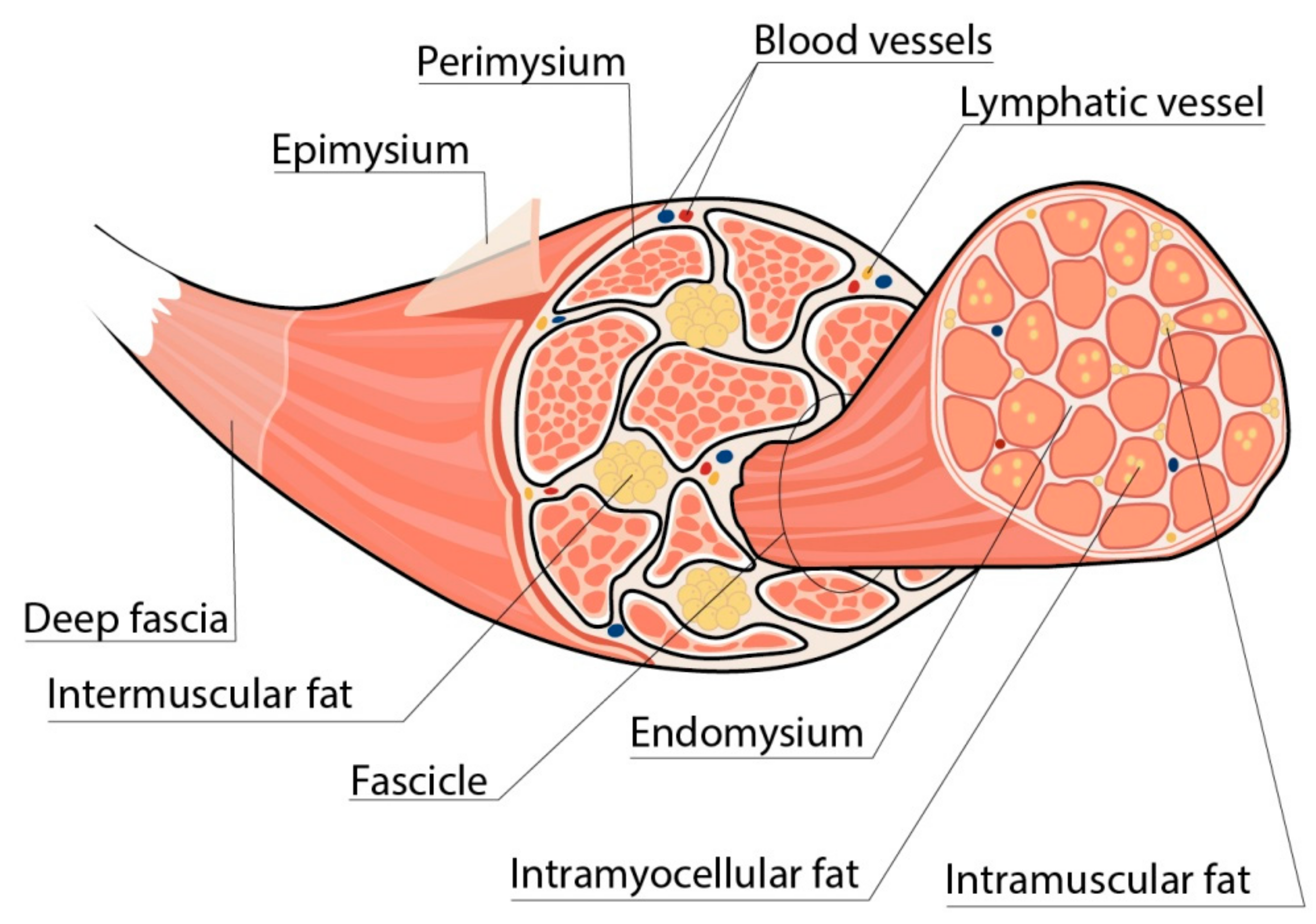
Figure 1. Localization of fat depots in skeletal muscles. Intermuscular fat depots represent adipocytes localized between muscle groups, whereas intramuscular fat depots are adipocytes located between the muscle fibers. Usually, these two depots are combined by the term “intermuscular fat”. Intramyocellular lipids denote lipid droplets stored within the myocytes.
Life 13 01415 g001
Figure 2. Possible explanations for “athlete’s paradox”. The term “athlete’s paradox” describes a condition characterized by high amounts of IMCL fat in the muscles observed both in patients with obesity and/or T2DM, and in endurance-trained athletes. In obese individuals, the IMCL depots are located mainly in fast-twitch muscle fibers and contain high amounts of ceramides and diacylglycerols (DAGs), as well as low concentrations of perilipin 5 (PLIN5) and triacyclglycerols (TAGs). In contrast, in endurance-trained athletes, IMCL droplets are situated primarily in slow-twitch muscle fibers and carry high amounts of TAGs and PLIN5 along with low concentrations of lipid metabolites, such as DAGs and ceramides. The intracellular localization of IMCL depots also differs between endurance-trained athletes and patients with obesity. In particular, in athletes, lipid droplets are located predominantly in close relation to mitochondria and the endoplasmic reticulum, while in obese individuals, they may be found in the sarcolemma, subsarcolemmal, or perinuclear regions. Overall, these differences determine a decrease in muscle oxidative capacity and insulin sensitivity in obese patients in contrast to the athletes, despite similar numbers of IMCL depots within the muscle fibers. Up-oriented arrows (↑) indicate up-regulation. Down-oriented arrows (↓) indicate downregulation. The number of arrows corresponds to the magnitude of the differences.
Life 13 01415 g002
Figure 3. Disturbed muscle metabolic flexibility in obesity and possible role of glucocorticoids. Disturbed muscle oxidative capacity is believed to be associated with both the change in the proportions of muscle fiber types and mitochondrial dysfunction. Patients with obesity have lower proportions of type I muscle fibers characterized by an increased number of mitochondria and, at the same time, greater proportions of type IIx fibers compared with lean individuals. Thus, obesity is associated with reduced muscle energetic efficiency with a predominance of glycolysis over oxidation processes. In addition, obesity is characterized by the decrease in mitochondrial surface area, substantially lower expression of mitochondria genes, and reduced activity of the mitochondrial electron transport chain. This causes incomplete β-oxidation and, consequently, the accumulation of lipid intermediates, capable of affecting mitochondrial morphology and function. Disturbed oxidative phosphorylation leads to the increased production of reactive oxygen species (ROS), which facilitate mitochondrial fragmentation. In these settings, the expression of peroxisome proliferator-activated receptor–gamma coactivator-1alpha (PGC-1α), which is initially lower in type IIx fibers compared to type I fibers, is being downregulated, further aggravating mitochondrial damage and dysfunction. It is well known that serum glucocorticoid (GC) levels are elevated in individuals with obesity, and this contributes to the development of insulin resistance, hyperglycemia, and dyslipidemia. Moreover, GCs appear to be involved in the disturbance of muscle metabolic flexibility in obesity. It is established that type IIx muscle fibers, being the predominant muscle fiber type in patients with obesity, are characterized by greater susceptibility to GCs, possibly due to the greater expression of GC receptors. Therefore, the negative effects of GCs on skeletal muscle may be more pronounced in patients with obesity. Additionally, GCs are known to increase the accumulation of lipid intermediates in skeletal muscle, thus facilitating mitochondria dysfunction and impairment of muscle energy metabolism. Up-oriented arrows (↑) indicate up-regulation. Down-oriented arrows (↓) indicate downregulation. The number of arrows corresponds to the magnitude of the differences.
Life 13 01415 g003
Figure 4. Key effects of estrogens, progesterone, and androgens on skeletal muscle. Obesity is known to be associated with reduced serum progesterone and estradiol concentrations in women, as well as with decreased production and secretion of androgens in men. Taking into account the beneficial effects of sex steroids on skeletal muscle, these changes in their serum concentrations may represent an additional mechanism contributing to the decline in muscle mass and function in obesity. AR = androgen receptor; ATP = adenosine triphosphate; ER = estrogen receptor; GCs = glucocorticoids; GH = growth hormone; IGF-1 = insulin growth factor 1; PGC-1α = peroxisome proliferator-activated receptor-gamma coactivator-1alpha; PR = progesterone receptor. Up-oriented arrows (↑) indicate up-regulation. Down-oriented arrows (↓) indicate downregulation.
Life 13 01415 g004
Table 1. Sex differences in muscle phenotypes.
ParameterMalesFemalesReference
Muscle fiber typeMore glycolytic fibers (fast-twitch) compared to femalesMore oxidative fibers (low-twitch) compared to males[118,120]
Satellite cellsGreater number of satellite cells and higher proliferation capacity compared to malesLess number of satellite cells and reduced proliferation capacity compared to females[121,122]
Susceptibility to atrophy More susceptible to inflammation-induced atrophyMore susceptible to disuse-induced atrophy[123,124]
Predominant mechanism of protein degradationAutophagyUbiquitin–proteasome system[125,126,127]
Age-related shift in myofiber typesTowards type 1 (slow-twitch) dominant compositionNo shift occurs[124]
Top-ranked differentially expressed process during aging Oxidative phosphorylationCell growth[124]
Disclaimer/Publisher’s Note: The statements, opinions and data contained in all publications are solely those of the individual author(s) and contributor(s) and not of MDPI and/or the editor(s). MDPI and/or the editor(s) disclaim responsibility for any injury to people or property resulting from any ideas, methods, instructions or products referred to in the content.