Article

Preliminary Survey of Pathogens in the Asian Honey Bee (Apis cerana) in Thailand

by
Patcharin Phokasem
1,2,
Chainarong Sinpoo
1,
Korrawat Attasopa
3,
Sasiprapa Krongdang
4,
Thunyarat Chantaphanwattana
1,
Tial C. Ling
1,
Jeffery S. Pettis
5,
Panuwan Chantawannakul
1,2,
Veeranan Chaimanee
6,* and
Terd Disayathanoowat
1,7,*
1
Bee Protection Laboratory, Department of Biology, Faculty of Science, Chiang Mai University, Chiang Mai 50200, Thailand
2
Environmental Science Research Center, Faculty of Science, Chiang Mai University, Chiang Mai 50200, Thailand
3
Department of Entomology and Plant Pathology, Faculty of Agriculture, Chiang Mai University, Chiang Mai 50200, Thailand
4
Faculty of Science and Social Sciences, Burapha University Sa Kaeo Campus, Sa Kaeo 27160, Thailand
5
Pettis and Assoc, LLC, Salisbury, MD 21801, USA
6
Department of Agro-Industrial Biotechnology, Maejo University Phrae Campus, Phrae 54140, Thailand
7
Research Center of Microbial Diversity and Sustainable Utilization, Chiang Mai University, Chiang Mai 50200, Thailand
*
Authors to whom correspondence should be addressed.
Life 2023, 13(2), 438; https://doi.org/10.3390/life13020438
Submission received: 19 December 2022 / Revised: 25 January 2023 / Accepted: 31 January 2023 / Published: 3 February 2023
(This article belongs to the Special Issue Genetics, Breeding and Reproduction of Honeybees)

Abstract

:
Widespread parasites, along with emerging threats, globalization, and climate change, have greatly affected honey bees’ health, leading to colony losses worldwide. In this study, we investigated the detection of biotic stressors (i.e., viruses, microsporidian, bacteria, and fungi) in Apis cerana by surveying the colonies across different regions of Thailand (Chiang Mai in the north, Nong Khai and Khon Kaen in the northeast, and Chumphon and Surat Thani in the south, in addition to the Samui and Pha-ngan islands). In this study, we detected ABPV, BQCV, LSV, and Nosema ceranae in A. cerana samples through RT-PCR. ABPV was only detected from the samples of Chiang Mai, whereas we found BQCV only in those from Chumphon. LSV was detected only in the samples from the Samui and Pha-ngan islands, where historically no managed bees are known. Nosema ceranae was found in all of the regions except for Nong Khai and Khon Kaen in northeastern Thailand. Paenibacillus larvae and Ascosphaera apis were not detected in any of the A. cerana samples in this survey. The phylogenetic tree analysis of the pathogens provided insights into the pathogens’ movements and their distribution ranges across different landscapes, indicating the flow of pathogens among the honey bees. Here, we describe the presence of emerging pathogens in the Asian honey bee as a valuable step in our understanding of these pathogens in terms of the decline in eastern honey bee populations.

Graphical Abstract

1. Introduction

The Asian honey bee, Apis cerana, is distributed throughout Asia. These bees provide not only important pollination services for plant ecosystems, but also economic value to society due to the production of honey [1]. Based on morphological characteristics [2], A. cerana is divided into four groups in Thailand: northern–central Thailand, southern Thailand, Samui Island, and Phuket Island). Beekeepers in Thailand have long keeping A. cerana using traditional log hives for more than 1000 years, and this traditional method of beekeeping continues today [1]. It is postulated that there is a great decline in pollinator populations and/or diversity around the globe [3], and that this is influenced by various factors, including habitat loss, pesticides, climate change, and the spread of emergent pathogens, parasites, and predators [4,5].
Honey bees have been found to suffer from various pathogens. The most serious pathogens occurring in honey bees include viruses, bacteria, microsporidians, and fungi [6]. These pathogens cause diseases that bring about large damage to the beekeeping industry, which can ultimately create a great economic losses worldwide [6,7,8]. The greatest concern for honey bees’ health is nosemosis (caused by Nosema spp.). This disease has caused great losses in some parts of Europe [8,9]. American foulbrood (AFB), caused by Paenibacillus larvae, is generally found in the European honey bee; however, in some parts of Asia where beekeeping overlaps with the ecological niche of A. cerana, interspecific pathogen transmission from Apis mellifera to A. cerana can be observed [10,11]. The fungal honey bee pathogen Ascosphaera apis is a common and widespread disease that can cause a severe decline in colony production [12]. Consequently, these biotic stressors can induce considerable losses to honey bee populations.
Although A. cerana is often considered to have lower pathogen prevalence compared to A. mellifera [11], most pathogens are capable of infecting multiple hosts in natural systems [13,14]. Many emerging diseases in animals are linked to the utilization of multiple hosts [13,14]. Previous studies have identified parasites and pathogens that attack honey bee colonies and are transmitted between different host species. For instance, viral diseases that occur frequently in A. mellifera have been found in A. cerana [15,16,17]. Similar patterns have also been found in various Bumbus species [18] and non-Apis hymenopteran species [19]. The reciprocal transmission of Varroa destructor and N. ceranae between A. cerana and A. mellifera has also been found to decrease honey quality and productivity [20,21,22,23]. Chinese sacbrood virus (CSBV) is a serious threat to A. cerana, and it has been detected in Chinese populations of A. mellifera [24]. These factors have caused a great decline in A. cerana populations over the past few years [25]. Therefore, serious colony losses worldwide have been correlated with the movement of pests and pathogens between different host populations.
Recently, many studies have discovered newly emergent pathogens (such as Apis mellifera filamentous virus and Lake Sinai Virus) in different honey bee populations [26]. Global assessment of the impacts of biotic stressors on honey bees can help in devising relevant and effective control strategies for pathogens that spread across different populations. To date, little information is known about the presence of honey bee pathogens in A. cerana populations. In this study, we evaluated the impacts of biotic stressors on Asian honey bees across different beekeeping sectors. The aim of this study was to assess the presence of 12 honey bee viruses: acute bee paralysis virus (ABPV), aphid lethal paralysis virus strain Brookings (ALP-Br), Big Sioux River virus (BSRV), black queen cell virus (BQCV), chronic bee paralysis virus (CBPV), deformed wing virus type A (DWV-A), deformed wing virus type B (DWV-B), Israeli acute paralysis virus (IAPV), Kashmir bee virus (KBV), sacbrood virus (SBV), slow bee paralysis virus (SBPV), and Lake Sinai virus (LSV). In addition, we aimed to examine the occurrence of four types of honey bee microsporidia (i.e., Nosema apis, N. ceranae, N. bombi, and Crithidia bombi), a fungus (i.e., Ascosphaera apis), and a bacterium (i.e., Paenibacillus larvae) in A. cerana collected from different regions of Thailand. We also aimed to compare the presence of pathogens in A. cerana between islands (the Samui and Pha-ngan islands) and mainland regions. Samui Island is in the Gulf of Thailand, approximately 35 km from the town of Surat Thani, whereas Pha-ngan Island is about 15 km from Samui Island [27]. On both islands, only native honey bees can be found. There are no records in terms of honey bee management on either island. A better understanding of A. cerana’s health and factors causing their colony decline and affecting their productivity in different geographic regions is a fundamental step in building beekeeping knowledge and strategies for conserving pollinator diversity via sustainable beekeeping.

2. Materials and Methods

2.1. Sample Collection

The samples of adult Apis cerana were collected from three different regions (northern, northeastern, and southern regions) and two islands (Samui and Pha-ngan islands) in Thailand from January to June 2021. Adult of A. cerana samples (n = 50–200 for each colony) were collected from 24 colonies in 10 different locations (Figure 1 and Supplementary Table S1). All samples were preserved in RNAlater (Invitrogen, Vilnius, Lithuania) and stored at −80 °C before further examination in the laboratory.

2.2. DNA Extraction and DNA Analyses for Species Identification

To confirm the morphospecies identification, molecular analyses were performed using highly conserved regions of the mitochondrial cytochrome c oxidase subunit 1 (COI) gene, which is referred to as the DNA barcoding region. Genomic DNA was isolated from the whole bodies of the A. cerana samples (Figure 1 and Supplementary Table S1) using a DNA purification kit (PureLink Genomic DNA Mini Kit, Invitrogen, Carlsband, CA, USA) according to the manufacturer’s instructions. The primer pair was used to amplify a partial fragment DNA of the COI gene (listed in Table S2). The PCR amplification was performed in 25 µL reactions containing 1X PCR buffer, 1.5 mM MgCl2, 0.2 mM dNTPs, 0.5 µM forward primer, 0.5 µM reverse primer, 1U Taq DNA polymerase (Invitrogen, Carlsband, CA, USA), and 50 ng of DNA template. The PCR cycling conditions were 94 °C for 5 min, and 40 cycles of 94 °C for 30 s, 55 °C for 30 s, and 72 °C for 45 s, followed by a final step at 72 °C for 7 min. The resulting PCR products were separated by size on 1.5% agarose gel electrophoresis, and the nucleotide sequences were analyzed to distinguish the honey bee species. The DNA sequences were deposited in GenBank with accession numbers (see Supplementary Figure S1 and Table S3).

2.3. RNA Extraction and cDNA Synthesis for Viral Detection

After confirming the A. cerana species, 10 adult workers from each colony were pooled and homogenized using a mortar and pestle with liquid nitrogen. Total RNA was extracted using TRIzol Reagent (Invitrogen, Carlsband, CA, USA) according to the manufacturer’s instructions. The concentration of RNA was measured for absorbance at 260 nm (A260), and its purity was assessed at a ratio of A260/A280 using a BioDrop-DUO UV/Vis spectrophotometer (BioDrop, Cambridge, UK). Four micrograms of RNA was reverse-transcribed into cDNA using Tetro Reverse Transcriptase (Bioline, Memphis, TN, USA). Both oligo(dT) and random hexamer primers were used in the reaction. The mixture was incubated at 25 °C for 10 min, followed by 45 °C for 30 min, and then the reaction was terminated at 85 °C for 5 min. The cDNA was obtained and stored at −20 °C before proceeding to the next step.

2.4. DNA Extraction for Non-Virus Analysis

Ten adult A. cerana workers from each colony were pooled and homogenized as described for the initial RNA extraction step above. Total genomic DNA was extracted using a DNA purification kit (PureLink Genomic DNA Mini Kit, Invitrogen, Carlsband, CA, USA) according to the manufacturer’s instructions. DNA samples were stored at −20 °C prior to molecular screening for microsporidia, fungi, and bacteria.

2.5. PCR Conditions

Each sample was screened for N. ceranae, N. apis, N. bombi, C. bombi, P. larvae, A. apis, and 12 honey bee viruses; ABPV, ALP-Br, BSRV, BQCV, CBPV, DWV-A, DWV-B, IAPV, KBV, SBV, SBPV, and LSV. The specific primers used for honey bee pathogens and housekeeping genes (i.e., β-actin and RPS5) are listed in Supplementary Table S2 [28,29,30,31,32,33,34,35,36,37,38,39,40,41,42,43,44,45]. The cDNA templates were diluted 5-fold. Total DNA was diluted to 100 ng/μL. PCRs were performed using Biometra thermal cyclers (Analytik Jena AG, Jena, Germany) in 25 µL volumes containing 1 µL of DNA or cDNA template, 2.5 µL of PCR buffer, 0.75 µL of MgCl2, 0.5 µL of dNTPs, 0.1 µL of Taq DNA polymerase (Invitrogen, Carlsband, CA, USA), and 1.25 µL of each forward and reverse primer (10 mM), plus 17.65 µL of water. Amplification was performed with the following thermal cycling profiles: 3 min incubation at 94 °C, followed by 40 cycles of 45 s at 94 °C for denaturation, 1 min at 52–56 °C for annealing (for ABPV, IAPV, and SBPV the annealing temperature was 52 °C; for DWA-A, DWV-B, SBV, BQCV KBV, and CBPV the annealing temperature was 55 °C; for LSV, ALP-Br, BSRV, N. ceranae, N. apis, N. bombi, C. bombi, P. larvae, and A. apis the annealing temperature was 56 °C), 1 min at 72 °C for extension, and then a final step of 10 min at 72 °C. In each run, PCR mixture without DNA was used as a negative control. The amplicons obtained were electrophoresed on 1.5% agarose gel to verify the size of the fragments with reference to a 100 bp ladder (GeneDireX, Taoyuan, Taiwan). The PCR products were cleaned up using ExoSAP reagent, and then the expected amplicons were nucleotide-sequenced bidirectionally by a commercial company (Macrogen, Seoul, South Korea).

2.6. Phylogenetic Tree Analysis

Fragment assembly of nucleotide sequences was performed using BioEdit software version 7.2.5 (Ibis Biosciences, Carlsbad, CA, USA) [46]. They were trimmed to equal size and aligned using MEGA X (iGEM, Boston, MA, USA). The obtained sequences were compared with the recorded viruses, Nosema, and COI gene in the GenBank database using the BLAST program (available from the National Center for Biotechnology Information (NCBI)). Evolutionary analyses were conducted in MEGA X [47] by using the maximum likelihood method and the Tamura–Nei model [48]. The initial tree was obtained automatically with neighbor-joining and BioNJ algorithms. The maximum likelihood trees were constructed using MEGA X. The appropriate substitution model was chosen as described in the legends of Figure 2, Figure 3, Figure 4, Figure 5 and Figure S1 for each virus, N. ceranae, and COI gene phylogeny. The bootstrap values of 1000 replicates were determined, and the percentage of replicates was shown in branches. The GenBank accession numbers from the isolates are given in the figures. The nucleotide sequences used for phylogenetic analysis were deposited in GenBank and assigned accession numbers. Related sequences of the viruses, Nosema, and bees used for constructing the phylogenetic trees were analyzed along with this study’s sequences (see Supplementary Figure S1 and Table S3).

3. Results

3.1. COI-Sequence-Based Characterization of A. cerana Samples

The bee phylogenetic tree based on the COI gene fragment estimated by the maximum likelihood method is presented in Figure S1. The tree implied the phylogenetic results of A. cerana subspecies appearing in Thailand. The samples from the northern and northeastern regions were identified as A. cerana indica, which were clustered with the same branch of A. cerana from India. The A. cerana samples from the southern regions and the Samui and Pha-ngan islands were found to be closely related to Apis nuluensis.

3.2. N. ceranae, P. larvae, and A. apis Frequencies in A. cerana Colonies

The results of N. ceranae prevalence are summarized in Table 1. N. ceranae-infected honey bees were detected among A. cerana workers from eight samples (33.33%). The average infection rate of N. ceranae in the southern region was 37.50%, while the average infection rate of N. ceranae in the northern region was 50.00%. Among southern locations with detectable N. ceranae, the highest prevalence for N. ceranae was found in Samui Island, with an infection rate of 25.00%. Nosema apis, N. bombi, C. bombi, P. larvae, and A. apis were not detected in any of the examined samples (Table 1).

3.3. Virus Frequencies in A. cerana Colonies

Prevalence data for 12 honey bee viruses (ABPV, ALP-Br, BSRV, BQCV, CBPV, DWV-A, DWV-B, IAPV, KBV, SBV, SBPV, and LSV) screened in 24 honey bee colonies from 10 locations are shown in Table 1. Honey bee viruses were detected in eight pools of 24 honey bee colonies. Of these eight positive pools, 29.17% of the viruses were collected in southern Thailand, followed by northern Thailand (4.17%). However, no positive viruses were found in the northeastern region. Of these viruses, only ABPV, BQCV, and LSV were detected in A. cerana. ABPV was found in one sample of A. cerana from the northern region (25.00%, Chiang Mai province). BQCV was found in one sample of A. cerana from the southern region (6.25%, Chumphon province). LSV was detected in six A. cerana samples from the southern region (37.50%, Samui and Pha-ngan islands).

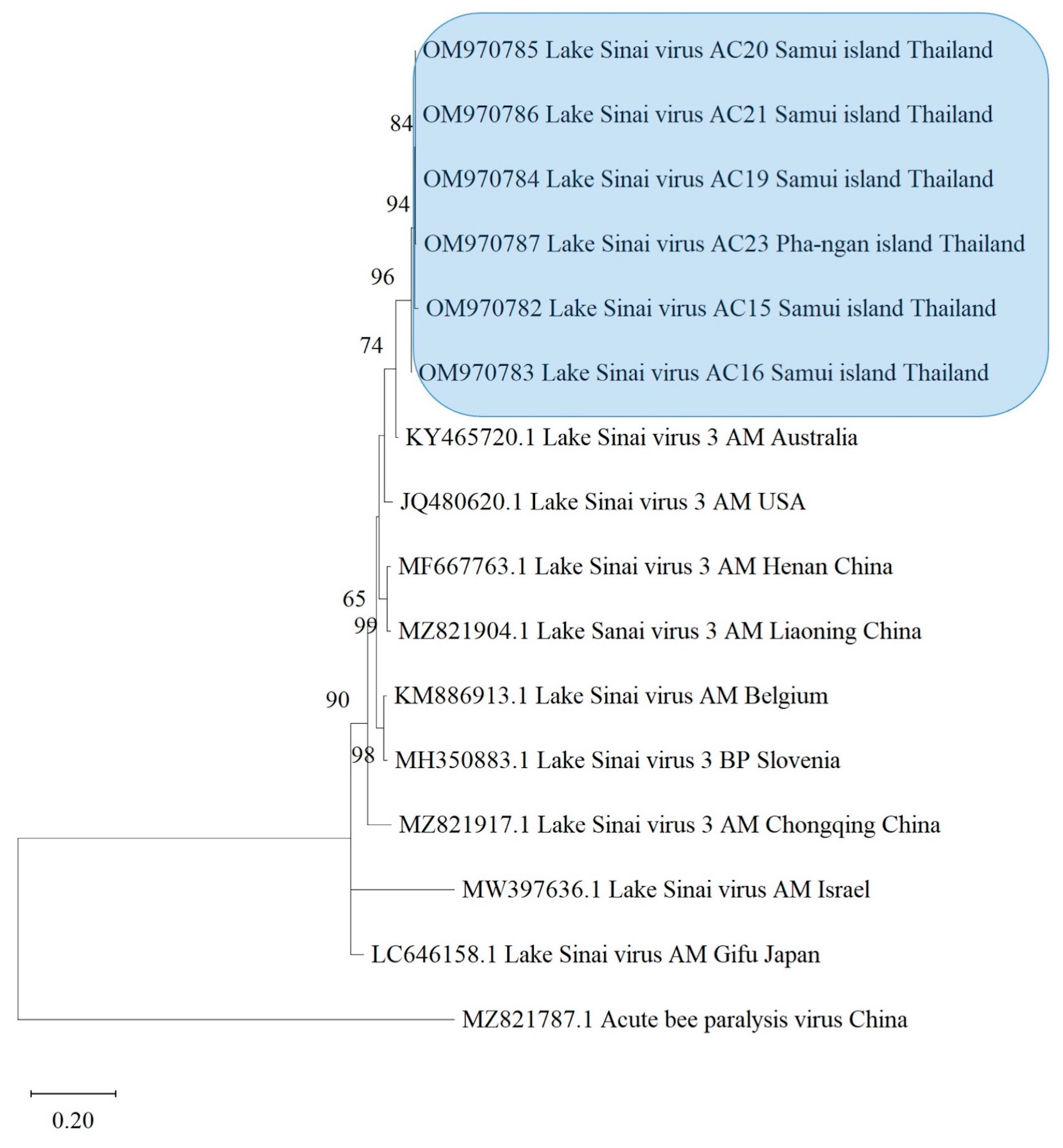
3.4. Phylogenetic Analysis

To study the genetic relationships and variability of the studied pathogens, the nucleotide sequences of pathogens were selected from the GenBank database, and the phylogenetic analysis was carried out via maximum likelihood (ML) estimation. The clustering pattern was constructed to assess the relationships between the samples of N. ceranae, ABPV, BQCV, and LSV from different geographic locations.
Nosema ceranae was only found in A. cerana from northern and southern Thailand. Based on closely related sequences of N. ceranae obtained from A. cerana, the phylogenetic tree was constructed. The N. ceranae isolated from northern and southern Thailand were part of the same cluster, which was different from the N. ceranae strain circulating in A. mellifera (Figure 2). However, the other cluster was N. ceranae detected in A. mellifera collected from Spain, China, France, and USA. The tree suggested that the epidemic of N. ceranae displayed some consistency across species between Asian and European honey bees.
LSV was found to be the most prevalent virus in A. cerana and was predominantly present in A. cerana from the southern region. The infection percentage of LSV accounted for 25.00% and affected the majority of A. cerana colonies. The phylogenetic tree of LSV was constructed from six isolates from A. cerana samples taken in this study. This also demonstrated that LSV has a genetic relationship according to host species isolation. LSV isolates from A. cerana colonies were clustered together in the phylogenetic tree, and they were collected from the southern region of Thailand, as well as the Samui and Pha-ngan islands. They were also nearly identical to LSV-3 isolated from A. mellifera from Australia, rather than other Asian isolates (Figure 3).
According to the phylogenetic trees based on the partial sequences of the capsid protein of BQCV, two clusters were shown according to the geographic locations (Figure 4). One unique group was formed by Asian countries, including Thailand, China, and South Korea, while another group was formed by USA isolates. The USA isolates obtained from Bombus impatiens formed a separate cluster. BQCV was randomly present among A. cerana, A. mellifera, and A. florea in the Asian cluster. The spread of BQCV also appeared in all regions, and the BQCV from Asian isolates was closely related to isolates from the USA. According to the BQCV tree, the isolates obtained from A. mellifera, A. cerana, and A. florea indicated that the Asian BQCV cluster (Thailand, China, South Korea) may be related to their sister taxon of BQCV isolated from Bombus impatiens (Figure 4).
According to the phylogenetic tree based on the RNA-dependent RNA polymerase region (RdRp) of ABPV, the sequences also formed distinct groups based on their geographical origins, regardless of honey bee species (Figure 5). Two clusters were formed: one comprising isolates from China, and one from the western isolates (i.e., Hungary and Poland). The phylogenetic tree of the ABPV isolates demonstrated that ABPV isolated from A. cerana was homogeneous and nearly identical to one of two distinct ABPV lineages found in A. mellifera (Figure 5).

4. Discussion

Our results provide an important overview of the distribution of pathogens of A. cerana across different regions of Thailand. A total of four pathogens (ABPV, BQCV, LSV, and N. ceranae) were found in adult A. cerana samples, even though these honey bee samples did not show any symptoms of disease. Our study showed, surprisingly, that A. cerana honey bees located in the Samui and Pha-ngan islands of Thailand, which were not managed by beekeepers, still had LSV infections in their colonies.
LSV was the most prevalent pathogen in A. cerana samples collected from the Samui and Pha-ngan islands (the distance of the two islands from mainland Thailand is approximately 35–55 km). Surprisingly, this virus was not found in the samples collected from mainland Thailand. LSV has also been detected in hornets [49], bumblebees [50,51], A. mellifera [52,53], Varroa destructor [54], and ants [55]. In addition, the V. destructor mite—an ectoparasite of honey bees—is known as a vector for LSV [56]. Our results are consistent with previous reports where LSV was detected in honey bees on Norfolk Island, Australia [52]. The distance of this island is approximately 1400 km from mainland Australia [57]. LSVs are very similar to chronic bee paralysis virus (CBPV) [58]. The recently described LSV was also found to be linked to a shift in gut bacterial composition that may be a biomarker of honey bee colony loss [59]. In addition, LSV1, LSV-2, LSV3, LSV6, and LSV 7 were recently discovered as honey bee viruses in the USA [59]. Phylogenetic analysis revealed one LSV-3 lineage in A. cerana that is closely related to LSV-3 from A. mellifera in Australia. In Thailand, there is no such record of the presence of LSV in A. mellifera or other arthropods. Therefore, our study is the first to report the presence of LSV-3 in A. cerana in Thailand. Experience would dictate that we need to be concerned about honey bee pathogens that can jump between Apis species.
ABPV is a common infective agent of A. mellifera colonies that is frequently detected in healthy colonies. This virus is one of the most serious problems in the beekeeping industry. It is assumed that this virus plays a role in causing the colony loss of A. mellifera across the globe [60]. In this study, ABPV was detected in only one of the A. cerana samples from the northern region (Chiang Mai province) in Thailand. ABPV and/or its strains KBV and IAPV were detected in Asian honey bees in South Korea, China, and Japan [15,16,61]. Previous studies have reported that ABPV was detected in A. mellifera in northern Thailand [62]. It is possible that ABPV from A. mellifera jumped to A. cerana. In Asia, A. mellifera colonies share the same habitats as A. cerana colonies [63]. Severe colony losses are often preceded by a rapid progression of paralysis caused by viruses of the ABPV complex [64]. According to our study, there is a great need to determine the virulence of ABPV in Asian honey bees.
BQCV (a member of the Dicistroviridae) is the most abundant of the honey bee viruses and is prevalent in covert infections of most Apis species, including both managed and wild species [63]. BQCV has been detected in A. cerana in China, South Korea, Vietnam, Japan, and Thailand [10,16,17,39,65,66]. In the current study, the prevalence of BQCV was found in only one sample among A. cerana samples from the southern region (Chumphon province) in Thailand. Previous findings suggested that BQCV was associated with Nosema in A. mellifera colonies in which serious clinical signs were observed [67]. Our results demonstrated the co-infection of BQCV and N. ceranae in the same colony of Asian honey bees located in the southern region.
Both BQCV and ABPV isolates obtained from A. cerana fell into the same cluster as those viruses isolated from A. mellifera. Previously, Sanpa and Chantawannakul [62] reported that DWV, ABPV, CBPV, KBV, and SBV were found in A. mellifera colonies in northern Thailand. BQCV also was detected in A. mellifera colonies in northern Thailand [65]. These findings could be a suitable way to explain our results of viral spillover from non-native to native honey bees. Further study is needed to determine whether the transfer of these pathogens from non-native to native honey bees could be ongoing, including interspecies transmission of parasites and a potentially crucial role of host–parasite interaction.
The microsporidia of Nosema spp. are obligate intracellular parasites [68]. Two species of Nosema have been described as infectors of honey bees (N. ceranae and N. apis) [20]. In the past, Nosema ceranae was found to parasitize only Asian honey bees, while N. apis was found to parasitize the European honey bee [69]. Nosema ceranae has infected A. mellifera and spread worldwide, leading to a decline in populations of N. apis [20,70,71,72,73]. Today, Nosema ceranae is the most common Nosema found in A. cerana and other Apis species [20,74,75,76]. The detection of N. ceranae has been reported in A. mellifera, A. cerana, A. dorsata [74,75,77], and Bombus spp. [78] in northern Thailand. Moreover, this parasite has been detected in A. cerana, A. florea, and the non-native A. mellifera in central Thailand [79]. Our results also showed that N. ceranae was detected in A. cerana samples from Chiang Mai, Chumphon, and Samui Island, but not in those collected from the northeast region. This finding is similar to that of Suraporn et al. [80], where Nosema was not detected in honey bees collected from the northeastern region of Thailand. However, these findings suggest that N. ceranae may be widespread and common in honey bees in Thailand, and potentially elsewhere in Southeast Asia. Furthermore, phylogenetic analyses can be used to separate specific taxa of N. ceranae in A. cerana and the other Apis isolates, as reported by Chaimanee et al. [75].

5. Conclusions

This study showed that the Asian honey bee (A. cerana) colonies distributed across several regions of Thailand were infected with several pathogens. The most prevalent pathogen was N. ceranae, followed by the viruses LSV, ABPV, and BQCV, in that order. The present study also reports the molecular characterization of LSV in A. cerana from Samui and Pha-ngan islands. Additionally, a low prevalence of ABPV and BQCV was observed in A. cerana. The phylogenetic tree analysis showed that pathogens can flow between host populations across the landscapes of different islands. Understanding the patterns of pathogen distribution will aid in disease control for honey bees in the future. The goal of this study was to highlight research findings that have contributed to our understanding of A. cerana colony health. Further investigation is needed to understand specific pathogens’ spillover processes. Increased knowledge of pathogen spillover will have important implications for the health and conservation of native honey bee species as well as other pollinators worldwide.

Supplementary Materials

The following supporting information can be downloaded at: https://www.mdpi.com/article/10.3390/life13020438/s1, Table S1: Sampling locations of specimens used in the present study, including geographic coordinates, elevations, and sample codes. Table S2: Primers used for molecular amplification of pathogens and mtDNA detection in the present study. Table S3: The sequences of Nosema spp., LSV, BQCV, ABPV, and bee specimens in our study, including other related sequences obtained from GenBank used in our phylogenetic tree analyses, showing GenBank accession numbers. Figure S1: The phylogenetic tree based on cytochrome c oxidase subunit I (COI) sequences of Apis cerana collected in Thailand using maximum likelihood analysis.

Author Contributions

Conceptualization, T.D., P.C. and V.C.; methodology, P.P., S.K. and K.A.; software, T.C.L.; validation, P.P. and J.S.P.; formal analysis, P.P., T.C. and K.A.; investigation, P.P., T.C. and C.S.; resources, T.D.; data curation, P.P. and T.C.L.; writing—original draft preparation, P.P., T.D., K.A. and S.K.; writing—review and editing, P.P., J.S.P., T.C.L., K.A., S.K., T.C., T.D., C.S. and V.C.; visualization, P.P., T.D. and P.C.; supervision, T.D. and P.C.; project administration, T.D. and P.C.; funding acquisition, T.D. and P.C. All authors have read and agreed to the published version of the manuscript.

Funding

This research was funded by the Mekong–Lancang Cooperation Special Fund: 2022.

Institutional Review Board Statement

This study did not require ethical approval.

Informed Consent Statement

Not applicable.

Data Availability Statement

Not applicable.

Acknowledgments

T.D., P.C., and P.P. were supported by Post-Doctoral Fellowship 2022, Office of Research Administration, Chiang Mai University, Thailand, grant number R000030564. This work was partially supported by Chiang Mai University. The Department of Biology, Faculty of Science, Chiang Mai University, Thailand, is also acknowledged.

Conflicts of Interest

The authors declare no conflict of interest.

References

  1. Chantawannakul, P.; Williams, G.; Neumann, P. Asian Beekeeping in the 21st Century; Springer: Berlin/Heidelberg, Germany, 2018; ISBN 978-981-10-8221-4. [Google Scholar]
  2. Sylvester, H.A.; Limbipichai, K.; Wongsiri, S.; Rinderer, T.E.; Mardan, M. Morphometric Studies of Apis cerana in Thailand and the Malaysian Peninsula. J. Apic. Res. 1998, 37, 137–145. [Google Scholar] [CrossRef]
  3. Potts, S.G.; Biesmeijer, J.C.; Kremen, C.; Neumann, P.; Schweiger, O.; Kunin, W.E. Global Pollinator Declines: Trends, Impacts and Drivers. Trends Ecol. Evol. 2010, 25, 345–353. [Google Scholar] [CrossRef] [PubMed]
  4. Gallai, N.; Salles, J.-M.; Settele, J.; Vaissière, B.E. Economic Valuation of the Vulnerability of World Agriculture Confronted with Pollinator Decline. Ecol. Econ. 2009, 68, 810–821. [Google Scholar] [CrossRef]
  5. Liu, Z.; Chen, C.; Niu, Q.; Qi, W.; Yuan, C.; Su, S.; Liu, S.; Zhang, Y.; Zhang, X.; Ji, T. Survey Results of Honey Bee (Apis mellifera) Colony Losses in China (2010–2013). J. Apic. Res. 2016, 55, 29–37. [Google Scholar] [CrossRef]
  6. Vanengelsdorp, D.; Meixner, M.D. A Historical Review of Managed Honey Bee Populations in Europe and the United States and the Factors That May Affect Them. J. Invertebr. Pathol. 2010, 103, S80–S95. [Google Scholar] [CrossRef]
  7. Gisder, S.; Genersch, E. Special Issue: Honey Bee Viruses. Viruses 2015, 7, 5603–5608. [Google Scholar] [CrossRef]
  8. Higes, M.; Martín-Hernández, R.; Botías, C.; Bailón, E.G.; González-Porto, A.V.; Barrios, L.; del Nozal, M.J.; Bernal, J.L.; Jiménez, J.J.; Palencia, P.G.; et al. How Natural Infection by Nosema ceranae Causes Honeybee Colony Collapse. Environ. Microbiol. 2008, 10, 2659–2669. [Google Scholar] [CrossRef]
  9. Higes, M.; Martín, R.; Meana, A. Nosema ceranae, a New Microsporidian Parasite in Honeybees in Europe. J. Invertebr. Pathol. 2006, 92, 93–95. [Google Scholar] [CrossRef]
  10. Forsgren, E.; Wei, S.; Guiling, D.; Zhiguang, L.; Van Tran, T.; Tang, P.T.; Truong, T.A.; Dinh, T.Q.; Fries, I. Preliminary Observations on Possible Pathogen Spill-over from Apis mellifera to Apis cerana. Apidologie 2015, 46, 265–275. [Google Scholar] [CrossRef]
  11. Lin, Z.; Page, P.; Li, L.; Qin, Y.; Zhang, Y.; Hu, F.; Neumann, P.; Zheng, H.; Dietemann, V. Go East for Better Honey Bee Health: Apis cerana Is Faster at Hygienic Behavior than A. mellifera. PLoS ONE 2016, 11, e0162647. [Google Scholar] [CrossRef] [Green Version]
  12. Jensen, A.B.; Aronstein, K.; Flores, J.M.; Vojvodic, S.; Palacio, M.A.; Spivak, M. Standard Methods for Fungal Brood Disease Research. J. Apic. Res. 2013, 52, 1–20. [Google Scholar] [CrossRef]
  13. Pedersen, A.B.; Altizer, S.; Poss, M.; Cunningham, A.A.; Nunn, C.L. Patterns of Host Specificity and Transmission among Parasites of Wild Primates. Int. J. Parasitol. 2005, 35, 647–657. [Google Scholar] [CrossRef]
  14. Woolhouse, M.E.; Taylor, L.H.; Haydon, D.T. Population Biology of Multihost Pathogens. Science 2001, 292, 1109–1112. [Google Scholar] [CrossRef]
  15. Ai, H.; Yan, X.; Han, R. Occurrence and Prevalence of Seven Bee Viruses in Apis mellifera and Apis cerana Apiaries in China. J. Invertebr. Pathol. 2012, 109, 160–164. [Google Scholar] [CrossRef]
  16. Choe, S.E.; Nguyen, L.T.K.; Noh, J.H.; Koh, H.B.; Jean, Y.H.; Kweon, C.H.; Kang, S.W. Prevalence and Distribution of Six Bee Viruses in Korean Apis cerana Populations. J. Invertebr. Pathol. 2012, 109, 330–333. [Google Scholar] [CrossRef]
  17. Li, J.; Qin, H.; Wu, J.; Sadd, B.M.; Wang, X.; Evans, J.D.; Peng, W.; Chen, Y. The Prevalence of Parasites and Pathogens in Asian Honeybees Apis cerana in China. PLoS ONE 2012, 7, e47955. [Google Scholar] [CrossRef]
  18. Peng, W.; Li, J.; Boncristiani, H.; Strange, J.P.; Hamilton, M.; Chen, Y. Host Range Expansion of Honey Bee Black Queen Cell Virus in the Bumble Bee, Bombus huntii. Apidologie 2011, 42, 650–658. [Google Scholar] [CrossRef]
  19. Singh, R.; Levitt, A.L.; Rajotte, E.G.; Holmes, E.C.; Ostiguy, N.; Vanengelsdorp, D.; Lipkin, W.I.; Depamphilis, C.W.; Toth, A.L.; Cox-Foster, D.L. RNA Viruses in Hymenopteran Pollinators: Evidence of Inter-Taxa Virus Transmission via Pollen and Potential Impact on Non-Apis Hymenopteran Species. PLoS ONE 2010, 5, e14357. [Google Scholar] [CrossRef]
  20. Klee, J.; Besana, A.M.; Genersch, E.; Gisder, S.; Nanetti, A.; Tam, D.Q.; Chinh, T.X.; Puerta, F.; Ruz, J.M.; Kryger, P.; et al. Widespread Dispersal of the Microsporidian Nosema ceranae, an Emergent Pathogen of the Western Honey Bee, Apis mellifera. J. Invertebr. Pathol. 2007, 96, 1–10. [Google Scholar] [CrossRef]
  21. Carreck, N.L.; Ball, B.V.; Martin, S.J. Honey Bee Colony Collapse and Changes in Viral Prevalence Associated With Varroa destructor. J. Apic. Res. 2010, 49, 93–94. [Google Scholar] [CrossRef]
  22. Genersch, E. Honey Bee Pathology: Current Threats to Honey Bees and Beekeeping. Appl. Microbiol. Biotechnol. 2010, 87, 87–97. [Google Scholar] [CrossRef] [PubMed]
  23. Higes, M.; Martín-Hernández, R.; Garrido-Bailón, E.; González-Porto, A.V.; García-Palencia, P.; Meana, A.; Del Nozal, M.J.; Mayo, R.; Bernal, J.L. Honeybee Colony Collapse Due to Nosema ceranae in Professional Apiaries. Environ. Microbiol. Rep. 2009, 1, 110–113. [Google Scholar] [CrossRef] [PubMed]
  24. Phokasem, P.; Liuhao, W.; Panjad, P.; Yujie, T.; Li, J.; Chantawannakul, P. Differential Viral Distribution Patterns in Reproductive Tissues of Apis mellifera and Apis cerana Drones. Front. Vet. Sci. 2021, 8, 608700. [Google Scholar] [CrossRef] [PubMed]
  25. Holly, T.-J.; Kaspar, T.-J. The Asian Honey Bee (Apis cerana) Is Significantly in Decline. Bee World 2016, 93, 90–97. [Google Scholar]
  26. Nanetti, A.; Bortolotti, L.; Cilia, G. Pathogens Spillover from Honey Bees to Other Arthropods. Pathogens 2021, 10, 1044. [Google Scholar] [CrossRef]
  27. Nazaruddin, D.A. Granite Landforms of Samui Island (Southern Thailand) from Geoheritage, Geoconservation and Geotourism Perspectives. Int. J. Geoheritage Park. 2020, 8, 75–86. [Google Scholar] [CrossRef]
  28. James, R.R.; Skinner, J.S. PCR Diagnostic Methods for Ascosphaera Infections in Bees. J. Invertebr. Pathol. 2005, 90, 98–103. [Google Scholar] [CrossRef]
  29. Dobbelaere, W.; de Graaf, D.C.; Peeters, J.E. Development of a Fast and Reliable Diagnostic Method for American Foulbrood Disease (Paenibacillus larvae Subsp. Larvae) Using a 16S RRNA Gene Based PCR. Apidologie 2001, 32, 363–370. [Google Scholar] [CrossRef]
  30. Klee, J.; Tek Tay, W.; Paxton, R.J. Specific and Sensitive Detection of Nosema Bombi (Microsporidia: Nosematidae) in Bumble Bees (Bombus Spp.; Hymenoptera: Apidae) by PCR of Partial RRNA Gene Sequences. J. Invertebr. Pathol. 2006, 91, 98–104. [Google Scholar] [CrossRef]
  31. Tay, W.T.; O’mahony, E.M.; Paxton, R.J. Complete RRNA Gene Sequences Reveal That the Microsporidium Nosema Bombi Infects Diverse Bumblebee (Bombus Spp.) Hosts and Contains Multiple Polymorphic Sites. J. Eukaryot. Microbiol. 2005, 52, 505–513. [Google Scholar] [CrossRef]
  32. Ravoet, J.; Maharramov, J.; Meeus, I.; De Smet, L.; Wenseleers, T.; Smagghe, G.; de Graaf, D.C. Comprehensive Bee Pathogen Screening in Belgium Reveals Crithidia mellificae as a New Contributory Factor to Winter Mortality. PLoS ONE 2013, 8, e72443. [Google Scholar] [CrossRef] [Green Version]
  33. Runckel, C.; Flenniken, M.L.; Engel, J.C.; Ruby, J.G.; Ganem, D.; Andino, R.; DeRisi, J.L. Temporal Analysis of the Honey Bee Microbiome Reveals Four Novel Viruses and Seasonal Prevalence of Known Viruses, Nosema, and Crithidia. PLoS ONE 2011, 6, e20656. [Google Scholar] [CrossRef]
  34. Gauthier, L.; Tentcheva, D.; Tournaire, M.; Dainat, B.; Cousserans, F.; Colin, M.E.; Bergoin, M. Viral Load Estimation in Asymptomatic Honey Bee Colonies Using the Quantitative RT-PCR Technique. Apidologie 2007, 38, 426–435. [Google Scholar] [CrossRef]
  35. Benjeddou, M.; Leat, N.; Allsopp, M.; Davison, S. Detection of Acute Bee Paralysis Virus and Black Queen Cell Virus from Honeybees by Reverse Transcriptase PCR. Appl. Environ. Microbiol. 2001, 67, 2384–2387. [Google Scholar] [CrossRef]
  36. Ribière, M.; Triboulot, C.; Mathieu, L.; Aurières, C.; Faucon, J.-P.; Pépin, M. Molecular Diagnosis of Chronic Bee Paralysis Virus Infection. Apidologie 2002, 33, 339–351. [Google Scholar] [CrossRef]
  37. Chen, Y.; Smith, I.; Collins, A.; Pettis, J.S.; Feldlaufer, M. Detection of Deformed Wing Virus Infection in Honey Bees, Apis mellifera L., in the United States. Am. Bee J. 2004, 144, 557–559. [Google Scholar]
  38. Zioni, N.; Soroker, V.; Chejanovsky, N. Replication of Varroa Destructor Virus 1 (VDV-1) and a Varroa Destructor Virus 1–Deformed Wing Virus Recombinant (VDV-1–DWV) in the Head of the Honey Bee. Virology 2011, 417, 106–112. [Google Scholar] [CrossRef]
  39. Yang, B.; Peng, G.; Li, T.; Kadowaki, T. Molecular and Phylogenetic Characterization of Honey Bee Viruses, Nosema Microsporidia, Protozoan Parasites, and Parasitic Mites in China. Ecol. Evol. 2013, 3, 298–311. [Google Scholar] [CrossRef]
  40. Stoltz, D.; Shen, X.-R.; Boggis, C.; Sisson, G. Molecular Diagnosis of Kashmir Bee Virus Infection. J. Apic. Res. 1995, 34, 153–160. [Google Scholar] [CrossRef]
  41. Chen, Y.; Zhao, Y.; Hammond, J.P.; Hsu, H.; Evans, J.D.; Feldlaufer, M.F. Multiple Virus Infections in the Honey Bee and Genome Divergence of Honey Bee Viruses. J. Invertebr. Pathol. 2004, 87, 84–93. [Google Scholar] [CrossRef]
  42. Manley, R.; Boots, M.; Wilfert, L. Condition-Dependent Virulence of Slow Bee Paralysis Virus in Bombus terrestris: Are the Impacts of Honeybee Viruses in Wild Pollinators Underestimated? Oecologia 2017, 184, 305–315. [Google Scholar] [CrossRef] [PubMed]
  43. Evans, J.D.; Pettis, J.S. Colony-Level Impacts of Immune Responsiveness in Honey Bees, Apis mellifera. Evolution 2005, 59, 2270–2274. [Google Scholar] [CrossRef]
  44. Simone, M.; Evans, J.D.; Spivak, M. Resin Collection and Social Immunity in Honey Bees. Evolution 2009, 63, 3016–3022. [Google Scholar] [CrossRef] [PubMed]
  45. Folmer, O.; Black, M.; Hoeh, W.; Lutz, R.; Vrijenhoek, R. DNA Primers for Amplification of Mitochondrial Cytochrome c Oxidase Subunit I from Diverse Metazoan Invertebrates. Mol. Mar. Biol. Biotechnol. 1994, 3, 294–299. [Google Scholar] [PubMed]
  46. Hall, T.A. BioEdit: A User-Friendly Biological Sequence Alignment Editor and Analysis Program for Windows 95/98/NT. Nucleic Acids Symp. Ser. 1999, 41, 95–98. [Google Scholar]
  47. Kumar, S.; Stecher, G.; Li, M.; Knyaz, C.; Tamura, K. MEGA X: Molecular Evolutionary Genetics Analysis across Computing Platforms. Mol. Biol. Evol. 2018, 35, 1547–1549. [Google Scholar] [CrossRef]
  48. Tamura, K.; Nei, M. Estimation of the Number of Nucleotide Substitutions in the Control Region of Mitochondrial DNA in Humans and Chimpanzees. Mol. Biol. Evol. 1993, 10, 512–526. [Google Scholar] [CrossRef]
  49. Yang, S.; Gayral, P.; Zhao, H.; Wu, Y.; Jiang, X.; Wu, Y.; Bigot, D.; Wang, X.; Yang, D.; Herniou, E.A.; et al. Occurrence and Molecular Phylogeny of Honey Bee Viruses in Vespids. Viruses 2020, 12, 6. [Google Scholar] [CrossRef]
  50. Dolezal, A.G.; Hendrix, S.D.; Scavo, N.A.; Carrillo-Tripp, J.; Harris, M.A.; Wheelock, M.J.; O’Neal, M.E.; Toth, A.L. Honey Bee Viruses in Wild Bees: Viral Prevalence, Loads, and Experimental Inoculation. PLoS ONE 2016, 11, e0166190. [Google Scholar] [CrossRef]
  51. Šimenc, L.; Kuhar, U.; Jamnikar-Ciglenečki, U.; Toplak, I. First Complete Genome of Lake Sinai Virus Lineage 3 and Genetic Diversity of Lake Sinai Virus Strains from Honey Bees and Bumble Bees. J. Econ. Entomol. 2020, 113, 1055–1061. [Google Scholar] [CrossRef]
  52. Malfroy, S.F.; Roberts, J.M.K.; Perrone, S.; Maynard, G.; Chapman, N. A Pest and Disease Survey of the Isolated Norfolk Island Honey Bee (Apis mellifera) Population. J. Apic. Res. 2016, 55, 202–211. [Google Scholar] [CrossRef]
  53. Čukanová, E.; Moutelíková, R.; Prodělalová, J. First Detection of Lake Sinai Virus in the Czech Republic: A Potential Member of a New Species. Arch. Virol. 2022, 167, 2213–2222. [Google Scholar] [CrossRef]
  54. Shojaei, A.; Nourian, A.; Khanjani, M.; Mahmoodi, P. The First Molecular Characterization of Lake Sinai Virus in Honey Bees (Apis mellifera) and Varroa destructor Mites in Iran. J. Apic. Res. 2021, 1–7. [Google Scholar] [CrossRef]
  55. Bigot, D.; Dalmon, A.; Roy, B.; Hou, C.; Germain, M.; Romary, M.; Deng, S.; Diao, Q.; Weinert, L.A.; Cook, J.M.; et al. The Discovery of Halictivirus Resolves the Sinaivirus Phylogeny. J. Gen. Virol. 2017, 98, 2864–2875. [Google Scholar] [CrossRef]
  56. Ravoet, J.; De Smet, L.; Wenseleers, T.; de Graaf, D.C. Genome Sequence Heterogeneity of Lake Sinai Virus Found in Honey Bees and Orf1/RdRP-Based Polymorphisms in a Single Host. Virus Res. 2015, 201, 67–72. [Google Scholar] [CrossRef]
  57. Holloway, J.D. Lepidoptera of Norfolk Island. Their Biogeography and Ecology; Springer Science & Business Media: Berlin/Heidelberg, Germany, 1977; ISBN 978-90-6193-124-9. [Google Scholar]
  58. Ribière, M.; Olivier, V.; Blanchard, P. Chronic Bee Paralysis: A Disease and a Virus like No Other? J. Invertebr. Pathol. 2010, 103, S120–S131. [Google Scholar] [CrossRef]
  59. Daughenbaugh, K.F.; Martin, M.; Brutscher, L.M.; Cavigli, I.; Garcia, E.; Lavin, M.; Flenniken, M.L. Honey Bee Infecting Lake Sinai Viruses. Viruses 2015, 7, 3285–3309. [Google Scholar] [CrossRef]
  60. Békési, L.; Ball, B.V.; Dobos-Kovács, M.; Bakonyi, T.; Rusvai, M. Occurrence of Acute Paralysis Virus of the Honey Bee (Apis mellifera) in a Hungarian Apiary Infested with the Parasitic Mite Varroa jacobsoni. Acta Vet. Hung. 1999, 47, 319–324. [Google Scholar] [CrossRef]
  61. Kojima, Y.; Toki, T.; Morimoto, T.; Yoshiyama, M.; Kimura, K.; Kadowaki, T. Infestation of Japanese Native Honey Bees by Tracheal Mite and Virus from Non-Native European Honey Bees in Japan. Microb. Ecol. 2011, 62, 895–906. [Google Scholar] [CrossRef]
  62. Sanpa, S.; Chantawannakul, P. Survey of Six Bee Viruses Using RT-PCR in Northern Thailand. J. Invertebr. Pathol. 2009, 100, 116–119. [Google Scholar] [CrossRef]
  63. Chantawannakul, P.; de Guzman, L.I.; Li, J.; Williams, G.R. Parasites, Pathogens, and Pests of Honeybees in Asia. Apidologie 2016, 47, 301–324. [Google Scholar] [CrossRef]
  64. de Miranda, J.R.; Cordoni, G.; Budge, G. The Acute Bee Paralysis Virus–Kashmir Bee Virus–Israeli Acute Paralysis Virus Complex. J. Invertebr. Pathol. 2010, 103, S30–S47. [Google Scholar] [CrossRef] [PubMed]
  65. Mookhploy, W.; Kimura, K.; Disayathanoowat, T.; Yoshiyama, M.; Hondo, K.; Chantawannakul, P. Capsid Gene Divergence of Black Queen Cell Virus Isolates in Thailand and Japan Honey Bee Species. J. Econ. Entomol. 2015, 108, 1460–1464. [Google Scholar] [CrossRef] [PubMed]
  66. Yuan, C.; Jiang, X.; Liu, M.; Yang, S.; Deng, S.; Hou, C. An Investigation of Honey Bee Viruses Prevalence in Managed Honey Bees (Apis mellifera and Apis cerana) Undergone Colony Decline. Open Microbiol. J. 2021, 15, 58–66. [Google Scholar] [CrossRef]
  67. Hong, I.-P.; Woo, S.-O.; Choi, Y.-S.; Han, S.-M.; Kim, N.-S.; Kim, H.-K.; Han, S.-H.; Lee, M.-Y.; Lee, M.-L.; Byeon, K.-H. Prevalence of Nosema and Virus in Honey Bee (Apis mellifera L.) Colonies on Flowering Period of Acacia in Korea. Mycobiology 2011, 39, 317–320. [Google Scholar] [CrossRef]
  68. Han, B.; Weiss, L.M. Microsporidia: Obligate Intracellular Pathogens Within the Fungal Kingdom. Microbiol. Spectr. 2017, 5. [Google Scholar] [CrossRef]
  69. Fries, I.; Feng, F.; da Silva, A.; Slemenda, S.B.; Pieniazek, N.J. Nosema ceranae n. Sp. (Microspora, Nosematidae), Morphological and Molecular Characterization of a Microsporidian Parasite of the Asian Honey Bee Apis cerana (Hymenoptera, Apidae). Eur. J. Protistol. 1996, 32, 356–365. [Google Scholar] [CrossRef]
  70. Paxton, R.J.; Klee, J.; Korpela, S.; Fries, I. Nosema ceranae Has Infected Apis mellifera in Europe since at Least 1998 and May Be More Virulent than Nosema apis. Apidologie 2007, 38, 558–565. [Google Scholar] [CrossRef]
  71. Fries, I. Nosema ceranae in European Honey Bees (Apis mellifera). J. Invertebr. Pathol. 2010, 103, S73–S79. [Google Scholar] [CrossRef]
  72. Natsopoulou, M.E.; McMahon, D.P.; Doublet, V.; Bryden, J.; Paxton, R.J. Interspecific Competition in Honeybee Intracellular Gut Parasites Is Asymmetric and Favours the Spread of an Emerging Infectious Disease. Proc. R. Soc. B Biol. Sci. 2015, 282, 20141896. [Google Scholar] [CrossRef]
  73. Martín-Hernández, R.; Botías, C.; Bailón, E.G.; Martínez-Salvador, A.; Prieto, L.; Meana, A.; Higes, M. Microsporidia Infecting Apis mellifera: Coexistence or Competition. Is Nosema ceranae Replacing Nosema apis? Environ. Microbiol. 2012, 14, 2127–2138. [Google Scholar] [CrossRef]
  74. Chaimanee, V.; Warrit, N.; Chantawannakul, P. Infections of Nosema ceranae in Four Different Honeybee Species. J. Invertebr. Pathol. 2010, 105, 207–210. [Google Scholar] [CrossRef]
  75. Chaimanee, V.; Chen, Y.; Pettis, J.S.; Scott Cornman, R.; Chantawannakul, P. Phylogenetic Analysis of Nosema ceranae Isolated from European and Asian Honeybees in Northern Thailand. J. Invertebr. Pathol. 2011, 107, 229–233. [Google Scholar] [CrossRef]
  76. Chaimanee, V.; Chantawannakul, P.; Chen, Y.; Evans, J.D.; Pettis, J.S. Differential Expression of Immune Genes of Adult Honey Bee (Apis mellifera) after Inoculated by Nosema ceranae. J. Insect Physiol. 2012, 58, 1090–1095. [Google Scholar] [CrossRef]
  77. Chaimanee, V.; Pettis, J.S.; Chen, Y.; Evans, J.D.; Khongphinitbunjong, K.; Chantawannakul, P. Susceptibility of Four Different Honey Bee Species to Nosema ceranae. Vet. Parasitol. 2013, 193, 260–265. [Google Scholar] [CrossRef]
  78. Sinpoo, C.; Disayathanoowat, T.; Williams, P.H.; Chantawannakul, P. Prevalence of Infection by the Microsporidian Nosema Spp. in Native Bumblebees (Bombus spp.) in Northern Thailand. PLoS ONE 2019, 14, e0213171. [Google Scholar] [CrossRef]
  79. Suwannapong, G.; Maksong, S.; Yemor, T.; Junsuri, N.; Benbow, M.E. Three Species of Native Thai Honey Bees Exploit Overlapping Pollen Resources: Identification of Bee Flora from Pollen Loads and Midguts from Apis cerana, A. dorsata and A. florea. J. Apic. Res. 2013, 52, 196–201. [Google Scholar] [CrossRef]
  80. Suraporn, S.; Natsopoulou, M.E.; Doublet, V.; McMahon, D.P.; Paxton, R.J. Nosema ceranae Is Not Detected in Honey Bees (Apis spp.) of Northeast Thailand. J. Apic. Res. 2013, 52, 259–261. [Google Scholar] [CrossRef]
Figure 1. Geographic locations of the sample collection sites (red dots) of Apis cerana in Thailand.
Life 13 00438 g001
Figure 2. The phylogenetic tree shows the relationships of Nosema ceranae isolates across different countries. The partial sequences of the 16S ribosomal RNA gene of N. ceranae were amplified from the collected Apis cerana. The tree was estimated using the maximum likelihood method. The phylogenetic tree was constructed using MEGA X with 1000 bootstrap replicates. The numbers at each node represent the bootstrap values as percentages. The taxon names in the green rectangle denote the samples from the northern region. The taxon names in the blue rectangle denote the samples from the southern region. Abbreviations: AC = Apis cerana; AM = Apis mellifera.
Life 13 00438 g002
Figure 3. The phylogenetic tree was estimated by the maximum likelihood method based on the capsid protein coding region of LSV found in Apis cerana in Thailand. The phylogenetic tree was constructed using MEGA X with 1000 bootstrap replicates. The numbers at each node represent the bootstrap values as percentages. The taxon names in the blue rectangle denote the samples from the southern region. Abbreviations: AC = Apis cerana; AM = Apis mellifera; BP = Bombus pascuorum.
Life 13 00438 g003
Figure 4. Maximum likelihood tree estimated based on the capsid protein coding region of BQCV found in Apis cerana in Thailand. The phylogenetic tree was constructed using MEGA X using a bootstrap value of 1000 replicates. The numbers at each node represent the bootstrap values as percentages. The taxon name in the blue rectangle denotes the samples from the southern region. Abbreviations: AC = Apis cerana; AM = Apis mellifera; AF = Apis florea; BI = Bombus impatiens.
Life 13 00438 g004
Figure 5. Maximum likelihood tree estimated based on the RNA-dependent RNA polymerase (RdRp) region of ABPV found in Apis cerana in Thailand. The phylogenetic tree was constructed using MEGA X using a bootstrap value of 1000 replicates. The numbers at each node represent the bootstrap values as percentages. The taxon name in the green rectangle denotes the samples from the northern region.
Life 13 00438 g005
Table 1. Frequencies of the prevalence of four pathogens recovered from Apis cerana in Thailand.
Sample CodeBacteriaFungiMicrosporidiansViruses
P. larvaeA. apisN. apisN. ceranaeN. bobiC. bobiLSVALPVBSRVABPVBQCVCBPVDWVADWVBIAPVKBVSBVSBPV
AC1---+--------------
AC2---+-----+--------
AC3------------------
AC4------------------
AC5------------------
AC6------------------
AC7------------------
AC8------------------
AC9---+--------------
AC10---+------+-------
AC11------------------
AC12------------------
AC13------------------
AC14---+--------------
AC15---+--+-----------
AC16---+--+-----------
AC17---+--------------
AC18------------------
AC19------+-----------
AC20------+-----------
AC21------+-----------
AC22------------------
AC23------+-----------
AC24------------------
Percentage (%)0%0%0%33%0%0%25%0%0%4%4%0%0%0%0%0%0%0%
Note: + = denotes presence, whereas - = denotes absence. Collection locations: AC1-2, Maerim; AC3-4, Pongyang; AC5-7, Si Chiang Mai; AC8, Mueang Khon Kaen; AC9-10, Mueang Chumphon; AC11-12, Ban Na San; AC14-17, Thong Krut; AC18, Taliang Ngam; AC19-21, Lipa Noi; AC22-24, Pha-ngan. Abbreviations: AC = Apis cerana.
Disclaimer/Publisher’s Note: The statements, opinions and data contained in all publications are solely those of the individual author(s) and contributor(s) and not of MDPI and/or the editor(s). MDPI and/or the editor(s) disclaim responsibility for any injury to people or property resulting from any ideas, methods, instructions or products referred to in the content.

