Article

Mdivi-1 Induced Mitochondrial Fusion as a Potential Mechanism to Enhance Stress Tolerance in Wheat

by
Daniya Rakhmatullina
1,
Anastasia Mazina
1,
Anastasia Ponomareva
1,
Svetlana Dmitrieva
1,
Richard Peter Beckett
2 and
Farida Minibayeva
1,3,*
1
Kazan Institute of Biochemistry and Biophysics, FRC Kazan Scientific Center, Russian Academy of Sciences, 420111 Kazan, Russia
2
School of Life Sciences, University of KwaZulu-Natal, Scottsville 3209, South Africa
3
Institute of Fundamental Medicine and Biology, Kazan (Volga Region) Federal University, 420008 Kazan, Russia
*
Author to whom correspondence should be addressed.
Life 2022, 12(9), 1386; https://doi.org/10.3390/life12091386
Submission received: 24 August 2022 / Revised: 2 September 2022 / Accepted: 5 September 2022 / Published: 6 September 2022
(This article belongs to the Special Issue Recent Study on Growth of Wheat under Stress)

Abstract

:

Simple Summary

Mitochondria play a key role in providing energy to cells. This paper is dedicated to elucidating mitochondria-dependent mechanisms that may enhance abiotic stress tolerance in wheat. Mitochondria are constantly undergoing dynamic processes of fusion and fission. In plants, stressful conditions tend to favor mitochondrial fusion processes. The role of mitochondrial fusion was studied by applying Mdivi-1, an inhibitor of mitochondrial fission, to wheat roots subjected to a wounding stress. Increased mitochondrial functional activity and upregulation of genes involved in energy metabolism suggest that mitochondrial fusion is associated with a general activation of energy metabolism. Controlling mitochondrial fusion rates could change the physiology of wheat plants by altering the energy status of the cell and helping to reduce the effects of stress.

Abstract

Mitochondria play a key role in providing energy to cells. These organelles are constantly undergoing dynamic processes of fusion and fission that change in stressful conditions. The role of mitochondrial fusion in wheat root cells was studied using Mdivi-1, an inhibitor of the mitochondrial fragmentation protein Drp1. The effect of the inhibitor was studied on mitochondrial dynamics in the roots of wheat seedlings subjected to a wounding stress, simulated by excision. Treatment of the stressed roots with the inhibitor increased the size of the mitochondria, enhanced their functional activity, and elevated their membrane potentials. Mitochondrial fusion was accompanied by a decrease in ROS formation and associated cell damage. Exposure to Mdivi-1 also upregulated genes encoding the glycolytic enzyme glyceraldehyde-3-phosphate dehydrogenase (GAPDH) and an energy sensor AMP-dependent protein sucrose non-fermenting-related kinase (SnRK1), suggesting that mitochondrial fusion is associated with a general activation of energy metabolism. Controlling mitochondrial fusion rates could change the physiology of wheat plants by altering the energy status of the cell and helping to mitigate the effects of stress.

1. Introduction

In eukaryotic cells, mitochondria perform many functions, including the production of ATP during oxidative phosphorylation, maintenance of cytosolic calcium levels, and the generation of reactive oxygen and nitrogen species [1]. Mitochondria are dynamic organelles that undergo fission and fusion; they can change their morphology, intracellular localization, and number. The size and number of mitochondria is mainly determined by the balance between their fusion and fission [2,3]. This balance corresponds to changes in environmental conditions and aims to optimize cellular metabolism [4]. A shift towards fusion leads to the formation of interconnected mitochondrial networks, while a shift towards fission induces the formation of more numerous morphologically and functionally different small spherical organelles [5,6]. In animals, it has been shown that such discrete mitochondria may become dysfunctional [1]. Furthermore, fusion of mitochondria can be both transient, when the morphology and composition of mitochondria do not change, or more prolonged, providing a dynamic network where the contents of the matrix and membrane proteins and lipids can be exchanged [7], maintaining the quality of mitochondria [8,9].
It has been demonstrated that the frequencies of fusion and fission events are closely related to the energy status of mitochondria [9,10]. ATP synthesis is carried out by an ATP synthase (complex V—oxidative phosphorylation systems) following the generation of a proton-motive force by protein complexes I–IV in the inner mitochondrial membrane. Studies using inhibitors of protein complexes [11,12,13] and uncouplers [14] have shown that a decrease in the activity of these complexes can cause changes in mitochondrial morphology. Many stresses are known to decrease the activity of the complexes, and therefore change mitochondrial morphology. For example, in mammals, interconnected mitochondrial networks are frequently present in cells with active respiration and metabolism, while fragmentation of mitochondria often occurs in response to stresses [15]. In plants, the formation of giant mitochondria occurs at critical times of the life cycle, and a variety of stresses and mitochondrial inhibitors have been reported to have a variety of effects on mitochondrial morphology [9]. Giant mitochondria are formed due to hypoxia or in response to reduced cytosolic sugar levels [16]. Recently, the formation of giant mitochondria was observed in etiolated cotyledons of Arabidopsis thaliana seedlings germinated in the dark, and their structure studied by making serial TEM sections [17]. The appearance of giant mitochondria occurs in response to the application of respiratory inhibitors. For example, [18] showed that elongated mitochondria were formed by treating Nitella flexilis cells with the mitochondrial poisons antimycin A and cyanide, and the inhibitor of electron transport in the chloroplast 3-(3,4-dichlorophenyl)-l,l-dimethylurea (DCMU) also induced the formation of elongated mitochondria. This was accompanied by a decrease in the activity of mitochondrial complexes III and IV and in photosynthetic activity. In the roots of wheat seedlings, inhibitors of various mitochondrial ETC complexes, e.g., rotenone and antimycin A, induced the temporary formation of giant mitochondria [19]. In contrast, cyanide and the uncouplers of oxidation and phosphorylation 2,4-dinitrophenol and carbonylcyanide-l-chlorophenylhydrazone (CCCP) had no effect on the size and shape of mitochondria in tobacco cultured cells [20]. Therefore, in plants the relationship between the activity of the mitochondrial complexes and mitochondrial architecture remains uncertain.
The control of mitochondrial fusion and fission has been evolutionarily conserved, and is very similar in all organisms, from yeast to humans [6]. The architecture and dynamic reorganization of the mitochondrial network (“mitochondrial dynamics”) are regulated by several recently identified proteins, most of which are dynamin-like proteins. These proteins belong to the GTPase families. For fusion, in tobacco the GTPase Miro2 in mitochondria interacts with the ER and appears to control mitochondria fusion and motility [21]. Conversely, the dynamin related protein (Drp1) participates in mitochondrial fragmentation. In mammalian cells, inhibition of Drp1 leads to elongation of mitochondria and delays their fragmentation during apoptosis [22]. Mitochondrial fusion is regulated by mitofusins (Mfn1, Mfn2), which regulate the fusion of the outer membrane, and the optical atrophy protein (Opa1), which promotes the fusion of the inner membrane. Collectively, these proteins play an important role in mitochondrial biogenesis, regulation of the cell cycle, cell survival, pathology, and death [23,24,25].
In Arabidopsis, homologues of the fragmentation protein DRP1 (AtDRP3A and AtDRP3B) have been identified [26]. These proteins are cytosolic and bind to the mitochondrial fragmentation site using the Elongated Mitochondria 1 (ELM1) protein, which is on the outer mitochondrial membrane [27]. For fusion, the precise mechanisms controlling the process in plants remain unclear [28]. While close homologues of the fusion proteins of the outer mitochondrial membrane are found in plants, they are localized in chloroplasts and determine thylakoid architecture [29]. Some results suggest that increased expression of the gene encoding the MELL1 protein, which controls the interaction of mitochondria and ER in plant cells, can lead to an increase in the size of mitochondria. However, this protein does not contain a GTPase domain, and the precise role of the protein in fusion is unknown [30].
Mitochondrial division inhibitor 3-(2,4-dichloro-5-methoxyphenyl)-2,3-dihydro-2-thioxo-4 (1H)-quinazolinone (Mdivi-1), an inhibitor of dynamin GTPases, has been widely used in animal cells to pharmacologically prevent mitochondrial fragmentation, e.g., in response to stress [31]. Mdivi-1 was first described by Cassidy-Stone et al., [32] as an inhibitor of Drp1 activity in yeast and mammalian cells. The authors demonstrated that Mdivi-1 prevents the oligomerization of Drp1 molecules into a ring, which occurs during mitochondrial fragmentation. Mdivi-1 promotes the rapid formation of a mitochondrial network [1] and prevents the degradation of mitochondria during autophagy [33]. Treating a variety of cells with this compound reduces the formation of reactive oxygen species (ROS) and inhibits apoptosis [34,35]. In plants, we have shown that treating wheat roots with Mdivi-1 induces the formation of “megamitochondria” [19]. The aim of the present work was to study the morphological and physiological consequences of mitochondrial fusion in plant cells in order to increase our understanding of the role of mitochondrial dynamics in the response of plants to stresses. Initially, the ultrastructure of megamitochondria was studied. Mitochondrial energy status and the expression of genes encoding the glycolytic enzyme glyceraldehyde-3-phosphate dehydrogenase (GAPDH) and the key energy sensor AMP-dependent protein sucrose non-fermenting-related kinase (SnRK1) were analyzed. Results suggested that mitochondrial fusion is accompanied with the redistribution of energy-producing metabolic pathways to sustain the supply of substrates to fused mitochondria. Furthermore, it was found that mitochondrial fusion lowers the level of oxidative stress and autophagy.

2. Materials and Methods

2.1. Materials

Seeds of wheat (Triticum aestivum L., cv. Kazanskaya Yubilejnaya) were distributed evenly in a single layer and sown on a tissue wetted with distilled water on a sheet of glass for 24 h. After that imbibed seeds were transferred to the containers with 0.25 mM CaCl2 solution and seedlings were grown hydroponically for 5 d at a light intensity of 100 W m−2 25 °C, with a 12 h photoperiod. Roots were excised from seedlings and incubated in 0.25 mM CaCl2 (control) and 0.1 μM Mdivi-1 (3-(2,4-dichloro-5-methoxyphenyl)-2,3-dihydro-2-thioxo-4(1H)-quinazolinone) (Sigma-Aldrich, Shanghai, China) for 1 to 5 h. Mdivi-1 was first dissolved in dimethylsulfoxide (DMSO, 1 mg in 100 μL), then brought to the working concentration with a solution of 0.25 mM CaCl2. A corresponding amount of DMSO was added to the control.

2.2. Confocal and Electron Microscopy

Longitudinal sections of root tips in the extension zone (0.5 cm) were stained with the fluorescent dyes tetramethyl rhodamine methyl ester (TMRM, Sigma, λab 540 nm/λem 573 nm) to visualize mitochondria and assess the membrane potential and with LysoTracker Red DND 99 (LT, Invitrogen, λab 577 nm/λem 590 nm) to visualize autophagosomes. Root sections were incubated in a solution containing 1 µM digitonin and 1 µM dye for 15 min, then rinsed three times with a solution containing 20 mM HEPES-Tris pH 7.4, 0.25 M sucrose, 5 mM EDTA, 0.1% BSA, 1 mM MgCl2. Fluorescence intensity was recorded using an LCM 510 META confocal microscope (Zeiss, Göttingen, Germany).
To study ultrastructure, 1–2 mm long root segments from the elongation zone were fixed with 2.5% glutaraldehyde solution in phosphate buffer pH 7.2 for 2 h. Samples were then dehydrated in ethyl alcohol of ascending concentration (30°, 40°, 50°, 60°, 70°, 96°), then acetone and propylene oxide. The samples were polymerized for 3 d in Epon-812 (Serva, Heidelberg, Germany) at temperatures from 37 °C to 60 °C. Sections were cut using an LKB-III ultramicrotome (Sweden) and stained with a saturated solution of aqueous uranyl acetate for 10 min at 60 °C, and then for 10 min with an aqueous solution of lead citrate. The preparations were examined using transmission electron microscopy (Jem-1200 EX, Jeol, Tokyo, Japan).

2.3. Respiration Rates

The oxygen consumption by roots was recorded by the manometric method in a Warburg apparatus. Wheat roots (150 mg) were placed in vials with the corresponding solutions (3 mL), and after incubation for 10 min respiration was measured every h for 5 h. Results were expressed as μL O2 h−1 g fresh mass.

2.4. H2O2 Content and Level of Lipid Peroxidation

The H2O2 content in the roots was determined spectrophotometrically (λ = 560 nm) using the xylenol orange method [36]. The determination of lipid peroxidation was assessed by the accumulation of the thiobarbituric acid (TBA) sensitive products [37].

2.5. Gene Expression by Quantitative Real-Time PCR

Total RNA was isolated from wheat roots using the RNeasy Plant Mini Kit (Qiagen, Hilden, Germany) according to the manufacturer’s protocols. RNA concentration and purity were assessed using the NanoDrop®ND-1000 spectrophotometer (Thermo Scientific, Wilmington, NC, USA) and the integrity was verified by 1% agarose gel electrophoresis. The reverse transcription (RT) reaction was performed using C1000 Touch™Thermal Cycler (Bio-Rad, Hercules, CA, USA) with the MMLV RT kit (Evrogen, Moscow, Russia). Real-time qPCR was performed using qPCRmix-HS SYBR (Evrogen, Moscow, Russia) and a Real-time CFX Connect (Bio-Rad, Hercules, CA, USA). The templates were amplified three times at 95 °C for 3 min followed by 40 cycles of amplification (94 °C for 10 s and 55/60 °C for 40 s). ADP-ribosylation factor (ARF) and RNase L inhibitor-like protein (RLI) genes were used as reference genes [38]. Sequences of primers for GAPDH genes were used from [39]. Primers for SnRK1 and ATG genes are shown in Supplementary Table S1.

2.6. Statistics

All measurements were taken out in at least 4 biological and 4 analytical replicates. Data are presented as means with standard deviations. Statistical analyses were carried out using the Student’s t-test at p ≤ 0.05.

3. Results

Mitochondria in the control roots excised from the wheat seedlings and incubated in 0.25 mM CaCl2 had an orthodox structure with an oval shape, medium density matrix, and numerous weakly expressed cristae (Figure 1a). Changes in mitochondrial morphology were visible after 1 h of exposure of roots to 0.1 μM Mdivi-1. In the presence of Mdivi-1, the size of mitochondria increased approximately 10 times from 0.5 to 1 µm in the control to 5 to 7 µm (Figure 1 and Figure 2). The ultrastructure of mitochondria in Mdivi-1 treated cells was variable. Some mitochondria maintained an orthodox structure with an oval shape (Figure 1b,c), while others became irregularly shaped with membranes forming constriction zones. This may reflect the fusion of the outer and inner membranes of mitochondria and the internal rearrangement of organelles.
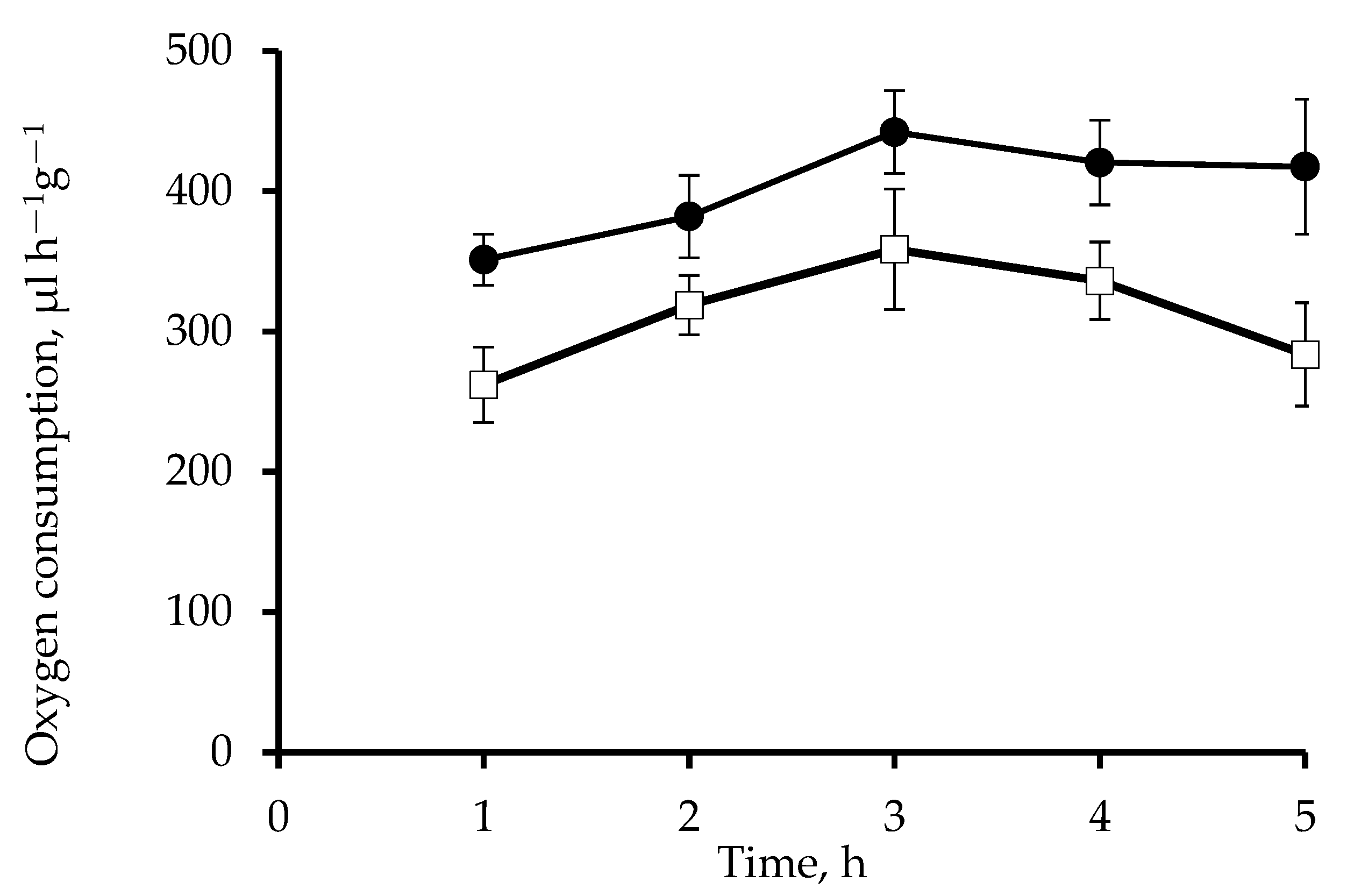
Incubation of roots of wheat seedlings in a solution of 0.1 µM Mdivi-1 for 5 h increased O2 consumption by approximately 30% (Figure 3). Respiration rates were consistently higher than those of the controls. The mitochondrial potential assessed with the voltage-dependent fluorescent dye TMRM was maintained at a high level during the entire time of exposure of roots to the inhibitor. In the control the membrane potential increased after 3 h and slightly decreased after 5 h (Figure 2).
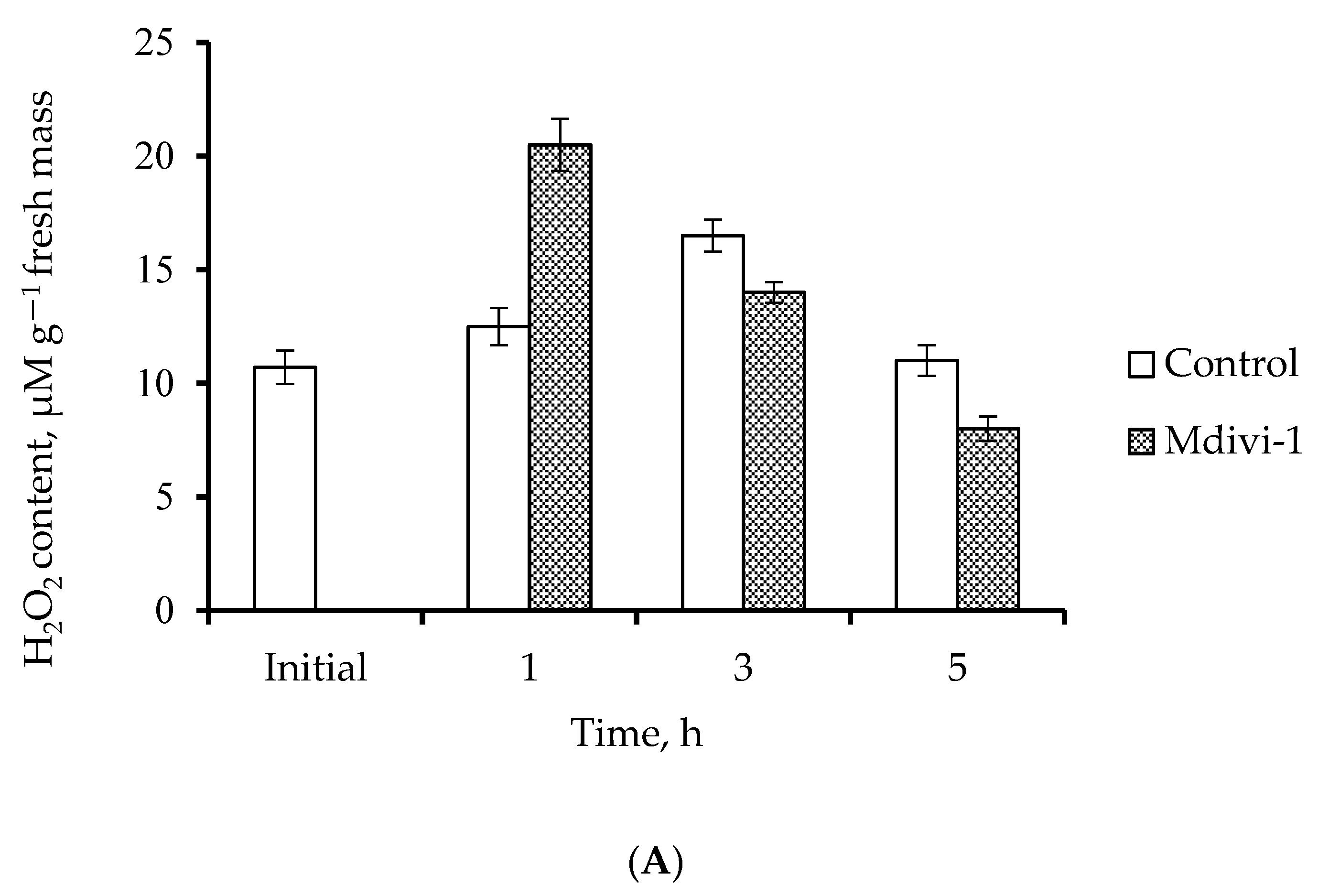
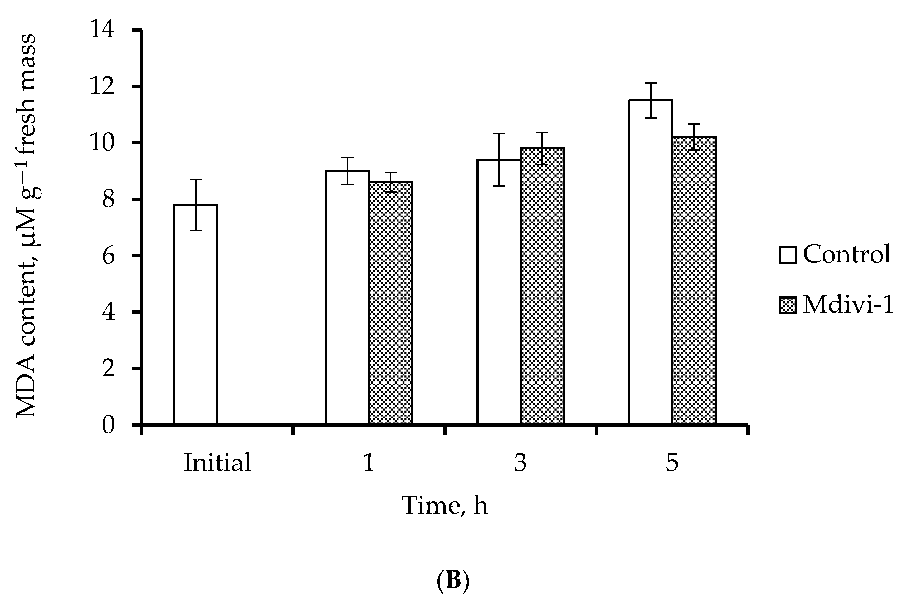
Analysis of ROS formation by the roots showed that incubation of wheat roots with 0.1 µM Mdivi-1 for 1 h sharply increased H2O2 content; after 5 h the content decreased to a level lower than that of the control (Figure 4A). In the control, a slight increase in the content of H2O2 was observed after 3 h of incubation. There were no significant changes in the MDA level both in the control and Mdivi-1 treated roots, indicating that lipid peroxidation had not increased (Figure 4B).
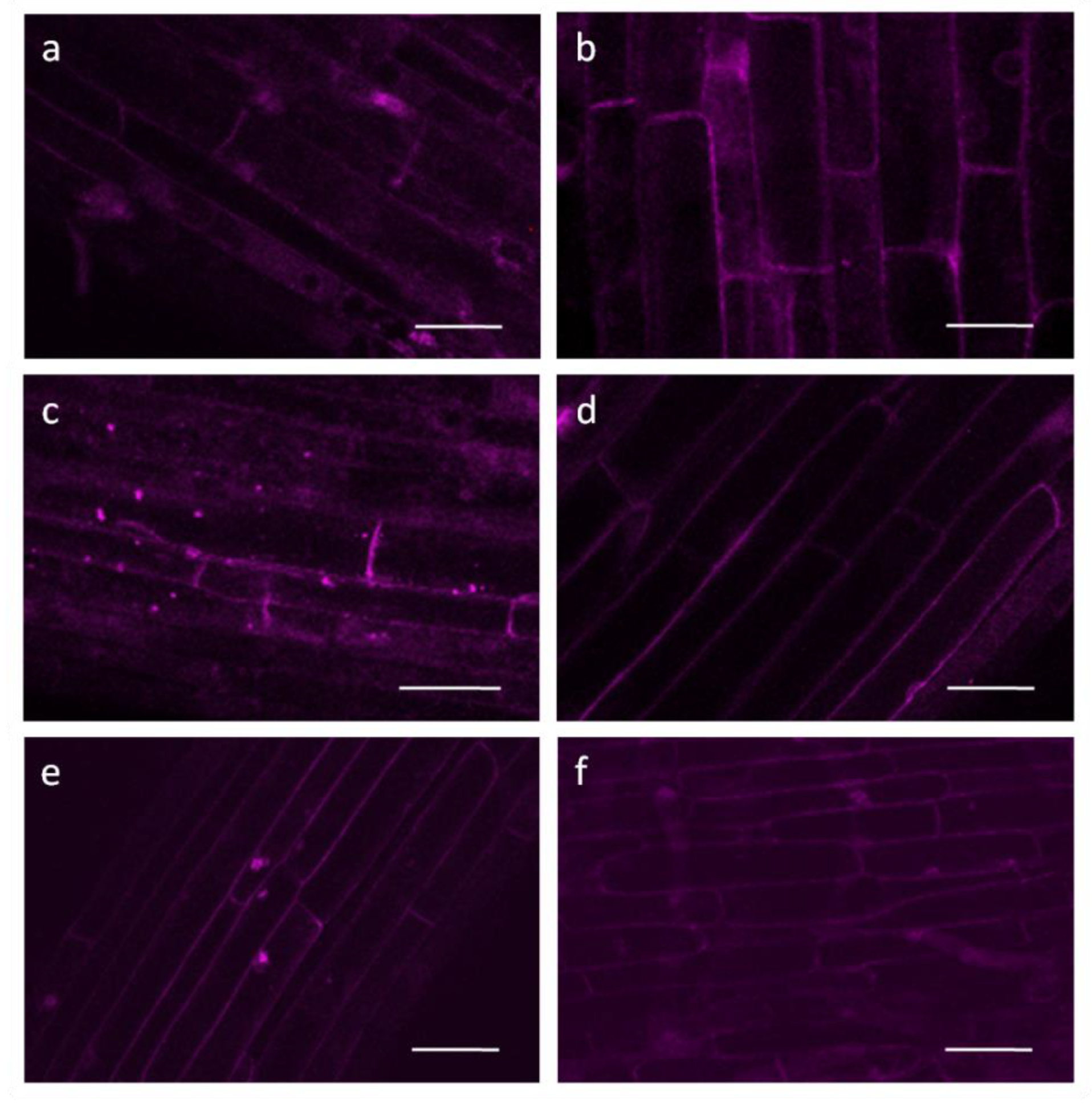
In the cells of the control roots, autophagosomes (visualized as bright LysoTracker stained dots) accumulated after incubation for 3 h in 0.25 mM CaCl2; their number decreased after incubation for 5 h (Figure 5). Using this dye results in some nonspecific background staining of cell walls, cytoplasm and perinuclear regions. No autophagosomes were visualized during the entire incubation of the roots with Mdivi-1 (Figure 5). Cell viability remained at control levels during the 24 h of incubation of the roots with Mdivi-1 (data not shown).
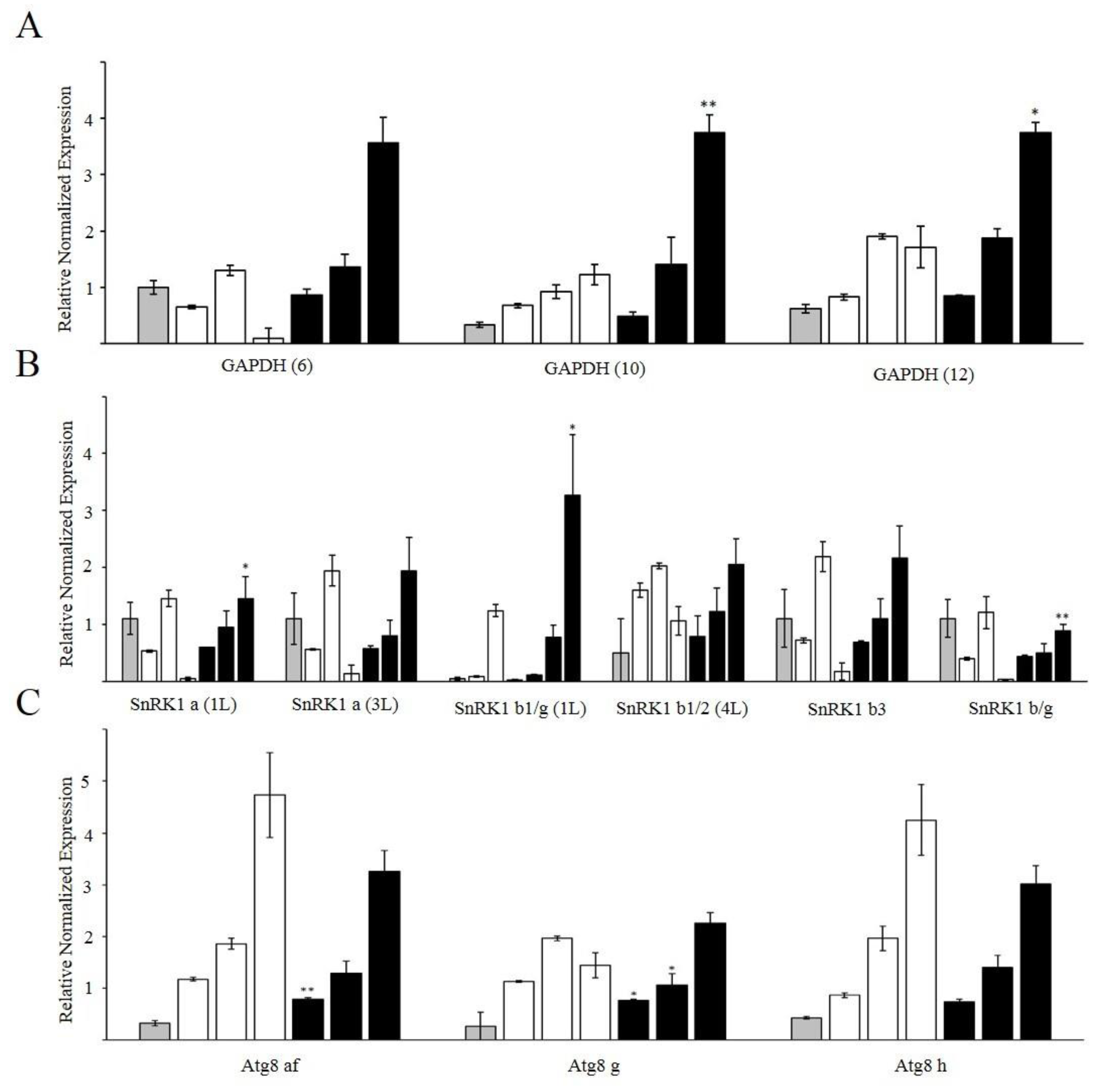
Gene expression analysis showed that the level of transcripts of the AMP-dependent protein kinase SnRK1 increased in the control roots after 3 h of incubation and then sharply decreased after incubation for 5 h (Figure 6A). In the presence of Mdivi-1, the expression of SnRK1 significantly increased after exposure for 5 h. Expression of genes encoding GAPDH isoforms increased after 5 h of exposure to Mdivi-1, while in the control no significant change in the expression of these genes occurred (Figure 6B). The level of transcripts of the autophagic marker protein ATG8 gradually increased in the control during incubation of roots in 0.25 mM CaCl2 for 1 to 5 h. While the expression of ATG8 was also upregulated during incubation of roots with Mdivi-1 for 1 to 5 h, the increase was significantly lower than that of the control (Figure 6C).

4. Discussion

The architecture of mitochondria and their dynamic reorganization are regulated by many factors, including dynamin-like proteins belonging to the GTPase family. In the present work, we demonstrated that the application of Mdivi-1, which inhibits dynamin-like protein Drp1, causes significant morphological and physiological consequences in the roots of wheat seedlings. While wounding stress in the control roots is accompanied by increased ROS formation and autophagy, Mdivi-1 treatment promotes mitochondrial fusion, increases energy production, and reduces ROS generation and autophagosome formation. Therefore, mitochondrial fusion appears to mitigate the negative effects of stress.

4.1. Mdivi-1 Changes the Ultrastructure and Energy Status of Mitochondria

Changes in mitochondrial morphology and activity (increased respiration and ∆ψH+) are often among the first markers of cellular stress [40,41]. In the cells of unstressed plants most mitochondria are small and discrete [42]. In response to stresses such as anoxia, elevated temperature or treatment with mitochondrial poisons, mitochondria can swell or increase in length [19,43,44,45]. Swelling of mitochondria can damage the outer, but not the inner membrane, and trigger the release of cytochrome c [46]. Low temperatures can induce either an increase in the size [47,48] or fragmentation of mitochondria [49]. Thus, careful study of mitochondria structure and function may provide early indicators of plant stress.
Interestingly, wounding stress in wheat roots on its own does not cause dramatic changes in the ultrastructure of mitochondria (Figure 1a), although after application of Mdivi-1 we observed the appearance of large elongated (Figure 1b) or spherical (Figure 1c) mitochondria. The morphology of mitochondrial cristae can be impaired during various stress conditions [50]. However, in our study, the morphology of cristae in large mitochondria was preserved. This indicates that despite causing gross morphological changes, Mdivi-1 did not damage the oxidative phosphorylation of fused mitochondria, because the surface area of cristae positively correlates with the amount of ATP produced by oxidative phosphorylation [50].
The dynamics of mitochondria, including fusion and fission processes, are closely related to their energy status [8]. For mammalian mitochondria, the literature is ambiguous on the effect of Mdivi-1 on enzyme activity of mitochondrial complexes, indicating both inhibition of ETC complex I [51] and an increase in the activity of mitochondrial complexes I, II, and IV [52]. Such differences may be due to the different concentrations of Mdivi-1 used in these studies. Treatment of wheat roots with 0.1 µM Mdivi-1 elevates the energy status of mitochondria, assessed here as increases in mitochondrial respiration rates (Figure 3) and the potential of the mitochondrial membranes (Figure 2). Therefore, fusion of mitochondria induced by Mdivi-1 appears to enhance the functioning of their ETC in wheat roots.
The increased mitochondrial activity that results from fusion requires additional resources, including those supplied by glycolysis. The glycolytic enzyme NAD-dependent glyceraldehyde-3-phosphate dehydrogenase (GAPDH) has a redox-sensitive cysteine in the catalytic center and can therefore reversibly change its own activity, thereby providing a link between redox status and energy metabolism. In Arabidopsis, GAPDH appears to act as an H2O2 sensor, triggering a protective response to oxidative stress and restoring cellular homeostasis [53]. When cysteine is in the oxidized state, GAPDH can be translocated into the nucleus and mediate signal transfer to induce defense genes [54]. When cysteine is in the reduced state, GAPDH can attach to the outer mitochondrial membrane via the VDAC3 protein, allowing glycolysis to take place next to mitochondria. It is likely that in wheat roots the increased mitochondrial activity induced by Mdivi-1 requires the increased import of glycolytic products such as pyruvate into the mitochondria. Therefore, it is important that the activity of GAPDH is increased, and/or the transcription of corresponding genes is upregulated. This is consistent with the observation in the present study of an upregulation of genes encoding GAPDH after 5 h of exposure of roots to Mdivi-1 (Figure 6A).
Mitochondrial fusion can also influence the key energy sensor SnRK1, the plant orthologue of AMPK (AMP activated protein kinase) in mammalian cells [55]. SnRK1 is an important metabolic regulator involved in stress signaling [56]. In mammalian cells AMPK can phosphorylate the mitochondrial outer membrane receptor MFF, which then binds Drp1, activating mitochondrial fission [57]. The upregulation of SnRK1 genes found in our experiments (Figure 6B) can reflect metabolic changes and energy shifts that occur in cells in response to Mdivi-1.

4.2. Mdivi-1 Treatment Reduces Oxidative Stress

Mitochondria are one of the main sources of ROS in the cells [58]. In our experiments, Mdivi-1 induced a sharp “burst” in H2O2 content after treatment of roots for 1 h (Figure 2a). This could be due to the activation of the electron transport chain. However, the subsequent reduction in H2O2 content (Figure 4A) and reduction of lipid peroxidation (Figure 4B) that occurred compared with the controls suggest that temporary rise in H2 fused mitochondria (Figure 3b,c) can effectively cope with oxidative stress. These results are consistent with the decrease in lipid peroxidation observed in Mdivi-1 treated human N2a cells [52]. Thus, by preventing fission GTPase inhibitors cause the appearance of enlarged mitochondria, stimulate ETC functioning and reduce the generation of ROS and associated cell damage.
A consequence of increased levels of ROS is an accumulation of oxidized macromolecules in cells. Previously we showed that the induction of oxidative stress in wheat roots by treating with prooxidant paraquat or antimycin A, an inhibitor of mitochondrial complex III, leads to the intensive formation of autolytic vacuoles (autophagosomes) and the activation of autophagic gene expression [59]. These changes are accompanied by a decrease in the membrane potential of mitochondria and the rate of oxygen consumption. In the present study, autophagosomes were formed in control roots after incubation for 3–5 h in 0.25 mM CaCl2 (Figure 5c,e). The initiation of autophagy was confirmed by the upregulation of genes that encode ATG8, a marker protein of autophagy (Figure 6C). ATG8 is a multifunctional ubiquitin-like protein required for the formation of the insulating membrane of autophagosomes, membrane docking, and fusion of the autophagosome with the vacuole/lysosome [60]. The activation of autophagy in the control may have occurred because of the need to degrade cellular structures damaged during the wounding stress caused by the excision of roots from the seedlings. Interestingly, no autophagosomes are observed in the cells of Mdivi-1 treated roots (Figure 5b,d,f), although some upregulation of ATG8 takes place (Figure 6C). Therefore, in the presence of Mdivi-1 stimulation of mitochondrial activity may reduce the need for autophagy.

5. Conclusions

Mitochondrial dynamics is a process that continuously takes place in a cell. The balance between fusion and fission of mitochondria aims to optimize cellular metabolism in response to changes in environmental conditions. In this study, pharmacological inhibition of mitochondrial fission with Mdivi-1 had morphological, physiological, and biochemical effects in T. aestivum roots. Enlarged mitochondria, some of irregular shape, were formed, and these mitochondria displayed enhanced functional activity. Upregulation of genes encoding the glycolytic enzyme GAPDH and the AMP-dependent protein kinase SnRK1 suggests that redistribution of energy-producing metabolic pathways occurs to sustain the supply of substrates to fused mitochondria. Controlling mitochondrial fusion rates could fundamentally change the physiology of wheat plants by altering the energy status of the cell. Compared to the untreated wounded control, the inhibition of mitochondrial fission had the effects of decreasing ROS formation and associated cell damage. Therefore, the inhibition of fission and resulting fusion of mitochondria might mitigate negative stress effects in wheat roots. In a broader context, mitochondrial fusion and fission events may provide important adaptive responses to environmental stresses, and the ability to control these processes could provide targets for the future breeding and genetic modification of crops.

Supplementary Materials

The following supporting information can be downloaded at: https://www.mdpi.com/article/10.3390/life12091386/s1, Table S1: Primers used for qPCR.

Author Contributions

Conceptualization, D.R. and F.M.; methodology, D.R. and A.P.; investigation, D.R., A.M., A.P. and S.D.; writing—original draft preparation, D.R.; writing—review and editing, R.P.B. and F.M.; project administration, F.M. All authors have read and agreed to the published version of the manuscript.

Funding

This research was funded by the state assignment of the FRC KazSC RAS.

Institutional Review Board Statement

Not applicable.

Informed Consent Statement

Not applicable.

Data Availability Statement

Not applicable.

Acknowledgments

F.M. thanks Kazan Federal University Strategic Academic Leadership Program (PRIORITY-2030) for partial support. The University of KwaZulu-Natal (South Africa) is thanked for granting sabbatical leave to R.P.B.

Conflicts of Interest

The authors declare no conflict of interest. The funders had no role in the design of the study; in the collection, analyses, or interpretation of data; in the writing of the manuscript, or in the decision to publish the results.

References

  1. Smith, G.; Gallo, G. To Mdivi-1 or Not to Mdivi-1: Is that the Question? Dev. Neurobiol. 2017, 77, 1260–1268. [Google Scholar] [CrossRef] [PubMed]
  2. Arimura, S.-I.; Yamamoto, J.; Aida, G.P.; Nakazono, M.; Tsutsumi, N. Frequent Fusion and Fission of Plant Mitochondria with Unequal Nucleoid Distribution. Proc. Natl. Acad. Sci. USA 2004, 101, 7805–7808. [Google Scholar] [CrossRef] [PubMed]
  3. Labbe, K.; Murley, A.; Nunnari, J. Determinants and Functions of Mitochondrial Behavior. Ann. Rev. Cell. Dev. Biol. 2014, 30, 357–391. [Google Scholar] [CrossRef] [PubMed]
  4. Flippo, K.H.; Strack, S. Mitochondrial Dynamics in Neuronal Injury, Development and Plasticity. J. Cell Sci. 2017, 130, 671–681. [Google Scholar] [CrossRef]
  5. Furt, F.; Moreau, P. Importance of Lipid Metabolism for Intracellular and Mitochondrial Membrane Fusion/Fission Prosses. Int. J. Biochem. Cell Biol. 2009, 41, 1828–1836. [Google Scholar] [CrossRef]
  6. Patrushev, M.V.; Mazunin, I.O.; Vinogradova, E.N.; Kamenski, P.A. Mitochondrial Fission and Fusion. Biochemistry 2015, 80, 1457–1464. [Google Scholar] [CrossRef]
  7. Liu, X.; Weaver, D.; Shirihai, O.; Hajnóczky, G. Mitochondrial ‘Kiss-and-Run’: Interplay Between Mitochondrial Motility and Fusion-Fission Dynamics. EMBO J. 2009, 28, 3074–3089. [Google Scholar] [CrossRef]
  8. Twig, G.; Hyde, B.; Shirihai, O.S. Mitochondrial Fusion Fission and Autophagy as a Quality Control Axis: The Bioenergetic View. Biochim. Biophys. Acta 2008, 1777, 1092–1109. [Google Scholar] [CrossRef]
  9. Rose, R.J. Contribution of Massive Mitochondrial Fusion and Subsequent Fission in the Plant Life Cycle to the Integrity of the Mitochondrion and Its Genome. Int. J. Mol. Sci. 2021, 22, 5429. [Google Scholar] [CrossRef]
  10. Benard, G.; Rossignol, R. Ultrastructure of the Mitochondrion and Its Bearing on Function and Bioenergetics. Antioxid. Redox Signal. 2008, 10, 1313–1342. [Google Scholar] [CrossRef]
  11. Ishihara, N.; Jofuku, A.; Eura, Y.; Mihara, K. Regulation of Mitochondrial Morphology by Membrane Potential, And Drp1-Dependent Division and Fzo1-Dependent Fusion Reaction in Mammalian Cells. Biochem. Biophys. Res. Commun. 2013, 301, 891–898. [Google Scholar] [CrossRef]
  12. Benard, G.; Bellance, N.; James, D.; Parrone, P.; Fernandez, H.; Letellier, T.; Rossignol, R. Mitochondrial Bioenergetics and Structural Network Organization. J. Cell Sci. 2007, 120, 838–848. [Google Scholar] [CrossRef]
  13. Liot, G.; Bossy, B.; Lubitz, S.; Kushnareva, Y.; Sejbuk, N.; Bossy-Wetzel, E. Complex II inhibition by 3-NP Causes Mitochondrial Fragmentation and Neuronal Cell Death Via An NMDA- And ROS-Dependent Pathway. Cell Death Differ. 2009, 16, 899–909. [Google Scholar] [CrossRef]
  14. Meeusen, S.; DeVay, R.; Block, J.; Cassidy-Stone, A.; Wayson, S.; McCaffery, J.M.; Nunnari, J. Mitochondrial Inner-Membrane Fusion and Crista Maintenance Requires the Dynamin-Related GTPase Mgm1. Cell 2006, 127, 383–395. [Google Scholar] [CrossRef] [PubMed]
  15. Galloway, C.A.; Lee, H.; Yoon, Y. Mitochondrial Morphology-Emerging Role in Bioenergetics. Free Radic. Biol. Med. 2012, 53, 2218–2228. [Google Scholar] [CrossRef]
  16. Jaipargas, E.A.; Barton, K.A.; Mathur, N.; Mathur, J. Mitochondrial Pleomorphy in Plant Cells Is Driven by Contiguous ER Dynamics. Front. Plant. Sci. 2015, 6, 783. [Google Scholar] [CrossRef]
  17. Fukushima, S.; Akita, K.; Takagi, T.; Kobayashi, K.; Moritoki, N.; Sugaya, H.; Arimura, S.-I.; Kuroiwa, H.; Kuroiwa, T.; Nagata, N. Existence of Giant Mitochondria-Containing Sheet Structures Lacking Cristae and Matrix in the Etiolated Cotyledon of Arabidopsis thaliana. Protoplasma 2022, 259, 731–742. [Google Scholar] [CrossRef]
  18. Foissner, I. Inhibitor Studies on Formation of Giant Mitochondria in Nitella flexilis. Phyton 1983, 23, 19–29. [Google Scholar]
  19. Rakhmatullina, D.; Ponomareva, A.; Gazizova, N.; Minibayeva, F. Mitochondrial Morphology and Dynamics in Triticum aestivum Roots in Response to Rotenone and Antimycin A. Protoplasma 2016, 253, 1299–1308. [Google Scholar] [CrossRef]
  20. Van Gestel, K.; Verbelen, J.P. Giant Mitochondria Are a Response to Low Oxygen Pressure in Cells of Tobacco (Nicotiana tabacum L.). J. Exp. Bot. 2002, 53, 1215–1218. [Google Scholar] [CrossRef]
  21. White, R.R.; Lin, C.; Leaves, I.; Castro, I.G.; Metz, J.; Bateman, B.C.; Botchway, S.W.; Ward, A.D.; Ashwin, P.; Sparkes, I. Miro2 Tethers the ER to Mitochondria to Promote Mitochondrial Fusion in Tobacco Leaf Epidermal Cells. Commun. Biol. 2020, 3, 161. [Google Scholar] [CrossRef] [PubMed] [Green Version]
  22. Lee, Y.J.; Jeong, S.Y.; Karbowski, M.; Smith, C.L.; Youle, R.J. Roles of the Mammalian Mitochondrial Fission and Fusion Mediators Fis1, Drp1, and Opa1 in Apoptosis. Mol. Biol. Cell. 2004, 15, 5001–5011. [Google Scholar] [CrossRef]
  23. Westermann, B. Bioenergetic Role of Mitochondrial Fusion and Fission. Biochim. Biophys. Acta 2012, 1817, 1833–1838. [Google Scholar] [CrossRef] [PubMed]
  24. Chan, D.C. Fusion and Fission: Interlinked Processes Critical for Mitochondrial Health. Ann. Rev. Genet. 2012, 46, 265–287. [Google Scholar] [CrossRef]
  25. Hoppins, S. The Regulation of Mitochondrial Dynamics. Curr. Opin. Cell Biol. 2014, 29, 46–52. [Google Scholar] [CrossRef]
  26. Wang, F.; Liu, P.; Zhang, Q.; Zhu, J.; Chen, T.; Arimura, S.-I.; Tsutsumi, N.; Lin, J. Phosphorylation and Ubiquitination of Dynamin-Related Proteins (AtDRP3A/3B) Synergically Regulate Mitochondrial Proliferation During Mitosis. Plant J. 2012, 72, 43–56. [Google Scholar] [CrossRef]
  27. Arimura, S.-I.; Fujimoto, M.; Doniwa, Y.; Kadoya, N.; Nakazono, M.; Sakamoto, W.; Tsutsumi, N. Arabidopsis ELONGATED MITOCHONDRIA1 Is Required for Localization of DYNAMIN-RELATED PROTEIN3A to Mitochondrial Fission Sites. Plant Cell 2008, 20, 1555–1566. [Google Scholar] [CrossRef]
  28. Pan, R.; Hu, J. The Conserved Fission Complex on Peroxisomes and Mitochondria. Plant Signal. Behav. 2011, 6, 870–872. [Google Scholar] [CrossRef]
  29. Gao, D.; Yang, J.; Wu, Y.; Wang, Q.; Wang, Q.; Lai, E.Y.; Zhu, J. Targeting Dynamin 2 As A Novel Pathway to Inhibit Cardiomyocyte Apoptosis Following Oxidative Stress. Cell Physiol. Biochem. 2016, 39, 2121–2134. [Google Scholar] [CrossRef]
  30. Mueller, S.J.; Reski, R. Mitochondrial Dynamics and the ER: The Plant Perspective. Front. Cell. Dev. Biol. 2015, 3, 78. [Google Scholar] [CrossRef]
  31. Lackner, L.; Nunnari, J. Small Molecule Inhibitors of Mitochondrial Division: Tools That Translate Basic Biological Research into Medicine. Chem. Biol. 2010, 17, 578–583. [Google Scholar] [CrossRef] [PubMed] [Green Version]
  32. Cassidy-Stone, A.; Chipuk, J.E.; Ingerman, E.; Song, C.; Yoo, C.; Kuwana, T.; Kurth, M.J.; Shaw, J.T.; Hinshaw, J.E.; Green, D.R.; et al. Chemical Inhibition of the Mitochondrial Division Dynamin Reveals Its Role in Bax/Bak-Dependent Mitochondrial Outer Membrane Permeabilization. Dev. Cell. 2008, 14, 193–204. [Google Scholar] [CrossRef] [PubMed]
  33. Vazquez-Martin, A.; Cufi, S.; Corominas-Faja, B.; Oliveras-Ferraros, C.; Vellon, L.; Menendez, J.A. Mitochondrial Fusion by Pharmacological Manipulation Impedes Somatic Cell Reprogramming to Pluripotency: New Insight into The Role of Mitophagy in Cell Stemness. Aging 2012, 4, 393–401. [Google Scholar] [CrossRef] [PubMed]
  34. Reddy, P.H. Inhibitors of Mitochondrial Fission as a Therapeutic Strategy for Diseases with Oxidative Stress and Mitochondrial Disfunction. J. Alzheimers Dis. 2014, 40, 245–256. [Google Scholar] [CrossRef] [PubMed]
  35. Rosdah, A.A.; Holien, K.J.; Delbridge, L.M.; Dusting, G.J.; Lim, S.Y. Mitochondrial fission—A Drug Target for Cytoprotection or Cytodestruction? Pharmacol. Res. Perspect. 2016, 4, e00235. [Google Scholar] [CrossRef] [PubMed]
  36. Wolff, S. Ferrous ion oxidation in presence of ferric ion indicator xylenol orange for measurement of hydroperoxides. Methods Enzymol. 1994, 233, 182–189. [Google Scholar]
  37. Kumar, G.; Knowles, N.R. Changes in Lipid Peroxidation and Lipolytic and Free-Radical Scavenging Enzyme Actvities During Aging and Sprouting of Potato (Solanum tuberosum) Seed-Tubers. Plant Physiol. 1993, 102, 115–124. [Google Scholar] [CrossRef]
  38. Paolacci, A.R.; Tanzarella, O.A.; Porceddu, E.; Ciaffi, M. Identification and Validation of Reference Genes for Quantitative RT-PCR Normalization in Wheat. BMC Mol. Biol. 2009, 10, 11. [Google Scholar] [CrossRef]
  39. Zeng, L.; Deng, R.; Guo, Z.; Yang, S.; Deng, X. Genome-Wide Identification and Characterization of Glyceraldehyde-3-Phosphate Dehydrogenase Genes Family in Wheat (Triticum aestivum). BMC Genomics 2016, 17, 240. [Google Scholar] [CrossRef]
  40. Scott, I.; Logan, D.C. Mitochondrial dynamics. In Plant Mitochondria; Kempken, F., Ed.; Springer: New York, NY, USA, 2011; pp. 31–63. [Google Scholar]
  41. Welchen, E.; Garcia, L.; Mansilla, N.; Gonzalez, D. Coordination of Plant Mitochondrial Biogenesis: Keeping Pace with Cellular Requirements. Front. Plant Sci. 2014, 1, 551. [Google Scholar] [CrossRef]
  42. Logan, D. The Dynamic Plant Chondriome. Semin. Cell Dev. Biol. 2010, 21, 550–557. [Google Scholar] [CrossRef] [PubMed]
  43. Virolainen, E.; Blokhina, O.; Fagerstedt, K. Ca2+-Induced High Amplitude Swelling and Cytochrome C Release from Wheat (Triticum aestivum L.) Mitochondria Under Anoxic Stress. Ann. Bot. 2002, 90, 509–516. [Google Scholar] [CrossRef]
  44. Vartapetian, B.B.; Andreeva, I.N.; Generosova, I.P.; Polyakova, L.I.; Maslova, I.P.; Dolgikh, Y.I.; Stepanova, A.Y. Functional Electron Microscopy in Studies of Plant Response and Adaptation to Anaerobic Stress. Ann. Bot. 2003, 91, 155–172. [Google Scholar] [CrossRef] [PubMed]
  45. Rurek, M.; Woyda-Ploszczyca, A.M.; Jarmuszkiewicz, W. Biogenesis of Mitochondria in Cauliflower (Brassica Oleracea Var. Botrytis) Curds Subjected to Temperature Stress and Recovery Involves Regulation of the Complexome, Respiratory Chain Activity, Organellar Translation and Ultrastructure. Biochim. Biophys. Acta 2015, 1847, 399–417. [Google Scholar] [CrossRef]
  46. Arpagaus, S.; Rawyler, A.; Braendle, R. Occurrence and Characteristics of the Mitochondrial Permeability Transition in Plants. J. Biol. Chem. 2002, 277, 1780–1787. [Google Scholar] [CrossRef]
  47. Murphy, C.; Wilson, J.M. Ultrastructural Features of Chilling–Injury in Episcia Reptans. Plant Cell Environ. 1981, 4, 261–265. [Google Scholar]
  48. Lee, S.H.; Singh, A.P.; Chung, G.C.; Kim, Y.S.; Kong, I. Chilling Root Temperature Causes Rapid Ultrastructural Changes in Cortical Cells in Cucumber (Cucumis sativus L.) Root Tips. J. Exp. Bot. 2002, 53, 2225–2237. [Google Scholar] [CrossRef]
  49. Arimura, S.-I.; Kurisu, R.; Sugaya, H.; Kadoya, N.; Tsutsumi, N. Cold Treatment Induces Transient Mitochondrial Fragmentation in Arabidopsis Thaliana in a Way That Requires DRP3A But Not ELM1 or an ELM1-Like Homologue ELM2. Int. J. Mol. Sci. 2017, 18, 2161. [Google Scholar] [CrossRef]
  50. Zick, M.; Rabl, R.; Reichert, A. Cristae formation–Linking Ultrastructure and Function of Mitochondria. Biochem. Biophys. Acta 2009, 1793, 5–19. [Google Scholar] [CrossRef]
  51. Bordt, E.A.; Clerc, P.; Roelofs, B.A.; Saladino, A.J.; Tretter, L.; Adam-Vizi, V.; Cherok, E.; Khalil, A.; Yadava, N.; Ge, S.X.; et al. The Putative Drp1 Inhibitor Mdivi-1 Is a Reversible Mitochondrial Complex I Inhibitor That Modulates Reactive Oxygen Species. Dev. Cell 2017, 40, 583–594. [Google Scholar] [CrossRef]
  52. Manczak, M.; Kandimalla, R.; Yin, X.; Reddy, P.H. Mitochondrial Division Inhibitor 1 Reduces Dynamin-Related Protein 1 and Mitochondrial Fission Activity. Hum. Mol. Genet. 2019, 28, 177–199. [Google Scholar] [CrossRef] [PubMed]
  53. Hancock, J.T.; Henson, D.; Nyirenda, M.; Desikan, R.; Harrison, J.; Lewis, M.; Hughes, J.; Neill, S.J. Proteomic Identification of Glyceraldehyde 3-Phosphate Dehydrogenase as an Inhibitory Target of Hydrogen Peroxide in Arabidopsis. Plant Physiol. Biochem. 2005, 43, 828–835. [Google Scholar] [CrossRef] [PubMed]
  54. Schneider, M.; Knuesting, J.; Birkholz, O.; Jürgen, J.; Scheibe, H.R. Cytosolic GAPDH as a Redox-Dependent Regulator of Energy Metabolism. BMC Plant Biol. 2018, 18, 184. [Google Scholar] [CrossRef] [Green Version]
  55. Wurzinger, B.; Nukarinen, E.; Nagele, T.; Weckwerth, W.; Teige, M. The SnRK1 Kinase as a Central Mediator of Energy Signaling Between Different Organelles. Plant Physiol. 2018, 176, 1085–1094. [Google Scholar] [CrossRef] [PubMed]
  56. Halford, N.G.; Hey, S. Snf1-Related Protein Kinases (SnRKs) Acts Within an Intricate Network That Links Metabolic and Stress Signaling in Plants. Biochem. J. 2009, 419, 247–259. [Google Scholar] [CrossRef] [PubMed]
  57. Toyama, E.Q.; Herzig, S.; Courchet, J.; Tommy, L.L.; Losón, O.C.; Hellberg, K.; Young, N.P.; Chen, H.; Polleux, F.; Chan, D.C.; et al. AMP-Activated Protein Kinase Mediates Mitochondrial Fission in Response to Energy Stress. Science 2016, 351, 275–281. [Google Scholar] [CrossRef]
  58. Zorov, D.B.; Juhaszova, M.; Sollott, S.J. Mitochondrial Reactive Oxygen Species (ROS) and ROS-Induced ROS Release. Physiol. Rev. 2014, 94, 909–950. [Google Scholar] [CrossRef]
  59. Minibayeva, F.; Dmitrieva, S.; Ponomareva, A.; Ryabovol, V. Oxidative Stress-Induced Autophagy in Plants: The Role of Mitochondria. Plant Physiol. Biochem. 2012, 59, 11–19. [Google Scholar] [CrossRef]
  60. Nakatogawa, H.; Ichimura, Y.; Ohsumi, Y. Atg8, a Ubiquitin-Like Protein Required for Autophagosome Formation, Mediates Membrane Tethering and Hemifusion. Cell 2007, 130, 165–178. [Google Scholar] [CrossRef] [Green Version]
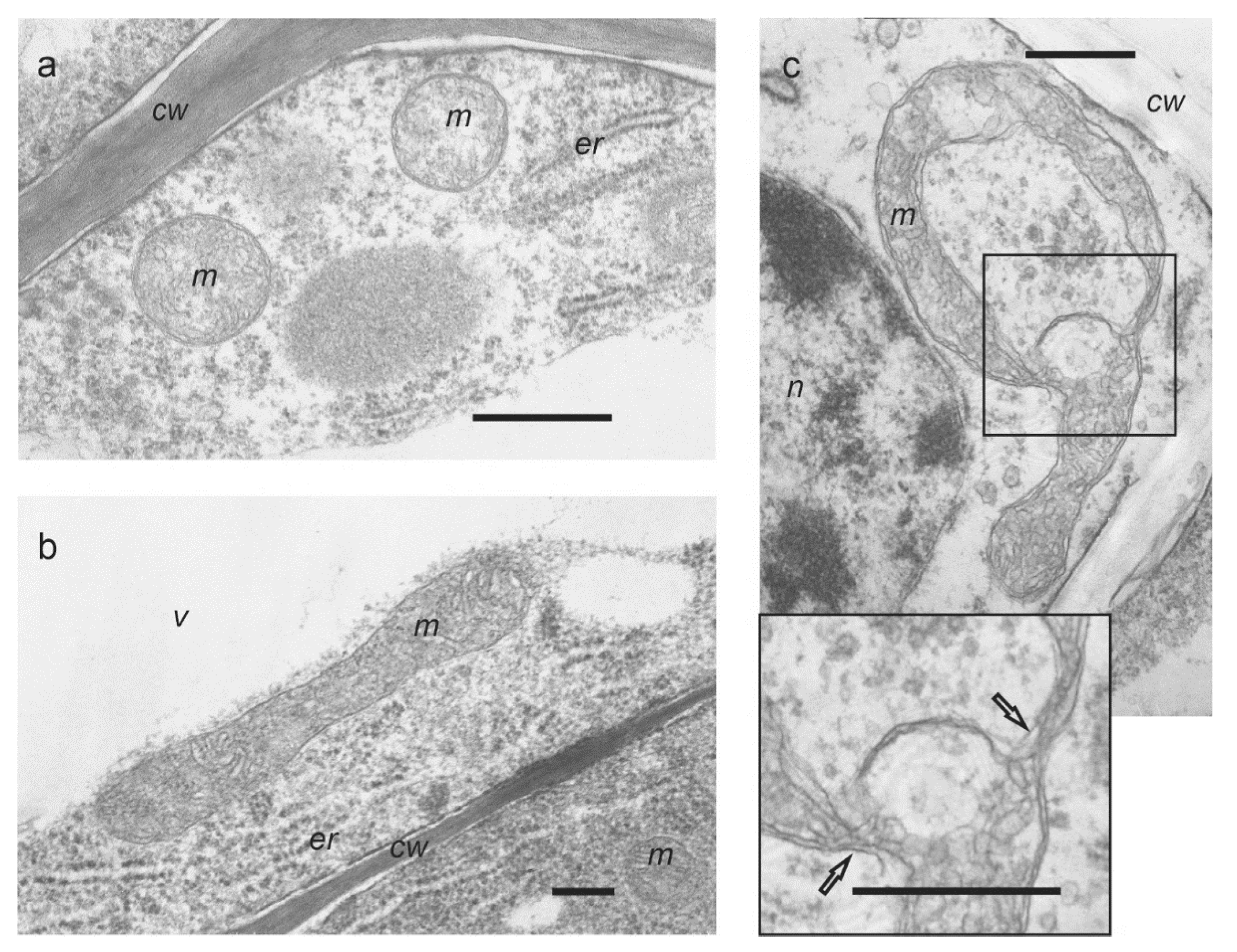
Figure 1. Ultrastructure of mitochondria in wheat roots: (a) orthodox mitochondria of oval shape in control roots (1 h); (b) enlarged mitochondrium in roots treated with 0.1 μM Mdivi-1 (1 h); (c) mitochondrium of irregular shape in Mdivi-1 treated roots (1 h). In the increased fragment, the arrows indicate constriction zones. cw—cell wall, er—endoplasmic reticulum, m—mitochondrium, n—nucleus, v—vacuole. Scale bar is 0.5 μm.
Life 12 01386 g001
Figure 2. Mitochondrial membrane potential visualized with TMRM: (a,c,e)—control for 1, 3, 5 h, respectively; (b,d,f)—roots treated with 0.1 μM Mdivi-1 for 1, 3, 5 h, respectively. Scale bar is 50 µm.
Life 12 01386 g002
Figure 3. Oxygen consumption of wheat roots treated with 0.1 μM Mdivi-1. Control—white squares, Mdivi-1—black circles. Treatment with Mdivi-1 had a significant effect, p ˂ 0.05.
Life 12 01386 g003
Figure 4. Content of H2O2 (A) and malondialdehyde (MDA) (B) in wheat roots following treatment with 0.1 μM Mdivi-1.
Life 12 01386 g004aLife 12 01386 g004b
Figure 5. Autophagosome formation visualized with LysoTracker Red: (a,c,e)—control for 1, 3, 5 h, respectively; (b,d,f)—roots treated with 0.1 μM Mdivi-1 for 1, 3, 5 h, respectively. Scale bar is 50 µm.
Life 12 01386 g005
Figure 6. Gene expression of wheat roots cells treated with 0.1 μM Mdivi-1: (A)—isoforms of glyceraldehyde-3-phosphate dehydrogenase (GAPDH); (B)—subunits of sucrose non-fermenting-related kinase (SnRK1); (C)—isoforms of ATG8 genes. Grey bar—intact roots, white bars—control roots for 1, 3, 5 h, black bars—roots treated with 0.1 μM Mdivi-1 for 1, 3, 5 h. * p ≤ 0.05; ** p ≤ 0.005.
Life 12 01386 g006
Publisher’s Note: MDPI stays neutral with regard to jurisdictional claims in published maps and institutional affiliations.