
Treating Spontaneous Intracranial Hypotension with an Anesthetic Modality: The Role of the Epidural Blood Patch †


 , and
, and 
Abstract
:1. Introduction
2. Cases Description
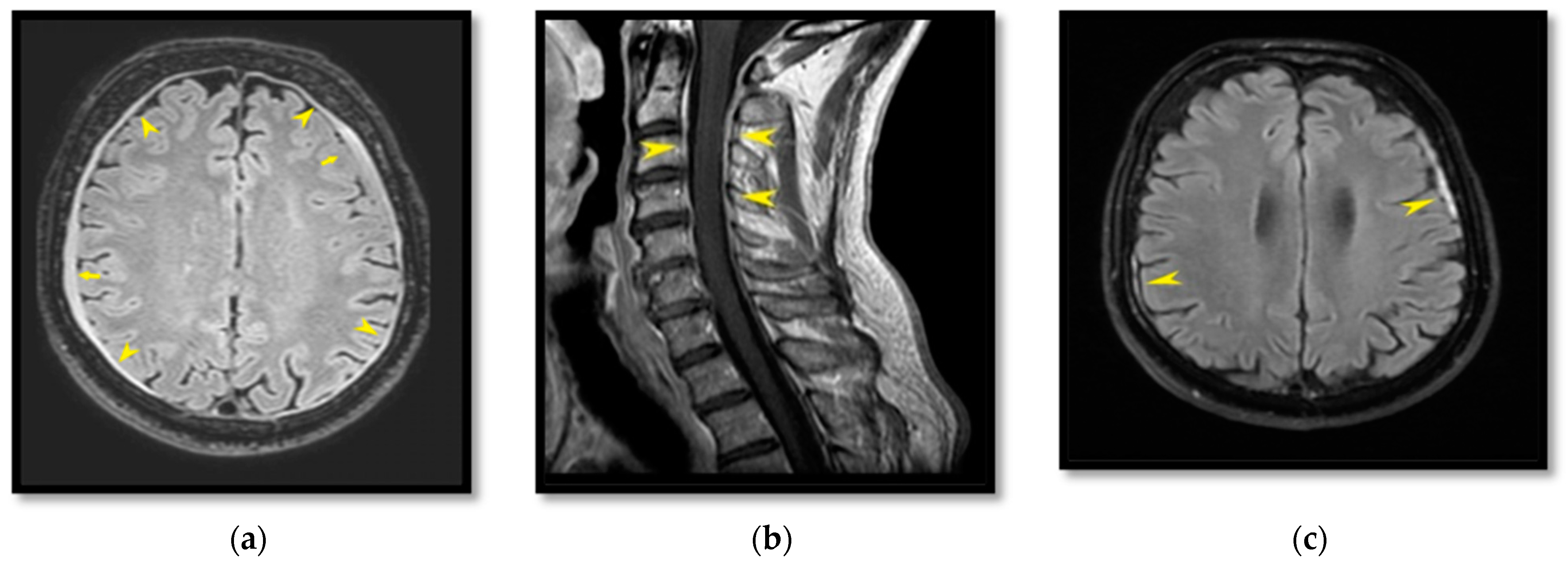
2.1. Patient I
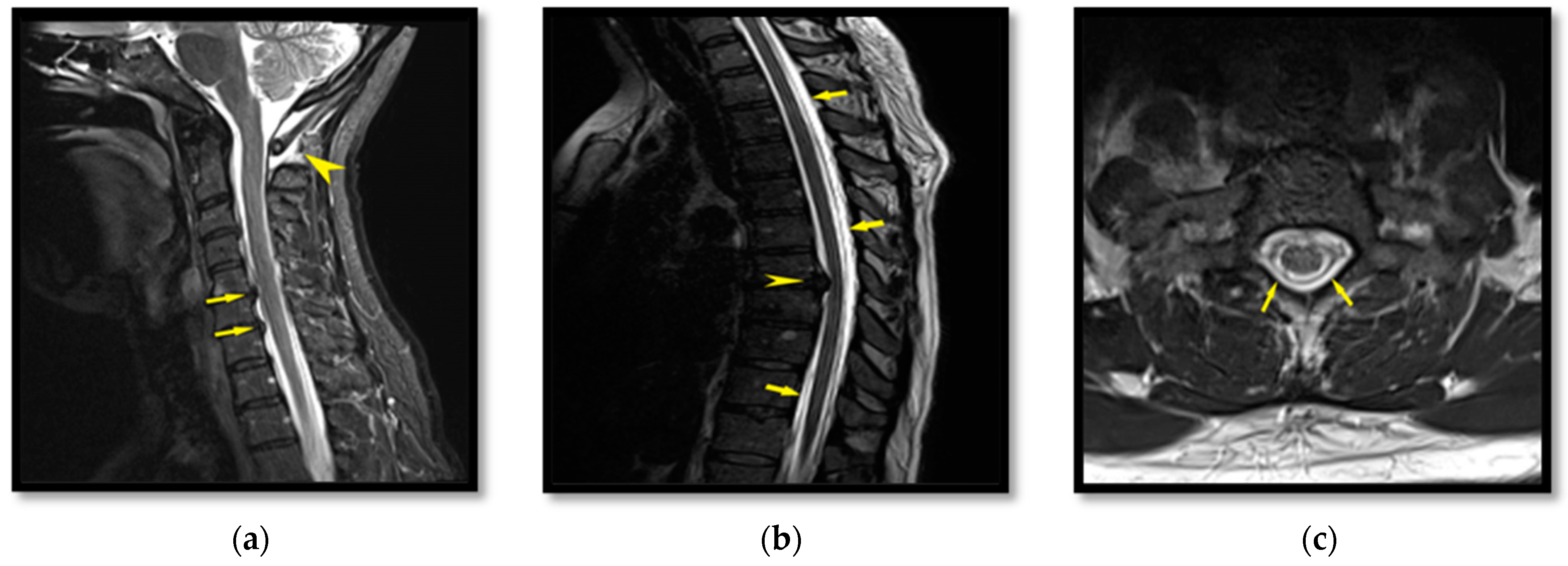
2.2. Patient II
2.3. Patient III
3. Discussion
4. Conclusions
Author Contributions
Funding
Informed Consent Statement
Data Availability Statement
Conflicts of Interest
References
- Davies, M.J.; Davies, M.A.; Sharpe, R.; Cordato, D.; Schwartz, R. Epidural Blood Patch as a Diagnostic and Therapeutic Intervention in Spontaneous Intracranial Hypotension: A Novel Approach to Management. World Neurosurg. 2020, 137, e242–e250. [Google Scholar] [CrossRef] [PubMed]
- Limaye, K.; Samant, R.; Lee, R.W. Spontaneous intracranial hypotension: Diagnosis to management. Acta Neurol. Belg. 2016, 116, 119–125. [Google Scholar] [CrossRef] [PubMed]
- Goldberg, J.; Häni, L.; Jesse, C.M.; Zubak, I.; Piechowiak, E.I.; Gralla, J.; Dobrocky, T.; Beck, J.; Raabe, A. Spontaneous Intracranial Hypotension Without CSF Leakage-Concept of a Pathological Cranial to Spinal Fluid Shift. Front. Neurol. 2021, 12, 760081. [Google Scholar] [CrossRef] [PubMed]
- Mokri, B. Spontaneous low pressure, low CSF volume headaches: Spontaneous CSF leaks. Headache J. Head Face Pain 2013, 53, 1034–1053. [Google Scholar] [CrossRef] [PubMed]
- Kranz, P.G.; Malinzak, M.D.; Amrhein, T.J.; Gray, L. Update on the Diagnosis and Treatment of Spontaneous Intracranial Hypotension. Curr. Pain Headache Rep. 2017, 21, 37. [Google Scholar] [CrossRef]
- Signorelli, F.; Caccavella, V.M.; Giordano, M.; Ioannoni, E.; Caricato, A.; Polli, F.M.; Olivi, A.; Montano, N. A systematic review and meta-analysis of factors affecting the outcome of the epidural blood patching in spontaneous intracranial hypotension. Neurosurg. Rev. 2021, 44, 3079–3085. [Google Scholar] [CrossRef]
- Wang, T.Y.; Karikari, I.O.; Amrhein, T.J.; Gray, L.; Kranz, P.G. Clinical outcomes following surgical ligation of cerebrospinal fluid-venous fistula in patients with spontaneous intracranial hypotension: A prospective case series. Oper. Neurosurg. Hagerstown 2020, 18, 239–245. [Google Scholar] [CrossRef] [Green Version]
- Gordon, N. Spontaneous intracranial hypotension. Dev. Med. Child Neurol. 2009, 51, 932–935. [Google Scholar] [CrossRef]
- Schievink, W.I. Spontaneous spinal cerebrospinal fluid leaks and intracranial hypotension. JAMA 2006, 295, 2286–2296. [Google Scholar] [CrossRef]
- Mokri, B. Cerebrospinal fluid volume depletion and its emerging clinical/imaging syndromes. Neurosurg. Focus. 2000, 9, e6. [Google Scholar] [CrossRef]
- Ljubisavljevic, S. Postdural puncture headache as a complication of lumbar puncture: Clinical manifestations, pathophysiology, and treatment. Neurol. Sci. 2020, 41, 3563–3568. [Google Scholar] [CrossRef] [PubMed]
- Ferrante, E.; Trimboli, M.; Rubino, F. Spontaneous intracranial hypotension: Review and expert opinion. Acta Neurol. Belg. 2020, 120, 9–18. [Google Scholar] [CrossRef] [PubMed]
- Schievink, W.I.; Maya, M.M.; Moser, F.G.; Tourje, J. Spectrum of subdural fluid collections in spontaneous intracranial hypotension. J. Neurosurg. 2005, 103, 608–613. [Google Scholar] [CrossRef] [PubMed] [Green Version]
- Schievink, W.I. Spontaneous spinal cerebrospinal fluid leaks: A review. Neurosurg. Focus. 2000, 9, e8. [Google Scholar] [CrossRef] [Green Version]
- Wang, S.J. Spontaneous intracranial hypotension. Contin. Minneap. Minn. 2021, 27, 746–766. [Google Scholar] [CrossRef]
- Kranz, P.G.; Stinnett, S.S.; Huang, K.T.; Gray, L. Spinal Meningeal Diverticula in Spontaneous Intracranial Hypotension: Analysis of Prevalence and Myelographic Appearance. Am. J. Neuroradiol. 2013, 34, 1284. [Google Scholar] [CrossRef] [PubMed] [Green Version]
- Kranz, P.G.; Gray, L.; Malinzak, M.D.; Amrhein, T.J. Spontaneous Intracranial Hypotension: Pathogenesis, Diagnosis, and Treatment. Neuroimaging Clin. N. Am. 2019, 29, 581–594. [Google Scholar] [CrossRef]
- Loya, J.J.; Mindea, S.A.; Yu, H.; Venkatasubramanian, C.; Chang, S.D.; Burns, T.C. Intracranial hypotension producing reversible coma: A systematic review, including three new cases. J. Neurosurg. 2012, 117, 615–628. [Google Scholar] [CrossRef] [Green Version]
- Headache Classification Committee of the International Headache Society (IHS). The International Classification of Headache Disorders, 3rd edition (beta version). Cephalalgia 2013, 33, 629–808. [Google Scholar] [CrossRef] [Green Version]
- Schievink, W.I.; Dodick, D.W.; Mokri, B.; Silberstein, S.; Bousser, M.G.; Goadsby, P.J. Diagnostic criteria for headache due to spontaneous intracranial hypotension: A perspective. Headache J. Head Face Pain 2011, 51, 1442–1444. [Google Scholar] [CrossRef]
- Mokri, B. Spontaneous cerebrospinal fluid leaks: From intracranial hypotension to cerebrospinal fluid hypovolemia—Evolution of a concept. Mayo Clin. Proc. 1999, 74, 1113–1123. [Google Scholar] [CrossRef] [PubMed]
- Upadhyaya, P.; Ailani, J. A review of spontaneous intracranial hypotension. Curr. Neurol. Neurosci. Rep. 2019, 19, 22. [Google Scholar] [CrossRef] [PubMed]
- D’Antona, L.; Jaime Merchan, M.A.; Vassiliou, A.; Watkins, L.D.; Davagnanam, I.; Toma, A.K.; Matharu, M.S. Clinical presentation, investigation findings, and treatment outcomes of spontaneous intracranial hypotension syndrome: A systematic review and meta-analysis. JAMA Neurol. 2021, 78, 329–337. [Google Scholar] [CrossRef] [PubMed]
- Ferrante, E.; Rubino, G.F.; Passarani, S.; Arpino, I. Spontaneous intracranial hypotension. J. Neurosurg. 2010, 113, 397–398. [Google Scholar] [CrossRef] [PubMed]
- Shima, K.; Ishihara, S.; Tomura, S. Pathophysiology and diagnosis of spontaneous intracranial hypotension. Acta Neurochir. Suppl. 2008, 102, 153–156. [Google Scholar] [CrossRef] [PubMed]
- Katz, D.; Beilin, Y. Review of the alternatives to epidural blood patch for treatment of postdural puncture headache in the parturient. Anesth. Analg. 2017, 124, 1219–1228. [Google Scholar] [CrossRef] [PubMed]
- Han, M.E.; Kim, H.J.; Lee, Y.S.; Kim, D.H.; Choi, J.T.; Pan, C.S.; Yoon, S.; Baek, S.Y.; Kim, B.S.; Kim, J.B.; et al. Regulation of cerebrospinal fluid production by caffeine consumption. BMC Neurosci. 2009, 10, 110. [Google Scholar] [CrossRef] [Green Version]
- Russo, C.; Buono, V.; Fenza, G.; Zandolino, A.; Serino, A.; Manto, A. Spontaneous intracranial hypotension: Two steroid-responsive cases. Pol. J. Radiol. 2018, 83, e229–e233. [Google Scholar] [CrossRef]
- Beards, S.C.; Jackson, A.; Griffiths, A.G.; Horsman, E.L. Magnetic resonance imaging of extradural blood patches: Appearances from 30 min to 18 h. Br. J. Anaesth. 1993, 71, 182–188. [Google Scholar] [CrossRef]
- Lander, C.J.; Korbon, G.A. Histopathologic consequences of epidural blood patch and epidurally administered Dextran 40. Anesthesiology 1988, 69, A410. [Google Scholar] [CrossRef]
- Cook, M.A.; Watkins-Pitchford, J.M. Epidural blood patch: A rapid coagulation response. Anesth. Analg. 1990, 70, 567–568. [Google Scholar] [CrossRef] [PubMed]
- Rajesh, M.C.; Noone, M.L.; Harish Babu, P.S. Epidural blood patch for spontaneous intracranial hypotension. Natl. Med. J. India 2019, 32, 288–289. [Google Scholar] [CrossRef] [PubMed]
- Ferrante, E.; Olgiati, E.; Sangalli, V.; Rubino, F. Early pain relief from orthostatic headache and hearing changes in spontaneous intracranial hypotension after epidural blood patch. Acta Neurol. Belg. 2016, 116, 503–508. [Google Scholar] [CrossRef] [PubMed]
- Ferrante, E.; Rubino, F.; Mongelli, M.; Arpino, I. Subarachnoideal blood spread following epidural blood patch given to treat spontaneous intracranial hypotension: Can it cause neurological complications? Clin. Neurol. Neurosurg. 2016, 140, 43–46. [Google Scholar] [CrossRef]
- Wu, J.W.; Hseu, S.S.; Fuh, J.L.; Lirng, J.F.; Wang, Y.F.; Chen, W.T.; Chen, S.P.; Wang, S.J. Factors predicting response to the first epidural blood patch in spontaneous intracranial hypotension. Brain 2017, 140, 344–352. [Google Scholar] [CrossRef]
- Niraj, G.; Critchley, P.; Kodivalasa, M.; Dorgham, M. Greater Occipital Nerve Treatment in the Management of Spontaneous Intracranial Hypotension Headache: A Case Report. Headache J. Head Face Pain 2017, 57, 952–955. [Google Scholar] [CrossRef]
- Furtado, I.F.; Pinto, M.M.; Amorim, P. Sphenopalatine Ganglion Block May Be an Efficient Treatment of Headache After Lumboperitoneal Shunt Placement: A Case Report. A A Pract. 2019, 12, 401–402. [Google Scholar] [CrossRef]
- Kamada, M.; Fujita, Y.; Ishii, R.; Endoh, S. Spontaneous intracranial hypotension successfully treated by epidural patching with fibrin glue. Headache J. Head Face Pain 2000, 40, 844–847. [Google Scholar] [CrossRef]
- Matsuhashi, A.; Takai, K.; Taniguchi, M. Microsurgical anatomy and treatment of dural defects in spontaneous spinal cerebrospinal fluid leaks. J. Neurosurg. Spine 2021, 34, 522–530. [Google Scholar] [CrossRef]


| Patient I | Patient II | Patient III | |
|---|---|---|---|
| Sex | Male | Male | Female |
| Age (years old) | 53 | 38 | 44 |
| Main symptoms | Dizziness | Headache, diplopia | Headache |
| Subdural hematoma | Yes | No | No |
| EBP level | L2–L3 | L1–L2 | T11–T12 |
| Amount of autologous blood infused (mL) | 35 | 30 | 43 |
| Complications | None | None | None |
| Response to EBP | Yes | Yes | Yes |
Publisher’s Note: MDPI stays neutral with regard to jurisdictional claims in published maps and institutional affiliations. |
© 2022 by the authors. Licensee MDPI, Basel, Switzerland. This article is an open access article distributed under the terms and conditions of the Creative Commons Attribution (CC BY) license (https://creativecommons.org/licenses/by/4.0/).
Share and Cite
Masourou, Z.; Papagiannakis, N.; Mantzikopoulos, G.; Mitsikostas, D.-D.; Theodoraki, K. Treating Spontaneous Intracranial Hypotension with an Anesthetic Modality: The Role of the Epidural Blood Patch. Life 2022, 12, 1109. https://doi.org/10.3390/life12081109
Masourou Z, Papagiannakis N, Mantzikopoulos G, Mitsikostas D-D, Theodoraki K. Treating Spontaneous Intracranial Hypotension with an Anesthetic Modality: The Role of the Epidural Blood Patch. Life. 2022; 12(8):1109. https://doi.org/10.3390/life12081109
Chicago/Turabian StyleMasourou, Zoi, Nikolaos Papagiannakis, Georgios Mantzikopoulos, Dimos-Dimitrios Mitsikostas, and Kassiani Theodoraki. 2022. "Treating Spontaneous Intracranial Hypotension with an Anesthetic Modality: The Role of the Epidural Blood Patch" Life 12, no. 8: 1109. https://doi.org/10.3390/life12081109
APA StyleMasourou, Z., Papagiannakis, N., Mantzikopoulos, G., Mitsikostas, D.-D., & Theodoraki, K. (2022). Treating Spontaneous Intracranial Hypotension with an Anesthetic Modality: The Role of the Epidural Blood Patch. Life, 12(8), 1109. https://doi.org/10.3390/life12081109