
Subfoveal Choroidal Thickness and Treatment Outcomes of Intravitreal Aflibercept for Branch Retinal Vein Occlusion


Abstract
1. Introduction
2. Materials and Methods
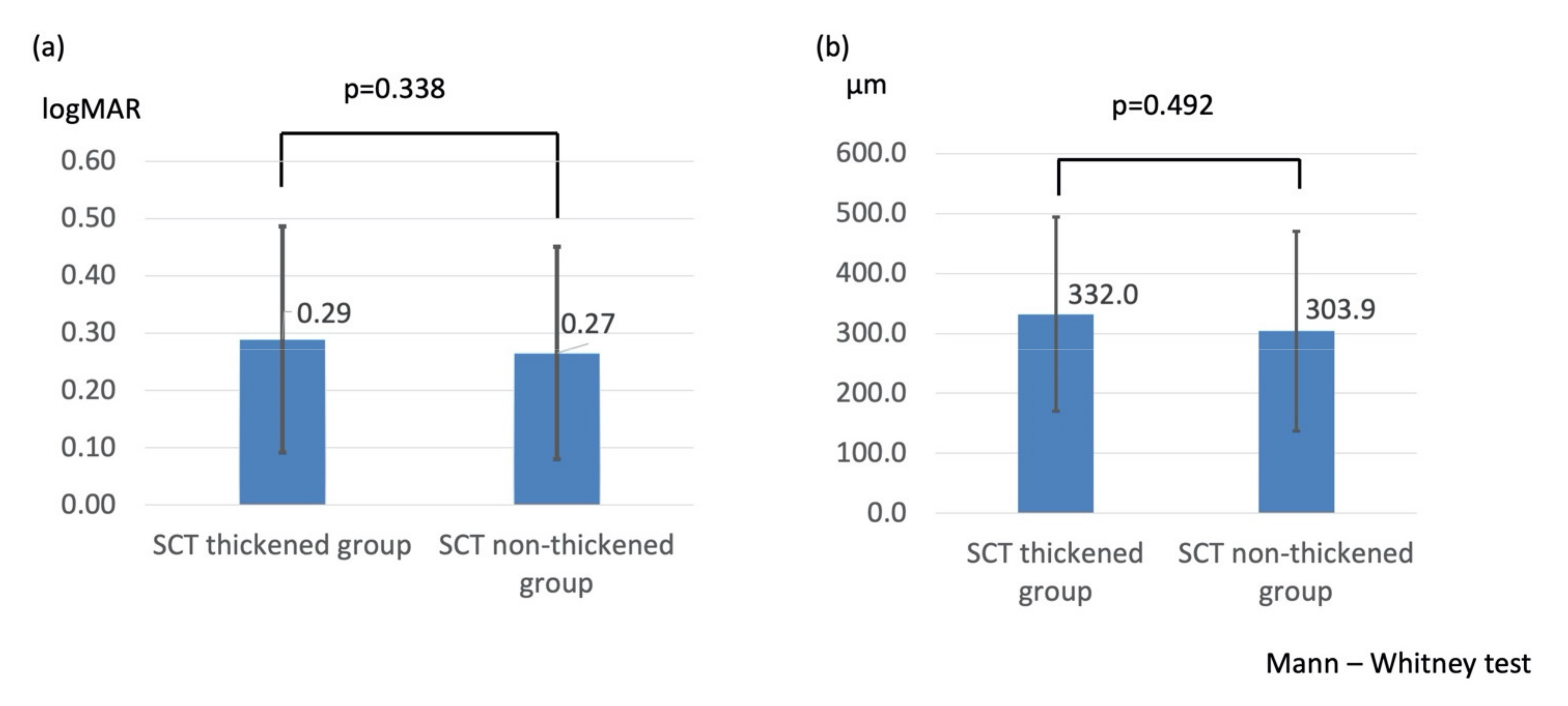
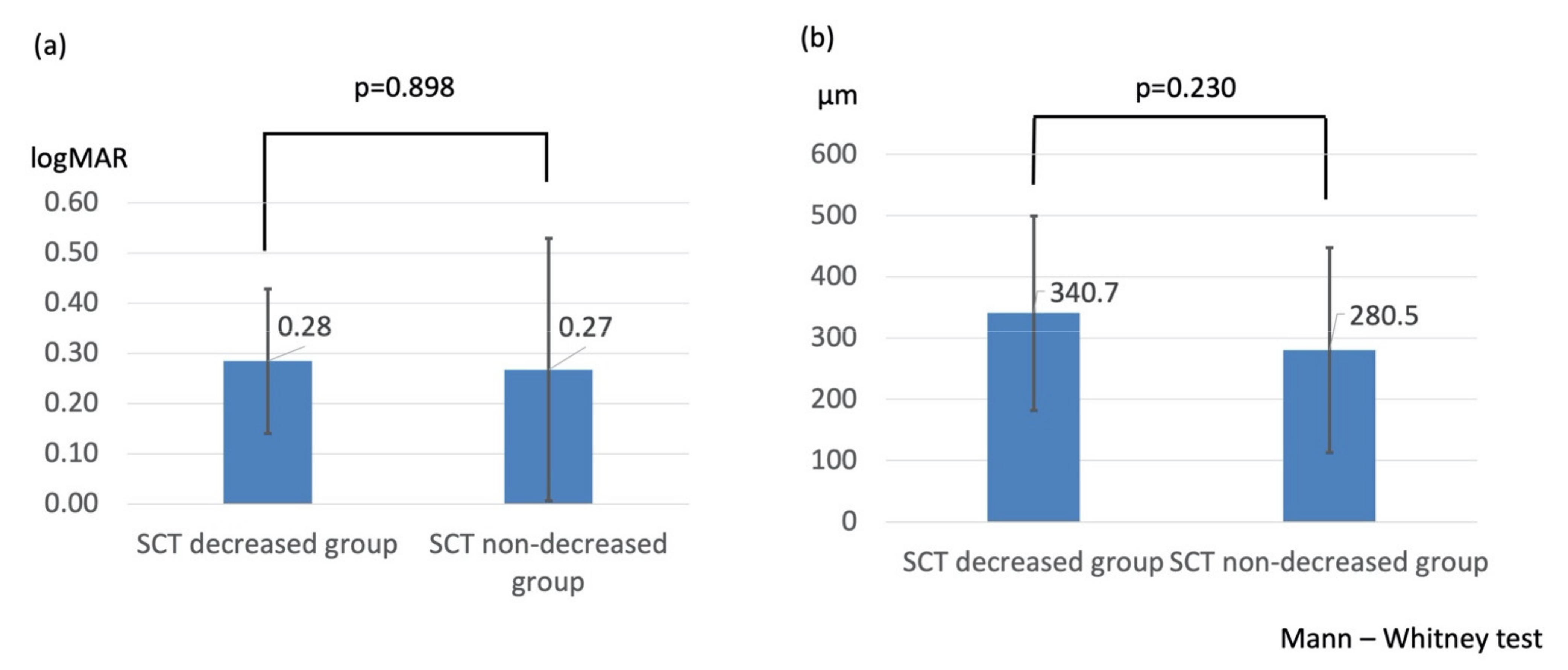
3. Results
4. Discussion
5. Conclusions
Author Contributions
Funding
Institutional Review Board Statement
Informed Consent Statement
Data Availability Statement
Acknowledgments
Conflicts of Interest
References
- Mitchell, P.; Smith, W.; Chang, A. Prevalence and associations of retinal vein occlusion in Australia. The Blue Mountains Eye Study. Arch. Ophthalmol. 1996, 114, 1243–1247. [Google Scholar] [CrossRef]
- Klein, R.; Klein, B.E.; Moss, S.E.; Meuer, S.M. The epidemiology of retinal vein occlusion: The Beaver Dam Eye Study. Trans. Am. Ophthalmol. Soc. 2000, 98, 133–141. [Google Scholar]
- Rogers, S.; McIntosh, R.L.; Cheung, N.; Lim, L.; Wang, J.J.; Mitchell, P.; Kowalski, J.W.; Nguyen, H.; Wong, T.Y. International Eye Disease Consortium. The prevalence of retinal vein occlusion: Pooled data from population studies from the United States, Europe, Asia, and Australia. Ophthalmology 2010, 117, 313–319. [Google Scholar] [CrossRef]
- Yasuda, M.; Kiyohara, Y.; Arakawa, S.; Hata, Y.; Yonemoto, K.; Doi, Y.; Iida, M.; Ishibashi, T. Prevalence and systemic risk factors for retinal vein occlusion in a general Japanese population: The Hisayama study. Investig. Ophthalmol Vis. Sci. 2010, 51, 3205–3209. [Google Scholar] [CrossRef] [PubMed]
- The Branch Vein Occlusion Study Group. BVOS: Argon laser photocoagulation for macular edema in branch vein occlusion. Am. J. Ophthalmol. 1984, 98, 271–282. [Google Scholar] [CrossRef]
- Ip, M.S.; Scott, I.U.; VanVeldhuisen, P.C.; Oden, N.L.; Blodi, B.A.; Fisher, M.; Singerman, L.J.; Tolentino, M.; Chan, C.K.; Gonzalez, V.H.; et al. A randomized trial comparing the efficacy and safety of intravitreal triamcinolone with standard care to treat vision loss associated with macular edema secondary to branch retinal vein occlusion: The Standard Care vs Corticosteroid for Retinal Vein Occlusion (SCORE) Study Report 6. Arch. Ophthalmol. 2009, 127, 1115–1128. [Google Scholar] [CrossRef]
- Campochiaro, P.A.; Heier, J.S.; Feiner, L.; Gray, S.; Saroj, N.; Rundle, A.C.; Murahashi, W.Y.; Rubio, R.G.; BRAVO Investigators. Ranibizumab for macular edema following branch retinal vein occlusion: Six-month primary end point results of a phase III study. Ophthalmology 2010, 117, 1102–1112. [Google Scholar] [CrossRef] [PubMed]
- Sakanishi, Y.; Lee, A.; Usui-Ouchi, A.; Ito, R.; Ebihara, N. Twelve-month outcomes in patients with retinal vein occlusion treated with low-frequency intravitreal ranibizumab. Clin. Ophthalmol. 2016, 10, 1161–1165. [Google Scholar] [CrossRef]
- Brown, D.M.; Campochiaro, P.A.; Bhisitkul, R.B.; Ho, A.C.; Gray, S.; Saroj, N.; Adamis, A.P.; Rubio, R.G.; Murahashi, W.Y. Sustained benefits from ranibizumab for macular edema following branch retinal vein occlusion: 12-month outcomes of a phase III study. Ophthalmology 2011, 118, 1594–1602. [Google Scholar] [CrossRef] [PubMed]
- Campochiaro, P.A.; Clark, W.L.; Boyer, D.S.; Heier, J.S.; Brown, D.M.; Vitti, R.; Kazmi, H.; Berliner, A.J.; Erickson, K.; Chu, K.W.; et al. Intravitreal aflibercept for macular edema following branch retinal vein occlusion: The 24-week results of the VIBRANT study. Ophthalmology 2015, 122, 538–544. [Google Scholar] [CrossRef]
- Wang, J.K.; Huang, T.L.; Su, P.Y.; Chang, P.Y.; Tseng, Y.Y. Intravitreal aflibercept for macular edema secondary to branch retinal vein occlusion in Chinese patients. Eye Sci. 2015, 30, 63–66. [Google Scholar] [PubMed]
- Noma, H.; Minamoto, A.; Funatsu, H.; Tsukamoto, H.; Nakano, K.; Yamashita, H.; Mishima, H.K. Intravitreal levels of vascular endothelial growth factor and interleukin-6 are correlated with macular edema in branch retinal vein occlusion. Graefes. Arch. Clin. Exp. Ophthalmol. 2006, 244, 309–315. [Google Scholar] [CrossRef] [PubMed]
- Dvorak, H.F.; Brown, L.F.; Detmar, M.; Dvorak, A.M. Vascular permeability factor/vascular endothelial growth factor, microvascular hyperpermeability, and angiogenesis. Am. J. Pathol. 1995, 146, 1029–1039. [Google Scholar] [PubMed]
- Weis, S.M.; Cheresh, D.A. Pathophysiological consequences of VEGF-induced vascular permeability. Nature 2005, 437, 497–504. [Google Scholar] [CrossRef]
- Shin, Y.U.; Lee, M.J.; Lee, B.R. Choroidal maps in different types of macular edema in branch retinal vein occlusion using swept-source optical coherence tomography. Am. J. Ophthalmol. 2015, 160, 328–334. [Google Scholar] [CrossRef] [PubMed]
- Okamoto, M.; Yamashita, M.; Sakamoto, T.; Ogata, N. Choroidal blood flow and thickness as predictors for response to anti-vascular endothelial growth factor therapy in macular edema secondary to branch retinal vein occlusion. Retina 2018, 38, 550–558. [Google Scholar] [CrossRef]
- Hasegawa, T.; Kawano, T.; Maruko, I.; Koizumi, H.; Iida, T. Clinical findings of eyes with macular edema associated with branch retinal vein occlusion refractory to ranibizumab. Retina 2018, 38, 1347–1353. [Google Scholar] [CrossRef]
- Tsuiki, E.; Suzuma, K.; Ueki, R.; Maekawa, Y.; Kitaoka, T. Enhanced depth imaging optical coherence tomography of the choroid in central retinal vein occlusion. Am. J. Ophthalmol. 2013, 156, 543–547. [Google Scholar] [CrossRef]
- Mrejen, S.; Spaide, R.F. Optical coherence tomography: Imaging of the choroid and beyond. Surv. Ophthalmol. 2013, 58, 387–429. [Google Scholar] [CrossRef]
- Konstantinidis, L.; Guex-Crosier, Y. Hypertension and the eye. Curr. Opin. Ophthalmol. 2016, 27, 514–521. [Google Scholar] [CrossRef]
- Tomiyasu, T.; Hirano, Y.; Yoshida, M.; Suzuki, N.; Nishiyama, T.; Uemura, A.; Yasukawa, T.; Ogura, Y. Microaneurysms cause refractory macular edema in branch retinal vein occlusion. Sci. Rep. 2016, 6, 29445. [Google Scholar] [CrossRef]
- Kang, H.M.; Choi, J.H.; Koh, H.J.; Lee, C.S.; Lee, S.C. Significant reduction of peripapillary choroidal thickness in patients with unilateral branch retinal vein occlusion. Retina 2018, 38, 72–78. [Google Scholar] [CrossRef]
- Chung, Y.K.; Shin, J.A.; Park, Y.H. Choroidal volume in branch retinal vein occlusion before and after intravitreal anti-VEGF injection. Retina 2015, 35, 1234–1239. [Google Scholar] [CrossRef] [PubMed]
- Kim, K.H.; Lee, D.H.; Lee, J.J.; Park, S.W.; Byon, I.S.; Lee, J.E. Regional choroidal thickness changes in branch retinal vein occlusion with macular edema. Ophthalmologica 2015, 234, 109–118. [Google Scholar] [CrossRef] [PubMed]
- Daizumoto, E.; Mitamura, Y.; Sano, H.; Akaiwa, K.; Niki, M.; Yamanaka, C.; Kinoshita, T.; Egawa, M.; Sonoda, S.; Sakamoto, T. Changes of choroidal structure after intravitreal aflibercept therapy for polypoidal choroidal vasculopathy. Br. J. Ophthalmol. 2017, 101, 56–61. [Google Scholar] [CrossRef]
- Gharbiya, M.; Giustolisi, R.; Marchiori, J.; Bruscolini, A.; Mallone, F.; Fameli, V.; Nebbioso, M.; Abdolrahimzadeh, S. Comparison of short-term choroidal thickness and retinal morphological changes after intravitreal anti-VEGF therapy with ranibizumab or aflibercept in treatment-naïve eyes. Curr. Eye Res. 2018, 43, 391–396. [Google Scholar] [CrossRef] [PubMed]
- Koizumi, H.; Kano, M.; Yamamoto, A.; Saito, M.; Maruko, I.; Sekiryu, T.; Okada, A.A.; Idea, T. Subfoveal choroidal thickness during aflibercept therapy for neovascular age-related macular degeneration: Twelve-month results. Ophthalmology 2016, 123, 617–624. [Google Scholar] [CrossRef]
- Brown, J.S.; Flitcroft, D.I.; Ying, G.; Francis, E.L.; Schmid, G.F.; Quinn, G.E.; Stone, R.A. In vivo human choroidal thickness measurements: Evidence for diurnal fluctuations. Investig. Ophthalmol. Vis. Sci. 2009, 50, 5–12. [Google Scholar] [CrossRef]
- Tan, C.S.; Ouyang, Y.; Ruiz, H.; Sadda, S.R. Diurnal variation of choroidal thickness in normal, healthy subjects measured by spectral domain optical coherence tomography. Investig. Ophthalmol. Vis. Sci. 2012, 53, 261–266. [Google Scholar] [CrossRef]
- Wei, W.B.; Xu, L.; Jonas, J.B.; Shao, L.; Du, K.F.; Wang, S.; Chen, C.X.; Xu, J.; Wang, Y.X.; Zhou, J.Q.; et al. Subfoveal choroidal thickness: The Beijing Eye Study. Ophthalmology 2013, 120, 175–180. [Google Scholar] [CrossRef]


| SCT Thickened Group | SCT Non-Thickened Group | ||
|---|---|---|---|
| n = 26 | n = 20 | p-Value | |
| Age (years) | 66.3 ± 9.7 | 66.3 ± 11.4 | 0.842 |
| Sex (male/female) | 12/14 | 9/11 | 1.000 |
| HT (+/−) | 16/10 | 14/6 | 1.000 |
| Right eye/left eye | 15/11 | 8/12 | 1.000 |
| BCVA (logMAR) | 0.41 ± 0.19 | 0.31 ± 0.23 | 0.143 |
| CFT (μm) | 564.3 ± 175.2 | 516.7 ± 135.4 | 0.236 |
| SCT (μm) Affected eye | 266.7 ± 59.1 | 213.5 ± 54.8 | 0.004 |
| Unaffected eye | 207.7 ± 54.5 | 236.6 ± 58.2 | 0.074 |
Publisher’s Note: MDPI stays neutral with regard to jurisdictional claims in published maps and institutional affiliations. |
© 2021 by the authors. Licensee MDPI, Basel, Switzerland. This article is an open access article distributed under the terms and conditions of the Creative Commons Attribution (CC BY) license (https://creativecommons.org/licenses/by/4.0/).
Share and Cite
Sakanishi, Y.; Morita, S.; Mashimo, K.; Tamaki, K.; Ebihara, N. Subfoveal Choroidal Thickness and Treatment Outcomes of Intravitreal Aflibercept for Branch Retinal Vein Occlusion. Life 2021, 11, 572. https://doi.org/10.3390/life11060572
Sakanishi Y, Morita S, Mashimo K, Tamaki K, Ebihara N. Subfoveal Choroidal Thickness and Treatment Outcomes of Intravitreal Aflibercept for Branch Retinal Vein Occlusion. Life. 2021; 11(6):572. https://doi.org/10.3390/life11060572
Chicago/Turabian StyleSakanishi, Yoshihito, Syu Morita, Keitaro Mashimo, Kazunori Tamaki, and Nobuyuki Ebihara. 2021. "Subfoveal Choroidal Thickness and Treatment Outcomes of Intravitreal Aflibercept for Branch Retinal Vein Occlusion" Life 11, no. 6: 572. https://doi.org/10.3390/life11060572
APA StyleSakanishi, Y., Morita, S., Mashimo, K., Tamaki, K., & Ebihara, N. (2021). Subfoveal Choroidal Thickness and Treatment Outcomes of Intravitreal Aflibercept for Branch Retinal Vein Occlusion. Life, 11(6), 572. https://doi.org/10.3390/life11060572