Article

Altered Blood Levels of Anti-Gal Antibodies in Alzheimer’s Disease: A New Clue to Pathogenesis?

1
Centre for Research and Training in Medicine of Aging, Department of Medicine and Health Sciences “V.Tiberio”, University of Molise, Località Tappino, 86100 Campobasso, Italy
2
Biocompatibility Innovation (BCI), via Lorenzo De Antoni 17/19, Este, 35042 Padova, Italy
*
Author to whom correspondence should be addressed.
Life 2021, 11(6), 538; https://doi.org/10.3390/life11060538
Submission received: 15 May 2021 / Revised: 5 June 2021 / Accepted: 6 June 2021 / Published: 9 June 2021

Abstract

:
Alzheimer’s disease is a neurodegenerative disorder whose pathological mechanisms, despite recent advances, are not fully understood. However, the deposition of beta amyloid -peptide and neuroinflammation, which is probably aggravated by dysbiotic microbiota, seem to play a key role. Anti-Gal are the most abundant xenoreactive natural antibodies. They are supposed to stem from immunization against the gut microbiota and have been implicated in the pathogenesis of several diseases, including multiple sclerosis. These antibodies target the alpha-Gal epitope, expressed on the terminal sugar units of glycoprotein or glycolipid of all mammals except apes, Old World monkeys and humans. The alpha-Gal is constitutively expressed in several bacteria constituting the brain microbiota, and alpha-Gal-like epitopes have been detected in gray matter, amyloid plaque, neurofibrillary tangles and corpora amylacea of the human brain, suggesting a potential link between anti-Gal and Alzheimer’s disease etiopathogenesis. For the first time, our study searched for possible alterations of anti-Gal immunoglobulin levels in Alzheimer’s disease patients. IgG and IgM blood levels were significantly lower, and IgA significantly higher in patients than in healthy subjects. These results suggest that such immunoglobulins might be implicated in Alzheimer’s disease pathogenesis and open new scenarios in the research for new biomarkers and therapeutic strategies.

1. Introduction

Alzheimer’s disease (AD), the most common form of dementia, is a progressive, irreversible and incurable neurodegenerative disease, which constitutes one of the major causes of dependency, disability and mortality [1]. There are over 50 million people worldwide living with dementia in 2020. This number will almost double every 20 years, reaching 82 million in 2030 and 152 million in 2050. AD accounts for 50–75% of dementia cases [2].
Despite the huge amount of research, the pathological mechanisms underlying the onset and progression of the disease are not fully understood. Neurodegeneration is most likely caused by an abnormal accumulation of the beta amyloid peptide which produces a cascade of events, including an inflammatory process mainly mediated by microglial activation and formation of neurofibrillary tangles that culminate in neuronal death. The triggering mechanism of this cascade is still unknown and therefore biomarkers with possible mechanistic insights into the AD pathophysiologic processes are urgently needed [3]. Recent works suggest a potential association between gut microbiota, neuroinflammation and AD, either directly owing to bacterial invasion of the brain due to leakage of the blood–brain barrier (BBB) and production of toxins and inflammation, or indirectly by modulating the immune response [4].
The α-Gal epitope (Galα1-3Galβ1-4GlcNAc-R) is a short carbohydrate sequence terminally located in the oligosaccharide unit of glycoproteins and glycolipids, commonly expressed at the cell surface of various microorganisms (protozoa, bacteria, fungi, viruses), marsupials and non-primate mammalians. This epitope is produced by the α1,3-galactosyltransferase (α1,3GT) enzyme [5,6,7,8,9]. In humans, the gene that encodes for this enzyme (α1,3GT or GGTA1 gene) is suppressed and the consequent absence of α-Gal on the surface of cells induces a massive production of anti-Gal antibodies [10]. Anti-Gal IgG, IgM and IgA, in fact, represent over 1% of all circulating immunoglobulins [11]. Their synthesis is thought to be mainly stimulated by chronic exposure to enteric bacteria, various pathogens and environmental antigens presenting the α-Gal epitope on the surface. Such induced immune response represents an evolutionary advantage for the human organism that acquires immune protection against these pathogens [12]. In effect, anti-Gal antibodies fulfil many important roles, such as ensuring immunity from infections originating from several micro-organisms; contributing to rejection in the case of animal-to-human cell, tissue and organ transplantation; accelerating the process of wound healing. However, circulating anti-Gal antibodies have been implicated in the pathogenesis of autoimmune diseases, such as Henoch–Schönlein purpura, IgA nephropathy, rheumatoid arthritis, Crohn’s disease and Graves’ disease [8,9,10,13,14]. Variations in the levels of anti-Gal have also been described in multiple sclerosis (MS) together with an altered microbiome, suggesting a link between anti-Gal antibodies and the disease [15,16].
In physiological conditions the anti-Gal antibodies cannot cross the BBB, so they are not present in the cerebrospinal fluid (CSF) of healthy individuals. However, varying titers of anti-Gal Ig were found in the CSF of patients with MS, Guillain-Barré syndrome and meningitis, suggesting that inflammatory processes might alter the BBB and favour the passage of blood Ig (including anti-Gal) into the CSF [17]. Furthermore, α-Gal-like epitopes have been detected in the brain, more precisely in the gray matter of healthy individuals who died accidentally [18], other than in the amyloid plaques, neurofibrillary tangles and corpora amylacea of patients with dementia, including AD [19]. Together with recent literature findings demonstrating the α-Gal epitope presence in several bacteria [20] constituting the brain microbiome [21], these observations suggest a potential relationship between anti-Gal antibodies and AD etiopathogenesis.
In this study we looked for the first time at possible alterations of anti-Gal Ig levels in AD patients, speculating on the potential role of such antibodies in the pathogenesis of the disease.

2. Materials and Methods

2.1. Patients and Healthy Controls

The participants in the study (n. = 60) were consecutively recruited at the Centre for Research and Training in Medicine of Aging of the University of Molise (Italy). The AD patients (n. = 30) fulfilled the National Institute of Aging and Alzheimer’s Association diagnostic criteria for “probable AD with documented decline” [22]. They scored < 24 on the Mini Mental State Examination and >0.5 on the Clinical Dementia Rating. To rule out other potential causes of cognitive impairment, all patients underwent blood tests (including full blood count, erythrocyte sedimentation rate, urea nitrogen and electrolytes, thyroid function, vitamin B12, and folate) and brain imaging. Thirty sex/age-matched cognitively healthy subjects (HS) were recruited as the control group. Since anti-Gal antibody levels can be altered in the context of different pathologies and treatments, subjects with pathologies, such as rheumatoid arthritis, interstitial cystitis, eosinophilic esophagitis [23], Henoch–Schönlein purpura, IgA nephropathy, Crohn’s disease and ulcerative colitis [24], or treated with anticancer such as Cetuximab (or Erbitux) [25], animal derived tissue patches, cartilaginous grafts or bioprostheses such as biological heart valves [26], were excluded. The clinical and demographic characteristics of the two groups of participants are summarized in Table 1.
The study was conducted in accordance with ethical principles stated in the Declaration of Helsinki, and with approved national and international guidelines for human research. The Institutional Review Board of the University of Molise approved the study (Prot. n. 007-08-2018). Written informed consent was obtained from participants or caregivers.

2.2. Blood Collection and Processing

The blood collection was performed between 8:00 and 8:30 a.m. after overnight fasting of at least 8–10 h. Venous blood was collected with a vacutainer system (Becton & Dickinson, Milan, Italy). To obtain the serum, the blood was centrifuged within 2 h at 1500× g for 10 min. The samples were then stored at −80 °C until their use.

2.3. Determination of α-Gal Antibody Titers

The evaluation of the different human anti α-Gal antibodies isotypes (IgG, IgM and IgA) was performed in duplicate and triplicate using a modified ELISA test (patent EP2626701). Data were expressed as the absorbance value. Each patient’s serum was diluted 50-fold with Phosphate-Buffered Saline (PBS, Merck Lifescience, Darmstadt, Germany) in a final volume of 2 mL.
Briefly, for each isotype, a Polysorp 96-well plate (Nunc, Rochester, NY, USA) was coated with 100 µL of α-Gal/human serum albumin (HSA) (Dextra Laboratories, Berkshire, UK), 5 µg/mL, for 2 h at 37 °C. After washing three times with PBS, the blocking procedure was performed using 300 µL per well of 2% HSA for 2 h, at RT in darkness. Wells were then washed three times, as above. A set of four wells for each column was loaded with 100 µL of a single diluted serum and the plate incubated for 2 h at 37 °C. After washing, the proper secondary horseradish peroxidase (HRP)-conjugate antibody (1:100) was loaded (anti-human IgG, anti-human IgM and anti-human IgA, Merck Lifescience, Darmsadt, Germany) and the plate incubated for 1 h at 37 °C. After washing, 100 µL of HRP substrate buffer was added to each well for 5 min, at RT, in the darkness. The plate absorbance was measured at 450 nm by a microplate spectrophotometer (Multiskan Sky, Thermo Fisher Scientific, Waltham, MA, USA).

2.4. Statistical Analysis
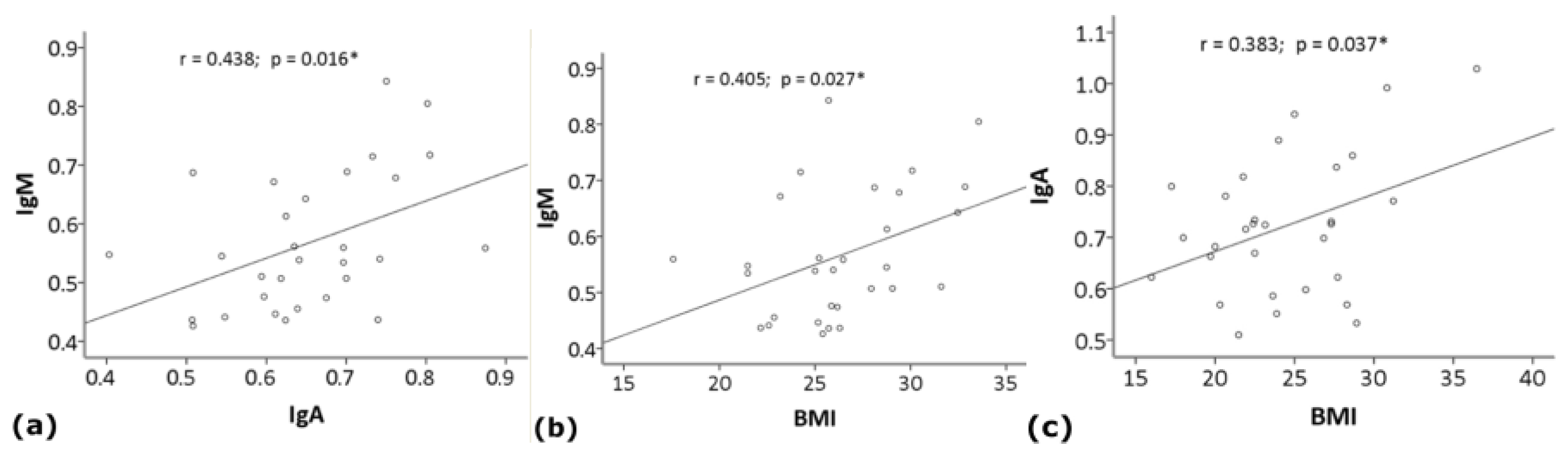
Data were analyzed using SPSS (v. 17.0) statistical software package (SPSS Inc., Chicago, IL, USA). Variables were examined for outliers and extreme values by means of box and normal quantile-quantile plots, and Shapiro–Wilk’s and Kolmogorov–Smirnov’s tests. When a normal distribution could not be accepted, variable transformations (square, square root, logarithmic, reciprocal of square root or reciprocal transformations) were reviewed and, if the normality could not be reached, (as for educational level and MMSE) nonparametric tests were used. One-way analysis of variance (ANOVA) was used to evaluate the differences between groups (AD vs. HS) in age, educational level, BMI and MMSE. Chi-square test was used to assess differences between groups in sex, blood group, comorbidity and drug intake. Group differences (HS vs. AD) were evaluated by uni- and multivariate analysis of covariance (ANCOVA), using age, sex, educational level, BMI, blood group, comorbidity and drug therapy as covariates. The assumption of the equality of variance was assessed by means of Levene’s test. Finally, correlation analysis was performed in each group by Pearson’s correlation coefficient (r) for normally distributed and Spearman rank correlation coefficient (rs) for not normally distributed variables, using Bonferroni’s correction for multiple comparisons. Correlation analysis was used to measure the strength and direction of association between IgG, IgM or IgA levels and age, sex, BMI, blood group, educational level, MMSE (as a measure of cognitive decline severity), comorbidity and drugs.

3. Results

Clinical and demographic characteristics of the subjects enrolled in the study are reported in Table 1. There were no significant differences between groups, excluding the higher educational level in HS.
Multivariate ANCOVA, including age, gender, educational level, BMI, blood group, comorbidity and drug therapy as covariates, showed a statistically significant difference (F = 13.566; df = 3.40; p < 0.001; partial η2 = 0.504) between groups (HS vs. AD). The results of univariate ANCOVA, as well as the serum levels of anti-Gal IgG, IgM and IgA are reported in Table 2.
IgG and IgM levels result significantly decreased, and IgA significantly increased, in AD patients compared to HS (Table 2; Figure 1).
In the HS group, the analysis showed a weak positive correlation between IgM and IgA (r = 0.438; p = 0.016) or BMI (r = 0.405; p = 0.027), not significant after Bonferroni’s correction (Figure 2). In the AD group, the analysis showed a weak positive correlation between IgA and BMI (r = 0.383; p = 0.037), not significant after Bonferroni’s correction (Figure 2), and a trend toward a positive correlation between IgG and IgM (r = 0.351; p = 0.057).
No significant relationship was found between Ig levels and age, sex, blood group, educational level, MMSE, comorbidity and drugs.

4. Discussion

This study demonstrated that anti-Gal IgG and IgM were significantly decreased, and IgA significantly increased in AD patients compared to HS. No significant relationship was found between Ig levels and age, sex, blood group, severity of disease as assessed by MMSE, level of education, co-morbidity and drugs in AD patients, except for a weak positive correlation between IgA and BMI. To our knowledge, this is the first study that has investigated the serum levels of anti-Gal antibodies in AD and, therefore, comparison studies are lacking. However, circulating anti-Gal IgG results reduced compared with those of age/gender-matched HS in patients with MS [15], an autoimmune, inflammatory and neurodegenerative disease [27]. The authors speculated that a modified gut microbiota, specifically characterized by low α-Gal-producing microorganisms, can modify the inflammation homeostasis of individuals genetically predisposed, increasing the risk of developing MS [15]. The finding that gut microbiota of MS patients is characterized by a significant decrease in microorganisms expressing the GGTA1 gene (which codes for the α-Gal epitope) supports this hypothesis [16]. In patients with inflammatory bowel diseases, such as Crohn’s disease and ulcerative colitis, anti-Gal specific IgA resulted in higher concentrations than in HS, while total IgM resulted significantly decreased [28]. The authors speculated that the increase in IgA was linked to the presence of a damaged intestinal barrier exposing the subject’s immune system to enteric bacteria presenting α-Gal. The reduction of total IgM was interpreted as a switching of the antibody class [28].
Although anti-Gal antibodies do not meet the definition of natural antibodies (i.e., antibodies present in a non-immunised organism from birth), many authors place them in this category given the numerous features in common [8,9,10,14]. IgM and IgG are the most widely described classes of natural antibodies since they are implicated in many infectious diseases and pathologies, such as neurological disorders, cancer, diabetes and cardiovascular diseases. Interestingly, lower levels of natural antibodies are generally negatively correlated with disease onset and progress, whereas high levels often correlate with protection or absence of disease [9,14]. An example is represented by the IgG antibodies against amyloid peptides that were reported to be reduced in AD patients compared to age-matched HS [29] and to decrease with normal ageing and advancing AD, especially those binding to assemblies of amyloidogenic peptides [30]. The literature available on IgA is scant in contrast to IgM and IgG natural antibodies. IgA functions as a potent pro-inflammatory agent, being a rapid activator of neutrophils and it is crucial in the first line of defence against pathogens. In particular, they are considered homeostatic immunoglobulins that neutralize microbiota and food antigens to prevent interactions with the host [9,31].
We speculated that the simultaneous decreases in anti-Gal IgM and IgG and the increase in anti-Gal IgA we observed in AD patients might reflect the class switching of anti-α-Gal B cells from the production of “natural” IgM antibodies to an IgA-mediated adaptive response. Significant increases in serum IgA levels were observed in AD patients in comparison with HS [32,33,34]. An example is the IgA antibodies directed against the N-methyl-d-Aspartate receptor (NMDAR), detected in 10% of AD patients as opposed to 2.8% of HS [35]. The NMDAR NR1 subunit was found to carry glycostructures recognized by antibodies raised against the Fuc alpha 1-2Gal epitope [36], a common constituent of A, B and O blood groups. Noticeably, most anti-B antibodies in the sera of A and O blood group carriers are cross-reactive anti-alpha-Gal antibodies [37]. It is also of note that IgM and IgA-enriched Ig preparations incited the production of higher levels of TNF-α and NO by primary rat microglial cells in vitro, compared with IgG [38], which highlights the possible role of IgA-mediated responses in the microglial activation and neuroinflammation associated with AD. The phlogosis of lymphoid tissue associated with abdominal fat, promoted by excess caloric intake, and/or with a gut microbial dysbiosis, might alter the normal balance between tolerance and reaction to ubiquitary antigens in the gut-associated lymphoid tissue (GALT) favouring the sensitization of α-Gal-specific T helper 2 cells. The positive correlation that we observed between IgA and BMI and the encouraging results of therapeutic approaches aimed at controlling metabolic imbalance and inflammation for halting the progression of AD [39] are noteworthy in this regard. B cells activated in the GALT may home into the marginal zone of the spleen [40], wherefrom IgA produced by GALT plasma cells in response to commensal or pathogenic bacteria may enter the blood and permeate the BBB, in the presence of phlogosis.
The mounting of such an adaptive response to microbial α-Gal epitopes in the GALT and the concomitant permeabilization of the BBB to anti-Gal IgA antibodies may damage cells and tissues in the central nervous system as a result of antibody- and complement-mediated responses to: (a) formerly tolerated α-Gal epitopes also expressed by the brain microbiome [20,21]; (b) self-antigens containing identical or similar oligosaccharide moieties linked to autologous peptides, on the base of epitopic mimicry or epitopic spreading.
Gut microbial dysbiosis, frequently found in patients suffering from neurodegenerative diseases including AD [41,42,43,44], and which promotes the growth of a more aggressive intestinal bacterial population (Table 3) [45,46,47,48], may lead to the secretion of amyloid, lipopolysaccharides and other toxic substances (e.g., beta-N-methylamino-L-alanine, saxitoxin and anatoxin-alpha) [49,50], which alter both the gastrointestinal permeability and BBB favouring the passage of immunoglobulins, microorganisms and other molecules (toxic or not) into the brain [51].
The binding of IgA to α-Gal-like epitopes present on gray matter [18] and/or on the bacteria within neurons [21] may trigger complement-mediated cytolysis and a neuroinflammation process that, in turn, may provoke or exacerbate the amyloid cascade. This hypothesis is supported by the finding that bacteria belonging mainly to the Enterobacteriaceae family, including Escherichia coli, Pasteurellaceae genera, including Haemophilus influenzae, and Lactobacillaceae family bearing the GGTA1 gene [20] have been found post-mortem in the brain of AD and control subjects and all contribute to the brain microbiome [21]. The observation that senile plaques and neurofibrillary tangles are diffusely stained by using lectins obtained from Griffonia simplicifolia and Amaranthus leucocarpus, which are specific to α-Gal-like epitopes [19,52], also supports our hypothesis. Further to this, corpora amylacea and spherical deposits, frequently found in the brain of demented patients, also react positively when stained by lectins. Corpora amylacea have long been assumed to be a hallmark of normal brain ageing, however, their role appears to be similar to that of senile plaques and neurofibrillary tangles in the extent of cognitive dysfunction of individuals with dementia [19].
We acknowledge some limitations of our study due to its small sample sizes and the observational nature of the results. However, this novel observation opens up new potential research questions and may be a basis for establishing a new etiological factor for AD. In particular, our findings offer support to speculations linking α-Gal, gut microbiota, neuroinflammation and AD. A further study with a larger sample size and a longitudinal design is needed to examine the potential predictive value of anti-Gal antibodies as a biomarker of AD development and progression.

5. Conclusions

This study, the first to investigate the serum titers of anti-Gal antibodies in AD, showed lower IgG and IgM and higher IgA levels in AD patients compared to healthy subjects. These results suggest a possible role of anti-Gal immunoglobulins in the pathogenesis of AD and support the theory of the association between host microbiota, neuroinflammation and dementia. If confirmed in a longitudinal study with a larger number of participants, these findings could open new scenarios in the knowledge of AD etiopathogenesis and the research for new biomarkers and therapeutic strategies.

Author Contributions

Conceptualization: A.D.C., A.G., F.G., F.N.; methodology: A.A. (Alessia Arcaro), F.N.; formal analysis: A.D.C.; investigation: A.A. (Antonella Angiolillo), A.A. (Alessia Arcaro), A.C.; writing—original draft preparation: A.D.C.; writing—review and editing: A.A. (Antonella Angiolillo), A.C., A.D.C., F.G., F.N.; validation: A.C.; visualization: A.A. (Antonella Angiolillo); supervision: A.D.C., A.G.; project administration: A.D.C., A.G., F.N. All authors have read and agreed to the published version of the manuscript.

Funding

This research received no external funding.

Institutional Review Board Statement

The study was conducted according to the guidelines of the Declaration of Helsinki and approved by the Institutional Review Board of University of Molise (protocol code 007-08-2018; 2 August 2018).

Informed Consent Statement

Written informed consent was obtained from all subjects involved in the study.

Data Availability Statement

The datasets used and analyzed during the current study are available from the corresponding author on reasonable request.

Acknowledgments

The authors are grateful to Santina Ciccotelli for her valuable assistance.

Conflicts of Interest

The authors declare no conflict of interest.

References

  1. Alzheimer’s Association. 2020 Alzheimer’s disease facts and figures. Alzheimer’s Dement. 2020, 16, 391–460. [Google Scholar] [CrossRef]
  2. Fleming, R.; Zeisel, J.; Bennet, K. World Alzheimer Report 2020, Vol I and II; Alzheimer’s Disease International: London, UK, 2020. [Google Scholar]
  3. McGrowder, D.A.; Miller, F.; Vaz, K.; Nwokocha, C.; Wilson-Clarke, C.; Anderson-Cross, M.; Brown, J.; Anderson-Jackson, L.; Williams, L.; Latore, L.; et al. Cerebrospinal Fluid Biomarkers of Alzheimer’s Disease: Current Evidence and Future Perspectives. Brain Sci. 2021, 11, 215. [Google Scholar] [CrossRef]
  4. González-Sanmiguel, J.; Schuh, C.M.A.P.; Muñoz-Montesino, C.; Contreras-Kallens, P.; Aguayo, L.G.; Aguayo, S. Complex Interaction between Resident Microbiota and Misfolded Proteins: Role in Neuroinflammation and Neurodegeneration. Cells 2020, 9, 2476. [Google Scholar] [CrossRef]
  5. Almeida, I.C.; Ferguson, M.A.; Schenkman, S.; Travassos, L.R. Lytic anti-alpha-galactosyl antibodies from patients with chronic Chagas’ disease recognize novel O-linked oligosaccharides on mucin-like glycosyl-phosphatidylinositol-anchored glycoproteins of Trypanosoma cruzi. Biochem. J. 1994, 304, 793. [Google Scholar] [CrossRef] [PubMed]
  6. Welsh, R.M.; O’Donnell, C.L.; Reed, D.J.; Rother, R.P. Evaluation of the Galalpha1-3Gal epitope as a host modification factor eliciting natural humoral immunity to enveloped viruses. J. Virol. 1998, 72, 4650–4656. [Google Scholar] [CrossRef] [Green Version]
  7. Han, W.; Cai, L.; Wu, B.; Li, L.; Xiao, Z.; Cheng, J.; Wang, P.G. The wciN gene encodes an α-1,3-galactosyltransferase involved in the biosynthesis of the capsule repeating unit of Streptococcus pneumoniae serotype 6B. Biochemistry 2012, 51, 5804–5810. [Google Scholar] [CrossRef] [Green Version]
  8. Huai, G.; Qi, P.; Yang, H.; Wang, Y. Characteristics of α-Gal epitope, anti-Gal antibody, α1,3 galactosyltransferase and its clinical exploitation. Int. J. Mol. Med. 2016, 37, 11–20. [Google Scholar] [CrossRef] [PubMed] [Green Version]
  9. Reyneveld, G.I.; Savelkoul, H.F.J.; Parmentier, H.K. Current Understanding of Natural Antibodies and Exploring the Possibilities of Modulation Using Veterinary Models. A Review. Front. Immunol. 2020, 11, 2139. [Google Scholar] [CrossRef]
  10. Galili, U. Discovery of the natural anti-Gal antibody and its past and future relevance to medicine. Xenotransplantation 2013, 20, 138–147. [Google Scholar] [CrossRef]
  11. Galili, U. Human Natural Antibodies to Mammalian Carbohydrate Antigens as Unsung Heroes Protecting against Past, Present, and Future Viral Infections. Antibodies 2020, 9, 25. [Google Scholar] [CrossRef]
  12. Galili, U. Significance of the evolutionary α1,3-galactosyltransferase (GGTA1) gene inactivation in preventing extinction of apes and old world monkeys. J. Mol. Evol. 2015, 80, 1–9. [Google Scholar] [CrossRef] [PubMed]
  13. Nguyen, T.G.; McKelvey, K.J.; March, L.M.; Hunter, D.J.; Xue, M.; Jackson, C.J.; Morris, J.M. Aberrant levels of natural IgM antibodies in osteoarthritis and rheumatoid arthritis patients in comparison to healthy controls. Immunol. Lett. 2016, 170, 27–36. [Google Scholar] [CrossRef] [PubMed]
  14. Palma, J.; Tokarz-Deptuła, B.; Deptuła, J.; Deptuła, W. Natural antibodies–facts known and unknown. Cent. Eur. J. Immunol. 2018, 43, 466–475. [Google Scholar] [CrossRef]
  15. Le Berre, L.; Rousse, J.; Gourraud, P.A.; Imbert-Marcille, B.M.; Salama, A.; Evanno, G.; Semana, G.; Nicot, A.; Dugast, E.; Guérif, P.; et al. Decrease of blood anti-α1,3 Galactose Abs levels in multiple sclerosis (MS) and clinically isolated syndrome (CIS) patients. Clin. Immunol. 2017, 180, 128–135. [Google Scholar] [CrossRef]
  16. Montassier, E.; Berthelot, L.; Soulillou, J.P. Are the decrease in circulating anti-α1,3-Gal IgG and the lower content of galactosyl transferase A1 in the microbiota of patients with multiple sclerosis a novel environmental risk factor for the disease? Mol. Immunol. 2018, 93, 162–165. [Google Scholar] [CrossRef] [PubMed]
  17. Galili, U.; Anaraki, F.; Thall, A.; Hill-Black, C.; Radic, M. One percent of human circulating B lymphocytes are capable of producing the natural anti-Gal antibody. Blood 1993, 82, 2485–2493. [Google Scholar] [CrossRef] [PubMed] [Green Version]
  18. Jaison, P.L.; Kannan, V.M.; Geetha, M.; Appukuttan, P. Epitopes recognized by serum anti-α-galactoside antibody are present on brain glycoproteins in man. J. Niosci. 1993, 18, 187–193. [Google Scholar] [CrossRef]
  19. Nishi, K.; Tanegashima, A.; Yamamoto, Y.; Ushiyama, I.; Ikemoto, K.; Yamasaki, S.; Nishimura, A.; Rand, S.; Brinkmann, B. Utilization of lectin-histochemistry in forensic neuropathology: Lectin staining provides useful information for postmortem diagnosis in forensic neuropathology. Leg. Med. 2003, 5, 117–131. [Google Scholar] [CrossRef]
  20. Montassier, E.; Al-Ghalith, G.A.; Mathé, C.; Le Bastard, Q.; Douillard, V.; Garnier, A.; Guimon, R.; Raimondeau, B.; Touchefeu, Y.; Duchalais, E.; et al. Distribution of Bacterial α1,3-Galactosyltransferase Genes in the Human Gut Microbiome. Front. Immunol 2020, 10, 3000. [Google Scholar] [CrossRef] [PubMed] [Green Version]
  21. Westfall, S.; Dinh, D.M.; Pasinetti, G.M. Investigation of Potential Brain Microbiome in Alzheimer’s Disease: Implications of Study Bias. J. Alzheimer’s Dis. 2020, 75, 559–570. [Google Scholar] [CrossRef] [PubMed]
  22. McKhann, G.M.; Knopman, D.S.; Chertkow, H.; Hyman, B.T.; Jack, C.R., Jr.; Kawas, C.H.; Klunk, W.E.; Koroshetz, W.J.; Manly, J.J.; Mayeux, R.; et al. The diagnosis of dementia due to Alzheimer’s disease: Recommendations from the National Institute on Aging-Alzheimer’s Association workgroups on diagnostic guidelines for Alzheimer’s disease. Alzheimer’s Dement. 2011, 7, 263–269. [Google Scholar] [CrossRef] [Green Version]
  23. Burk, C.M.; Beitia, R.; Lund, P.K.; Dellon, E.S. High rate of galactose-alpha-1,3-galactose sensitization in both eosinophilic esophagitis and patients undergoing upper endoscopy. Dis. Esophagus 2016, 29, 558–562. [Google Scholar] [CrossRef] [PubMed] [Green Version]
  24. Safaie, P.; Ham, M.; Kuang, P.; Mehta, A.S.; Wang, M.; Cheifetz, A.S.; Robson, S.; Lau, D.; Block, T.M.; Moss, A.C. Lectin-reactive anti-α-gal in patients with Crohn’s disease: Correlation with clinical phenotypes. Inflamm. Bowel Dis. 2013, 19, 2796–2800. [Google Scholar] [CrossRef] [Green Version]
  25. Chinuki, Y.; Morita, E. Alpha-Gal-containing biologics and anaphylaxis. Allergol. Int. 2019, 68, 296–300. [Google Scholar] [CrossRef] [PubMed]
  26. Naso, F.; Stefanelli, U.; Buratto, E.; Lazzari, G.; Perota, A.; Galli, C.; Gandaglia, A. Alpha-Gal Inactivated Heart Valve Bioprostheses Exhibit an Anti-Calcification Propensity Similar to Knockout Tissues. Tissue Eng. Part A 2017, 23, 1181–1195. [Google Scholar] [CrossRef] [PubMed]
  27. Celarain, N.; Tomas-Roig, J. Aberrant DNA methylation profile exacerbates inflammation and neurodegeneration in multiple sclerosis patients. J. Neuroinflamm. 2020, 17, 21. [Google Scholar] [CrossRef]
  28. Mangold, A.; Lebherz, D.; Papay, P.; Liepert, J.; Hlavin, G.; Lichtenberger, C.; Adami, A.; Zimmermann, M.; Klaus, D.; Reinisch, W.; et al. Anti-Gal titers in healthy adults and inflammatory bowel disease patients. Transp. Proc. 2011, 43, 3964–3968. [Google Scholar] [CrossRef]
  29. Weksler, M.E.; Relkin, N.; Turkenich, R.; La Russe, S.; Zhou, L.; Szabo, P. Patients with Alzheimer disease have lower levels of serum anti-amyloid peptide antibodies than healthy elderly individuals. Exp. Gerontol. 2002, 37, 943–948. [Google Scholar] [CrossRef]
  30. Britschgi, M.; Olin, C.E.; Johns, H.T.; Takeda-Uchimura, Y.; LeMieux, M.C.; Rufibach, K.; Rajadas, J.; Zhang, H.; Tomooka, B.; Robinson, W.H.; et al. Neuroprotective natural antibodies to assemblies of amyloidogenic peptides decrease with normal aging and advancing Alzheimer’s disease. Proc. Natl. Acad. Sci. USA 2009, 106, 12145–12150. [Google Scholar] [CrossRef] [Green Version]
  31. Heineke, M.H.; van Egmond, M. Immunoglobulin A: Magic bullet or Trojan horse? Eur. J. Clin. Investig. 2017, 47, 184–192. [Google Scholar] [CrossRef] [Green Version]
  32. Leblhuber, F.; Walli, J.; Tilz, G.P.; Wachter, H.; Fuchs, D. Systemische Veränderungen des Immunsystems bei Patienten mit Alzheimer-Demenz [Systemic changes of the immune system in patients with Alzheimer’s dementia]. Dtsch. Med. Wochenschr. 1998, 123, 787–791. [Google Scholar] [CrossRef]
  33. de la Rubia Ortí, J.E.; Sancho Castillo, S.; Benlloch, M.; Julián Rochina, M.; Corchón Arreche, S.; García-Pardo, M.P. Impact of the Relationship of Stress and the Immune System in the Appearance of Alzheimer’s Disease. J. Alzheimer’s Dis. 2017, 55, 899–903. [Google Scholar] [CrossRef]
  34. de la Rubia Ortí, J.E.; Prado-Gascó, V.; Sancho Castillo, S.; Julián-Rochina, M.; Romero Gómez, F.J.; García-Pardo, M.P. Cortisol and IgA are Involved in the Progression of Alzheimer’s Disease. A Pilot Study. Cell. Mol. Neurobiol. 2019, 39, 1061–1065. [Google Scholar] [CrossRef]
  35. Doss, S.; Wandinger, K.P.; Hyman, B.T.; Panzer, J.A.; Synofzik, M.; Dickerson, B.; Mollenhauer, B.; Scherzer, C.R.; Ivinson, A.J.; Finke, C.; et al. High prevalence of NMDA receptor IgA/IgM antibodies in different dementia types. Ann. Clin. Transl. Neurol. 2014, 1, 822–832. [Google Scholar] [CrossRef] [PubMed]
  36. Smalla, K.H.; Angenstein, F.; Richter, K.; Gundelfinger, E.D.; Staak, A. Identification of fucose alpha(1-2) galactose epitope-containing glycoproteins from rat hippocampus. Neuroreport 1998, 9, 813–817. [Google Scholar] [CrossRef]
  37. Galili, U.; Buehler, J.; Shohet, S.B.; Macher, B.A. The human natural anti-Gal IgG. III. The subtlety of immune tolerance in man as demonstrated by crossreactivity between natural anti-Gal and anti-B antibodies. J. Exp. Med. 1987, 165, 693–704. [Google Scholar] [CrossRef]
  38. Pul, R.; Nguyen, D.; Schmitz, U.; Marx, P.; Stangel, M. Comparison of intravenous immunoglobulin preparations on microglial function in vitro: More potent immunomodulatory capacity of an IgM/IgA-enriched preparation. Clin. Neuropharmacol. 2002, 25, 254–259. [Google Scholar] [CrossRef]
  39. Bredesen, D.E.; Amos, E.C.; Canick, J.; Ackerley, M.; Raji, C.; Fiala, M.; Ahdidan, J. Reversal of cognitive decline in Alzheimer’s disease. Aging 2016, 8, 1250–1258. [Google Scholar] [CrossRef] [PubMed] [Green Version]
  40. Vossenkämper, A.; Blair, P.A.; Safinia, N.; Fraser, L.D.; Das, L.; Sanders, T.J.; Stagg, A.J.; Sanderson, J.D.; Taylor, K.; Chang, F.; et al. A role for gut-associated lymphoid tissue in shaping the human B cell repertoire. J. Exp. Med. 2013, 210, 1665–1674. [Google Scholar] [CrossRef] [PubMed] [Green Version]
  41. Gerhardt, S.; Mohajeri, M.H. Changes of Colonic Bacterial Composition in Parkinson’s Disease and Other Neurodegenerative Diseases. Nutrients 2018, 10, 708. [Google Scholar] [CrossRef] [PubMed] [Green Version]
  42. Szablewski, L. Human Gut Microbiota in Health and Alzheimer’s Disease. J. Alzheimer’s Dis. 2018, 62, 549–560. [Google Scholar] [CrossRef] [PubMed]
  43. Angelucci, F.; Cechova, K.; Amlerova, J.; Hort, J. Antibiotics, gut microbiota, and Alzheimer’s disease. J. Neuroinflamm. 2019, 16, 108. [Google Scholar] [CrossRef]
  44. Liu, S.; Gao, J.; Zhu, M.; Liu, K.; Zhang, H.L. Gut Microbiota and Dysbiosis in Alzheimer’s Disease: Implications for Pathogenesis and Treatment. Mol. Neurobiol. 2020, 57, 5026–5043. [Google Scholar] [CrossRef]
  45. Pluta, R.; Ułamek-Kozioł, M.; Januszewski, S.; Czuczwar, S.J. Gut microbiota and pro/prebiotics in Alzheimer’s disease. Aging 2020, 12, 5539–5550. [Google Scholar] [CrossRef]
  46. Zhang, M.; Zhao, D.; Zhou, G.; Li, C. Dietary Pattern, Gut Microbiota, and Alzheimer’s Disease. J. Agric. Food Chem. 2020, 68, 12800–12809. [Google Scholar] [CrossRef]
  47. Megur, A.; Baltriukienė, D.; Bukelskienė, V.; Burokas, A. The Microbiota-Gut-Brain Axis and Alzheimer’s Disease: Neuroinflammation Is to Blame? Nutrients 2020, 13, 37. [Google Scholar] [CrossRef]
  48. Miyake, S.; Kim, S.; Suda, W.; Oshima, K.; Nakamura, M.; Matsuoka, T.; Chihara, N.; Tomita, A.; Sato, W.; Kim, S.W.; et al. Dysbiosis in the Gut Microbiota of Patients with Multiple Sclerosis, with a Striking Depletion of Species Belonging to Clostridia XIVa and IV Clusters. PLoS ONE 2015, 10, e0137429. [Google Scholar] [CrossRef] [Green Version]
  49. Brenner, S.R. Blue-green algae or cyanobacteria in the intestinal micro-flora may produce neurotoxins such as Beta-N-Methylamino-L-Alanine (BMAA) which may be related to development of amyotrophic lateral sclerosis, Alzheimer’s disease and Parkinson-Dementia-Complex in humans and Equine Motor Neuron Disease in horses. Med. Hypotheses 2013, 80, 103. [Google Scholar] [CrossRef]
  50. Schwartz, K.; Boles, B.R. Microbial amyloids--functions and interactions within the host. Curr. Opin. Microbiol. 2013, 16, 93–99. [Google Scholar] [CrossRef] [Green Version]
  51. Kesika, P.; Suganthy, N.; Sivamaruthi, B.S.; Chaiyasut, C. Role of gut-brain axis, gut microbial composition, and probiotic intervention in Alzheimer’s disease. Life Sci. 2021, 264, 118627. [Google Scholar] [CrossRef]
  52. Guevara, J.; Espinosa, B.; Zenteno, E.; Vázguez, L.; Luna, J.; Perry, G.; Mena, R. Altered glycosylation pattern of proteins in Alzheimer disease. J. Neuropathol. Exp. Neurol. 1998, 57, 905–914. [Google Scholar] [CrossRef] [PubMed]
Figure 1. Serum levels (O.D. 450 nm) of anti-Gal IgG (a), IgM (b) and IgA (c) in the study groups. Box-plots show median (horizontal line in the box), 25th and 75th percentiles (edges of box), maximum and minimum values (whiskers) and outliers (°) of Ig levels in Alzheimer’s disease (AD) and healthy subjects (HS) groups (see Table 2 for statistical details).
Life 11 00538 g001
Figure 2. Scatter plots showing the correlation between serum levels (O.D. 450 nm) of (a) anti-Gal IgM and IgA (y = 0.49x + 0.25; R2 = 0.192) or (b) BMI (y = 0.01x + 0.24; R2 = 0.164) in healthy subjects, and (c) IgA and BMI (y = 0.01x + 0.45; R2 = 0.147) in Alzheimer’s disease patients. *, not significant after Bonferroni’s correction.
Life 11 00538 g002
Table 1. Demographic and clinical variables of study groups.
VariablesAD
(N. 30)
HS
(N. 30)
F (1,59)/X2 #p
Females/males (N.)19/1115/151.0860.297
Age (mean ± SD, y)
(range, y)
83.77 ± 5.89
(70–96)
80.83 ± 6.04
(70–93)
3.6310.062
Education level (mean + SD, y)9.10 ± 5.2711.77 ± 4.194.7080.034
BMI (mean ± SD, kg/m2)24.37 ± 4.5626.37 ± 3.703.4750.067
MMSE (score)17.88 ± 6.8530.07 ± 1.3191.632<0.001
Blood group (N; %)
013; 43.3%14; 46.6%0.0670.795
A13; 43.3%8; 26.6%1.8320.176
B3; 10%6; 20%1.1760.278
AB1; 3.3%2; 6.6%0.3510.554
Medical history (N; %)
Smoke *3; 10%3; 10%0.0001.000
Dyslipidemia11; 36.6%12; 40%0.0710.791
Diabetes8; 26.6%6; 20%0.3730.542
Hypertension17; 56.6%19; 63.3 %0.2780.598
Myocardial infarction3; 10%3; 10%0.0001.000
TIA/Stroke3; 10%1; 3.33%1.0710.301
Drugs (N; %)
Antihypertensive17; 56.6%18; 60%0.0690.793
Lipid-lowering10; 30%11; 36.6%0.0730.787
Hypoglycemic8; 26.6%6; 20%0.3730.542
Antiacid12; 40%11; 36.6%0.0710.791
Antiplatelet13; 43.3%12; 40%0.0690.793
Anti-inflammatory3; 10%3; 10%0.0001.000
*, current smoker; AD, Alzheimer’s disease; HS, healthy subjects; BMI, Body mass index; MMSE, Mini Mental State Examination; TIA, transient ischemic attack. #, As described in the Methods section, F pertains to evaluation of age, schooling, BMI, MMSE, while X2 applies to all other parameters.
Table 2. Serum levels (mean ± standard deviation) of anti-Gal immunoglobulins in Alzheimer’s disease (AD) and healthy subjects (HS) groups and results of univariate ANCOVA (see statistical analysis section for details).
Ig (O.D. 450 nm)ADHSPartial η2Fdfp
IgG0.696 ± 0.180.837 ± 0.160.1215.784(1, 42)0.021
IgA0.721 ± 0.130.652 ± 0.130.1195.661(1, 42)0.022
IgM0.413 ± 0.980.56 ± 0.110.41930.313(1, 42)<0.001
Table 3. Variations in the intestinal bacterial population characterizing the gut microbiota dysbiosis in the presence of AD. Trends can be defined from data collection available in the literature.
Micro-OrganismsIncreasingDecreasingReferences
CyanobacteriaX [42,50]
Chlamydia pneumoniaeX [42,43,45]
Borrelia burgdorferiX [43,45]
Escherichia coliX [43,44,45,46,47]
ShigellaX [43,44,45,46,47]
Enterococcus X[41,43,44,46,47]
Blautia glucerasea/productaX [41,48]
Clostridium perfringens/saccharoliticum X[41,44,45,47]
Gemellaceae (Gemella)X [41,48]
Mogibacteriaceae X[41,48]
Veillonellaceae (Dialister) X[41]
Tissirellaceae X[41,48]
Bifidobacteriaceae (Bifidobacterium) X[41]
Bacterodaceae (Bacteroides)X [41,44,45,46,47]
AkkermansiaX [46]
Bacillus subtilisX [46,47]
Klebsiella pneumoniaX [44,46]
Mycobacterium spp.X [46]
Staphylococcus aureusX [44,46]
Streptococcus spp.X [46]
Fusobacteriaceae X[47]
PrevotellaceaeX [47]
Publisher’s Note: MDPI stays neutral with regard to jurisdictional claims in published maps and institutional affiliations.

