Article

Measuring Proviral HIV-1 DNA: Hurdles and Improvements to an Assay Monitoring Integration Events Utilising Human Alu Repeat Sequences

by
Eva Malatinkova
1,†,
Jordan Thomas
2,†,
Ward De Spiegelaere
3,
Sofie Rutsaert
1,
Anna Maria Geretti
4,5,
Georgios Pollakis
2,
William A. Paxton
2,
Linos Vandekerckhove
1 and
Alessandra Ruggiero
2,6,*
1
HIV Cure Research Center, Department of Internal Medicine, Faculty of Medicine and Health Sciences, Ghent University, B-9000 Ghent, Belgium
2
Department of Clinical Infection, Microbiology and Immunology, Institute of Infection, Veterinary and Ecological Sciences, University of Liverpool, Liverpool L69 7BE, UK
3
Laboratory of Veterinary Morphology, Faculty of Veterinary Sciences, Ghent University, B-9820 Ghent, Belgium
4
Fondazione PTV and Faculty of Medicine, University of Rome Tor Vergata, 00133 Rome, Italy
5
School of Immunology & Microbial Sciences, King’s College London, London WC2R 2LS, UK
6
Department Neurosciences, Biomedicine and Movement Sciences, School of Medicine-University of Verona, 37129 Verona, Italy
*
Author to whom correspondence should be addressed.
These authors contributed equally.
Life 2021, 11(12), 1410; https://doi.org/10.3390/life11121410
Submission received: 30 November 2021 / Revised: 13 December 2021 / Accepted: 13 December 2021 / Published: 16 December 2021
(This article belongs to the Section Epidemiology)

Abstract

:
Integrated HIV-1 DNA persists despite antiretroviral therapy and can fuel viral rebound following treatment interruption. Hence, methods to specifically measure the integrated HIV-1 DNA portion only are important to monitor the reservoir in eradication trials. Here, we provide an up-to-date overview of the literature on the different approaches used to measure integrated HIV-1 DNA. Further, we propose an implemented standard-curve free assay to quantify integrated HIV-1 DNA, so-called Alu-5LTR PCR, which utilises novel primer combinations. We tested the Alu-5LTR PCR in 20 individuals on suppressive ART for a median of nine years; the results were compared to those produced with the standard-free Alu-gag assay. The numbers of median integrated HIV-1 DNA copies were 5 (range: 1–12) and 14 (5–26) with the Alu-gag and Alu-5LTR, respectively. The ratios between Alu-gag vs Alu-5LTR results were distributed within the cohort as follows: most patients (12/20, 60%) provided ratios between 2–5, with 3/20 (15%) and 5/20 (25%) being below or above this range, respectively. Alu-5LTR assay sensitivity was also determined using an “integrated standard”; the data confirmed the increased sensitivity of the assay, i.e., equal to 0.25 proviruses in 10,000 genomes. This work represents an improvement in the field of measuring proviral HIV-1 DNA that could be employed in future HIV-1 persistence and eradication studies.

1. Introduction

Current antiretroviral therapy (ART) is ineffective at eradicating human immunodeficiency virus type-1 (HIV-1) infection, as the virus stably integrates its DNA into the genome of long-lived target cells, which include CD4 T cells, monocytes, and macrophages [1,2,3,4,5,6]. Specific T cell subsets are thought to be key hosts of the HIV-1 latent reservoir, and therefore, important contributors to viral persistence. For example, follicular T cells (Tfh) within the B cell follicle have recently been shown to be enriched in replication competent provirus [7,8,9,10,11]. Similarly, a high proportion of regulatory T cells (Treg) and Th17 cells has been shown to harbour HIV-1 provirus [12,13,14,15,16]. Recently, CD32+ T cells have been identified as a reservoir of proviral DNA [17,18]; however, the contribution of these cells to the viral reservoir remains controversial [19,20,21,22]. Despite the vast majority of integrated HIV-1 DNA sequences being replication deficient due to mutations introduced during reverse transcription [23,24,25,26], the small proportion of provirus that retains replication competence is sufficient to facilitate virus rebound within weeks of ART cessation [27,28,29]. The latent reservoir (LR) is therefore considered the primary barrier to HIV-1 eradication and must be specifically and accurately measured in order to assess the efficacy of novel cure strategies [6].
Cell-based assays, such as the quantitative viral outgrowth assay (QVOA), measure the production of HIV-1 from stimulated CD4 T cells, and are considered the gold standard method for LR quantitation due to their ability to measure only replication-competent provirus [20,30,31,32,33,34,35]. The first generation QVOA assays are known to underestimate the size of the replication competent reservoir by excluding intact proviruses that require multiple rounds of stimulation for viral outgrowth [24]. Additionally, they are laborious and time consuming, requiring large amounts of patient material [25]. Next generation QVOAs aim to reduce sample material and the time to assay read out [36,37]. Nevertheless, a recent analysis of QVOA revealed significant variation in sample quantification from multiple labs, suggesting that meaningful comparisons of results from clinical studies may be limited [38]. Alternative methods to quantify intracellular HIV-1 reservoirs have been developed with the view to improve practicality whilst maintaining accuracy. One explored possibility is the measurement of cell-associated RNA as a surrogate of virus replication following cell stimulation; however, this does not indicate the replication competency of the virus [39,40,41].
With improvements in sequencing techniques, some have proposed the use of next generation sequencing (NGS) to measure the proviral reservoir [42,43,44,45,46,47]. NGS can be used both to characterise viral sequences and define integration sites, that can be used to track the clonal expansion and persistence of infected cells, as well as integration site data, which can be used to identify proviruses that affect the growth and survival of the infected cells [48]. Methods such as Full-Length Individual Proviral Sequencing (FLIPS) have been developed to identify functional proviruses [42], but without identifying the provirus integration site [49]. As described in Gao H et al. [47], the ligase-mediated (LM) PCR [50], linear-amplification-mediated (LAM) PCR [51], linker-mediated PCR [52], inverse PCR [53], and, most recently, nonrestrictive (nr) LAM PCR [54,55] can be used to characterise HIV-1 provirus integration sites, but these approaches do not provide full genome sequences. More recently, matched integration site and proviral sequencing (MIP-Seq) and multiple-displacement amplification-single-genome sequencing (MDA-SGS) have made it possible to sequence the individual proviruses to be linked to the integration site in the genome [43,49,56]. Multiple displacement amplification (MDA) facilitates amplification of the whole genome from patient samples, producing 1000–10,000 identical copies of an individual cell’s genome, including any proviral sequence that it may harbour [43]. As described in Einkauf et al., this has been combined with viral genome sequencing using primers spanning near-full-length HIV-1 [5,57], and with chromosomal integration site analysis based on integration site loop amplification (ISLA) [58], ligation-mediated PCR (LM-PCR) [59], or nonrestrictive linear amplification-mediated PCR (nrLAM-PCR) [55]. Results generated from MDA have highlighted that prolonged ART enriches for intact integrated HIV-1 genomes into nongenic regions or in the reverse orientation to the host gene, enforcing deep viral latency [43,56]. This year, Artesi M. et al. presented a novel approach called Pooled CRISPR Inverse PCR sequencing (PCIP-seq), which allows for the simultaneous identification of the integration site and tracking of clone abundance while also sequencing the provirus inserted at that position [49]. Nevertheless, the feasibility of use of NGS-based assays in clinical settings may be limited due to large sample volumes and costs.
A widely used alternative is the measurement of intracellular HIV-1 DNA using PCR based assays, even though they do not distinguish defective proviruses [4,6]. Total HIV-1 DNA in infected cells can be found in two forms: integrated HIV-1 DNA (provirus) and unintegrated HIV-1 DNA, which includes linear or dead-end circular episomal DNA that recombine to form 1-or 2-LTR circles [6,53,60]. Total HIV-1 DNA quantification assays are high-throughput and relatively inexpensive; however, substantial variation exists when comparing different assays due to the diversity of the HIV-1 genome and consequent primer/target mismatches [61,62]. Unlike total HIV-1 DNA quantification methods, measurements of proviral HIV-1 DNA can be more challenging, because they rely on the amplification of the junction between human genome and HIV using an HIV-1 specific primer and a primer that anneals to human Alu sequences that are interspersed within the human genome at about 5 kb distance from each other [4,52,63,64,65,66,67]. One rigorous study by Erikson [68] compared all the methods used to measure the HIV-1 reservoir, showing that total and integrated HIV-1 DNA quantification assays correlate significantly in ART suppressed patients, likely due to the paucity of unintegrated DNA forms in the absence of ongoing replication [68]. This analysis also demonstrated that measuring integrated HIV-1 DNA using the so-called Alu-gag PCR correlated well with results obtained from QVOA, and together, these findings suggest that integrated HIV-1 DNA quantification may be the most appropriate PCR-based method for LR measurement [68].
One hurdle in measuring integrated HIV-1 is represented by the natural heterogeneity of HIV-1 integration which results in poor PCR efficiency. As such, if using a quantification standard to measure integrations, this should contain multiple integration sites [64]. To obviate to this, we recently optimised the Alu-gag assay for standard-free quantification of integrated HIV-1 DNA in peripheral blood mononuclear cells (PBMC) using Poisson statistics [4,65,67]; of note, this assay was demonstrated to correlate well with QVOA, thus, providing some indications on the replication competency of the virus [69]. Another hurdle is the linear amplification of nonintegrated DNA forms, though this issue has been addressed by simultaneous amplification with only the HIV-1 primer to distinguish them from integrated provirus [70,71]. Because of these limitations, the Alu-gag HIV-1 PCR assay has been demonstrated to detect 10% of integration events, translating to 0.5 proviruses in 10,000 genomes [65,71]. This limit of detection is a reflection of the efficiency of PCR which is determined by different factors, including primer mismatches and the distance of HIV-1 integration sites to nearby Alu sequences [67]. The quantification of integrated HIV-1 DNA has already provided important information on the mechanisms of persistence [4,68,69,71,72,73,74,75,76], and it is frequently used in clinical HIV-1 cure trials [26,77,78,79,80,81]. Its use, in combination with NGS, has been suggested as the most appropriate method for LR quantification [82].
Indeed, whilst measurements of total and integrated HIV-1 DNA are equivalent in ART-suppressed patients, measuring the integrated portion of the viral genome becomes crucial in eradication strategies aiming at reducing the viral reservoir. In this brief report, we present our in-house optimisation method to improve the sensitivity of the standard-free version of the Alu-gag PCR assay. We have tested a different primer combination, widely validated to detect different subtypes ensuring wide application of the assay [83], with the aim of detecting an elevated number of integrated HIV-1 DNA molecules.

2. Materials and Methods

All experiments were performed in accordance with relevant guidelines and regulations.

2.1. Integrated HIV-1 DNA Quantification by qPCR

Integrated HIV-1 DNA was quantified with the Alu-gag and Alu-5LTR assays using the same genomic DNA aliquots. Alu-PCR repetitive sampling was performed as previously described [4,65,67] using the primers listed in Table S1. Briefly, isolated genomic DNA undergoes first PCR (PCR1) amplification targeting both human Alu and HIV-1 sequence (run in 40 replicates), allowing for the exponential amplification of integrated HIV-1 DNA. Here, unintegrated HIV-1 DNA forms are amplified in a linear fashion and can be discriminated from integrated amplicons through performing an additional amplification with only the HIV primer being run in parallel (herein referred to as ‘HIV-1 control’; run in 20 replicates per sample and standard). Subsequently, such pre-amplified HIV-1 DNA is quantified using HIV-1 specific qPCR. The mean cycle of quantification (Cq) obtained by the ‘HIV-1 control’ provides the background signal that is used to set the threshold under which a sample can be defined as positive for integration [67]. All PCR1 reactions were run in 20 µL containing 250 nM and 1500 nM of forward and reverse primers, respectively, 1× Promega GoTaq mix and 0.02 U/µL GoTaq polymerase (Promega), 10nM dNTPs (Promega) and DNA extract ranging from 50–250 ng/reaction depending on sample availability. The DNA input was normalised over cellular HIV-1 DNA with the same quantity used for both the Alu-gag and Alu-5LTR amplifications. The thermocycling conditions used in this assay were previously described by Liszewski [65]. In brief, they comprised 2′ at 95 °C; 40 cycles of the following steps: 95 °C for 15″–50 °C for 15″–70 °C for 3.30′; followed by 15″ at 70 °C. Subsequently, 2 µL of PCR product was used as input for the qPCR containing 400 nM of primers, 200 nM of probe, 1X qPCR SuperMix with ROX (Roche Molecular System) and water to a final volume of 10 µL. qPCR was performed on 2 qPCR machines following validation of the systems (data not shown): Roche qPCR System (supported by Light-Cycler 480 SW 1.5 software, Roche, Basel, Switzerland) and AB 7900HT System (supported by SDS 2.2.1 software, Applied Biosystem). Thermocycling conditions were 2′ at 50 °C and 5′ at 95 °C, followed by 45 cycles at 95 °C for 15″, and 60 °C for 1′. Integrated HIV-1 DNA copy numbers were quantified by inputting the Cq values, obtained from the qPCR into a predesigned computational template [67]. We previously demonstrated that the calculation of the errors based on the Poisson distribution lost power when the input of positive wells was minor compared to that of four Alu-HIV-1 positive wells, and thus, that a minimum of four positive wells is required for reliable quantification. In the original calculation sheet, a number of integrations per cell were corrected by 0.1 to account for the limit of quantification of the Alu-gag assay, that is, 10% of the total HIV-1 integration events. Here, the results are provided as the number of integrated HIV-1 DNA copies per replicate, which did not include the correction factor. This decision was taken to avoid confusion regarding the correction factor which is a direct reflection of the limit of quantification, and as such, is expected to be different between the two integration assays.

2.2. Patients Tested with the Alu-gag and Alu-LTR Assay

For this study, we used PBMC obtained from 20 HIV-1 infected patients undergoing fully suppressive ART that were described previously in reservoir quantification studies [61,73,84,85]. In particular, the studies were approved either by UK Central Ethics Committee or by Ethics Committee of Ghent University Hospital (Reference numbers: B670201317826, B670201525241). All patients provided written informed consent. Patient characteristics are summarised in Supplementary Tables S1 and S2. PMBCs were isolated by density gradient centrifugation and stored at −80 °C or −150 °C prior to genomic DNA isolation, as previously described [73,74].

2.3. Standard for Integrated HIV-1 DNA Assay

A previously validated integration standard was provided by Prof Una O’Doherty (Center for Aids Research at the University of Pennsylvania) which carried one single copy of integrated HIV-1 DNA with no traces of unintegrated HIV-1 DNA per single CEM-ss cell [65]. This standard was used to compare the Alu-gag and Alu-5LTR integrated HIV-1 DNA assays, following in-house measurements of the HIV-copies/µL using ddPCR and qPCR. Furthermore, this standard was diluted at one or two copies per replicate in the DNA of an equivalent of 40,000 uninfected genomes (266 ng of Human Genomic DNA, Sigma, Burlington, MA, USA) to analyse assay sensitivity.
An alternative integration standard was generated in-house utilising latently-infected J-LAT cells. These cells carry a single copy of HIV-1 DNA per cell with no detectable virus replication occurring during passage [86]. To partly compensate for the clonality of the HIV-1 integration sites, different J-LAT clones carrying unique integrations sites were obtained from the NIH AIDS reagent programme: 6.3 (cat#9846), 8.4 (cat#9847), 9.2 (cat#9848), 10.6 (cat#9849), 15.4 (cat#9850). Cells were maintained as indicated by the manufacturer. At passage number 2, 5 × 10⁶ cells were frozen in DMSO as performed with PBMCs. Genomic DNA was isolated using QIAGEN blood extraction kit (QIAGEN) according to manufacturer instructions and total HIV-1 DNA was measured by qPCR [87]. Twenty µL of genomic DNA isolated from each of the different cell clones were merged to generate the “J-LAT standard”. Following total HIV-1 DNA quantification [62,87], we tested three different concentrations of this standard: one, two and five copies diluted in 7500 uninfected cells.

2.4. Statistics

Data distribution was assessed through descriptive scatter plots. Bland Altman analysis is the preferred statistical method to investigate agreement and bias between the two quantitative assays. By comparing the mean and standard deviation of each measurement, agreement between the two methods can be investigated. Initial validations of the assumption of constant variance and constant difference between Alu-gag and Alu-5LTR methods were performed by regressing the absolute values of the residuals of the averages and the differences of the averages, respectively. The power of the linear statistics was 81%. Linear correlation and Bland Altman analyses were performed using the Methcomp package (version 1.22.2) in R, as previously described [88].

3. Results

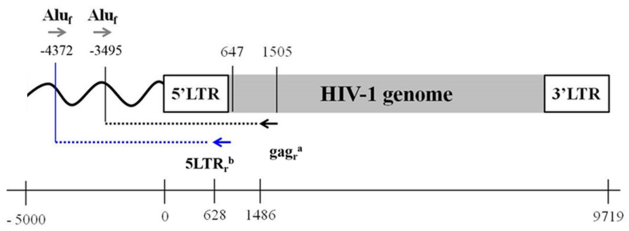
3.1. Design an Improved HIV-1 Integration Assay (Alu-5LTR)

In order to improve the previously used Alu-gag standard-free assay, we applied an alternative primer combination to the first PCR (Figure 1) using primers that were previously validated in other approaches employed to measure integrated HIV-1 DNA [61].
In particular, we replaced the HIV-primer (which binds the gag region in the Alu-gag assay) with a well-known validated primer that is located at the border between the LTR and splice donor 1 of the HIV-1 sequence, closer to the 5′-end of the HIV-1 genome (647 bp) [83] (Figure 1). Using a Polymerase that amplifies up to 5 kb, we then allowed for amplification of the integrated HIV-1 DNA closer to human Alu sequence, thus improving the number of integration events that could be detected. The choice of the alternative HIV primer was made by cross-validation of the primer sequence using a combined in silico and in vivo evaluation using different HIV-1 DNA primers [61] that allowed for reliable amplification of different HIV-1 subtypes. We established that the efficiency of the second PCR was similar between the validated standard-free Alu-gag and the novel proposed assay, as well as being like other validated assays and with little room for improvement. However, in order to avoid overlaps with the primers in the PCR1 in the LTR region, we decided to use the primers described in Avettand-Fènoël 2009 [83]. Meanwhile, we decided to use the same probe of the Alu-gag assay in order to run the same patient sample in the same detection plate to ensure higher consistency.

3.2. Testing of Alu-5LTR in Patients with Undetectable Viral Load Undergoing Suppressive Antiretroviral Therapy

We then tested the two assays in 20 patients undergoing suppressive ART. We obtained quantification of integrated HIV-1 DNA with both methods in 20/20 samples (100%, Figure 2a). Overall median integrated HIV-1 copies per replicate were 5 (range: 1–12) and 14 (range: 5–26) with the Alu-gag and Alu-5LTR assays, respectively. The median ratio between the integrated HIV-1 DNA copies measured with Alu-5LTR vs Alu-gag was 3.5 (range: 1.4–6.3). Most samples (12/20, 60%) had ratios between 2 and 5, with a few being below (3/20, 15%) or above (5/20, 25%) this range (Figure 2b). Additionally, we tested the HIV-1 DNA standard and found that the median ratio between integrated HIV-1 DNA copies measured with the Alu-5LTR vs Alu-gag was 2.8 (Figure 2b).

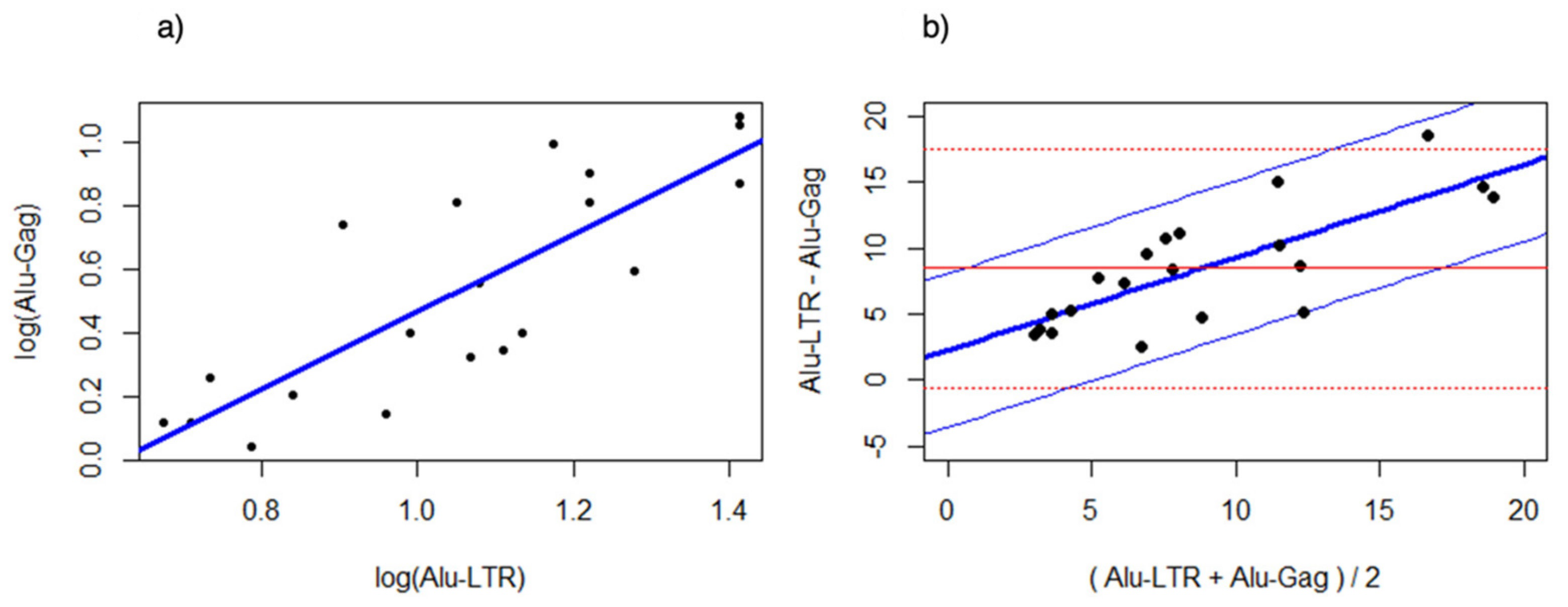
3.3. Bland Altman Analysis of the Alu-5LTR vs. Alu-gag Assays

Linear correlation of log-transformed data for both assays demonstrated a positive correlation (slope: 1.2125, R2 = 0.68, p = 8.55 × 10⁻⁶) (Figure 3a).
Initial assumption testing for the Bland Altman analysis revealed that the variation was constant (p = 0.176). However, the assumption for constant difference between the Alu-5LTR and Alu-gag method was violated. Indeed, a Bland Altman analysis of the nontransformed data showed that the differences between both methods increased when the average estimated value increased (Figure 3b). Hence, the bias between the Alu-5LTR and Alu-gag was proportional and not constant. Regression analysis of the differences over the average values between Alu-5LTR and Alu-gag revealed that the Alu-5LTR quantified, on average, two times more integrated HIV-1 DNA compared to the Alu-gag assay (Figure 3b. Alu-LTR = 3.47 + 2.09 Alu-gag; standard error at 95% confidence ± 4.49; p = 5.48 × 10⁻⁵).

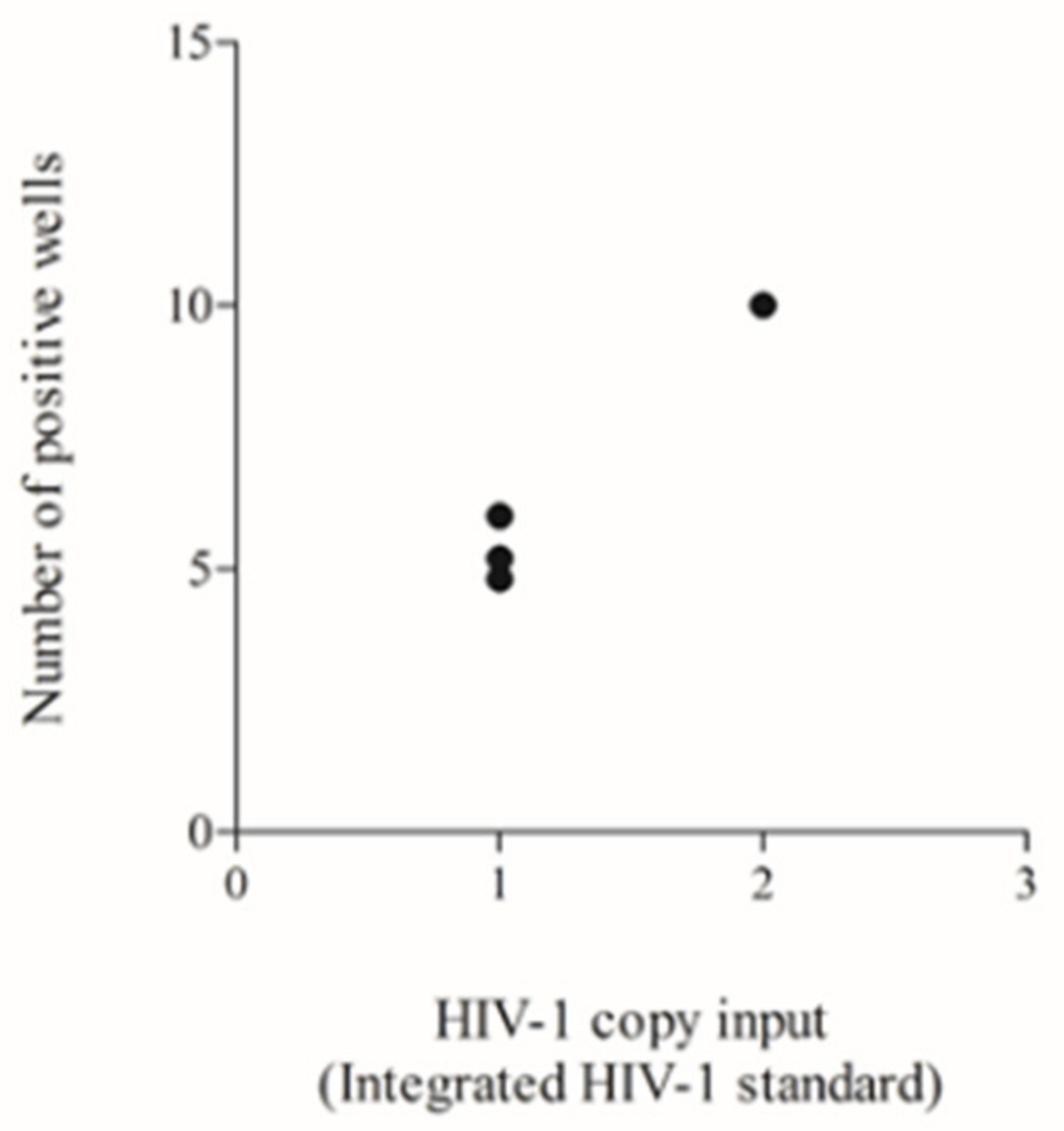
3.4. Testing of a Previously Used HIV-1 DNA Standard to Define the Lower Input Needed to Allow Reliable Poisson Quantification

To define the sensitivity of the assay, we tested different inputs of the ‘integration standard’ and analysed the number of positive wells among the 40 replicates of Alu-gag or Alu-5LTR. As explained in the methods section, four replicates needed to be scored as “positive for integrated HIV-1 DNA” to achieve reliable Poisson quantification. Indeed, calculation of the errors for each measurement with less than four positive replicates would be unreliable, leading to false-positive or negative results, as discussed previously [67]. In this type of experiment, we aimed to identify which HIV-1 DNA input led to as few as four positive wells being classified as positive.
We initially tested two HIV-1 copies diluted in the human genome equivalent to 40,000 cells, which is equal to 0.5 proviruses in 10,000 cells and which confirmed the sensitivity of the Alu-gag, as previously demonstrated [65]. This input produced 10 positive replicates, which is above the threshold of the four positive reactions required for reliable quantification [67]. Therefore, we reduced the input down to one copy in 40,000 cells, which is equivalent to 0.25 proviruses in 10,000 cells, and repeated the test in three independent runs. In these additional tests, we obtained five and six positive reactions in 2/3 (67%) and 1/3 (33%) experiments, respectively (Figure 4). This number of positive wells was still above the threshold of four positive reactions [67], and thus, this result demonstrated that the Alu-LTR assay can reliably detect fewer HIV-1 DNA input copies compared to the Alu-gag assay. We did not perform further testing with inputs lower than 0.25 proviruses in 10,000 cells, because we wanted to avoid obtaining unreliable results below the validated threshold (i.e., four positive wells).

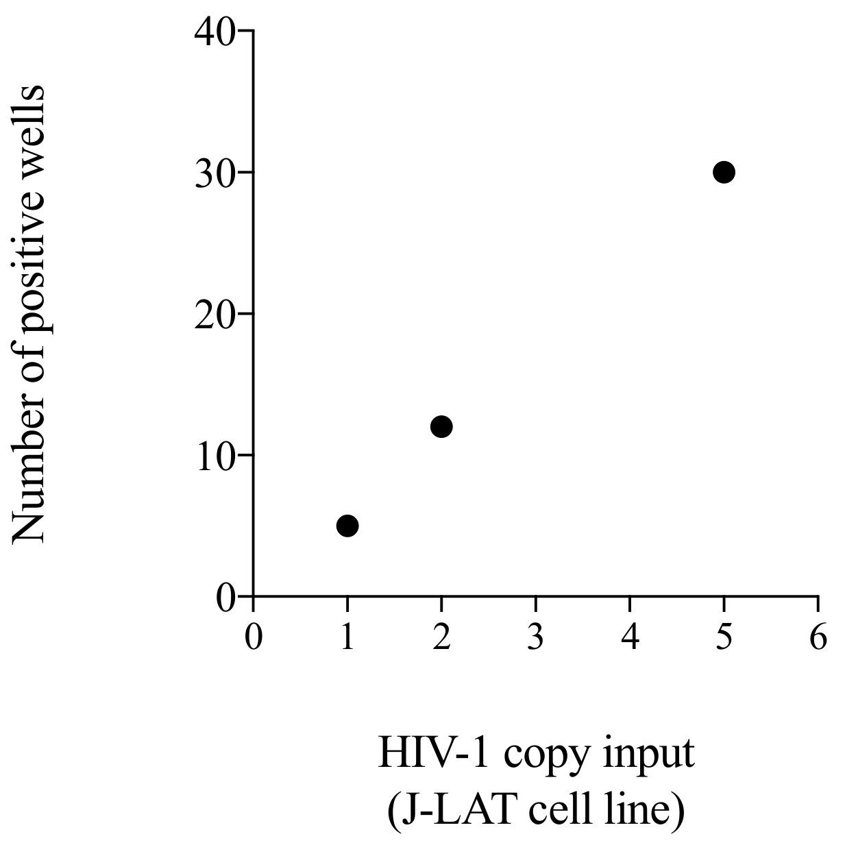
3.5. Testing of a Cellular Standard to Be Used for the Initial Setting of the Alu-5LTR Assay: Towards Cross-Laboratory Harmonisation

To enable interlaboratory validation of the assay, we generated a calibrator based on a combination of J-LAT cell lines that would allow us to compare assay sensitivity in different laboratory settings. We tested three different HIV-1 DNA inputs: the lowest, the highest and one midpoint sampling. By comparing the number of wells scoring positive for integrated HIV-1 DNA copies, we determined that the number of positive wells and HIV-1 DNA input correlated well through linear regression analysis (Figure 5, R square = 0.99). As expected, we found that the number of positive wells increased with elevated HIV-1 DNA input: one, two and five HIV-1 copy inputs produced 5, 12, and 30 positive wells, respectively.
Number of positive wells obtained by the testing of one, two or five copies of J-LAT standard with the Alu-5LTR assay. Each black dot represents one single run. Correlation analysis.

4. Discussion

Measuring integrated HIV-1 proviral DNA is essential for the study of reservoir dynamics in eradication studies. However, the qPCR quantification assays currently available are challenging, and any improvements would aid the field in better monitoring the beneficial effects of strategies aimed at HIV-1 cure. Here, we outline the difficulties posed in measuring HIV-1 integration within cells and in monitoring the LR. Further, we present improvements to a PCR-based integrated HIV-1 DNA assay with increased sensitivity to proviral DNA in PBMC. We utilised primer sets binding to a highly conserved region of the HIV-1 genome, closer to the 5′ integration site, in order to improve the efficiency of PCR amplification. Our results suggested that this method was more sensitive in quantifying integrated HIV-1 DNA in patients undergoing effective ART.
Over time, different approaches have been used to quantify integrated HIV-1 DNA levels [65] via qPCR. Of all the methods, the Alu-HIV-1 PCR assay demonstrates a good compromise between accuracy and clinical application [89]. One of these assays is the Alu-gag, which we improved to be standard-free and to achieve a 10% threshold of detecting integration events [67]. We developed this assay further to improve the sensitivity of the assay changing the HIV-1 primer, binding toward the end of the viral genome. By using the same approach used in the previously published Alu-gag assay, we would expect to produce fragments of a similar length but which were more likely to contain both the Alu and the HIV-1 sequences, ensuring detection of the integrated HIV-1 DNA. Our results on both patient testing and standards confirmed our assumption. In the majority of tested patients, our Alu-5LTR assay improved the quantification of integrated HIV-1 DNA by two to five-fold compared to the Alu-gag method. The results on the ‘integration standard’ with the two assays indicated that Alu-5LTR provided a two-fold increase in sensitivity. This improved sensitivity also provides a lower limit of detection with the Alu-5LTR assay, i.e., 0.25 proviruses in 10,000 genomes, which is two times lower compared to the previously described Alu-gag assay.
Our data indicated that the Alu-5LTR assay showed increased efficiency of detection in some patients, but not in others. This difference does not seem to be associated with HIV-1 subtype, considering that the majority of patients were infected with subtype B. A Bland Altman analysis revealed that the increased difference between the two assays was correlated linearly with a higher average level of detected integrated HIV-1 DNA. The reason for these differences should be discussed in the context of recent works describing the composition, characterisation and heterogeneity of the HIV-1 genomes in different patients. It has been demonstrated that the majority of HIV-1 proviral reservoir sequences harbour deletions within the HIV-1 genome, including Gag [25,42,90,91]. Thus, it may be possible that LTR-based assays lead to more accurate quantification because they overcome the hurdle of primer mismatch caused by deletion in Gag. Nonetheless, our 5-LTR assay showed superiority in quantification also in the “integrated standard”, which can be presumed to contain intact gag regions. Another aspect that should be discussed is the clonal expansion of HIV-infected cells, as demonstrated by Maldarelli [92]. This clonal expansion may lead to nonrandomly distributed integration sites, and may bias integrated HIV-1 quantification in some patients with high clonality of infected cells. We could speculate that some cellular populations carry conserved integration sites with specific distribution with regard to the distance from an Alu sequence. In this case, if the whole cell population has integration sites that are very close to an Alu sequence, using the Alu-gag or the Alu-5LTR should not make a substantial difference on the outcome. In contrast, if the HIV-1 is integrated at a considerable distance from an Alu site, the use of the Alu-5LTR could represent an improvement. However, since we do not have data on the HIV-1 integration sites of the presently studied patients, we cannot prove this assumption. The debate on the lack of heterogeneity of the integration sites also affects the interpretation of the correction factor used in the calculation of the integrated HIV-1 DNA in cells [65,67,89]. In fact, if it is correct that some patients have HIV-1 integration sites that may facilitate or inhibit the detection of the integrated HIV-1 DNA, it cannot be assumed that the assay performs with the same level of sensitivity for each single patient. Hence, we would not encourage the use of integrated HIV-1 data for absolute quantification to discriminate individual patients, but rather, recommend the use this assay to investigate longitudinal changes within patients or relative changes between larger patient cohorts.
One previous study explored the possibility of targeting the LTR region in PCR1 [63]; this assay has been used in other works [52,93]. The author described a two-step method for the quantification of integrated HIV-1 DNA, where PCR1 was run with a primer binding to one region of the HIV-1 LTR very close to the one we targeted [63]. Of note, in that study, the detection limit was 1.2 proviruses in 10,000 cells. In another study, Vandergeeten et al. [52] demonstrated a quantification of integrated HIV-1 DNA as low as 10 copies/106 cells in 1/31 patients (3.2%); however, the quantification was performed utilising purified CD4 T-cells, which are a demonstrated source of the virus reservoir, and hence, carry many more copies of HIV-1 DNA. Our approach adds to previous methods by demonstrating improved sensitivity in PBMC and not purified CD4-T cells when using an amplification PCR setting that includes one single primer binding the HIV-1 LTR sequence and one single primer binding the Alu sequence. Furthermore, we used an innovative quantitative method that avoids the use of a standard due to the use of the Poisson distribution statistics [67], rendering the assay more applicable on a larger scale. However, it must be noted that for a fair and direct comparison with Vandergeeten’s method, the Alu-5LTR assay should be performed with purified CD4 T cells, or alternatively, Vandergeeten’s method could be used to test the same PBMC samples reported in this manuscript. Unfortunately, neither were accessible due to a lack of samples.
Despite the standard-free setting of this assay, we acknowledge the fundamental importance of testing an integration standard for a reliable methodology setup in different laboratories. With this in mind, we designed an integrated HIV-1 DNA standard obtained by mixing different clones of the J-LAT cells. This cell line was selected because it harboured numerous integrated HIV-1 DNA copies that did not change over cell passaging [86]. Furthermore, each clone carries HIV provirus in distinct genome locations, expecting different HIV-Alu distances, thus serving as an ideal standard representing the random integration observed in patients. Such a standard is easy to generate and can be used to calibrate the validation of this assay. Nonetheless, we acknowledge that this is only the first step towards the setting up of these novel methods, and that further validations are needed.
This study presents some limitations. First, even if we used widely validated primers known to detect different subtypes [61], primer fidelity is a hurdle when quantifying a highly variant virus such as HIV-1 [65,83]. This should be considered when studying subjects infected with various HIV subtypes. Further, this study did now allow for comparison with QVOA because of sample unavailability. The results obtained using this in vitro assay will provide valuable information, even though it is important to highlight that these methods need to be validated with clinical outcomes such as time to viral rebound in treatment interruption studies, given that QVOA is an imperfect measure.

5. Conclusions

Here, we have demonstrated that our primer combination improved the quantification of integrated HIV-1 DNA copies as compared to any previously used methodology using peripheral blood. Additionally, our method has the advantage of avoiding the CD4 T-cell purification step. Furthermore, achieving improved sensitivity lowers the level of patient material that is needed for this assay to be conducted, with consequent benefits for settings with limited sample availability. We applied the Poisson principles for quantification, which simplified the procedure by avoiding having to use a standard that needs to be included in each separate run. Undoubtedly, future modifications will be made to the many assays quantifying the variant virus life-cycle stages and which will be utilised in assaying the effectiveness of strategies aimed at restricting, or indeed, eradicating HIV-1.

Supplementary Materials

The following are available online at https://www.mdpi.com/article/10.3390/life11121410/s1; Table S1. Primer sequences (5′-3′) used in the integrated HIV-1 DNA assays, Table S2. Patients’ information, Table S3. Patients’ information.

Author Contributions

Conceptualisation, W.D.S. and A.R.; Data curation, J.T. and A.R.; Formal analysis, E.M., W.D.S. and A.R.; Funding acquisition, W.A.P., L.V. and A.R.; Investigation, E.M., J.T., S.R. and A.R.; Supervision, A.M.G., G.P., W.A.P. and L.V.; Writing–original draft, E.M., J.T. and A.R.; Writing–review & editing, E.M., J.T., W.D.S., S.R., A.M.G., G.P., W.A.P., L.V. and A.R. All authors have read and agreed to the published version of the manuscript.

Funding

The research leading to these results has received funding from the European Community’s Seventh Framework Programme under grant agreement nr. HEALTH-F3-2012-305578 (PathCo) and the H2020 program under grant agreement nr. 681032 (EHVA). A. Ruggiero received an EFIS-Immunology Letters (IL) Short-Term Fellowship to support her staying at the University of Ghent. E. Malatinkova received Special Research Fund (BOF) by Ghent University (BOF, 01P06816). L. Vandekerckhove is funded by the Research Foundation Flanders (FWO, 1.8.020.09.N.00). S. Rutsaert received a strategic basic research fund of the Research Foundation – Flanders (FWO, 1S32916N). The funding sources had no role in the writing of the manuscript or the decision to submit for publication. The corresponding authors had full access to all the data in the study and had the final responsibility for the decision to submit for publication.

Institutional Review Board Statement

These studies were approved either by UK Central Ethics Committee or by Ethics Committee of Ghent University Hospital (Reference numbers: B670201317826, B670201525241).

Informed Consent Statement

All participants signed informed consent statement.

Data Availability Statement

All data available are contained in this manuscript.

Acknowledgments

We are grateful to patients and hospital staff for their collaboration in the recruitment. We thank Una O’Doherty (Center for Aids Research at the University of Pennsylvania) for the integration standard. The following reagents were obtained through the NIH AIDS Reagent Program, Division of AIDS, NIAID, NIH: J-Lat Full Length Clone (#6, #8.4, #9.2, #10.6, #15.4) from Eric Verdin.

Conflicts of Interest

A.M.G. is currently employed as Expert Scientist at Roche Pharma Research and Early Discovery (pRED); Roche had no involvement in the work. Other authors have no conflict of interest to declare.

References

  1. Palmer, S.; Maldarelli, F.; Wiegand, A.; Bernstein, B.; Hanna, G.J.; Brun, S.C.; Kempf, D.J.; Mellors, J.W.; Coffin, J.M.; King, M.S. Low-level viremia persists for at least 7 years in patients on suppressive antiretroviral therapy. Proc. Natl. Acad. Sci. USA 2008, 105, 3879–3884. [Google Scholar] [CrossRef] [PubMed] [Green Version]
  2. Yukl, S.A.; Shergill, A.K.; Ho, T.; Killian, M.; Girling, V.; Epling, L.; Li, P.; Wong, L.K.; Crouch, P.; Deeks, S.G.; et al. The distribution of HIV DNA and RNA in cell subsets differs in gut and blood of HIV-positive patients on ART: Implications for viral persistence. J. Infect. Dis. 2013, 208, 1212–1220. [Google Scholar] [CrossRef] [PubMed] [Green Version]
  3. Sturdevant, C.B.; Joseph, S.B.; Schnell, G.; Price, R.W.; Swanstrom, R.; Spudich, S. Compartmentalized replication of R5 T cell-tropic HIV-1 in the central nervous system early in the course of infection. PLoS Pathog. 2015, 11, e1004720. [Google Scholar] [CrossRef] [PubMed] [Green Version]
  4. Alexaki, A.; Liu, Y.; Wigdahl, B. Cellular reservoirs of HIV-1 and their role in viral persistence. Curr. HIV Res. 2008, 6, 388–400. [Google Scholar] [CrossRef] [Green Version]
  5. Lee, E.; von Stockenstrom, S.; Morcilla, V.; Odevall, L.; Hiener, B.; Shao, W.; Hartogensis, W.; Bacchetti, P.; Milush, J.; Liegler, T.; et al. Impact of Antiretroviral Therapy Duration on HIV-1 Infection of T Cells within Anatomic Sites. J. Virol. 2020, 94, e01270-19. [Google Scholar] [CrossRef] [Green Version]
  6. Thomas, J.; Ruggiero, A.; Paxton, W.A.; Pollakis, G. Measuring the Success of HIV-1 Cure Strategies. Front. Cell. Infect. Microbiol. 2020, 10, 134. [Google Scholar] [CrossRef]
  7. Banga, R.; Procopio, F.A.; Noto, A.; Pollakis, G.; Cavassini, M.; Ohmiti, K.; Corpataux, J.M.; de Leval, L.; Pantaleo, G.; Perreau, M. PD-1(+) and follicular helper T cells are responsible for persistent HIV-1 transcription in treated aviremic individuals. Nat. Med. 2016, 22, 754–761. [Google Scholar] [CrossRef]
  8. Banga, R.; Procopio, F.A.; Ruggiero, A.; Noto, A.; Ohmiti, K.; Cavassini, M.; Corpataux, J.M.; Paxton, W.A.; Pollakis, G.; Perreau, M. Blood CXCR3(+) CD4 T Cells Are Enriched in Inducible Replication Competent HIV in Aviremic Antiretroviral Therapy-Treated Individuals. Front. Immunol. 2018, 9, 144. [Google Scholar] [CrossRef]
  9. Banga, R.; Rebecchini, C.; Procopio, F.A.; Noto, A.; Munoz, O.; Ioannidou, K.; Fenwick, C.; Ohmiti, K.; Cavassini, M.; Corpataux, J.M.; et al. Lymph node migratory dendritic cells modulate HIV-1 transcription through PD-1 engagement. PLoS Pathog. 2019, 15, e1007918. [Google Scholar] [CrossRef] [Green Version]
  10. Buzon, M.J.; Martin-Gayo, E.; Pereyra, F.; Ouyang, Z.; Sun, H.; Li, J.Z.; Piovoso, M.; Shaw, A.; Dalmau, J.; Zangger, N.; et al. Long-term antiretroviral treatment initiated at primary HIV-1 infection affects the size, composition, and decay kinetics of the reservoir of HIV-1-infected CD4 T cells. J. Virol. 2014, 88, 10056–10065. [Google Scholar] [CrossRef] [Green Version]
  11. Buzon, M.J.; Sun, H.; Li, C.; Shaw, A.; Seiss, K.; Ouyang, Z.; Martin-Gayo, E.; Leng, J.; Henrich, T.J.; Li, J.Z.; et al. HIV-1 persistence in CD4+ T cells with stem cell-like properties. Nat. Med. 2014, 20, 139–142. [Google Scholar] [CrossRef]
  12. Tran, T.A.; de Goer de Herve, M.G.; Hendel-Chavez, H.; Dembele, B.; Le Nevot, E.; Abbed, K.; Pallier, C.; Goujard, C.; Gasnault, J.; Delfraissy, J.F.; et al. Resting regulatory CD4 T cells: A site of HIV persistence in patients on long-term effective antiretroviral therapy. PloS ONE 2008, 3, e3305. [Google Scholar] [CrossRef]
  13. Alvarez, Y.; Tuen, M.; Shen, G.; Nawaz, F.; Arthos, J.; Wolff, M.J.; Poles, M.A.; Hioe, C.E. Preferential HIV infection of CCR6+ Th17 cells is associated with higher levels of virus receptor expression and lack of CCR5 ligands. J. Virol. 2013, 87, 10843–10854. [Google Scholar] [CrossRef] [Green Version]
  14. Christensen-Quick, A.; Lafferty, M.; Sun, L.; Marchionni, L.; DeVico, A.; Garzino-Demo, A. Human Th17 Cells Lack HIV-Inhibitory RNases and Are Highly Permissive to Productive HIV Infection. J. Virol. 2016, 90, 7833–7847. [Google Scholar] [CrossRef] [Green Version]
  15. Sun, H.; Kim, D.; Li, X.; Kiselinova, M.; Ouyang, Z.; Vandekerckhove, L.; Shang, H.; Rosenberg, E.S.; Yu, X.G.; Lichterfeld, M. Th1/17 Polarization of CD4 T Cells Supports HIV-1 Persistence during Antiretroviral Therapy. J. Virol. 2015, 89, 11284–11293. [Google Scholar] [CrossRef] [Green Version]
  16. Caruso, M.P.; Falivene, J.; Holgado, M.P.; Zurita, D.H.; Laufer, N.; Castro, C.; Nico, A.; Maeto, C.; Salido, J.; Perez, H.; et al. Impact of HIV-ART on the restoration of Th17 and Treg cells in blood and female genital mucosa. Sci. Rep. 2019, 9, 1978. [Google Scholar] [CrossRef]
  17. Darcis, G.; Kootstra, N.A.; Hooibrink, B.; van Montfort, T.; Maurer, I.; Groen, K.; Jurriaans, S.; Bakker, M.; van Lint, C.; Berkhout, B.; et al. CD32(+)CD4(+) T Cells Are Highly Enriched for HIV DNA and Can Support Transcriptional Latency. Cell Rep. 2020, 30, 2284–2296.E3. [Google Scholar] [CrossRef] [Green Version]
  18. Descours, B.; Petitjean, G.; Lopez-Zaragoza, J.L.; Bruel, T.; Raffel, R.; Psomas, C.; Reynes, J.; Lacabaratz, C.; Levy, Y.; Schwartz, O.; et al. CD32a is a marker of a CD4 T-cell HIV reservoir harbouring replication-competent proviruses. Nature 2017, 543, 564–567. [Google Scholar] [CrossRef]
  19. Abdel-Mohsen, M.; Kuri-Cervantes, L.; Grau-Exposito, J.; Spivak, A.M.; Nell, R.A.; Tomescu, C.; Vadrevu, S.K.; Giron, L.B.; Serra-Peinado, C.; Genesca, M.; et al. CD32 is expressed on cells with transcriptionally active HIV but does not enrich for HIV DNA in resting T cells. Sci. Transl. Med. 2018, 10. [Google Scholar] [CrossRef] [Green Version]
  20. Badia, R.; Ballana, E.; Castellvi, M.; Garcia-Vidal, E.; Pujantell, M.; Clotet, B.; Prado, J.G.; Puig, J.; Martinez, M.A.; Riveira-Munoz, E.; et al. CD32 expression is associated to T-cell activation and is not a marker of the HIV-1 reservoir. Nat. Commun. 2018, 9, 2739. [Google Scholar] [CrossRef] [Green Version]
  21. Bertagnolli, L.N.; White, J.A.; Simonetti, F.R.; Beg, S.A.; Lai, J.; Tomescu, C.; Murray, A.J.; Antar, A.A.R.; Zhang, H.; Margolick, J.B.; et al. The role of CD32 during HIV-1 infection. Nature 2018, 561, E17–E19. [Google Scholar] [CrossRef] [PubMed]
  22. Martin, G.E.; Pace, M.; Thornhill, J.P.; Phetsouphanh, C.; Meyerowitz, J.; Gossez, M.; Brown, H.; Olejniczak, N.; Lwanga, J.; Ramjee, G.; et al. CD32-Expressing CD4 T Cells Are Phenotypically Diverse and Can Contain Proviral HIV DNA. Front. Immunol. 2018, 9, 928. [Google Scholar] [CrossRef] [PubMed] [Green Version]
  23. Sanchez, G.; Xu, X.; Chermann, J.C.; Hirsch, I. Accumulation of defective viral genomes in peripheral blood mononuclear cells of human immunodeficiency virus type 1-infected individuals. J. Virol. 1997, 71, 2233–2240. [Google Scholar] [CrossRef] [PubMed] [Green Version]
  24. Ho, Y.C.; Shan, L.; Hosmane, N.N.; Wang, J.; Laskey, S.B.; Rosenbloom, D.I.; Lai, J.; Blankson, J.N.; Siliciano, J.D.; Siliciano, R.F. Replication-competent noninduced proviruses in the latent reservoir increase barrier to HIV-1 cure. Cell 2013, 155, 540–551. [Google Scholar] [CrossRef] [Green Version]
  25. Bruner, K.M.; Murray, A.J.; Pollack, R.A.; Soliman, M.G.; Laskey, S.B.; Capoferri, A.A.; Lai, J.; Strain, M.C.; Lada, S.M.; Hoh, R.; et al. Defective proviruses rapidly accumulate during acute HIV-1 infection. Nat. Med. 2016, 22, 1043–1049. [Google Scholar] [CrossRef] [Green Version]
  26. Azzoni, L.; Foulkes, A.S.; Papasavvas, E.; Mexas, A.M.; Lynn, K.M.; Mounzer, K.; Tebas, P.; Jacobson, J.M.; Frank, I.; Busch, M.P.; et al. Pegylated Interferon alfa-2a monotherapy results in suppression of HIV type 1 replication and decreased cell-associated HIV DNA integration. J. Infect. Dis. 2013, 207, 213–222. [Google Scholar] [CrossRef] [Green Version]
  27. Chun, T.W.; Davey, R.T., Jr.; Engel, D.; Lane, H.C.; Fauci, A.S. Re-emergence of HIV after stopping therapy. Nature 1999, 401, 874–875. [Google Scholar] [CrossRef]
  28. Davey, R.T., Jr.; Bhat, N.; Yoder, C.; Chun, T.W.; Metcalf, J.A.; Dewar, R.; Natarajan, V.; Lempicki, R.A.; Adelsberger, J.W.; Miller, K.D.; et al. HIV-1 and T cell dynamics after interruption of highly active antiretroviral therapy (HAART) in patients with a history of sustained viral suppression. Proc. Natl. Acad. Sci. USA 1999, 96, 15109–15114. [Google Scholar] [CrossRef] [Green Version]
  29. Li, J.Z.; Etemad, B.; Ahmed, H.; Aga, E.; Bosch, R.J.; Mellors, J.W.; Kuritzkes, D.R.; Lederman, M.M.; Para, M.; Gandhi, R.T. The size of the expressed HIV reservoir predicts timing of viral rebound after treatment interruption. Aids 2016, 30, 343–353. [Google Scholar] [CrossRef] [Green Version]
  30. Siliciano, J.D.; Siliciano, R.F. Enhanced culture assay for detection and quantitation of latently infected, resting CD4+ T-cells carrying replication-competent virus in HIV-1-infected individuals. Methods Mol. Biol. 2005, 304, 3–15. [Google Scholar] [CrossRef]
  31. Laird, G.M.; Eisele, E.E.; Rabi, S.A.; Lai, J.; Chioma, S.; Blankson, J.N.; Siliciano, J.D.; Siliciano, R.F. Rapid quantification of the latent reservoir for HIV-1 using a viral outgrowth assay. PLoS Pathog. 2013, 9, e1003398. [Google Scholar] [CrossRef] [Green Version]
  32. Rosenbloom, D.I.; Elliott, O.; Hill, A.L.; Henrich, T.J.; Siliciano, J.M.; Siliciano, R.F. Designing and Interpreting Limiting Dilution Assays: General Principles and Applications to the Latent Reservoir for Human Immunodeficiency Virus-1. Open Forum Infect. Dis. 2015, 2, ofv123. [Google Scholar] [CrossRef] [Green Version]
  33. Fun, A.; Mok, H.P.; Wills, M.R.; Lever, A.M. A highly reproducible quantitative viral outgrowth assay for the measurement of the replication-competent latent HIV-1 reservoir. Sci. Rep. 2017, 7, 43231. [Google Scholar] [CrossRef] [Green Version]
  34. Passaes, C.P.B.; Bruel, T.; Decalf, J.; David, A.; Angin, M.; Monceaux, V.; Muller-Trutwin, M.; Noel, N.; Bourdic, K.; Lambotte, O.; et al. Ultrasensitive HIV-1 p24 Assay Detects Single Infected Cells and Differences in Reservoir Induction by Latency Reversal Agents. J. Virol. 2017, 91, e02296-16. [Google Scholar] [CrossRef] [Green Version]
  35. Massanella, M.; Yek, C.; Lada, S.M.; Nakazawa, M.; Shefa, N.; Huang, K.; Richman, D.D. Improved assays to measure and characterize the inducible HIV reservoir. Ebiomedicine 2018, 36, 113–121. [Google Scholar] [CrossRef] [Green Version]
  36. Sanyal, A.; Mailliard, R.B.; Rinaldo, C.R.; Ratner, D.; Ding, M.; Chen, Y.; Zerbato, J.M.; Giacobbi, N.S.; Venkatachari, N.J.; Patterson, B.K.; et al. Novel assay reveals a large, inducible, replication-competent HIV-1 reservoir in resting CD4(+) T cells. Nat. Med. 2017, 23, 885–889. [Google Scholar] [CrossRef]
  37. Stone, M.; Rosenbloom, D.; Bacchetti, P.; Deng, X.; Dimapasoc, M.; Keating, S.; Bakkour, S.; Richman, D.; Mellors, J.; Deeks, S.; et al. Assessing suitability of next-generation viral outgrowth assays as proxies for classic QVOA to measure HIV-1 latent reservoir size. J. Infect. Dis. 2020, 224, 1209–1218. [Google Scholar] [CrossRef] [Green Version]
  38. Rosenbloom, D.I.S.; Bacchetti, P.; Stone, M.; Deng, X.; Bosch, R.J.; Richman, D.D.; Siliciano, J.D.; Mellors, J.W.; Deeks, S.G.; Ptak, R.G.; et al. Assessing intra-lab precision and inter-lab repeatability of outgrowth assays of HIV-1 latent reservoir size. PLoS Comput. Biol. 2019, 15, e1006849. [Google Scholar] [CrossRef] [Green Version]
  39. Procopio, F.A.; Fromentin, R.; Kulpa, D.A.; Brehm, J.H.; Bebin, A.G.; Strain, M.C.; Richman, D.D.; O’Doherty, U.; Palmer, S.; Hecht, F.M.; et al. A Novel Assay to Measure the Magnitude of the Inducible Viral Reservoir in HIV-infected Individuals. Ebiomedicine 2015, 2, 874–883. [Google Scholar] [CrossRef] [Green Version]
  40. Frank, I.; Acharya, A.; Routhu, N.K.; Aravantinou, M.; Harper, J.L.; Maldonado, S.; Sole Cigoli, M.; Semova, S.; Mazel, S.; Paiardini, M.; et al. A Tat/Rev Induced Limiting Dilution Assay to Measure Viral Reservoirs in Non-Human Primate Models of HIV Infection. Sci. Rep. 2019, 9, 12078. [Google Scholar] [CrossRef] [Green Version]
  41. Bertoldi, A.; D’Urbano, V.; Bon, I.; Verbon, A.; Rokx, C.; Boucher, C.; van Kampen, J.J.A.; Gruters, R.A.; Gallinella, G.; Calza, L.; et al. Development of C-TILDA: A modified TILDA method for reservoir quantification in long term treated patients infected with subtype C HIV-1. J. Virol. Methods 2020, 276, 113778. [Google Scholar] [CrossRef] [PubMed]
  42. Hiener, B.; Horsburgh, B.A.; Eden, J.S.; Barton, K.; Schlub, T.E.; Lee, E.; von Stockenstrom, S.; Odevall, L.; Milush, J.M.; Liegler, T.; et al. Identification of Genetically Intact HIV-1 Proviruses in Specific CD4(+) T Cells from Effectively Treated Participants. Cell Rep. 2017, 21, 813–822. [Google Scholar] [CrossRef] [PubMed] [Green Version]
  43. Einkauf, K.B.; Lee, G.Q.; Gao, C.; Sharaf, R.; Sun, X.; Hua, S.; Chen, S.M.; Jiang, C.; Lian, X.; Chowdhury, F.Z.; et al. Intact HIV-1 proviruses accumulate at distinct chromosomal positions during prolonged antiretroviral therapy. J. Clin. Investig. 2019, 129, 988–998. [Google Scholar] [CrossRef] [PubMed] [Green Version]
  44. Kuo, H.H.; Banga, R.; Lee, G.Q.; Gao, C.; Cavassini, M.; Corpataux, J.M.; Blackmer, J.E.; Zur Wiesch, J.S.; Yu, X.G.; Pantaleo, G.; et al. Blood and lymph node dissemination of clonal genome-intact HIV-1 DNA sequences during suppressive antiretroviral therapy. J. Infect. Dis. 2020, 222, 655–660. [Google Scholar] [CrossRef]
  45. Pinzone, M.R.; Bertuccio, M.P.; VanBelzen, D.J.; Zurakowski, R.; O’Doherty, U. Next-Generation Sequencing in a Direct Model of HIV Infection Reveals Important Parallels to and Differences from In Vivo Reservoir Dynamics. J. Virol. 2020, 94, e01900-19. [Google Scholar] [CrossRef]
  46. Lambrechts, L.; Cole, B.; Rutsaert, S.; Trypsteen, W.; Vandekerckhove, L. Emerging PCR-Based Techniques to Study HIV-1 Reservoir Persistence. Viruses 2020, 12, 149. [Google Scholar] [CrossRef] [Green Version]
  47. Gao, H.; Hawkins, T.; Jasti, A.; Chen, Y.H.; Mockaitis, K.; Dinauer, M.; Cornetta, K. Development and Evaluation of Quality Metrics for Bioinformatics Analysis of Viral Insertion Site Data Generated Using High Throughput Sequencing. Biomedicines 2014, 2, 195–210. [Google Scholar] [CrossRef] [Green Version]
  48. Wells, D.W.; Guo, S.; Shao, W.; Bale, M.J.; Coffin, J.M.; Hughes, S.H.; Wu, X. An analytical pipeline for identifying and mapping the integration sites of HIV and other retroviruses. BMC Genom. 2020, 21, 216. [Google Scholar] [CrossRef] [Green Version]
  49. Artesi, M.; Hahaut, V.; Cole, B.; Lambrechts, L.; Ashrafi, F.; Marcais, A.; Hermine, O.; Griebel, P.; Arsic, N.; van der Meer, F.; et al. PCIP-seq: Simultaneous sequencing of integrated viral genomes and their insertion sites with long reads. Genome Biol. 2021, 22, 97. [Google Scholar] [CrossRef]
  50. Schmidt, M.; Hoffmann, G.; Wissler, M.; Lemke, N.; Mussig, A.; Glimm, H.; Williams, D.A.; Ragg, S.; Hesemann, C.U.; von Kalle, C. Detection and direct genomic sequencing of multiple rare unknown flanking DNA in highly complex samples. Hum. Gene Ther. 2001, 12, 743–749. [Google Scholar] [CrossRef]
  51. Wu, C.; Jares, A.; Winkler, T.; Xie, J.; Metais, J.Y.; Dunbar, C.E. High efficiency restriction enzyme-free linear amplification-mediated polymerase chain reaction approach for tracking lentiviral integration sites does not abrogate retrieval bias. Hum. Gene Ther. 2013, 24, 38–47. [Google Scholar] [CrossRef]
  52. Vandergeeten, C.; Fromentin, R.; Merlini, E.; Lawani, M.B.; DaFonseca, S.; Bakeman, W.; McNulty, A.; Ramgopal, M.; Michael, N.; Kim, J.H.; et al. Cross-clade ultrasensitive PCR-based assays to measure HIV persistence in large-cohort studies. J. Virol. 2014, 88, 12385–12396. [Google Scholar] [CrossRef] [Green Version]
  53. Chun, T.W.; Carruth, L.; Finzi, D.; Shen, X.; DiGiuseppe, J.A.; Taylor, H.; Hermankova, M.; Chadwick, K.; Margolick, J.; Quinn, T.C.; et al. Quantification of latent tissue reservoirs and total body viral load in HIV-1 infection. Nature 1997, 387, 183–188. [Google Scholar] [CrossRef]
  54. Gabriel, R.; Eckenberg, R.; Paruzynski, A.; Bartholomae, C.C.; Nowrouzi, A.; Arens, A.; Howe, S.J.; Recchia, A.; Cattoglio, C.; Wang, W.; et al. Comprehensive genomic access to vector integration in clinical gene therapy. Nat. Med. 2009, 15, 1431–1436. [Google Scholar] [CrossRef]
  55. Paruzynski, A.; Arens, A.; Gabriel, R.; Bartholomae, C.C.; Scholz, S.; Wang, W.; Wolf, S.; Glimm, H.; Schmidt, M.; von Kalle, C. Genome-wide high-throughput integrome analyses by nrLAM-PCR and next-generation sequencing. Nat. Protocol 2010, 5, 1379–1395. [Google Scholar] [CrossRef]
  56. Patro, S.C.; Brandt, L.D.; Bale, M.J.; Halvas, E.K.; Joseph, K.W.; Shao, W.; Wu, X.; Guo, S.; Murrell, B.; Wiegand, A.; et al. Combined HIV-1 sequence and integration site analysis informs viral dynamics and allows reconstruction of replicating viral ancestors. Proc. Natl. Acad. Sci. USA 2019, 116, 25891–25899. [Google Scholar] [CrossRef]
  57. Lee, G.Q.; Bangsberg, D.R.; Mo, T.; Lachowski, C.; Brumme, C.J.; Zhang, W.; Lima, V.D.; Boum, Y.; Mwebesa, B.B.; Muzoora, C.; et al. Prevalence and clinical impacts of HIV-1 intersubtype recombinants in Uganda revealed by near-full-genome population and deep sequencing approaches. AIDS 2017, 31, 2345–2354. [Google Scholar] [CrossRef]
  58. Wagner, T.A.; McLaughlin, S.; Garg, K.; Cheung, C.Y.; Larsen, B.B.; Styrchak, S.; Huang, H.C.; Edlefsen, P.T.; Mullins, J.I.; Frenkel, L.M. HIV latency. Proliferation of cells with HIV integrated into cancer genes contributes to persistent infection. Science 2014, 345, 570–573. [Google Scholar] [CrossRef] [Green Version]
  59. Serrao, E.; Cherepanov, P.; Engelman, A.N. Amplification, Next-generation Sequencing, and Genomic DNA Mapping of Retroviral Integration Sites. J. Vis. Exp. 2016, 53840. [Google Scholar] [CrossRef] [Green Version]
  60. Rouzioux, C.; Avettand-Fenoel, V. Total HIV DNA: A global marker of HIV persistence. Retrovirology 2018, 15, 30. [Google Scholar] [CrossRef] [Green Version]
  61. Rutsaert, S.; De Spiegelaere, W.; Van Hecke, C.; De Scheerder, M.A.; Kiselinova, M.; Vervisch, K.; Trypsteen, W.; Vandekerckhove, L. In-depth validation of total HIV-1 DNA assays for quantification of various HIV-1 subtypes. Sci. Rep. 2018, 8, 17274. [Google Scholar] [CrossRef]
  62. Thomas, J.; Ruggiero, A.; Procopio, F.A.; Pantaleo, G.; Paxton, W.A.; Pollakis, G. Comparative analysis and generation of a robust HIV-1 DNA quantification assay. J. Virol. Methods 2018, 263, 24–31. [Google Scholar] [CrossRef]
  63. Brussel, A.; Delelis, O.; Sonigo, P. Alu-LTR real-time nested PCR assay for quantifying integrated HIV-1 DNA. Methods Mol. Biol. 2005, 304, 139–154. [Google Scholar] [CrossRef]
  64. Agosto, L.M.; Yu, J.J.; Dai, J.; Kaletsky, R.; Monie, D.; O’Doherty, U. HIV-1 integrates into resting CD4+ T cells even at low inoculums as demonstrated with an improved assay for HIV-1 integration. Virology 2007, 368, 60–72. [Google Scholar] [CrossRef] [Green Version]
  65. Liszewski, M.K.; Yu, J.J.; O’Doherty, U. Detecting HIV-1 integration by repetitive-sampling Alu-gag PCR. Methods 2009, 47, 254–260. [Google Scholar] [CrossRef] [Green Version]
  66. Brady, T.; Kelly, B.J.; Male, F.; Roth, S.; Bailey, A.; Malani, N.; Gijsbers, R.; O’Doherty, U.; Bushman, F.D. Quantitation of HIV DNA integration: Effects of differential integration site distributions on Alu-PCR assays. J. Virol. Methods 2013, 189, 53–57. [Google Scholar] [CrossRef] [Green Version]
  67. De Spiegelaere, W.; Malatinkova, E.; Lynch, L.; Van Nieuwerburgh, F.; Messiaen, P.; O’Doherty, U.; Vandekerckhove, L. Quantification of Integrated HIV DNA by Repetitive-Sampling Alu-HIV PCR on the Basis of Poisson Statistics. Clin. Chem. 2014, 60, 886–895. [Google Scholar] [CrossRef] [Green Version]
  68. Eriksson, S.; Graf, E.H.; Dahl, V.; Strain, M.C.; Yukl, S.A.; Lysenko, E.S.; Bosch, R.J.; Lai, J.; Chioma, S.; Emad, F.; et al. Comparative analysis of measures of viral reservoirs in HIV-1 eradication studies. PLoS Pathog. 2013, 9, e1003174. [Google Scholar] [CrossRef]
  69. Kiselinova, M.; De Spiegelaere, W.; Buzon, M.J.; Malatinkova, E.; Lichterfeld, M.; Vandekerckhove, L. Integrated and Total HIV-1 DNA Predict Ex Vivo Viral Outgrowth. PLoS Pathog. 2016, 12, e1005472. [Google Scholar] [CrossRef] [Green Version]
  70. O’Doherty, U.; Swiggard, W.J.; Jeyakumar, D.; McGain, D.; Malim, M.H. A sensitive, quantitative assay for human immunodeficiency virus type 1 integration. J. Virol. 2002, 76, 10942–10950. [Google Scholar] [CrossRef] [Green Version]
  71. Yu, J.J.; Wu, T.L.; Liszewski, M.K.; Dai, J.; Swiggard, W.J.; Baytop, C.; Frank, I.; Levine, B.L.; Yang, W.; Theodosopoulos, T.; et al. A more precise HIV integration assay designed to detect small differences finds lower levels of integrated DNA in HAART treated patients. Virology 2008, 379, 78–86. [Google Scholar] [CrossRef] [PubMed] [Green Version]
  72. Mexas, A.M.; Graf, E.H.; Pace, M.J.; Yu, J.J.; Papasavvas, E.; Azzoni, L.; Busch, M.P.; Di Mascio, M.; Foulkes, A.S.; Migueles, S.A.; et al. Concurrent measures of total and integrated HIV DNA monitor reservoirs and ongoing replication in eradication trials. Aids 2012, 26, 2295–2306. [Google Scholar] [CrossRef] [PubMed]
  73. Malatinkova, E.; De Spiegelaere, W.; Bonczkowski, P.; Kiselinova, M.; Vervisch, K.; Trypsteen, W.; Johnson, M.; Verhofstede, C.; de Looze, D.; Murray, C.; et al. Impact of a decade of successful antiretroviral therapy initiated at HIV-1 seroconversion on blood and rectal reservoirs. eLife 2015, 4, e09115. [Google Scholar] [CrossRef] [PubMed] [Green Version]
  74. Ruggiero, A.; De Spiegelaere, W.; Cozzi-Lepri, A.; Kiselinova, M.; Pollakis, G.; Beloukas, A.; Vandekerckhove, L.; Strain, M.; Richman, D.; Phillips, A.; et al. During Stably Suppressive Antiretroviral Therapy Integrated HIV-1 DNA Load in Peripheral Blood is Associated with the Frequency of CD8 Cells Expressing HLA-DR/DP/DQ. EBioMedicine 2015, 2, 1153–1159. [Google Scholar] [CrossRef] [Green Version]
  75. Ganor, Y.; Real, F.; Sennepin, A.; Dutertre, C.A.; Prevedel, L.; Xu, L.; Tudor, D.; Charmeteau, B.; Couedel-Courteille, A.; Marion, S.; et al. HIV-1 reservoirs in urethral macrophages of patients under suppressive antiretroviral therapy. Nat. Microbiol. 2019, 4, 633–644. [Google Scholar] [CrossRef]
  76. Tremeaux, P.; Lenfant, T.; Boufassa, F.; Essat, A.; Melard, A.; Gousset, M.; Delelis, O.; Viard, J.P.; Bary, M.; Goujard, C.; et al. Increasing contribution of integrated forms to total HIV DNA in blood during HIV disease progression from primary infection. Ebiomedicine 2019, 41, 455–464. [Google Scholar] [CrossRef] [Green Version]
  77. Rasmussen, T.A.; Tolstrup, M.; Brinkmann, C.R.; Olesen, R.; Erikstrup, C.; Solomon, A.; Winckelmann, A.; Palmer, S.; Dinarello, C.; Buzon, M.; et al. Panobinostat, a histone deacetylase inhibitor, for latent-virus reactivation in HIV-infected patients on suppressive antiretroviral therapy: A phase 1/2, single group, clinical trial. Lancet HIV 2014, 1, e13–e21. [Google Scholar] [CrossRef]
  78. Jacobson, J.M.; Routy, J.P.; Welles, S.; DeBenedette, M.; Tcherepanova, I.; Angel, J.B.; Asmuth, D.M.; Stein, D.K.; Baril, J.G.; McKellar, M.; et al. Dendritic Cell Immunotherapy for HIV-1 Infection Using Autologous HIV-1 RNA: A Randomized, Double-Blind, Placebo-Controlled Clinical Trial. J. Acquir. Immune Defic. Syndr. 2016, 72, 31–38. [Google Scholar] [CrossRef] [Green Version]
  79. Tapia, G.; Hojen, J.F.; Okvist, M.; Olesen, R.; Leth, S.; Nissen, S.K.; VanBelzen, D.J.; O’Doherty, U.; Mork, A.; Krogsgaard, K.; et al. Sequential Vacc-4x and romidepsin during combination antiretroviral therapy (cART): Immune responses to Vacc-4x regions on p24 and changes in HIV reservoirs. J. Infect. 2017, 75, 555–571. [Google Scholar] [CrossRef]
  80. Vibholm, L.; Schleimann, M.H.; Hojen, J.F.; Benfield, T.; Offersen, R.; Rasmussen, K.; Olesen, R.; Dige, A.; Agnholt, J.; Grau, J.; et al. Short-Course Toll-Like Receptor 9 Agonist Treatment Impacts Innate Immunity and Plasma Viremia in Individuals With Human Immunodeficiency Virus Infection. Clin. Infect. Dis. 2017, 64, 1686–1695. [Google Scholar] [CrossRef]
  81. Moron-Lopez, S.; Navarro, J.; Jimenez, M.; Rutsaert, S.; Urrea, V.; Puertas, M.C.; Torrella, A.; De Clercq, L.; Ribas, B.P.; Galvez, C.; et al. Switching From a Protease Inhibitor-based Regimen to a Dolutegravir-based Regimen: A Randomized Clinical Trial to Determine the Effect on Peripheral Blood and Ileum Biopsies From Antiretroviral Therapy-suppressed Human Immunodeficiency Virus-infected Individuals. Clin. Infect. Dis. 2019, 69, 1320–1328. [Google Scholar] [CrossRef]
  82. Pinzone, M.R.; O’Doherty, U. Measuring integrated HIV DNA ex vivo and in vitro provides insights about how reservoirs are formed and maintained. Retrovirology 2018, 15, 22. [Google Scholar] [CrossRef] [Green Version]
  83. Avettand-Fenoel, V.; Chaix, M.L.; Blanche, S.; Burgard, M.; Floch, C.; Toure, K.; Allemon, M.C.; Warszawski, J.; Rouzioux, C.; The French Pediatric Cohort Study ANRS—CO 01 Group. LTR real-time PCR for HIV-1 DNA quantitation in blood cells for early diagnosis in infants born to seropositive mothers treated in HAART area (ANRS CO 01). J. Med. Virol. 2009, 81, 217–223. [Google Scholar] [CrossRef]
  84. Avettand-Fènoël, V.; Hocqueloux, L.; Ghosn, J.; Cheret, A.; Frange, P.; Melard, A.; Viard, J.P.; Rouzioux, C. Total HIV-1 DNA, a Marker of Viral Reservoir Dynamics with Clinical Implications. Clin. Microbiol. Rev. 2016, 29, 859–880. [Google Scholar] [CrossRef] [Green Version]
  85. Ruggiero, A.; Cozzi-Lepri, A.; Beloukas, A.; Richman, D.; Khoo, S.; Phillips, A.; Geretti, A.M.; Group, E.S. Factors Associated with Persistence of Plasma HIV-1 RNA during Long-term Continuously Suppressive Firstline Antiretroviral Therapy. Open Forum Infect. Dis. 2018, 5, ofy032. [Google Scholar] [CrossRef] [Green Version]
  86. Symons, J.; Chopra, A.; Malatinkova, E.; De Spiegelaere, W.; Leary, S.; Cooper, D.; Abana, C.O.; Rhodes, A.; Rezaei, S.D.; Vandekerckhove, L.; et al. HIV integration sites in latently infected cell lines: Evidence of ongoing replication. Retrovirology 2017, 14, 2. [Google Scholar] [CrossRef] [Green Version]
  87. Van der Sluis, R.M.; van Montfort, T.; Centlivre, M.; Schopman, N.C.; Cornelissen, M.; Sanders, R.W.; Berkhout, B.; Jeeninga, R.E.; Paxton, W.A.; Pollakis, G. Quantitation of HIV-1 DNA with a sensitive TaqMan assay that has broad subtype specificity. J. Virol. Methods 2013, 187, 94–102. [Google Scholar] [CrossRef]
  88. Carstensen, B. Comparing methods of measurement: Extending the LoA by regression. Stat. Med. 2010, 29, 401–410. [Google Scholar] [CrossRef]
  89. Ruggiero, A.; Malatinkova, E.; Rutsaert, S.; Paxton, W.A.; Vandekerckhove, L.; Spiegelaere, W.D. Utility of integrated HIV-1 DNA quantification in cure studies. Future Virol. 2017, 12, 215–225. [Google Scholar] [CrossRef]
  90. Rainwater-Lovett, K.; Ziemniak, C.; Watson, D.; Luzuriaga, K.; Siberry, G.; Petru, A.; Chen, Y.; Uprety, P.; McManus, M.; Ho, Y.C.; et al. Paucity of Intact Non-Induced Provirus with Early, Long-Term Antiretroviral Therapy of Perinatal HIV Infection. PloS ONE 2017, 12, e0170548. [Google Scholar] [CrossRef]
  91. Pollack, R.A.; Jones, R.B.; Pertea, M.; Bruner, K.M.; Martin, A.R.; Thomas, A.S.; Capoferri, A.A.; Beg, S.A.; Huang, S.H.; Karandish, S.; et al. Defective HIV-1 Proviruses Are Expressed and Can Be Recognized by Cytotoxic T Lymphocytes, which Shape the Proviral Landscape. Cell Host Microbe 2017, 21, 494–506. [Google Scholar] [CrossRef] [Green Version]
  92. Maldarelli, F.; Wu, X.; Su, L.; Simonetti, F.R.; Shao, W.; Hill, S.; Spindler, J.; Ferris, A.L.; Mellors, J.W.; Kearney, M.F.; et al. HIV latency. Specific HIV integration sites are linked to clonal expansion and persistence of infected cells. Science 2014, 345, 179–183. [Google Scholar] [CrossRef] [Green Version]
  93. Murray, J.M.; Zaunders, J.J.; McBride, K.L.; Xu, Y.; Bailey, M.; Suzuki, K.; Cooper, D.A.; Emery, S.; Kelleher, A.D.; Koelsch, K.K.; et al. HIV DNA subspecies persist in both activated and resting memory CD4+ T cells during antiretroviral therapy. J. Virol. 2014, 88, 3516–3526. [Google Scholar] [CrossRef] [Green Version]
Figure 1. Primer combinations for the PCR1 step in the Alu-gag and Alu-5LTR assays. Figure depicting the different primers used in the Alu-gag and in the Alu-5LTR assay for the PCR1 step. a gag reverse primer that binds HIV-1 genome at 1505–1486 bp; b 5LTR reverse primer that binds HIV-1 genome at 647–628 bp. The black and the blue dashed lines represent 5 kb long fragments that are the longest that can be amplified by the polymerase used in these assays. With the Alu-gag assay integration events that are up to 3495 bp away from the human Alu sequence can be amplified, whereas with the Alu-5LTR assay integration events that are up to 4372 bp from an Alu sequence can be amplified. LTR = long terminal repeats, f = forward, r = reverse.
Life 11 01410 g001
Figure 2. Alu-LTR assay showed higher sensitivity in measuring integrated HIV-1 DNA as compared to Alu-gag assay. (a) integrated HIV-1 DNA copies per replicate measured with Alu-gag (black-empty dots) and Alu-5LTR (black-empty diamonds) assays. (b) representation of ratios between measurements obtained with Alu-5LTR and Alu-gag assays. Each black-empty dot or triangle represents one patient, the black filled dot or triangles represents the integrated standard. The patients and integrated standard are divided into 3 groups based on the ratios (<2, 2–5 and >5), as shown on x-axis.
Life 11 01410 g002
Figure 3. (a) Linear regression analysis of the log transformed data from Alu-gag and Alu-5LTR assays. The trend-line is marked in blue. (b) Bland Altman plot of the nontransformed data, showing the difference between both methods (thick blue line) and the 95% confidence intervals (thin blue lines) when constant difference would be assumed. The red lines indicate the differences and the 95% confidence interval in case the difference between both methods would be constant.
Life 11 01410 g003
Figure 4. Testing of a previously used HIV-1 DNA standard to define the lower HIV-1 DNA input for reliable Poisson quantification. Number of positive wells obtained by testing one and two copies of integrated HIV-1 standard. Each black dot represents one single run.
Life 11 01410 g004
Figure 5. Testing of a standard based on J-LAT cells.
Life 11 01410 g005
Publisher’s Note: MDPI stays neutral with regard to jurisdictional claims in published maps and institutional affiliations.

