
Chronic, Mild Vestibulopathy Leads to Deficits in Spatial Tasks that Rely on Vestibular Input While Leaving Other Cognitive Functions and Brain Volumes Intact


 and
and 
Abstract
:1. Introduction
2. Materials and Methods
2.1. Ethical Approval
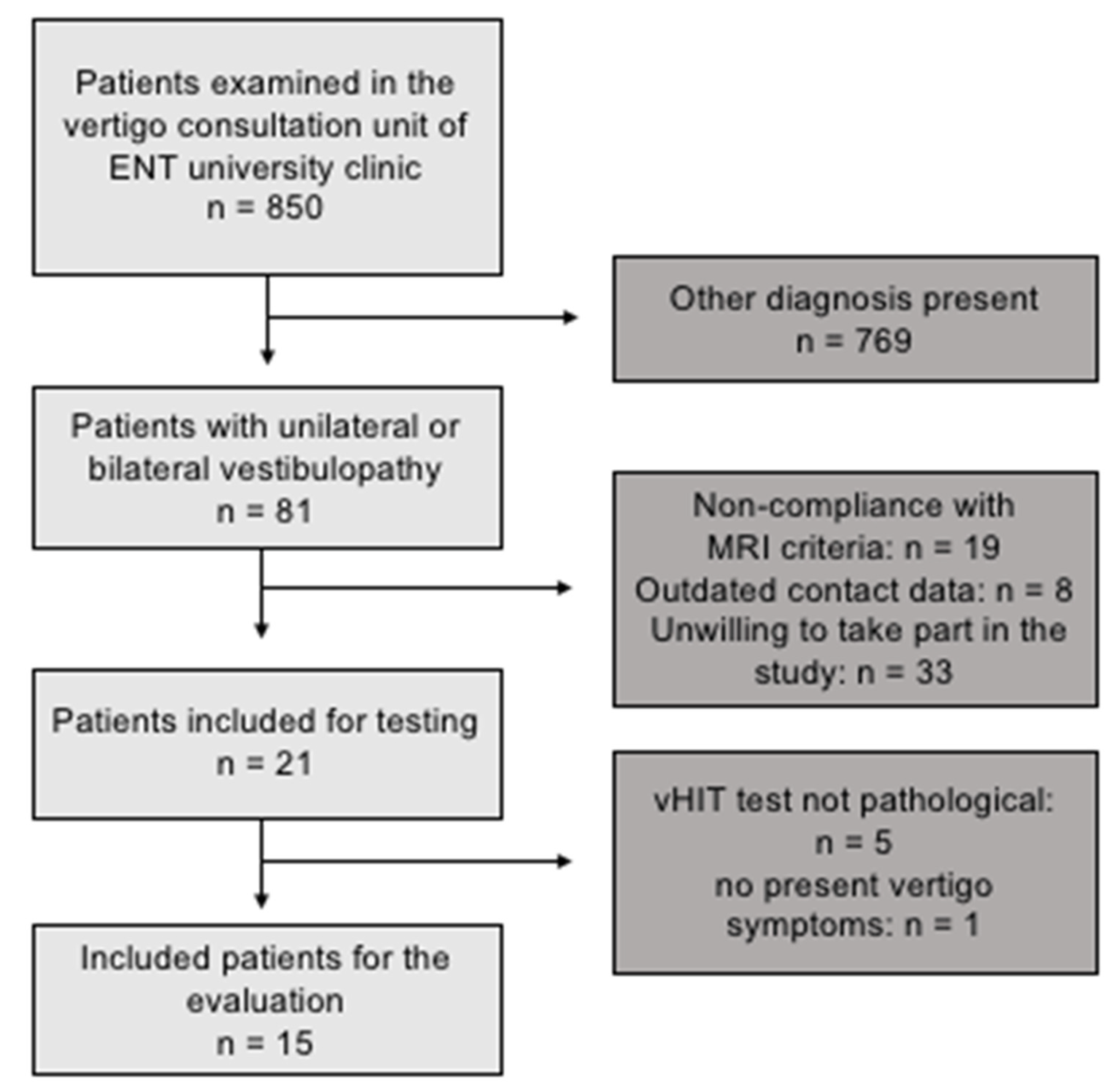
2.2. Participants
2.3. Vestibular Non-Cognitive Tasks
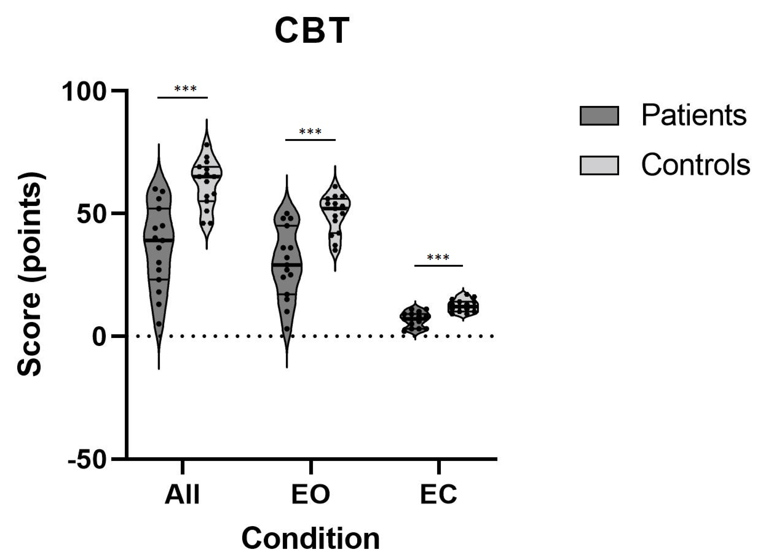
2.3.1. Clinical Balance Test (CBT)
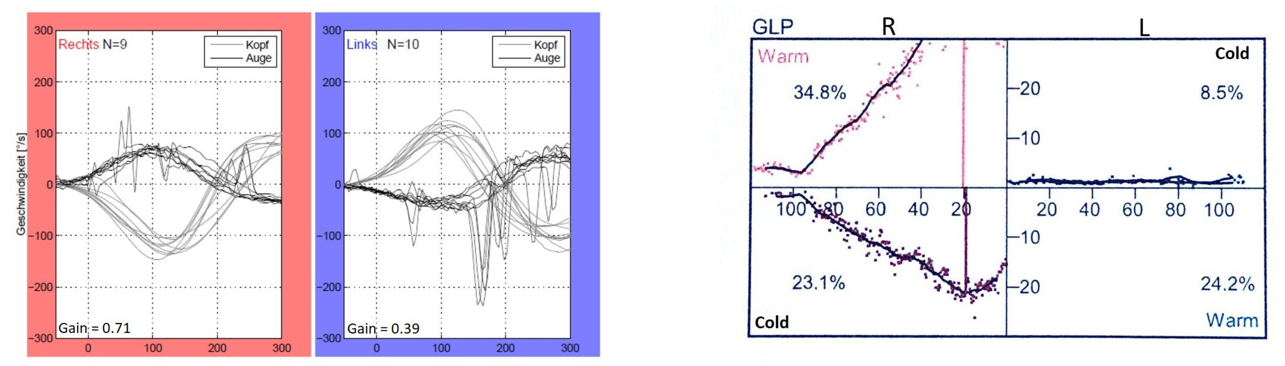
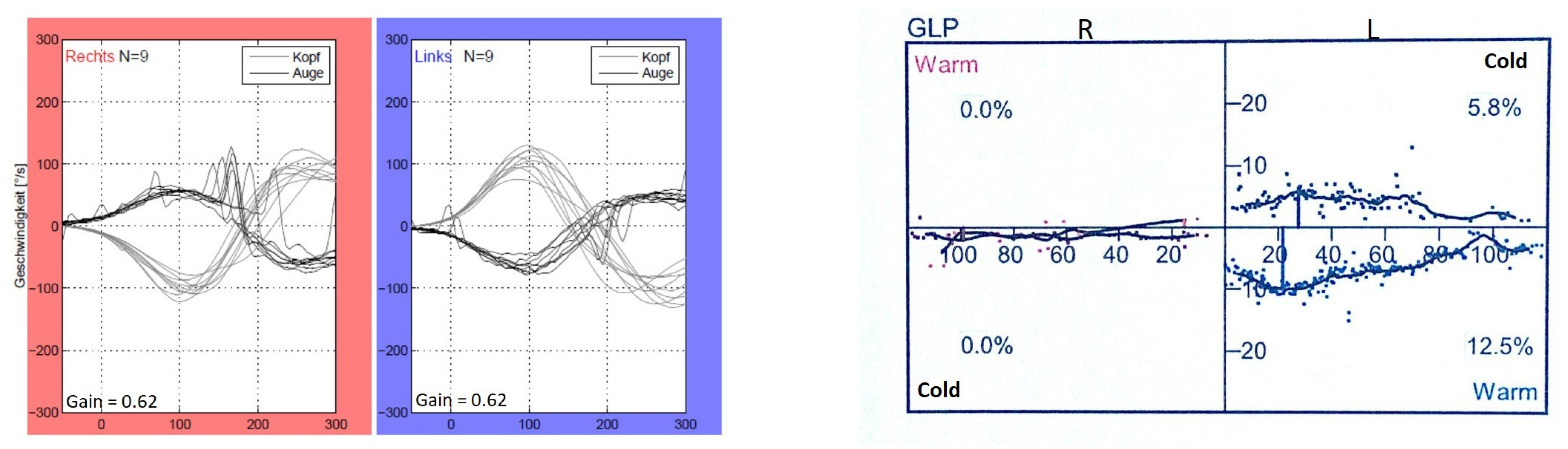
2.3.2. vHIT
2.3.3. Vestibular Caloric Stimulation
2.4. Vestibular Cognitive Tasks
2.4.1. Triangle Completion Test (TCT)
2.4.2. Rotational Memory (RM)
2.5. Visual (Non-Vestibular) Cognitive Tasks
BIS-4—Visuo-Constructive and Spatial Abilities
2.6. General Cognitive Task
d2-R—Attention and Concentration Abilities
2.7. Structural Brain Analyses (VBM)
2.8. Statistical Analysis
3. Results
3.1. Vestibular Non-Cognitive Task (CBT)
3.2. Vestibular Cognitive Tasks (TCT and RM)
3.3. Visual (Non-Vestibular) Cognitive Tasks (BIS-4)
3.4. General Cognitive Task (D2-R)
3.5. Whole-Brain Analysis
Region of Interest (ROI) Analyses
4. Discussion
Author Contributions
Funding
Institutional Review Board Statement
Informed Consent Statement
Data Availability Statement
Conflicts of Interest
References
- Yang, T.; Xirasagar, S.; Cheng, Y.; Wu, C.; Kuo, N.; Lin, H. Peripheral Vestibular Disorders: Nationwide Evidence From Taiwan. Laryngoscope 2020, 131, 639–643. [Google Scholar] [CrossRef] [PubMed]
- Zingler, V.C.; Weintz, E.; Jahn, K.; Huppert, D.; Cnyrim, C.; Brandt, T.; Strupp, M. Causative factors, epidemiology, and follow-up of bilateral vestibulopathy. Ann. N. Y. Acad. Sci. 2009, 1164, 505–508. [Google Scholar] [CrossRef] [PubMed]
- Strupp, M.; Kim, J.-S.; Murofushi, T.; Straumann, D.; Jen, J.C.; Rosengren, S.M.; Della Santina, C.C.; Kingma, H. Bilateral vestibulopathy: Diagnostic criteria consensus document of the classification committee of the barany society. J. Vestib. Res. 2017, 27, 177–189. [Google Scholar] [CrossRef] [Green Version]
- Guinand, N.; Boselie, F.; Guyot, J.-P.; Kingma, H. Quality of life of patients with bilateral vestibulopathy. Ann. Otol. Rhinol. Laryngol. 2012, 121, 471–477. [Google Scholar] [CrossRef] [PubMed]
- Moser, M.-B.; Rowland, D.C.; Moser, E.I. Place cells, grid cells, and memory. Cold Spring Harb. Perspect. Biol. 2015, 7, a021808. [Google Scholar] [CrossRef] [PubMed] [Green Version]
- Moser, E.I.; Kropff, E.; Moser, M.-B. Place Cells, Grid Cells, and the Brain’s Spatial Representation System. Annu. Rev. Neurosci. 2008, 31, 69–89. [Google Scholar] [CrossRef] [Green Version]
- Kaski, D.; Quadir, S.; Nigmatullina, Y.; Malhotra, P.; Bronstein, A.M.; Seemungal, B.M. Temporoparietal encoding of space and time during vestibular-guided orientation. Brain 2016, 139, 392–403. [Google Scholar] [CrossRef] [Green Version]
- Hitier, M.; Besnard, S.; Smith, P.F. Vestibular pathways involved in cognition. Front. Integr. Neurosci. 2014, 8, 59. [Google Scholar] [CrossRef]
- Stackman, R.W.; Clark, A.S.; Taube, J.S. Hippocampal spatial representations require vestibular input. Hippocampus. 2002, 12, 291–303. [Google Scholar] [CrossRef] [Green Version]
- Russell, N.A.; Horii, A.; Smith, P.F.; Darlington, C.L.; Bilkey, D.K. Lesions of the vestibular system disrupt hippocampal theta rhythm in the rat. J. Neurophysiol. 2006, 96, 4–14. [Google Scholar] [CrossRef] [PubMed] [Green Version]
- Péruch, P.; Lopez, C.; Redon-Zouiteni, C.; Escoffier, G.; Zeitoun, A.; Sanjuan, M.; Devèze, A.; Magnan, J.; Borel, L. Vestibular information is necessary for maintaining metric properties of representational space: Evidence from mental imagery. Neuropsychologia 2011, 49, 3136–3144. [Google Scholar] [CrossRef] [PubMed]
- Schautzer, F.; Hamilton, D.; Kalla, R.; Strupp, M.; Brandt, T. Spatial memory deficits in patients with chronic bilateral vestibular failure. Ann. N. Y. Acad. Sci. 2003, 1004, 316–324. [Google Scholar] [CrossRef] [PubMed]
- Hüfner, K.; Hamilton, D.A.; Kalla, R.; Stephan, T.; Glasauer, S.; Ma, J.; Brüning, R.; Markowitsch, H.J.; Labudda, K.; Schichor, C.; et al. Spatial memory and hippocampal volume in humans with unilateral vestibular deafferentation. Hippocampus 2007, 17, 471–485. [Google Scholar] [CrossRef]
- Dordevic, M.; Taubert, M.; Müller, P.; Kaufmann, J.; Hökelmann, A.; Müller, N.G. Brain Gray Matter Volume Is Modulated by Visual Input and Overall Learning Success but Not by Time Spent on Learning a Complex Balancing Task. J. Clin. Med. 2018, 8, 9. [Google Scholar] [CrossRef] [PubMed] [Green Version]
- Dordevic, M.; Hökelmann, A.; Müller, P.; Rehfeld, K.; Müller, N.G. Improvements in Orientation and Balancing Abilities in Response to One Month of Intensive Slackline-Training. A Randomized Controlled Feasibility Study. Front. Hum. Neurosci. 2017, 11, 55. [Google Scholar] [CrossRef] [PubMed] [Green Version]
- Dordevic, M.; Taubert, M.; Müller, P.; Riemer, M.; Kaufmann, J.; Hökelmann, A.; Müller, N.G. Which Effects on Neuroanatomy and Path-Integration Survive? Results of a Randomized Controlled Study on Intensive Balance Training. Brain Sci. 2020, 10, 210. [Google Scholar] [CrossRef] [Green Version]
- Dobbels, B.; Peetermans, O.; Boon, B.; Mertens, G.; Van de Heyning, P.; Van Rompaey, V. Impact of bilateral vestibulopathy on spatial and nonspatial cognition: A systematic review. Ear Hear. 2019, 40, 757–765. [Google Scholar] [CrossRef]
- Hong, S.-K.; Kim, J.H.; Kim, H.-J.; Lee, H.-J. Changes in the gray matter volume during compensation after vestibular neuritis: A longitudinal VBM study. Restor. Neurol. Neurosci. 2014, 32, 663–673. [Google Scholar] [CrossRef] [PubMed]
- Brandt, T.; Schautzer, F.; Hamilton, D.A.; Brüning, R.; Markowitsch, H.J.; Kalla, R.; Darlington, C.; Smith, P.; Strupp, M. Vestibular loss causes hippocampal atrophy and impaired spatial memory in humans. Brain 2005, 128, 2732–2741. [Google Scholar] [CrossRef]
- Göttlich, M.; Jandl, N.M.; Sprenger, A.; Wojak, J.F.; Münte, T.F.; Krämer, U.M.; Helmchen, C. Hippocampal gray matter volume in bilateral vestibular failure. Hum. Brain Mapp. 2016, 37, 1998–2006. [Google Scholar] [CrossRef]
- Kremmyda, O.; Hüfner, K.; Flanagin, V.; Hamilton, D.A.; Linn, J.; Strupp, M.; Jahn, K.; Brandt, T.; Kremmyda, O.; Hüfner, K.; et al. Beyond Dizziness: Virtual Navigation, Spatial Anxiety and Hippocampal Volume in Bilateral Vestibulopathy. Front. Hum. Neurosci. 2016, 10, 139. [Google Scholar] [CrossRef] [PubMed] [Green Version]
- Helmchen, C.; Klinkenstein, J.C.; Krüger, A.; Gliemroth, J.; Mohr, C.; Sander, T. Structural brain changes following peripheral vestibulo-cochlear lesion may indicate multisensory compensation. J. Neurol. Neurosurg. Psychiatry 2011, 82, 309–316. [Google Scholar] [CrossRef] [PubMed] [Green Version]
- Zheng, Y.; Balabhadrapatruni, S.; Baek, J.H.; Chung, P.; Gliddon, C.; Zhang, M.; Darlington, C.L.; Napper, R.; Strupp, M.; Brandt, T.; et al. The effects of bilateral vestibular loss on hippocampal volume, neuronal number, and cell proliferation in rats. Front. Neurol. 2012, 3, 20. [Google Scholar] [CrossRef] [PubMed] [Green Version]
- Popp, P.; Wulff, M.; Finke, K.; Rühl, M.; Brandt, T.; Dieterich, M. Cognitive deficits in patients with a chronic vestibular failure. J. Neurol. 2017, 264, 554–563. [Google Scholar] [CrossRef]
- Zu Eulenburg, P.; Stoeter, P.; Dieterich, M. Voxel-based morphometry depicts central compensation after vestibular neuritis. Ann. Neurol. 2010, 68, 241–249. [Google Scholar] [CrossRef]
- Dordevic, M.; Schrader, R.; Taubert, M.; Müller, P.; Hökelmann, A.; Müller, N.G. Vestibulo-Hippocampal Function Is Enhanced and Brain Structure Altered in Professional Ballet Dancers. Front. Integr. Neurosci. 2018, 12, 50. [Google Scholar] [CrossRef]
- Beckmann, J.F.; Guthke, J.; Jäger, A.O.; Süß, H.-M.; Beauducel, A. Berliner Intelligenzstruktur-Test (BIS), Form 4. Diagnostica 1999, 45, 56–61. [Google Scholar] [CrossRef]
- Brickenkamp, R.; Cubero, N.S. Test. de Atención D2; TEA Ediciones SA: Barcelona, Spain, 2009. [Google Scholar]
- Yato, Y.; Hirose, S.; Wallon, P.; Mesmin, C.; Jobert, M. d2-R test for Japanese adolescents: Concurrent validity with the attention deficit-hyperactivity disorder rating scale. Pediatr. Int. 2019, 61, 43–48. [Google Scholar] [CrossRef] [Green Version]
- Agrawal, Y.; Van de Berg, R.; Wuyts, F.; Walther, L.; Magnusson, M.; Oh, E.; Sharpe, M.; Strupp, M. Presbyvestibulopathy: Diagnostic criteria Consensus document of the classification committee of the Bárány Society. J. Vestib. Res. 2019, 29, 161–170. [Google Scholar] [CrossRef] [Green Version]
- Matiño-Soler, E.; Esteller-More, E.; Martin-Sanchez, J.C.; Martinez-Sanchez, J.M.; Perez-Fernandez, N. Normative data on angular vestibulo-ocular responses in the yaw axis measured using the video head impulse test. Otol. Neurotol. 2015, 36, 466–471. [Google Scholar] [CrossRef]
- Bachmann, K.; Sipos, K.; Lavender, V.; Hunter, L.L. Video head impulse testing in a pediatric population: Normative findings. J Am Acad Audiol 2018, 29, 417–426. [Google Scholar] [CrossRef] [PubMed]
- Yang, C.J.; Lee, J.Y.; Kang, B.C.; Lee, H.S.; Yoo, M.H.; Park, H.J. Quantitative analysis of gains and catch-up saccades of video-head-impulse testing by age in normal subjects. Clin. Otolaryngol. 2016, 41, 532–538. [Google Scholar] [CrossRef] [PubMed]
- McGarvie, L.A.; MacDougall, H.G.; Halmagyi, G.M.; Burgess, A.M.; Weber, K.P.; Curthoys, I.S. The video head impulse test (vHIT) of semicircular canal function—age-dependent normative values of VOR gain in healthy subjects. Front. Neurol. 2015, 6, 154. [Google Scholar] [CrossRef] [Green Version]
- Baier, B.; zu Eulenburg, P.; Best, C.; Geber, C.; Müller-Forell, W.; Birklein, F.; Dieterich, M. Posterior insular cortex—A site of vestibular-somatosensory interaction? Brain Behav. 2013, 3, 519–524. [Google Scholar] [CrossRef] [PubMed]
- Müller, N.G.; Riemer, M.; Brandt, L.; Wolbers, T. Erratum to: Repetitive transcranial magnetic stimulation reveals a causal role of the human precuneus in spatial updating. Sci. Rep. 2018, 8, 13720. [Google Scholar] [CrossRef]
- Wolbers, T.; Hegarty, M.; Büchel, C.; Loomis, J.M. Spatial updating: How the brain keeps track of changing object locations during observer motion. Nat. Neurosci. 2008, 11, 1223–1230. [Google Scholar] [CrossRef]
- Baloh, R.W.; Jacobson, K.M.; Beykirch, K.; Honrubia, V. Static and dynamic posturography in patients with vestibular and cerebellar lesions. Arch. Neurol. 1998, 55, 649–654. [Google Scholar] [CrossRef]
- Dordevic, M.; Gruber, J.; Schmitt, F.C.; Mueller, N. Impairments in path integration, rotational memory and balancing in patients with temporal lobe epilepsy. BMJ Neurol. Open 2020, 2, e000077. [Google Scholar] [CrossRef]
- Gill-Body, K.M.; Beninato, M.; Krebs, E.D. Relationship among balance impairments, functional performance, and disability in people with peripheral vestibular hypofunction. Phys. Ther. 2000, 80, 748–758. [Google Scholar] [CrossRef] [Green Version]
- Previc, F.H.; Krueger, W.W.; Ross, R.A.; Roman, M.A.; Siegel, G. The relationship between vestibular function and topographical memory in older adults. Front. Integr. Neurosci. 2014, 8, 46. [Google Scholar] [CrossRef] [PubMed] [Green Version]
- Bigelow, R.T.; Agrawal, Y. Vestibular involvement in cognition: Visuospatial ability, attention, executive function, and memory. J. Vestib. Res. 2015, 25, 73–89. [Google Scholar] [CrossRef] [PubMed]
- Guidetti, G.; Monzani, D.; Trebbi, M.; Rovatti, V. Impaired navigation skills in patients with psychological distress and chronic peripheral vestibular hypofunction without vertigo. Acta Otorhinolaryngol. Ital. 2008, 28, 21–25. [Google Scholar] [PubMed]
- Dobbels, B.; Mertens, G.; Gilles, A.; Claes, A.; Moyaert, J.; Van De Berg, R.; Van De Heyning, P.; Vanderveken, O.; Van Rompaey, V. Cognitive function in acquired bilateral vestibulopathy: A cross-sectional study on cognition, hearing, and vestibular loss. Front. Neurosci. 2019, 13, 340. [Google Scholar] [CrossRef] [PubMed]
- Hüfner, K.; Binetti, C.; Hamilton, D.A.; Stephan, T.; Flanagin, V.L.; Linn, J.; Labudda, K.; Markowitsch, H.; Glasauer, S.; Jahn, K.; et al. Structural and functional plasticity of the hippocampal formation in professional dancers and slackliners. Hippocampus 2011, 21, 855–865. [Google Scholar] [CrossRef]
- Péruch, P.; Borel, L.; Gaunet, F.; Thinus-Blanc, G.; Magnan, J.; Lacour, M. Spatial performance of unilateral vestibular defective patients: In nonvisual versus visual navigation. J. Vestib. Res. 1999, 9, 37–47. [Google Scholar] [CrossRef] [PubMed]
- Dieterich, M.; Bense, S.; Lutz, S.; Drzezga, A.; Stephan, T.; Bartenstein, P.; Brandt, T. Dominance for vestibular cortical function in the non-dominant hemisphere. Cereb. Cortex 2003, 13, 994–1007. [Google Scholar] [CrossRef] [PubMed] [Green Version]
- Becker-Bense, S.; Dieterich, M.; Buchholz, H.-G.; Bartenstein, P.; Schreckenberger, M.; Brandt, T. The differential effects of acute right- vs. left-sided vestibular failure on brain metabolism. Brain Struct. Funct. 2014, 219, 1355–1367. [Google Scholar] [CrossRef]
- Grabherr, L.; Cuffel, C.; Guyot, J.-P.; Mast, F.W. Mental transformation abilities in patients with unilateral and bilateral vestibular loss. Exp. Brain Res. 2011, 209, 205–214. [Google Scholar] [CrossRef] [Green Version]
- Helmchen, C.; Ye, Z.; Sprenger, A.; Münte, T.F. Changes in resting-state fMRI in vestibular neuritis. Brain Struct. Funct. 2014, 219, 1889–1990. [Google Scholar] [CrossRef] [PubMed]

| Patient (P) | Age | Gender | Years of Education | Time since First Symptoms | Symptoms at the Time of Testing | Affected Side | Calorics Right/Left * | VOR Gain Right/Left |
|---|---|---|---|---|---|---|---|---|
| P1 | 58 | F | 13 | 2 years | Daily dizziness, decreased concentration | Bilateral (L > R) | 6.9/1.9 | 0.87/0.67 |
| P2 | 51 | M | 12 | 4 years | Daily dizziness | Bilateral (R > L) | 2.0/5.1 | 0.55/0.7 |
| P3 | 75 | M | 13 | 23 years | Dizziness when eyes closed | Right | 1.8/6.8 | 0.62/1 |
| P4 | 59 | M | 16 | 3 years | Daily dizziness, decreased concentration and memory | Bilateral (L > R) | 8.5/3.1 | 0.69/0.31 |
| P5 | 58 | F | 15 | 11 years | Daily dizziness, decreased concentration and memory | Bilateral (L > R) | 5.0/2.1 | 0.86/0.67 |
| P6 | 50 | M | 12 | 3.5 years | Daily dizziness, decreased concentration and memory | Bilateral | 3.2/1.9 | 0.50/0.63 |
| P7 | 66 | M | 14 | 11 years | Daily dizziness | Left | 14.6/3.2 | 0.89/0.47 |
| P8 | 57 | M | 15 | 2 years | Daily dizziness | Bilateral (L > R) | 4.5/2.0 | 0.60/0.68 |
| P9 | 52 | M | 16 | 4 years | Dizziness when stressed | Bilateral (R > L) | 1.6/8.0 | 0.62/0.62 |
| P10 | 47 | F | 12 | 5 years | Daily dizziness | Bilateral (L > R) | 5.7/1.5 | 0.71/0.64 |
| P11 | 47 | M | 12 | 11 months | Daily dizziness | Left | 29.2/1.7 | 0.71/0.39 |
| P12 | 41 | M | 17 | 9 months | Daily dizziness | Bilateral (L > R) | 5.9/2.0 | 0.6/0.35 |
| P13 | 74 | M | 17 | 2 years | Daily dizziness | Bilateral | No data available | 0.62/0.62 |
| P14 | 55 | M | 12 | 3 years | Daily dizziness, tendency to fall, decreased attention | Bilateral (L > R) | 4.6/2.7 | HIT data not analyzable |
| P15 | 75 | F | 13 | 7 years | Daily dizziness decreased concentration | Bilateral | 1.1/1.5 | HIT data not analyzable |
| Test | Condition(s) | Mean ± SD Controls | Mean ± SD Patients | p-Value | Effect Size (d) |
|---|---|---|---|---|---|
| Clinical Balance Test (CBT) | All Conditions | 62.1 ± 9.6 | 36.5 ± 17.0 | 0.000 *** | 1.86 |
| Open Eyes | 49.7 ± 7.8 | 29.7 ± 14.5 | 0.000 *** | 1.72 | |
| Closed Eyes | 12.4 ± 2.5 | 6.8 ± 3.0 | 0.000 *** | 2.03 |
| Test | Condition(s) | Mean ± SD Controls | Mean ± SD Patients | p-Value | Effect Size (d) | |
|---|---|---|---|---|---|---|
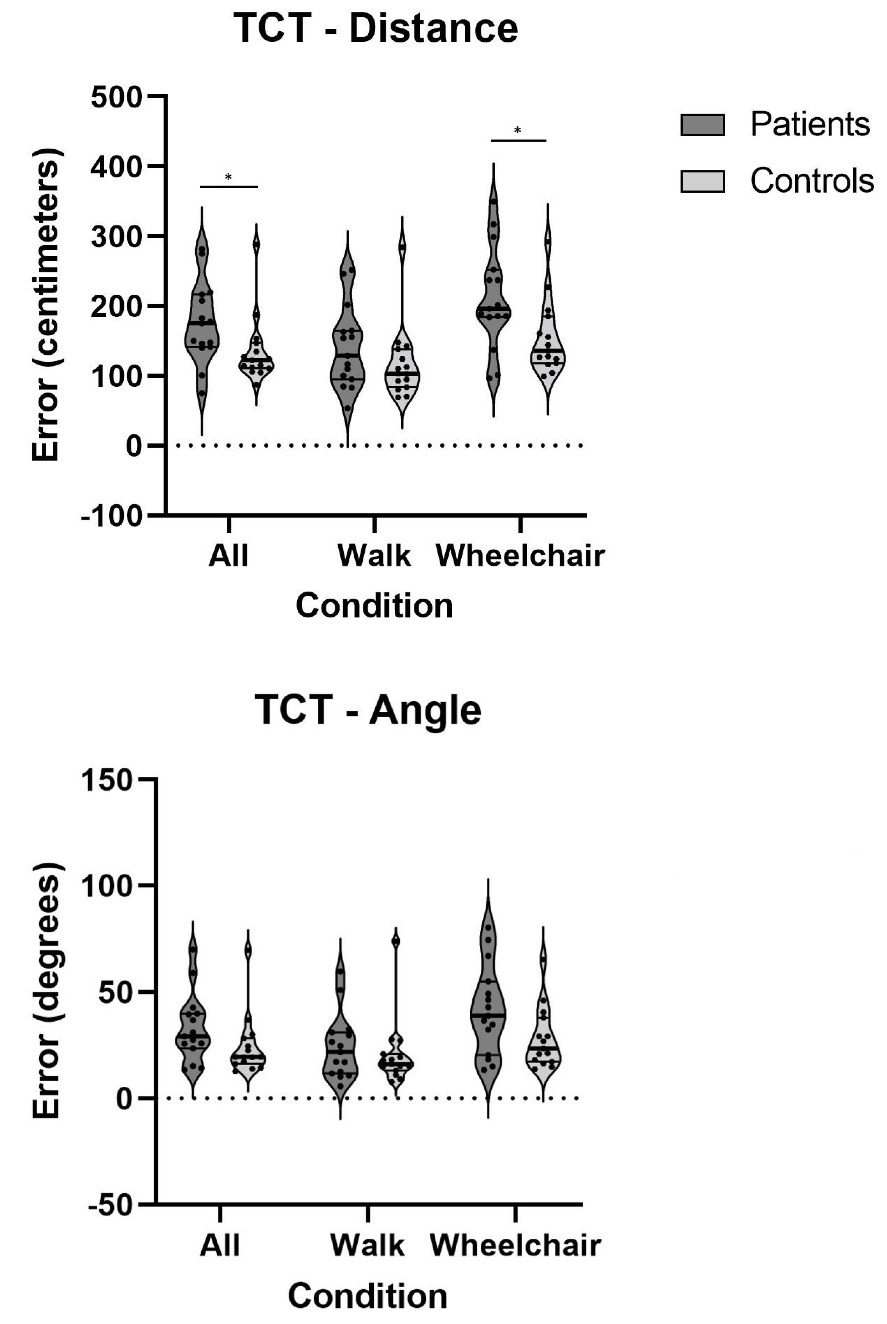
| Triangle Completion Test (TCT) | Angle | All Conditions | 24.3 ± 14.2 | 33.0 ± 15.9 | 0.056 | 0.57 |
| Walk | 20.6 ± 15.8 | 24.2 ± 15.2 | 0.436 | 0.23 | ||
| Wheelchair | 28.1 ± 14.2 | 41.7 ± 21.0 | 0.067 | 0.76 | ||
| Distance | All Conditions | 135.5 ± 48.6 | 175.9 ± 57.5 | 0.023 * | 0.76 | |
| Walk | 117.0 ± 52.5 | 140.6 ± 58.6 | 0.174 | 0.42 | ||
| Wheelchair | 154.0 ± 52.1 | 211.3 ± 73.1 | 0.019 * | 0.90 | ||
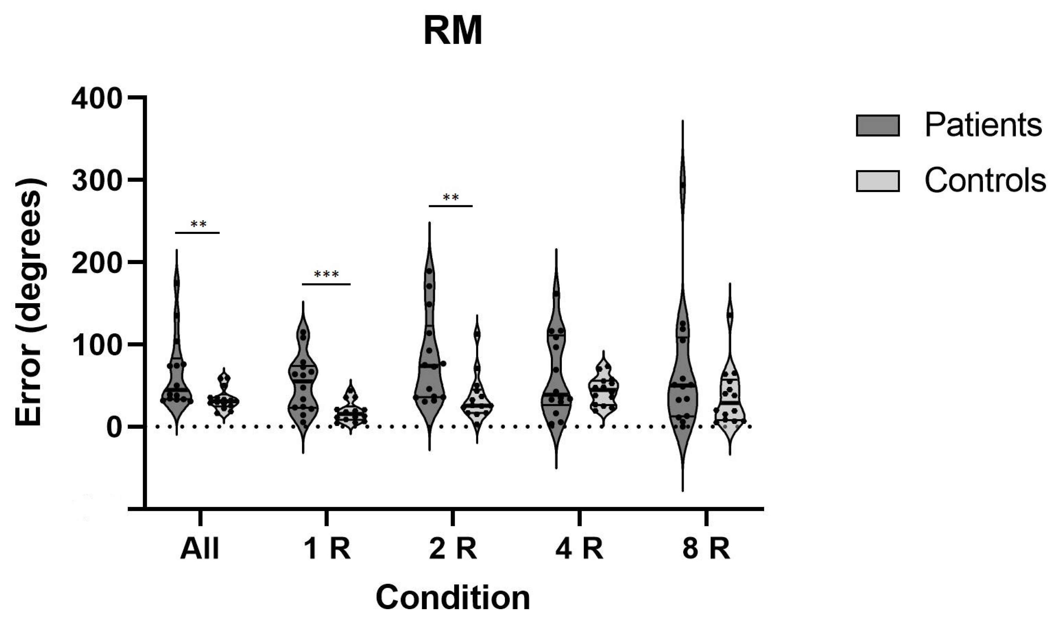
| Rotational Memory (RM) | All Conditions | 33.8 ± 13.4 | 66.4 ± 44.3 | 0.004 ** | 0.99 | |
| One Rotation | 18.3 ± 12.4 | 52.8 ± 34.1 | 0.001 ** | 1.34 | ||
| Two Rotations | 35.6 ±27.9 | 82.7 ± 54.1 | 0.002 ** | 1.10 | ||
| Four Rotations | 43.9 ± 17.0 | 62.2 ± 49.6 | 0.769 | 0.49 | ||
| Eight Rotations | 37.5 ± 35.4 | 67.9 ± 76.7 | 0.401 | 0.51 | ||
| Test | Condition(s) | Mean ± SD Controls | Mean ± SD Patients | p-Value | Effect Size (d) |
|---|---|---|---|---|---|
| BIS-4 | Correct (in %) | 47.1 ± 9.4 | 53.4 ± 13.2 | 0.187 | 0.55 |
| Wrong (quantity) | 3.0 ± 1.8 | 2.1 ± 1.2 | 0.202 | 0.60 | |
| Missed (in %) | 52.4 ± 9.6 | 46.6 ± 13.2 | 0.202 | 0.50 | |
| AN_1 | 18.8 ± 18.2 | 27.7 ± 21.5 | 0.367 | 0.45 | |
| AW_1 | 24.3 ± 17.9 | 27.1 ± 27.9 | 0.902 | 0.12 |
| Test | Condition(s) | Mean ± SD Controls | Mean ± SD Patients | p-Value | Effect Size (d) |
|---|---|---|---|---|---|
| D2-R | E% | 14.3 ± 18.9 | 19.7 ± 15.4 | 0.067 | 0.31 |
| PTO | 130.2 ± 33.2 | 122.1 ± 34.2 | 0.436 | 0.24 | |
| CP | 110.6 ± 47.6 | 101.2 ± 41.1 | 0.486 | 0.26 | |
| EO | 13.9 ± 15.2 | 17.7 ± 13.0 | 0.250 | 0.33 | |
| EC | 1.7 ± 2.9 | 3.5 ± 7.5 | 0.202 | 0.21 |
Publisher’s Note: MDPI stays neutral with regard to jurisdictional claims in published maps and institutional affiliations. |
© 2021 by the authors. Licensee MDPI, Basel, Switzerland. This article is an open access article distributed under the terms and conditions of the Creative Commons Attribution (CC BY) license (https://creativecommons.org/licenses/by/4.0/).
Share and Cite
Dordevic, M.; Sulzer, S.; Barche, D.; Dieterich, M.; Arens, C.; Müller, N.G. Chronic, Mild Vestibulopathy Leads to Deficits in Spatial Tasks that Rely on Vestibular Input While Leaving Other Cognitive Functions and Brain Volumes Intact. Life 2021, 11, 1369. https://doi.org/10.3390/life11121369
Dordevic M, Sulzer S, Barche D, Dieterich M, Arens C, Müller NG. Chronic, Mild Vestibulopathy Leads to Deficits in Spatial Tasks that Rely on Vestibular Input While Leaving Other Cognitive Functions and Brain Volumes Intact. Life. 2021; 11(12):1369. https://doi.org/10.3390/life11121369
Chicago/Turabian StyleDordevic, Milos, Sabrina Sulzer, Doreen Barche, Marianne Dieterich, Christoph Arens, and Notger G. Müller. 2021. "Chronic, Mild Vestibulopathy Leads to Deficits in Spatial Tasks that Rely on Vestibular Input While Leaving Other Cognitive Functions and Brain Volumes Intact" Life 11, no. 12: 1369. https://doi.org/10.3390/life11121369
APA StyleDordevic, M., Sulzer, S., Barche, D., Dieterich, M., Arens, C., & Müller, N. G. (2021). Chronic, Mild Vestibulopathy Leads to Deficits in Spatial Tasks that Rely on Vestibular Input While Leaving Other Cognitive Functions and Brain Volumes Intact. Life, 11(12), 1369. https://doi.org/10.3390/life11121369