Markers of Endothelial Injury and Dysfunction in Early- and Late-Onset Preeclampsia


Abstract
:1. Introduction
2. Materials and Methods
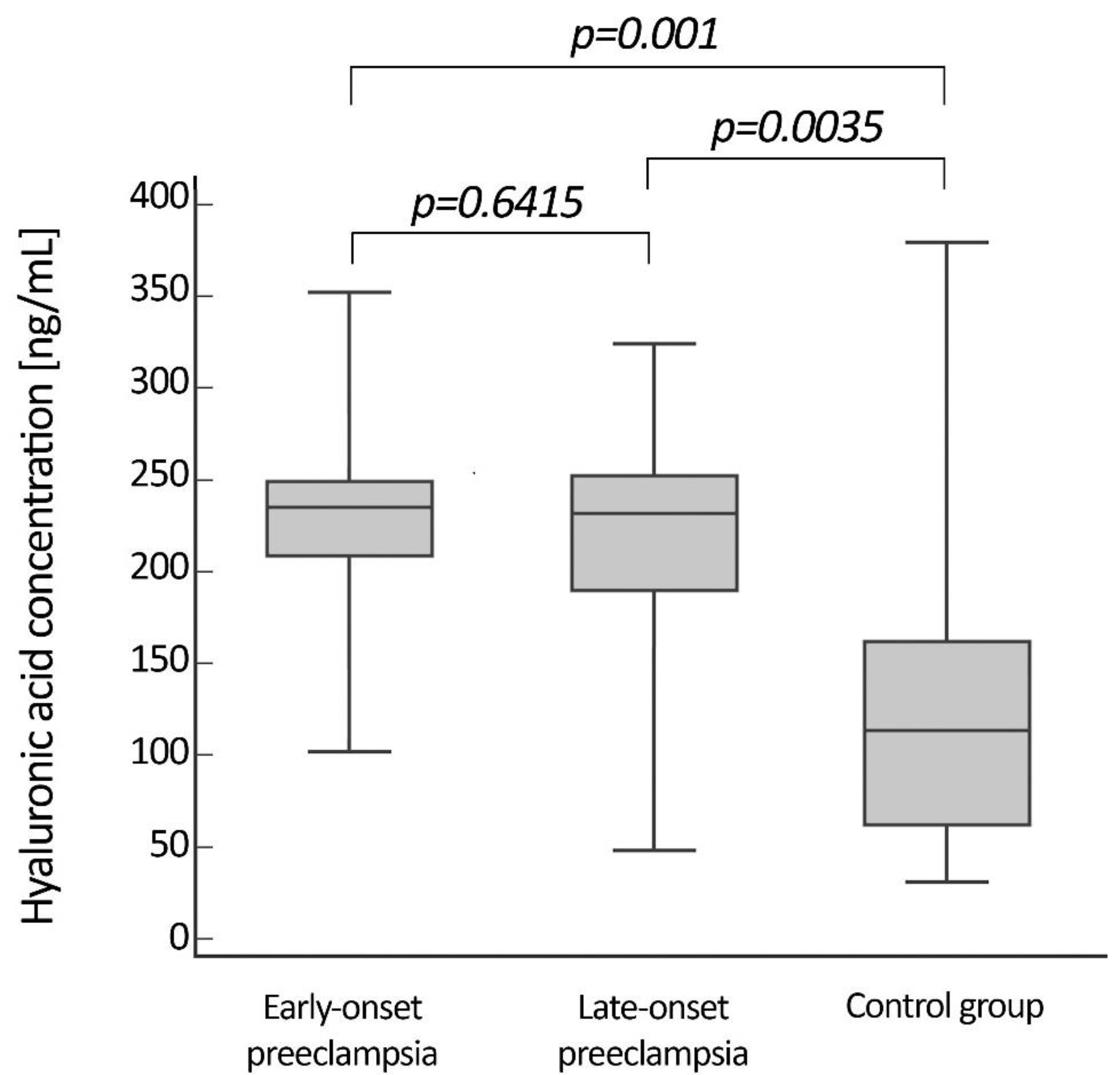
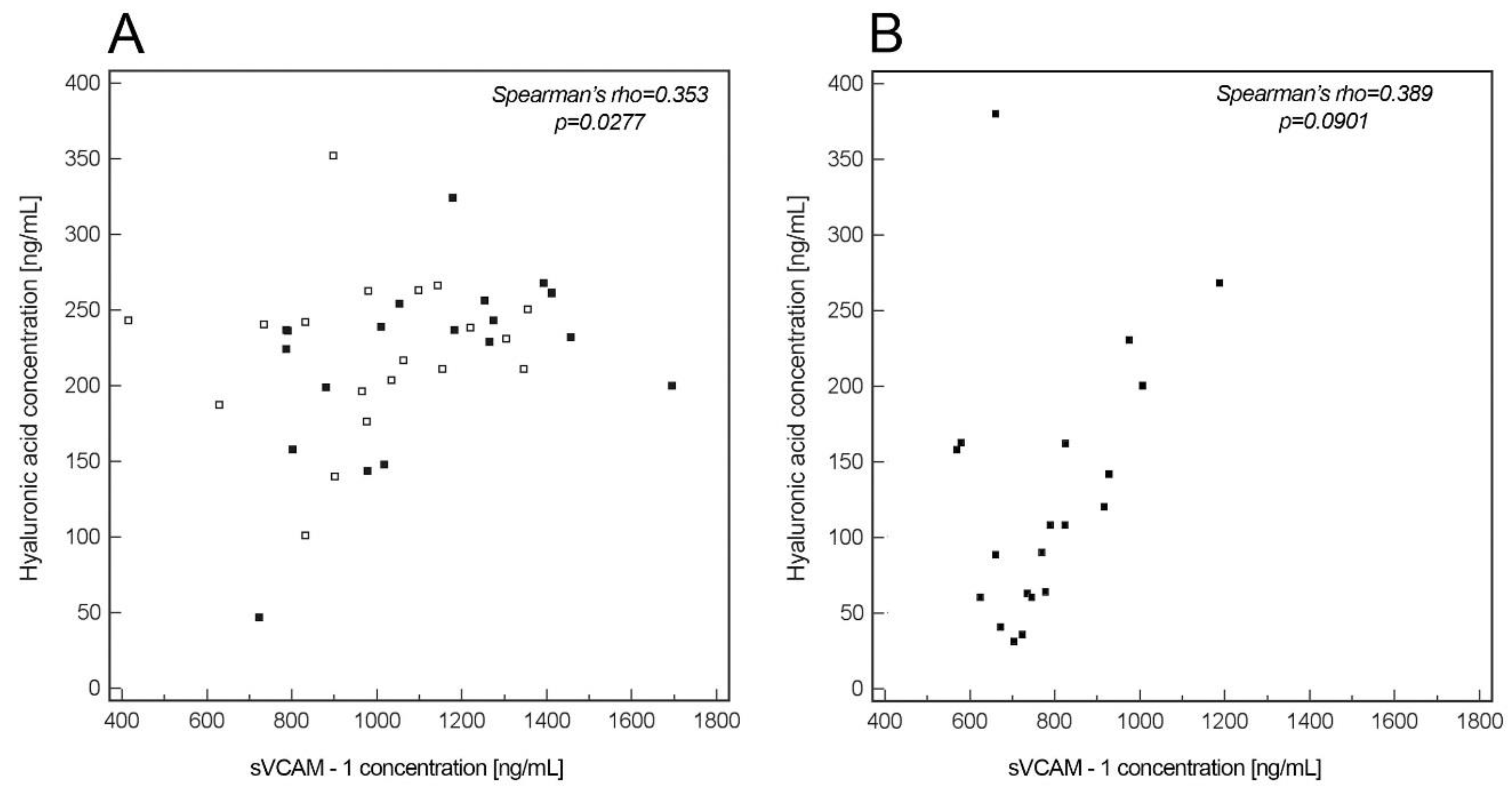
3. Results
4. Discussion
5. Conclusions
Author Contributions
Funding
Conflicts of Interest
References
- Roberts, J.M.; Hubel, C.A. The two stage model of preeclampsia: Variations on the theme. Placenta 2009, 30 (Suppl. A), S32–S37. [Google Scholar] [CrossRef] [PubMed] [Green Version]
- Wójtowicz, A.; Zembala-Szczerba, M.; Babczyk, D.; Kołodziejczyk-Pietruszka, M.; Lewaczyńska, O.; Huras, H. Early- and Late-Onset Preeclampsia: A Comprehensive Cohort Study of Laboratory and Clinical Findings according to the New ISHHP Criteria. Int. J. Hypertens. 2019, 2019, 4108271–4108279. [Google Scholar] [CrossRef] [PubMed]
- Kornacki, J.; Skrzypczak, J. Preeclampsia-two manifestations of the same disease. Ginekol. Pol. 2008, 79, 432–437. [Google Scholar] [PubMed]
- Romao, M.; Weel, I.C.; Lifshitz, S.J.; Peracoli, M.T.S.; Witkin, S.S. Elevated hyaluronan and extracellular matrix metalloproteinase inducer levels in women with preeclampsia. Arch. Gynecol. Obstet. 2014, 289, 575–579. [Google Scholar] [CrossRef]
- Weissgerber, T.L.; Garcia-Valencia, O.; Milic, N.M.; Codsi, E.; Cubro, H.; Nath, M.; White, W.M.; Nath, K.A.; Garovic, V.D. Early Onset Preeclampsia Is Associated With Glycocalyx Degradation and Reduced Microvascular Perfusion. J. Am. Hear. Assoc. 2019, 8, e010647. [Google Scholar] [CrossRef] [Green Version]
- Kornacki, J.; Wirstlein, P.; Wender-Ozegowska, E. Levels of syndecan-1 and hyaluronan in early- and late-onset preeclampsia. Pregnancy Hypertens. 2019, 18, 108–111. [Google Scholar] [CrossRef]
- Docheva, N.; Romero, R.; Chaemsaithong, P.; Tarca, A.L.; Bhatti, G.; Pacora, P.; Panaitescu, B.; Chaiyasit, N.; Chaiworapongsa, T.; Maymon, E.; et al. The Profiles of Soluble Adhesion Molecules in the “Great Obstetrical Syndromes”. J. Matern. Neonatal Med. 2018, 32, 2113–2136. [Google Scholar] [CrossRef] [PubMed]
- Kim, S.Y.; Ryu, H.M.; Yang, J.H.; Kim, M.Y.; Ahn, H.K.; Lim, H.J.; Shin, J.S.; Woo, H.J.; Park, S.Y.; Kim, Y.M.; et al. Maternal Serum Levels of VCAM-1, ICAM-1 and E-selectin in Preeclampsia. J. Korean Med. Sci. 2004, 19, 688–692. [Google Scholar] [CrossRef] [PubMed] [Green Version]
- Rios, D.R.A.; Alpoim, P.N.; Godoi, L.C.; Perucci, L.O.; de Sousa, L.P.; Gomes, K.B.; Dusse, L.M.S. Increased Levels of sENG and sVCAM-1 and Decreased Levels of VEGF in Severe Preeclampsia. Am. J. Hypertens. 2015, 29, 1307–1310. [Google Scholar] [CrossRef] [PubMed]
- Dogan, E.; Cansum Demir, S.; Kucukgoz Gulec, U. Maternal Soluble Vascular Cytoplasmic Adhesion molecule-1 and Fibronectin Levels in Early- and Late-Onset Preeclamptic Pregnancies. Clin. Exp. Obstet. Gynecol. 2014, 41, 681–684. [Google Scholar] [PubMed]
- Hypertension in Pregnancy; American College of Obstetricians and Gynecologists. Report of the American College of Obstetricians and Gynecologists’ Task Force on Hypertension in Pregnancy. Obstet. Gynecol. 2013, 122, 1122–1131. [Google Scholar]
- Figueras, F.; Gratacos, E. Update on the diagnosis and classification of fetal growth restriction and proposal of a stage-based management protocol. Fetal Diagn. Ther. 2014, 36, 86–98. [Google Scholar] [CrossRef] [PubMed]
- Venkatesha, S.; Toporsian, M.; Lam, C.; Hanai, J.; Mammoto, T.; Kim, Y.M.; Bdolah, Y.; Lim, K.H.; Yuan, H.-T.; Libermann, T.A.; et al. Soluble endoglin contributes to the pathogenesis of preeclampsia. Nat. Med. 2006, 12, 642–649. [Google Scholar] [CrossRef] [PubMed]
- Zachary, I.; Mathur, A.; Yla-Herttuala, S.; Martin, J. Vascular Protection A Novel Nonangiogenic Cardiovascular Role for Vascular Endothelial Growth Factor. Artherioscler. Thromb. Vasc. Biol. 2000, 20, 1512–1520. [Google Scholar] [CrossRef] [PubMed] [Green Version]
- Geldenhuys, J.; Rossouw, T.M.; Lombaard, H.A.; Ehlers, M.M.; Kock, M. Disruption in the Regulation of Immune Responses in the Placental Subtype of Preeclampsia. Front. Immunol. 2018, 9, 1659. [Google Scholar] [CrossRef] [PubMed]
- Laresgoiti-Servitje, E. A leading role for the immune systemin the pathophysiology of preeclampsia. J. Leukoc. Biol. 2013, 94, 247–257. [Google Scholar] [CrossRef]
- Chaiworapongsa, T.; Chaemsaithong, P.; Yeo, L.; Romero, R. Preeclampsia part 1: Current understanding of its pathophysiology. Nat. Rev. Nephrol. 2014, 10, 466–480. [Google Scholar] [CrossRef] [Green Version]
- Austgulen, R.; Lien, E.; Vince, G.; Redman, C.W. Increased maternal plasma levels of soluble adhesion molecules (ICAM-1, VCAM-1, E-selectin) in preeclampsia. Eur. J. Obstet. Gynecol. Reprod. Biol. 1997, 71, 53–58. [Google Scholar] [CrossRef]
- Yuksel, M.A.; Tüten, A.; Oncul, M.; Acikgoz, A.S.; Yuksel, I.T.; Toprak, M.S.; Ekmekçi, H.; Ekmekçi, O.B.; Madazli, R. Serum endocan concentration in women with pre-eclampsia. Arch. Gynecol. Obstet. 2015, 292, 69–73. [Google Scholar] [CrossRef]
- González-Chávez, A.; Elizondo-Argueta, S.; Gutiérrez-Reyes, G.; Leon-Pedroza, J.I. Pathophysiological implications between chronic inflammation and the development of diabetes and obesity. Cirugía Cir. 2011, 79, 209–216. [Google Scholar]
- Dogne, S.; Flamion, B.; Caron, N. Endothelial Glycocalyx as a Shield against Diabetic Vascular Complications Involvement of Hyaluronan and Hyaluronidases. Arterioscler. Thromb. Vasc. Biol. 2018, 38, 1427–1439. [Google Scholar] [CrossRef] [PubMed]
- Kim, Y.-H.; Nijst, P.; Kiefer, K.; Tang, W. Endothelial Glycocalyx as Biomarker for Cardiovascular Diseases: Mechanistic and Clinical Implications. Curr. Heart Fail. Rep. 2017, 14, 117–126. [Google Scholar] [CrossRef] [PubMed] [Green Version]
- Ziganshina, M.M.; Yarotskaya, E.L.; Bovin, N.V.; Pavlovich, S.V.; Sukhikh, G.T. Can Endothelial Glycocalyx Be a Major Morphological Substrate in Pre-Eclampsia? Int. J. Mol. Sci. 2020, 21, 3048. [Google Scholar] [CrossRef] [PubMed]
- Deanfield, J.E.; Halcox, J.P.; Rabelink, T.J. Endothelial Function and Dysfunction. Circulation 2007, 115, 1285–1295. [Google Scholar] [CrossRef] [PubMed]
- Zhou, C.; Yan, Q.; Zou, Q.-Y.; Zhong, X.-Q.; Tyler, C.T.; Magness, R.R.; Bird, I.M.; Zheng, J. Sexual Dismorphisms of Preeclampsia-Dysregulated Transcriptomic Profiles and Cell Function in Fetal Endothelial Cells. Hypertension 2019, 74, 154–163. [Google Scholar] [CrossRef] [PubMed]
- Andersen, L.B.; Joergensen, J.S.; Herse, F.; Andersen, M.S.; Christesen, H.T.; Dechend, R. The association between angiogenic markers and fetal sex: Implications for preeclampsia research. J. Reprod. Immunol. 2016, 117, 24–29. [Google Scholar] [CrossRef] [PubMed]
Publisher’s Note: MDPI stays neutral with regard to jurisdictional claims in published maps and institutional affiliations. |


| Early-Onset PE (EOP) (n = 20) | Late-Onset PE (LOP) (n = 20) | Normal Pregnancy (NP) (n = 20) | P EOP vs. LOP | P EOP vs. NP | P LOP vs. NP | |
|---|---|---|---|---|---|---|
| Age (years) median (range) | 28 (25–41) | 31 (19–40) | 32 (27–36) | 0.103 | 0.029 | 0.314 |
| Parity n (%) 0 ≥1 | 8 (40) 12 (60) | 13 (65) 7 (35) | 11 (55) 9 (45) | 0.2802 | ||
| BMI at onset of preeclampsia median (range) | 26.4 (21.8–40.6) | 34.3 (22.7–53.2) | 25 (22–29) | 0.0168 | 0.2420 | 0.0009 |
| Gestational age at blood sampling (weeks) median (range) | 28 (24–32) | 33 (34–38) | 31 (23–37) | 0.0009 | 0.0248 | 0.0511 |
| Gestational age at delivery (weeks) median (range) | 32 (26–37) | 37 (34–39) | 39 (37- 41) | 0.0005 | 0.0007 | 0.01 |
| Mode of delivery n (%) Vaginal delivery Caesarean section | 0 20 (100) | 4 (20) 16 (80) | 8 (60) 12 (40) | 0.0209 with Yates correction | ||
| FGR n (%) | 12 (60%) | 4 (20%) | 0.0098 | |||
| Fetal distress as an indication to caesarean section n (%) | 13 (65%) | 2 (10%) | 0.0003 | |||
| Mean systolic pressure (mmHg) median (range) | 150 (130–180) | 150 (130–180) | 110 (100–120) | 0.3107 | 0.0009 | 0.0009 |
| Mean diastolic pressure (mmHg) median (range) | 90 (90–110) | 100 (85–130) | 70 (60–80) | 0.3980 | 0.0009 | 0.0004 |
| Proteinuria (g/24 h) median (range) | 3.78 (0.3–10.5) | 3.86 (0.4–12.9) | 0.6917 | |||
| Newborn’s birth weight (g) mean | 1319 ± 568 | 2636 ± 683 | 3437 ± 554 | 0.0008 | 0.0009 | 0.0006 |
| Fetal gender n (%) Boy Girl | 9 (45%) 11 (55%) | 10 (50%) 10 (50%) | 10 (50%) 10 (50%) | 0.9354 | ||
© 2020 by the authors. Licensee MDPI, Basel, Switzerland. This article is an open access article distributed under the terms and conditions of the Creative Commons Attribution (CC BY) license (http://creativecommons.org/licenses/by/4.0/).
Share and Cite
Kornacki, J.; Wirstlein, P.; Wender-Ozegowska, E. Markers of Endothelial Injury and Dysfunction in Early- and Late-Onset Preeclampsia. Life 2020, 10, 239. https://doi.org/10.3390/life10100239
Kornacki J, Wirstlein P, Wender-Ozegowska E. Markers of Endothelial Injury and Dysfunction in Early- and Late-Onset Preeclampsia. Life. 2020; 10(10):239. https://doi.org/10.3390/life10100239
Chicago/Turabian StyleKornacki, Jakub, Przemysław Wirstlein, and Ewa Wender-Ozegowska. 2020. "Markers of Endothelial Injury and Dysfunction in Early- and Late-Onset Preeclampsia" Life 10, no. 10: 239. https://doi.org/10.3390/life10100239
APA StyleKornacki, J., Wirstlein, P., & Wender-Ozegowska, E. (2020). Markers of Endothelial Injury and Dysfunction in Early- and Late-Onset Preeclampsia. Life, 10(10), 239. https://doi.org/10.3390/life10100239