Article

Identification of Mineral Pigments on Red- and Dark-Decorated Prehistoric Pottery from Bulgaria

1
Georgi Nadjakov Institute of Solid State Physics, Bulgarian Academy of Sciences, 72 Tzarigradsko Chaussee, Blvd., 1784 Sofia, Bulgaria
2
Department of Prehistory, Archaeological Museum “Maritza-Iztok”, 6260 Radnevo, Bulgaria
*
Author to whom correspondence should be addressed.
Minerals 2025, 15(8), 877; https://doi.org/10.3390/min15080877
Submission received: 30 July 2025 / Revised: 15 August 2025 / Accepted: 18 August 2025 / Published: 20 August 2025
(This article belongs to the Special Issue Mineral Pigments: Properties Analysis and Applications)

Abstract

Identifying the mineral pigments used in the decoration of prehistoric pottery is a significant step for understanding the evolution of the technological practices over time. On the Balkan Peninsula during late prehistory, the techniques used for red and dark-colored decorations underwent a significant transformation. In the Early Neolithic period, pottery was often decorated with dark-toned paints, ranging from deep red to brown. However, this approach declined noticeably during the Chalcolithic period, when red pigment pseudo-incrustation became the predominant decorative method. This study aims to identify the mineral pigments used in red and dark decorations on Neolithic and Chalcolithic pottery from Bulgaria and to trace possible technological, regional, or chronological variations in their composition. A total of 34 ceramic sherds, decorated in shades from red to brown and black, were analyzed using two complementary spectroscopic techniques: laser-induced breakdown spectroscopy (LIBS) and Fourier-transform infrared spectroscopy (FTIR). LIBS data were further evaluated using principal component analysis (PCA) to classify materials based on elemental composition. The results indicate that red decorations are consistently composed of hematite and remain compositionally stable regardless of the region, time period, or application technique. In contrast, dark decorations contain various combinations of iron oxides (magnetite and hematite) and manganese oxides, often including barium-rich manganese compounds—potentially indicating pigment provenance. Additionally, the dark decorations display regional differences.

1. Introduction

Ceramic archeological artifacts are crucial for understanding the everyday life of ancient communities, as they represent the most ubiquitous findings that are discovered almost everywhere around the world. This valuable archeo-material provides extensive information about the technological capabilities of a particular cultural group, as well as the esthetic preferences and stylistic trends of their time [1,2].
Since the earliest stages of human cultural development, people have searched for natural sources of colors, both organic and inorganic, to create pigments, a practice that has accompanied the making of marks and decorated objects from prehistoric times onward. The use of pigments for decoration is a universal phenomenon found throughout the world’s prehistory, reflecting both esthetic traditions and technological knowledge shared by diverse cultures [3]. Decorative materials in colors that contrasted with the base were often applied to enhance visual appeal. The decoration of ceramic wares varied widely in terms of application techniques and ornamental designs. The use of colorful decorations in prehistoric contexts serves as a key indicator for identifying regional differences and expressing cultural identity. In prehistoric times, these pigments came in a range of colors, including white, black, yellow, and various shades of brown and red. They were applied as an additional layer that was distinct in color from the pre-treated underlying surface known as engobe. Engobe refers to a thin, clay-based coating applied to pottery to improve smoothness and enhance surface texture. By the end of the Early Neolithic period in the Balkan Peninsula, red and dark colors—particularly in shades of brown and black—were commonly applied as paint on a polished surface to adorn ceramic vessels. This method of applying decorations, known as the paint technique, declined during the Chalcolithic period, when encrusting red pigments became the dominant decoration technique. A specific method of incrustation involving rubbing a red powder with a powdery consistency onto a previously roughened surface or applying a very dilute solution after the vessel has been fired is the so-called pseudo-incrustation method [4].
The adoption of analytical techniques in archeology has expanded research beyond cultural classification based on typology and decorative styles of pottery, enabling the study of raw material composition as well. Spectroscopic techniques, in particular, allow for the identification of the chemical and mineral composition of pigments—information that cannot be determined through visual inspection alone.
In the prehistory of the Balkan Peninsula, coloristic decorations played a vital role in shaping regional and cultural characteristics across Southeastern Europe and served as a basis for relative chronology. Archaeometric analysis of the mineral pigments used for decoration offers complementary technological and material insights. Over the past three decades, a growing number of published studies have examined the archaeometric analysis of prehistoric red and dark pigments from this region. A significant volume of research has been conducted in Greece [5,6,7,8,9,10,11] and Romania [12,13,14,15,16,17]. Despite the limited number, studies have also been published from Hungary [18,19], Albania [20,21,22], Serbia [23,24,25], and Croatia [26]. The archaeometric research on pigments from the Neolithic and Chalcolithic periods in Bulgaria is still in its early stages and remains relatively sporadic. Some research teams have conducted archaeometric analyses of the red and dark decoration compositions on Neolithic and Chalcolithic ceramic vessels from several settlements across Bulgaria [27,28,29,30,31].
This study aims to identify the red and dark (brown to black) mineral pigments used for decoration on Neolithic and Chalcolithic pottery from the territory of Bulgaria and to assess a possible regional variation in their composition, with particular attention to identifying region-specific pigment types that may serve as provenance indicators. A total of 34 decorated pottery fragments from nine archeological deposits, located in four geographical zones in Bulgaria, were analyzed using two complementary spectroscopic techniques: laser-induced breakdown spectroscopy (LIBS) and Fourier-transformed infrared spectroscopy in attenuated total reflection mode (ATR-FTIR). The data obtained from the LIBS analyses are treated with the multivariate statistical technique, principal component analysis (PCA), to explore the variances in the elemental content of the decorations.

2. Materials and Methods

2.1. Analyzed Decorated Sherds

The ceramic artifacts analyzed in this study were selected to reflect the decorative styles typical of Neolithic and Chalcolithic cultural groups across different regions of Bulgaria. Their dating is based on the Bulgarian periodization method [32]. Most of the fragments are small (~2–5 cm2) to medium (~6–12 cm2) in size and do not retain identifiable vessel shapes. This study includes artifacts from nine archeological sites located within four distinct geographical regions of Bulgaria. Figure 1 presents a map showing the locations of these sites, and the main characteristics of the ceramic shards are summarized in Table 1.
The southwest area is represented by twelve sherds from four archeological sites, all located along the Struma River valley in the Blagoevgrad district: three sherds from settlement near Strumsko, dated to the Late Neolithic (~5100–4900 BC) to Early Chalcolithic (~4900–4700 BC) period (sherds U6, U7, and U8) [33]; one sherd from a settlement near Balgarchevo village, dated to the end of the Late Neolithic period (sherd U9) [34]; three sherds from a Late Neolithic settlement near Damyanitsa village (sherds U10, U11, and U13) [35]; and five sherds from a settlement (Topolnica–Promachon) near Topolnica village, dated to the Late Neolithic period (sherds N1, N2, N4, N5, and N7) [33]. The sherds U6, U7, U8, and U9 belong to the Balgarchevo I culture, and the sherds U10, U11, U13, N1, N2, N4, N5, and N7 are affiliated with the Topolnica–Acropotamos culture.
From the region of northwestern Bulgaria, twelve sherds from two archeological sites are provided. Ten of the sherds are from a settlement in the Malo Pole area near Gradeshnitsa village, Vratsa District (sherds B1, B3–11), dated to the Early Neolithic period (~6200–5500 BC) [36]. These ten sherds represent the Gradeshnitsa culture. The other two sherds are from a Late Chalcolithic settlement (~4400–3900 BC) in the Kaleto area near the city of Mezdra (sherds B25 and B26) and represent the final stage of the Krivodol–Sălcuţa–Bubani culture [37].
The central Bulgaria region is represented by six sherds from two archeological sites located in the Thracian valley. Five of them are from the settlement mound in the Starozagorski Bani site, Stara Zagora district (sherds S15, S16, S20, S21, and S26). These sherds are dated to the last phase of the Late Chalcolithic period and represent the Karanovo VI culture [38]. One sherd (S5) is from the archeological site near Madzherito village, dated to the Early Neolithic period [39]. This sherd represents the Karanovo I culture, specifically the Azmak version.
From the eastern region, four sherds are provided from the Kozarevo Mound, which is located near the city of Kableshkovo, Bourgas District (K 2-5) [40]. These sherds are dated to Phase III of the Late Chalcolithic period.
When identifying complex inorganic materials such as mineral pigments, it is essential to use a combination of analytical techniques to complement their results. Considering the valuable nature of archeological artifacts, the use of non-invasive methods is required. To acquire comprehensive information, two spectroscopic techniques were employed. LIBS was used to determine the chemical elements in the pigments, while FTIR, which provides insights into the molecular composition, was used to complement the LIBS results.

2.2. Laser-Induced Breakdown Spectroscopy (LIBS)

Among the key benefits that make LIBS competitive with other analytical techniques in archaeometry are its micro-destructive nature, the lack of requirement for prior sample preparation, its ability to detect multiple elements simultaneously, the option for depth profiling without the need for object fractionation, and its suitability for in situ analysis—particularly important when laboratory-based analysis is not achievable. LIBS has demonstrated its ability to successfully study archeological pigments [41,42].
The LIBS technique involves directing a focused, high-energy laser pulse onto the surface of a sample. The laser pulse duration is about a few nanoseconds. The intense energy at the focal point rapidly heats the material, causing a minute portion to be ablated and form a plasma plume composed of atoms and ions from the sample’s elements. Once the laser pulse ends, electrons in the plasma recombine with ions, and the excited species relax, releasing light that is characteristic of the elements present. This emitted light is then transmitted to a spectrometer using an optical fiber. LIBS can be used to analyze a wide variety of materials irrespective of their phase [43,44].
In this study, LIBS measurements were carried out using a portable LIBSCAN25+ system (Applied Photonics Ltd., Skipton, UK). The setup features a Q-switched Nd:YAG laser operating at the fundamental wavelength of 1064 nm, with a pulse duration of 10 ns and a standard output energy of 50 mJ per pulse. It also includes six spectrometers covering the 200–750 nm spectral range, along with beam focusing and light collection optics. The laser beam was focused onto the sample surface using a lens with a 90 mm focal length. Plasma emission was collected via six optical assemblies, three designed for the UV-VIS range and three for the VIS–NIR range, and transmitted to the spectrometers through optical fibers.
For the experiments, the samples were irradiated using reduced pulse energy of 8 mJ to ensure ablation of only the decoration layer, without affecting the underlying engobe. The depth of the crater obtained under these experimental conditions is approximately in the range of 1–5 µm. Ablation was conducted under ambient air at atmospheric pressure. To minimize interference from the initial continuum emission of the plasma, signal acquisition was delayed by 1.3 microseconds following each laser pulse, with a detection window (gate) of 1 millisecond. Spectral data collection was managed using the LIBSoft V14 software. Analyses were carried out on both the decorations and the engobes of the pottery sherds. For the identification of the chemical elements from the detected spectral lines the National Institute of Standards and Technology (NIST) database was used [45].
The LIBS data were statistically evaluated using principal component analysis (PCA) to group the fragments based on similarities and differences in their elemental compositions.
PCA is a technique for reducing the complexity of large datasets by converting the original variables into a smaller number of uncorrelated variables known as principal components. These components represent the most important patterns of variation within the data, enabling more straightforward analysis and visualization with minimal loss of meaningful information [46,47,48]. The PCA score plot displays the similarities and differences between sherds, showing how they are distributed in the space defined by the principal components. In this current study, each dot on the score plot represents a sherd. The closer the dots are, the more similar the characteristics are in terms of elemental content. The PCA loading plot shows the relationship between the variables and the PCs, indicating how strongly each variable influences each PC. The statistical analysis in our study was performed using the OriginPro 2018 (9.55) software.

2.3. Fourier-Transformed Infrared Spectroscopy in Attenuated Total Reflection Mode (ATR-FTIR)

ATR-FTIR is a prominent analytical method for determining the mineral composition of archeological ceramics [49,50,51]. It works by detecting the characteristic absorption of infrared radiation by the functional groups within the material, which are influenced by its molecular structure, bond vibrations, and crystal lattice. This technique is valued for its simplicity, speed, and reliability. It offers notable advantages over other mineralogical methods by enabling the identification of both crystalline and pseudo-amorphous phases in ceramic components. Additionally, FTIR is considered non-invasive due to its minimal sample requirements [52].
In this study, ATR-FTIR was employed to identify the mineral compounds present in the pigments. The analyses were conducted using a Perkin Elmer Spectrum Two FTIR spectrometer equipped with a PIKE GladiATR accessory and a monolithic diamond ATR crystal (Pike Technologies, Fitchburg, WI, USA) at room temperature. For each sample, a small portion of the decorative layer was carefully scraped, finely ground in an agate mortar, and placed directly onto the diamond crystal without additional preparation. Spectra were collected in the mid-infrared (MIR) range (4000–400 cm−1), averaging 32 scans at a resolution of 4 cm−1. Baseline correction was carried out using the instrument’s software, and spectral data were plotted using OriginPro Version 2024 (10.1) (OriginLab Corporation, Northampton, MA, USA). Interpretation was performed by comparing the results with established reference studies.

3. Results

3.1. Elemental Composition

The elemental analyses with LIBS are constrained to qualitative and semi-quantitative elemental determination due to the absence of suitable matrix-matched calibration standards for pigments and ceramic materials. Based on the analytical results, all examined ceramic sherds display an identical elemental content. The predominant elements detected include calcium, aluminum, iron, magnesium, silicon, potassium, and sodium. Additionally, manganese, strontium, barium, and titanium were also identified.
All the detected elements are present in each of the analyzed sherds, both in the engobes and the decorations. Nevertheless, the relative intensities of the spectral emission lines of the detected elements varied in each sherd, indicating different concentrations of these elements.
Due to the similar elemental composition of all decorations, distinguishing between various mineral pigments is challenging. This necessitates the application of the multivariate technique PCA for the statistical treatment of the data obtained from the LIBS analysis, which helps classify the sherds according to the differences and similarities in their chemical content. Classification of the sherds is based on variations in the spectral line intensities of the constituent elements. The input data for conducting PCA consisted of the intensities of selected spectral lines emitted by each element. Given that spectral line intensities depend not only on elemental concentrations but also on the matrix effects and experimental conditions, normalization of the measured peak areas is required. This normalization was achieved by dividing the integrated peak area of each selected spectral line by the total light intensity emitted from the plasma in each recorded spectrum [53].
The characteristic atomic and ionic emission lines used for elemental identification and for conducting PCA are presented in Table 2. The choice of appropriate spectral lines for PCA is guided by two primary considerations: the spectral lines must be free from overlap with lines of other elements, and the strongest analytical lines of certain elements—those most susceptible to self-absorption effects—should be avoided.
The PCA technique enables the extraction of complex information from multiple datasets by visualizing variations in the relative elemental composition across different sherds. Figure 2 shows the resulting biplot of the first two principal components, which explain 53.10% of the cumulative variance (PC 1 explains 30.39% of the variation, while PC 2 explained 22.71% of the variation). The biplot combines the score plot and the loading plot into a single graph. Although all of the decorations have an identical elemental composition and the score plot does not show apparent clustering, some distinctive characteristics can still be observed.
The score plot reveals a slight separation between the red and dark decorations, indicating differences in their mineral composition despite the overall similarity in elemental content among all the sherds. All of the red-decorated sherds, except K4 and K5, along with some of the dark-decorated ones, are located in the negative region of PC2. In contrast, most of the dark-decorated ones are located in the positive region.
Most of the scores representing red decorations are clustered together, indicating significant similarity in their chemical composition. It is also observed that the dark decorations are more dispersed, implying that a greater diversity of substances has been used for the dark colors.
Another distinctive feature that can be noticed on the score plot is that the sherds from Kozareva Mound (K2, K3, K4, and K5) are, to some degree, separated from the rest. They are located in the most positive part of PC1. The loading plot shows that PC1 is most influenced by iron and silicon. This indicates that the decorations of these sherds—both red and dark—are distinguished from the others by a higher content of these elements.
The loading plot reveals that the loadings associated with barium and manganese exhibit a strong positive correlation, indicating that in samples with increased manganese content, barium levels are also increased—and vice versa. Most of the dark-decorated sherds are positioned in the positive side of PC2, which is most influenced by barium and manganese. This suggests that, in these dark decorations, the coloration is most likely achieved through the application of mineral compounds containing manganese oxides enriched with barium.
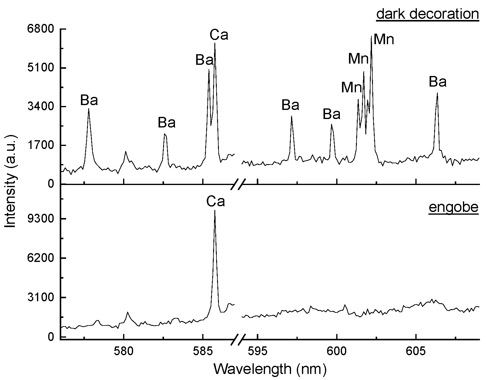
It is quite possible that barium naturally exists in clays as a minor component. To make sure that the increased levels of barium and manganese in the decorations—and the resulting dark color—are due to their intentional addition rather than being inherent to the clay, the spectra of the dark decorative areas were compared with those of the corresponding engobe layers. The analysis revealed that the spectral lines of barium and manganese are significantly weaker—or entirely absent—in the engobes compared to the decorations. This confirms the suggestion that the dark color is due to the presence of minerals containing barium and manganese. Figure 3 presents an example spectra obtained from one of the dark-decorated sherds (sherd N4), illustrating this finding. The spectral regions from 576.00 nm to 587.00 nm and from 594.00 nm to 609.00 nm, in which some of the barium and manganese spectral lines are emitted, obtained from the decoration and the engobe, are compared.
Most of the dark-decorated sherds exhibited an increased amount of barium-enriched manganese minerals compared to their engobes. The exceptions are sherds B5, B6, B9, B10, N2, S5, K2, and K3. In the decorations of sherds B5, B6, B9, and B10, only manganese is increased, indicating the use of manganese oxides as a pigment. In sherd N2, the decoration shows increased concentrations of both manganese and iron, most likely indicating the use of umber—a natural earth pigment containing iron oxide and manganese oxide known for its brownish color [54]. In sherds S5, K2, and K3, only iron is increased, suggesting that the dark color in these objects was achieved using iron oxides.
The other strong positive correlation observed on the loading plot is between calcium and strontium. The simultaneous increase in calcium and strontium can be explained by the fact that during the growth of calcite crystals, it is common for calcium atoms to be substituted by strontium cations (Sr2⁺) due to the close similarity in their ionic radii and charges, as well as the ability of carbonates to interact with various divalent metals [55,56,57]. Therefore, the strong positive correlation between these two elements suggests the presence of calcite in some of the decorations.
The PCA biplot shows that most of the red decorations exhibit a higher titanium content compared to the dark decorations. When comparing the spectra of the decorations to those of the corresponding engobes, it is evident that the titanium spectral lines in the red decorations are as strong as those in the engobes. In contrast, in the dark decorations, they appear weaker. To illustrate this, Figure 4 shows the spectral region from 497.5 nm to 501.00 nm, highlighting several atomic spectral lines of titanium obtained from two examples. The spectra of the red (Figure 4a) and dark (Figure 4b) decorations, along with their corresponding engobes, are compared.
In general, the red-decorated objects exhibit a significant similarity between the decoration and the engobe material, with the iron content being slightly higher in the decoration. The only exceptions are the sherds from Kozareva Mound (K2, K3, K4, and K5), which display a significantly higher iron content in the decoration compared to the engobe. In contrast, the dark decorations exhibit a slightly more diverse composition, primarily due to elevated levels of barium, manganese, and, in some sherds, iron.
The spectral lines of aluminum, silicon, magnesium, potassium, and sodium are almost equally strong both in the decorations and engobes. Considering that these elements are natural constituents of clays, it can be assumed that the coloring mineral, most probably red ocher, was mixed with clay.

3.2. Molecular Composition

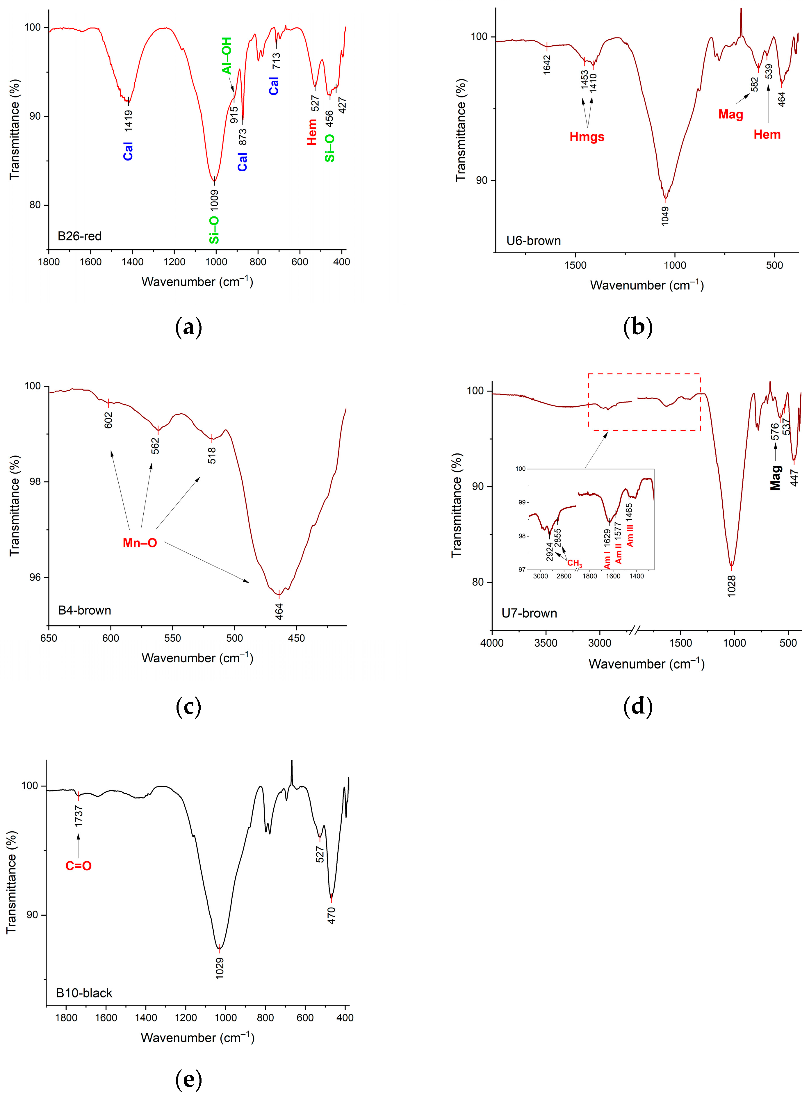
The ATR-FTIR analysis provided additional details on the mineralogical composition of the paints used to decorate the ancient pottery. The spectra obtained displayed complex and partially overlapping characteristic bands, making interpretation challenging. Representative spectra are shown in Figure 5. All the detected minerals are summarized in Table 3.
All spectra were dominated by characteristic vibrations of absorbed water, which are seen as a very broad band around 3400 cm−1 and attributed to O–H stretching and another broad band around 1640 cm−1 due to the H–O–H bending mode of water [58]. Quartz was also present in all spectra, identified by its characteristic doublet at 778–799 cm−1, with the peak at 695 cm−1 and the shoulder at 1164 cm−1 [59]. The presence of aluminosilicate clay minerals, such as illite, montmorillonite, and kaolinite, was indicated by a strong, broad band in the 800–1200 cm−1 range centered between 1005 and 1049 cm−1, corresponding to Si–O stretching vibrations, as well as Si–O–Si bending features around 450 and 420 cm−1 (Figure 5a). Furthermore, a weak band at ~915 cm−1, attributed to Al–OH vibrations in phyllosilicates, was observed in some samples (Figure 5a) [60].
In some spectra, characteristic bands of carbonate species, likely from calcite (CaCO3), were also observed. These included a broad band near 1420 cm−1 and two narrow bands at 873 and 713 cm−1, corresponding to the asymmetric stretching of the carbonate ion (ν3), out-of-plane bending (ν2), and in-plane bending (ν4) vibrations, respectively [61]. Additionally, the presence of hydromagnesite (4MgCO3·Mg(OH)2·4H2O) was inferred by two narrow bands at 1410 and 1453 cm−1, which split the carbonate stretching vibration (Figure 5b) [62].
Characteristic spectral features associated with iron- and manganese-based metal oxides were consistently detected in the 900–400 cm−1 region, which are typically responsible for the red to brown/black hues of the decorative pigments. Hematite (Fe2O3) was clearly identified in red paints through its prominent absorption band at 524–536 cm−1 (Figure 5a). Its second notable band falls within 430–470 cm−1, overlapping with the regions of clays and feldspars, complicating precise attribution [61]. In the dark-colored pigments, hematite was often found alongside magnetite (Fe3O4), with a characteristic peak between 556 and 585 cm−1 (Figure 5b,d) [63]. Manganese oxides were also detected in some dark samples by bands around 602, 562, 518, and 464 cm−1 due to Mn–O vibrations (Figure 5c) [64,65].
Traces of organic compounds were detected in several samples. In two samples (U7 and B7), weak features of proteinaceous materials were identified by the asymmetric and symmetric –CH2– stretching modes at 2984 and 2855 cm−1, along with amide I, II, and III bands at 1629, 1577, and 1465 cm−1, respectively (Figure 5d). In two other samples (B10 and N2), a weak broad absorption at 1737 cm−1, corresponding to the C=O stretching mode of lipids, was detected (Figure 5e), suggesting the presence of oil or resinous materials [66].

4. Discussion

This archaeometric study of red- and dark-decorated prehistoric pottery sherds revealed a notable similarity in the composition of the decorative layers. In addition to the coloring agent, earth pigments typically contain a combination of accessory components such as clays, carbonates, and quartz [67]. All of the analyzed samples contained clay minerals, including aluminosilicates, as well as quartz and various metal oxides. The clay material is a carrier to which the coloring agent is added.
The results from both analytical methods indicated that in the red decorations, iron oxides, specifically hematite, are the predominant constituent. The elemental analyses disclosed an increased amount of titanium in all the engobes and in nearly all of the red decorations compared to the dark ones. The increased titanium amount can be attributed to the fact that this element is a common trace element in clay minerals. It is typically present as titanium dioxide (TiO2) or as a substituent within the crystal lattice of clay minerals. The concentration of titanium can vary depending on the type and origin of the clay [68,69,70].
The elemental analysis revealed that sherds from the eastern region, specifically the Kozarevo Mound, generally exhibit the highest concentrations of iron and silicon compared to samples from the other studied regions. This could be a reflection of a regional characteristic of the clay which might naturally contain more iron oxides and silicate minerals.
In some of the red-decorated sherds, the presence of calcite is observed. Although calcite is typically associated with the formation of white rather than red or dark coloration, its occurrence in these samples may not be accidental. Calcite can be intentionally added to alter the mechanical properties of the material; for example, it may serve as an extender in pigments [71] or as a temper when added to clay [72]. Nevertheless, it is more likely that the calcite originates naturally from the raw materials used. Many studies report the natural presence of calcite in red pigments [73,74,75,76,77], including in pottery from Bulgaria [28].
Similarly, calcite is also detected in some of the dark-decorated sherds, where its presence is again more plausibly attributed to the natural composition of the raw materials rather than deliberate addition. Additionally, in two dark-decorated sherds from the southwestern region, the carbonate mineral hydromagnesite is identified. These minerals may also derive from natural raw materials, but they can alternatively form through post-burial precipitation or as an alteration product [78].
The red pigment used across Southeastern Europe during the Neolithic and Chalcolithic periods shows significant permanence, with hematite being the predominant ingredient. The use of cinnabar on a limited number of archeological findings has only been reported in Hungary [19] and in the western part of the Balkans, Serbia [23], and Thessaly, Greece [11].
Unlike the red decorations, the brown ones demonstrate a slightly greater variety in their composition. Molecular analysis primarily identifies hematite and magnetite, and in some samples, these are mixed with manganese oxides. The elemental analyses and the resulting loadings in PCA revealed a robust positive correlation between barium and manganese in most of the dark decorations. The simultaneous increase in barium and manganese contents most likely indicates the presence of psilomelane, a widespread group of hard, black, hydrous barium-bearing manganese oxides/hydroxides, including primarily romanechite (Ba,H2O)2(Mn4+,Mn3+)5O10 and hollandite Ba(Mn4+,Mn2+)8O16. Also, psilomelane is often associated with wad, an old mining term for a black earth pigment rich in manganese. Wad has no defined chemical composition and typically consists of hydrated manganese oxides (such as romanechite, pyrolusite (MnO2), birnessite ((Na,Ca)0.5(Mn4+,Mn3+)2O4·1.5H2O), todorokite (Ca,K,Na,Mg,Ba,Mn) (Mn,Mg,Al)6O12.3H2O), and hydrated iron oxides [75,77].
All sherds showing notable increases in barium and manganese contents are located in western Bulgaria—specifically, the Vratsa and Mezdra districts in northwestern Bulgaria and the Struma River valley in the southwest. Given the known presence of wad in southwestern Bulgaria and also that the river is relatively close to these two regions, there are deposits of romanechite and hollandite [79]. The presence of barium-bearing manganese minerals in the dark decorations can be considered as a local characteristic of the mineral sources and could therefore be a valuable indicator of origin.
To the best of our knowledge, the use of barium-bearing manganese oxides/hydroxides, specifically those from the psilomelane group, has not yet been identified as a dark pigment within the territory of the Balkan Peninsula. Their use has been reported in Paleolithic rock art in southwestern Europe [80,81,82].
In some of the dark-decorated objects from northwestern Bulgaria, an increased iron content was detected along with an increased manganese content. This suggests that the dark pigment used in these cases was umber, a natural earth pigment mixture consisting primarily of iron oxides with manganese oxides.
The results from both methods show that the dark decorations on sherds from central (Madzherito) and eastern (Kozarevo Mound) Bulgaria consist primarily of iron-bearing minerals (hematite and magnetite) mixed with clay.
The molecular analysis detected organic components in some of the sherds. Proteinaceous materials were identified in two of the dark decorations. Traces of lipids were detected in two other sherds, both in red and dark decorations, suggesting the presence of oil or resinous materials. These organics could point to ancient food processing, old repairs, or waterproofing purposes [83,84]. Alternatively, they may have served as binding agents to enhance the adhesion of pigments to the ceramic surface [85,86].
Across Southeastern Europe, dark colors ranging from brown to black were produced using various inorganic materials in combination with specific firing conditions. One of the primary raw materials used was manganese-enriched earth (umber). This material has been mainly identified in Romania [14,15,16], Albania [21,22], and Greece (Thessaly) [8]. Another method for achieving dark coloration involved using iron-containing minerals fired in a reducing atmosphere, a technique also documented in many parts of the Balkan Peninsula, particularly in Romania [14,15,16], Albania [21,22], and Greece (Thessaly and Eastern Macedonia) [6,7].
There are some publications on the use of carbon black pigments, specifically graphite, on the territory of Southeast Europe [7,9,15,87,88,89]. In Bulgaria, the use of graphite has been documented in objects from the western region [33,90]. A specific technique involving carbon black has been identified in Croatia, where soot was used to achieve a black color [26].
In general, the mineral pigments identified in Neolithic and Chalcolithic pottery from Bulgaria display a high degree of consistency, closely matching those found across Southeastern Europe. In both Bulgaria and the wider region, red coloration was predominantly achieved with hematite, with cinnabar appearing only rarely in the Western Balkans. The dark pigments, on the other hand, exhibit greater diversity, including mixtures of iron oxides (magnetite and hematite), manganese oxide-rich earths, and, in some areas, graphite. The only notable regional distinction, according to our results, is the use of barium-rich manganese oxides in western Bulgaria. This overall uniformity in pigment use, combined with localized variations, points to shared technological traditions alongside region-specific resource exploitation in the Balkans during these periods. 5. Conclusions
This study presents the results of archaeometric research on mineral pigments in red- and dark-decorated prehistoric pottery sherds from various geographical regions of present-day Bulgaria. A combined spectroscopic approach was utilized to identify the earth pigments used for decoration.
The results revealed compositional similarities across the analyzed samples. In all of the decorations, the mineral pigment is mixed with a clay carrier material. The red decorations are consistently dominated by hematite. Dark decorations, on the other hand, demonstrate greater compositional diversity, with various combinations of iron oxides (magnetite and hematite) and manganese oxides being detected. Strong correlation between barium and manganese in many dark-decorated sherds indicated the use of barium-bearing manganese oxide (assuming the presence of romanechite, hollandite, or todorokite in the raw material). This pigment type was identified in dark decorations from western Bulgaria (Vratsa District and the Struma River valley), suggesting local sourcing and potential as a provenance marker. Some sherds from northwestern Bulgaria (Vratsa District) exhibited high levels of both iron and manganese, suggesting the use of umber as a pigment. In contrast, the dark-decorated sherds from central (Stara Zagora District) and eastern (Bourgas District) Bulgaria were primarily composed of magnetite and hematite mixed with clay.
In general, the red pigments show considerable conservative tendency regardless of the region, period, or decorative technique. However, the dark decorations show a clear regional differentiation in the mineral pigments used.
This archaeometric study has revealed important insights into the raw materials used in the red and dark decorations of Neolithic and Chalcolithic pottery from various geographical regions of Bulgaria, providing a basis for elucidating and tracing the spread of the technology of coloristic decorations and regional characteristics during late prehistory in Southeastern Europe.

Author Contributions

Conceptualization, V.T., V.A., A.P. and V.M.; formal analysis, V.T. and V.A.; investigation, V.T. and V.A.; resources, A.P.; writing—original draft preparation, V.T. and V.A.; writing—review and editing, V.T., V.A., V.M. and A.P.; visualization, V.T.; project administration, V.T. and V.M. All authors have read and agreed to the published version of the manuscript.

Funding

This research received no external funding.

Data Availability Statement

The original contributions presented in the study are included in the article; further inquiries can be directed to the corresponding author.

Acknowledgments

Fourier-transform infrared spectroscopy was performed at the Department of Optoelectronic Methods and Techniques for Artwork Restoration and Conservation, National Institute of Research and Development for Optoelectronics-INOE 2000.

Conflicts of Interest

The authors declare no conflicts of interest.

References

  1. Sinopoli, C.M. Approaches to Archaeological Ceramics, 1st ed.; Springer: New York, NY, USA, 1991. [Google Scholar]
  2. Rosiak, A.; Józefowska, A.; Jałowiecka, M.; Kałuzna-Czaplinska, J. Advanced Characterization of Archaeological Ceramics. Annu. Rev. Mater. Res. 2025, 55, 443–468. [Google Scholar] [CrossRef]
  3. Barnett, J.R.; Miller, S.; Pearce, E. Colour and art: A brief history of pigments. Opt. Laser Technol. 2006, 38, 445–453. [Google Scholar] [CrossRef]
  4. Pirovska, A. Technique and Technology of Colorful Decoration in Prehistory. Ph.D. Thesis, Faculty of History, Sofia University, “St. Kliment Ohridski”, Sofia, Bulgaria, 2024. [Google Scholar]
  5. Iordanidis, A.; Garcia-Guinea, J. A preliminary investigation of black, brown and red coloured potsherds from ancient upper Macedonia, northern Greece. Mediterr. Archaeol. Archaeom. 2011, 11, 85–97. [Google Scholar]
  6. Yiouni, P. Painted pottery from East Macedonia, in North Greece: Technological analysis of decorative techniques. Doc. Praehist. 2000, XXVII, 199–214. [Google Scholar]
  7. Yiouni, P. Surface Treatment of Neolithic Vessels from Macedonia and Thrace. Annu. Br. Sch. Athens 2001, 96, 1–25. [Google Scholar] [CrossRef]
  8. Jones, R. The Decoration and Firing of Ancient Greek Pottery: A Review of Recent Investigations. Adv. Archaeomaterials 2021, 2, 67–127. [Google Scholar] [CrossRef]
  9. Sakalis, A.J.; Kazakis, N.A.; Merousis, N.; Tsirliganis, N.C. Study of Neolithic pottery from Polyplatanos (Imathia) using micro X-ray fluorescence spectroscopy, stereoscopic microscopy and multivariate statistical analysis. J. Cult. Herit. 2013, 14, 485–498. [Google Scholar] [CrossRef]
  10. Tsirtsoni, Z.; Malamidou, D.; Kilikoglou, V.; Karatasios, I.; Lespez, L. Black-on-red painted pottery production and distribution in late Neolithic Macedonia. In Archaeometric and Archaeological Approaches to Ceramics; Waksman, S.Y., Ed.; BAR Publishing: Oxford, UK, 2007; pp. 57–62. [Google Scholar]
  11. Rondiri, V.; Asderaki-Tzoumerkioti, E. The ‘management’ of painted and monochrome pottery of Neolithic Thessaly, Central Greece: Technology and provenance. In Proceedings of the 6th Symposium of the Hellenic Society for Archaeometry (Bar International Series 2780); Photos-Jones, E., Bassiakos, Y., Filippaki, E., Hein, A., Karatasios, I., Kilikoglou, V., Kouloumpi, E., Eds.; British Archaeological Reports (BAR): Oxford, UK, 2016; pp. 3–12. [Google Scholar]
  12. Spataro, M.; Cubas, M.; Craig, O.E.; Chapman, J.C.; Boroneanţ, A.; Bonsall, C. Production and function of Neolithic black-painted pottery from Schela Cladovei (Iron Gates, Romania). Archaeol. Anthrop. Sci. 2019, 11, 6287–6304. [Google Scholar] [CrossRef]
  13. Opris, V.; Dragos, M.; Andrei, R.; Straticiuc, M.; Simion, C.; Stanculescu, I.; Miu, L.; Dinca, L. Archaeometric Studies of Boian Pottery from Nanov-‘Vistireasa 3’ (Teleorman County, Romania, c. 4800-4500 cal BC). In Bridging Science and Heritage in the Balkans: Studies in Archaeometry, Cultural Heritage and Conservation; Palincaș, N., Ponta, C., Eds.; Archaeopress Publishing Ltd.: Oxford, UK, 2019; pp. 92–102. [Google Scholar] [CrossRef]
  14. Buzgar, N.; Apopei, A.I.; Buzatu, A. Characterization and source of Cucuteni black pigment (Romania): Vibrational spectrometry and XRD study. J. Archaeol. Sci. 2013, 40, 2128–2135. [Google Scholar] [CrossRef]
  15. Buzgar, N.; Bodi, G.; Aştefanei, D.; Buzatu, A. The Raman study of white, red and black pigments used in Cucuteni Neolithic painted ceramics. ANALELE ŞTIINŢIFICE ALE UNIVERSITĂŢII „AL. I. CUZA” IAŞI”, Geologie 2010, 56, 5–14. [Google Scholar]
  16. Buzgar, N.; Bodi, G.; Buzatu, A.; Apopei, A.I.; Aștefanei, D. Raman and XRD studies of black pigment from Cucuteni ceramics. ANALELE ŞTIINŢIFICE ALE UNIVERSITĂŢII „AL. I. CUZA” IAŞI”,Geologie 2010, 56, 95–108. [Google Scholar]
  17. Oancea, A.V.; Bodi, G.; Nica, V.; Ursu, L.E.; Drobota, M.; Cotofana, C.; Vasiliu, A.L.; Simionescu, B.C.; Olaru, M. Multianalytical characterization of Cucuteni pottery. J. Eur. Ceram. Soc. 2017, 37, 5079–5098. [Google Scholar] [CrossRef]
  18. Szíki, G.Á.; Biró, K.T.; Uzonyi, I.; Dobos, E.; Kiss, Á.Z. Investigation of incrusted pottery found in the territory of Hungary by micro-PIXE method. Nucl. Instrum. Meth. B 2003, 210, 478–482. [Google Scholar] [CrossRef]
  19. Mihály, J.; Komlósi, V.; Tóth, A.; Tóth, Z.; Ilon, G. Vibrational spectroscopic study of pigment raw materials and painted ceramics excavated at Szombathely-Oladi plató, Hungary. In Vessels: Inside and Outside, Proceedings of the Conference EMAC ’07. 9th European Meeting on Ancient Ceramics, Hungarian National Museum, Budapest, Hungary, 24–27 October 2007; Biró, K.T., Szilágyi, V., Kreiter, A., Eds.; The Hungarian National Museum: Budapest, Hungary, 2009; pp. 47–51. [Google Scholar]
  20. Ndreçka, E.; Civici, N.; Beqiraj, E.; Gjipali, I. Results from Multi Technique Investigation of Pottery from Different Early Neolithic Sites in Albania. J. Mater. Sci. Chem. Eng. 2017, 5, 10–26. [Google Scholar] [CrossRef]
  21. Ndreçka, E.; Civici, N.; Gjipali, I.; Niccolai, F.; Ridolfi, S. Investigation of Pottery from Different Neolithic Sites in Southeast Albania Using Various Analytical Techniques. J. Mater. Sci. Chem. Eng. 2017, 5, 71–89. [Google Scholar] [CrossRef]
  22. Elezi, G. Sociocultural Dimensions of Production, Use, and Circulation of Late Neolithic Pottery from Southern Balkans. Ph.D. Thesis, University of California, Los Angeles, CA, USA, 2020. [Google Scholar]
  23. Mioč, U.B.; Colomban, P.; Sagon, G.; Stojanović, M.; Rosić, A. Ochre decor and cinnabar residues in Neolithic pottery from Vinča, Serbia. J. Raman Spectrosc. 2004, 35, 843–846. [Google Scholar] [CrossRef]
  24. Gajič-Kvaščev, M.; Stojanoviš, M.M.; Šmit, Ž.; Kantarelou, V.; Karydas, A.G.; Šljivar, D.; Milovanovič, D.; Andrič, V. New evidence for the use of cinnabar as a colouring pigment in the Vinča culture. J. Archaeol. Sci. 2012, 39, 1025–1033. [Google Scholar] [CrossRef]
  25. Perisic, N.; Stojanović, M.M.; Andrić, V.; Mioc, U.; Damjanović-Vasilić, L. Physicochemical characterisation of pottery from the Vinča culture, Serbia, regarding the firing temperature and decoration techniques. J. Serb. Chem. Soc. 2016, 81, 1415–1426. [Google Scholar] [CrossRef]
  26. Lukačević, I.; Rajković, D. Non-invasive Analyses of Ancient Ceramics Colorants. Croat. Chem. Acta 2015, 88, 53–58. [Google Scholar] [CrossRef]
  27. Pernicheva–Perets, L.; Grębska–Kulow, M.; Kulov, L. Balgarchevo. The Prehistoric Settlement, Volume I; Craft House Bulgaria: Sofia, Bulgaria, 2011. [Google Scholar]
  28. Dzhanfezova, T.; Tzvetanova, Y.; Bakamska, A. Prehistoric colour palettes decoded by painted pottery analysis: The vivid past of the Early Neolithic Galabnik settlement mound (southwestern Bulgaria) over time. J. Archaeol. Sci. Rep. 2025, 63, 105088. [Google Scholar] [CrossRef]
  29. Hristov, M. Early Chalcolithic pottery from the Region of Panagyurishte. In Early Symbolic Systems for Communication in Southeast Europe; Nikolova, L., Ed.; International Series 1139; British Archaeological Reports: Oxford, UK, 2003; pp. 606–607. [Google Scholar]
  30. Georgieva, P.; Kostov, R. Geoarchaeological and archaeomineralogical studies of the Kozareva mogila—A settlement and a necropolis, district of Burgas. Geol. Miner. Resour. 2014, 5, 17–23. [Google Scholar]
  31. Dzhanfezova, T.; Doherty, C.; Elenski, N. Shaping a Future of Painting: The Early Neolithic Pottery from Dzhulyunitsa, North Central Bulgaria. Bulg. E-J. Archaeol. 2014, 4, 137–159. [Google Scholar]
  32. Boyadzhiev, Y. The transition between Neolithic and Chalcolithic on the territory of Bulgaria. In the Neolithic and Eneolithic in Southeast Europe. New Approaches to Dating and Cultural Dynamics in the 6th to 4th Millennium BC. Prähistorische Archäologie in Südost Europa, Band 28; Schier, W., Draşovean, F., Eds.; Verlag Marie Leidorf GmbH: Rahden, Germany, 2014; pp. 49–68. [Google Scholar]
  33. Vajsov, I. Promachon-Topolnica. A typology of painted decorations and its use as a chronological marker. In the Struma/Strymon River Valley in Prehistory. In the Steps of James Harvey Gaul, 2; Todorova, H., Stefanivic, M., Ivanov, G., Eds.; Museum of History-Kyustendil: Sofia, Bulgaria, 2007; pp. 79–120. [Google Scholar] [CrossRef]
  34. Grębska-Kulow, M.; Zidarov, P. The Routes of Neolithisation: The Middle Struma Valley from a Regional Perspective. Open Archaeol. 2021, 7, 1000–1014. [Google Scholar] [CrossRef]
  35. Grębska-Kulow, M.; Vajsov, I.; Ivanov, G.; Chukalev, K.; Evlogiev, Y. Archaeological research of the prehistoric settlement Damyanitsa 2017, Stage 1. In Archaeological Discoveries and Excavations in 2017; Vagalinski, L., Ed.; National Archaeological Institute with Museum, Bulgarian Academy of Sciences: Sofia, Bulgaria, 2018; pp. 30–33. [Google Scholar]
  36. Nikolov, B. An early Neolithic settlement near the village of Gradeshnitsa, Vratsa District. In Archaeology; National Archaeological Institute with Museum, Bulgarian Academy of Sciences: Sofia, Bulgaria, 1975; Volume 17. [Google Scholar]
  37. Ganetsovski, G. Regular arhaeological excavation of the prehistoric layer at Kaleto. Multilayer site in the town of mezdra, Vratsa district. In Archaeological Discoveries and Excavations in 2014; Kabakchieva, G., Ed.; National Archaeological Institute with Museum, Bulgarian Academy of Sciences: Sofia, Bulgaria, 2015; pp. 79–81. [Google Scholar]
  38. Kalchev, P. The Transition from Late Neolithic to Early Chalcolithic in the Stara Zagora Area. In Prehistoric Thrace; Nikolov, V., Băčvarov, K., Kalchev, P., Eds.; Institute of Archaeology with Museum—BAS, Regional Museum of History—Stara Zagora: Sofia-Stara Zagora, Bulgaria, 2004; pp. 215–226. [Google Scholar]
  39. Kalchev, P.; Andreeva, D. Rescue archeological excavations at a prehistoric site near the village of Madzherito, Stara Zagora municipality. In Archaeological Discoveries and Excavations in 2016; Vagalinski, L., Ed.; National Archaeological Institute with Museum, Bulgarian Academy of Sciences: Sofia, Bulgaria, 2017; pp. 69–72. [Google Scholar]
  40. Georgieva, P.; Danov, V.; Russeva, V.; Gurova, M.; Parvanov, S.; Rachev, R. Kozareva mogila: A tell and a cemetery. In Archaeological Discoveries and Excavations in 2017; Vagalinski, L., Ed.; National Archaeological Institute with Museum, Bulgarian Academy of Sciences: Sofia, Bulgaria, 2018; pp. 65–68. [Google Scholar]
  41. Siozos, P.; Philippidis, A.; Anglos, D. Portable laser-induced breakdown spectroscopy/diffuse reflectance hybrid spectrometer for analysis of inorganic pigment. Spectrochim. Acta B 2017, 137, 93–100. [Google Scholar] [CrossRef]
  42. Syta, O.; Wagner, B.; Bulska, E.; Zielińska, D.; Żukowska, G.Z.; Gonzalez, J.; Russo, R. Elemental imaging of heterogeneous inorganic archaeological samples by means of simultaneous laser induced breakdown spectroscopy and laser ablation inductively coupled plasma mass spectrometry measurements. Talanta 2018, 179, 784–791. [Google Scholar] [CrossRef]
  43. Singh, J.P.; Thakur, S.N. Laser Induced Breakdown Spectroscopy, 1st ed.; Elsevier: Amsterdam, The Netherlands, 2007. [Google Scholar]
  44. Cremers, D.A.; Radziemski, L.J. Handbook of Laser-Induced Breakdown Spectroscopy, 2nd ed.; John Wiley & Sons Ltd.: West Sussex, UK, 2013. [Google Scholar]
  45. Kramida, A.; Ralchenko, Y.; Reader, J.; NIST ASD Team. NIST Atomic Spectra Database (version 5.10); National Institute of Standards and Technology: Gaithersburg, MD, USA, 2023. Available online: https://www.nist.gov/pml/atomic-spectra-database (accessed on 2 June 2025).
  46. Erdem, A.; Çilingiroglu, A.; Giakoumaki, A.; Castanys, M.; Kartsonaki, E.; Fotakis, C.; Anglos, D. Characterization of Iron age pottery from eastern Turkey by laser-induced breakdown spectroscopy (LIBS). J. Archaeol. Sci. 2008, 35, 2486–2494. [Google Scholar] [CrossRef]
  47. Schröder, S.; Pavlov, S.G.; Rauschenbach, I.; Jessberger, E.K.; Hübers, H.W. Detection and identification of salts and frozen salt solutions combining laser-induced breakdown spectroscopy and multivariate analysis methods: A study for future martian exploration. Icarus 2013, 223, 61–73. [Google Scholar] [CrossRef]
  48. Smith, L.A. A Tutorial on Principal Components Analysis; Computer Science Technical Report OUCS-2002-12; University of Otago: Dunedin, New Zealand, 2002. [Google Scholar]
  49. Akyuz, S.; Guliyev, F.; Celik, S.; Ozel, A.E.; Alakbarov, V. Investigations of the Neolithic potteries of 6th millennium BC from Göytepe-Azerbaijan by vibrational spectroscopy and chemometric techniques. Vib. Spectrosc. 2019, 105, 102980. [Google Scholar] [CrossRef]
  50. Bintintan, A.; Gligor, M.; Radulescu, C.; Dulama, I.D.; Lucian Olteanu, R.; Teodorescu, S.; Stirbescu, R.M.; Bucurica, I.A. Multielemental and Chemical Characterization of Eneolithic Petresti Painted Pottery from the Alba Iulia-Lumea Noua Archaeological Site, Romania. Anal. Lett. 2019, 52, 2348–2364. [Google Scholar] [CrossRef]
  51. Ménager, M.; Esquivel, P.F.; Conejo, P.S. The use of FT-IR spectroscopy and SEM/EDS characterization of slips and pigments to determine the provenances of archaeological ceramics: The case of Guanacaste ceramics (Costa Rica). Microchem. J. 2021, 162, 105838. [Google Scholar] [CrossRef]
  52. Hunt, A.M.W. The Oxford Handbook of Archaeological Ceramic Analysis, 1st ed.; Hunt, A.M.W., Ed.; Oxford University Press: Oxford, UK, 2017. [Google Scholar]
  53. Kuzmanovic, M.; Stancalie, A.; Milovanovic, D.; Staicu, A.; Damjanovic-Vasilic, L.; Rankovic, D.; Savovic, J. Analysis of lead-based archaeological pottery glazes by laser induced breakdown spectroscopy. Opt. Laser Technol. 2021, 134, 106599. [Google Scholar] [CrossRef]
  54. Sharma, A.; Singh, M.R. A Review on Historical Earth Pigments Used in India’s Wall Paintings. Heritage 2021, 4, 1970–1994. [Google Scholar] [CrossRef]
  55. Cubillas, P.; Hu, X.; Higgins, S.R. Strontium incorporation during calcite growth: Implications for chemical mapping using friction force microscopy. Chem. Geol. 2015, 411, 274–282. [Google Scholar] [CrossRef]
  56. Lerouge, C.; Gaucher, E.C.; Tournassat, C.; Negrel, P.; Crouzet, C.; Guerrot, C.; Gautier, A.; Michel, P.; Vinsot, A.; Buschaert, S. Strontium distribution and origins in a natural clayey formation (Callovian-Oxfordian, Paris Basin, France): A new sequential extraction procedure. Geochim. Cosmochim. Acta 2010, 74, 2926–2942. [Google Scholar] [CrossRef]
  57. Lindroos, A.J.; Aro, L. Natural factors influencing strontium concentrations in bulk and throughfall deposition, soil solution and litterfall in forest ecosystems on Olkiluoto Island, southwestern Finland. Boreal Environ. Res. 2019, 24, 233–242. [Google Scholar]
  58. Shoval, S. Fourier Transform Infrared Spectroscopy (FTIR) in Archaeological Ceramic Analysis. In the Oxford Handbook of Archaeological Ceramic Analysis; Hunt, A., Ed.; Oxford University Press: Oxford, UK, 2016; pp. 509–530. [Google Scholar]
  59. De Benedetto, G.E.; Laviano, R.; Sabbatini, L.; Zambonin, P.G. Infrared Spectroscopy in the Mineralogical Characterization of Ancient Pottery. J. Cult. Herit. 2002, 3, 177–186. [Google Scholar] [CrossRef]
  60. Madejová, J.; Gates, W.P.; Petit, S. IR Spectra of Clay Minerals. Dev. Clay. Sci. 2017, 8, 107–149. [Google Scholar] [CrossRef]
  61. Genestar, C.; Pons, C. Earth Pigments in Painting: Characterisation and Differentiation by Means of FTIR Spectroscopy and SEM-EDS Microanalysis. Anal. Bioanal. Chem. 2005, 382, 269–274. [Google Scholar] [CrossRef]
  62. Molefe, D.M.; Labuschagne, J.; Focke, W.W.; Van Der Westhuizen, I.; Ofosu, O. The Effect of Magnesium Hydroxide, Hydromagnesite and Layered Double Hydroxide on the Heat Stability and Fire Performance of Plasticized Poly(Vinyl Chloride). J. Fire. Sci. 2015, 33, 493–510. [Google Scholar] [CrossRef]
  63. Cortea, I.M.; Chiroşca, A.; Angheluţǎ, L.M.; Seriţan, G. INFRA-ART: An Open Access Spectral Library of Art-Related Materials as a Digital Support Tool for Cultural Heritage Science. J. Comput. Cult. Herit. 2023, 16, 1–11. [Google Scholar] [CrossRef]
  64. Buciuman, F.; Patcas, F.; Craciun, R.; Zahn, D.R.T. Vibrational Spectroscopy of Bulk and Supported Manganese Oxides. Phys. Chem. Chem. Phys. 1999, 1, 185–190. [Google Scholar] [CrossRef]
  65. Taranu, B.O.; Novaconi, S.D.; Ivanovici, M.; Gonçalves, J.N.; Rus, F.S. α-MnO2 Nanowire Structure Obtained at Low Temperature with Aspects in Environmental Remediation and Sustainable Energy Applications. Appl. Sci. 2022, 12, 6821. [Google Scholar] [CrossRef]
  66. Derrick, M.R.; Stulik, D.; Landry, J.M. Scientific Tools for Conservation-Infrared Spectroscopy in Conservation Science; The Getty Conservation Institute: Los Angeles, CA, USA, 1999. [Google Scholar]
  67. Siddall, R. Mineral Pigments in Archaeology: Their Analysis and the Range of Available Materials. Minerals 2018, 8, 201. [Google Scholar] [CrossRef]
  68. Mclaughlin, R.J.W. Iron and titanium oxides in soil clays and silts. Geochim. Cosmochim. Acta 1954, 5, 85–96. [Google Scholar] [CrossRef]
  69. Hradil, D.; Hradilová, J.; Bezdicka, P. Clay Minerals in European Painting of the Mediaeval and Baroque Periods. Minerals 2020, 10, 255. [Google Scholar] [CrossRef]
  70. Casale, S.; Donner, N.; Braekmans, D.; Geurds, A. Geochemical and petrographic assessment of clay outcrops and archaeological ceramics from the pre-hispanic site of Aguas Buenas (cal 400–1250 CE), Central Nicaragua. Microchem. J. 2020, 156, 104829. [Google Scholar] [CrossRef]
  71. Domingo, I.; García-Borja, P.; Roldán, C. Identification, processing and use of red pigments (hematite and cinnabar) in the valencian early neolithic (Spain). Archaeometry 2012, 54, 868–892. [Google Scholar] [CrossRef]
  72. Shoval, S.; Gaft, M.; Beck, P.; Kirsh, Y. Thermal behaviour of limestone and monocrystalline calcite tempers during firing and their use in ancient vessels. J. Therm. Anal. 1993, 40, 263–273. [Google Scholar] [CrossRef]
  73. Toschi, F.; Paladini, A.; Colosi, F.; Cafarelli, P.; Valentini, V.; Falconieri, M.; Gagliardi, S.; Santoro, P. A multi-technique approach for the characterization of Roman mural paintings. Appl. Surf. Sci. 2013, 284, 291–296. [Google Scholar] [CrossRef]
  74. Capel, J.; Huertas, F.; Pozzuoli, A.; Linares, J. Red ochre decorations in Spanish Neolithic ceramics: A mineralogical and technological study. J. Archaeol. Sci. 2006, 33, 1157–1166. [Google Scholar] [CrossRef]
  75. Cortea, I.M.; Ghervase, L.; Radvan, R.; Seritan, G. Assessment of Easily Accessible Spectroscopic Techniques Coupled with Multivariate Analysis for the Qualitative Characterization and Differentiation of Earth Pigments of Various Provenance. Minerals 2022, 12, 755. [Google Scholar] [CrossRef]
  76. Angeli, L.; Arias, C.; Cristoforetti, G.; Fabbri, C.; Legnaioli, S.; Palleschi, V.; Radi, G.; Salvetti, A.; Tognoni, E. Spectroscopic Techniques Applied to the Study of Italian Painted Neolithic Potteries. Laser Chem. 2006, 2006, 61607. [Google Scholar] [CrossRef]
  77. Domingo, I.; Chieli, A. Characterizing the pigments and paints of prehistoric artists. Archaeol. Anthropol. Sci. 2021, 13, 196. [Google Scholar] [CrossRef]
  78. Fabbri, B.; Gualtieri, S.; Shoval, S. The Presence of Calcite in Archeological Ceramics. J. Eur. Ceram. Soc. 2014, 34, 1899–1911. [Google Scholar] [CrossRef]
  79. Kostov, I.; Breskovska, V.; Mincheva-Stefanova, J.; Kirov, G.N. Minerals in Bulgaria. Original Title: Mineralite v Bulgaria; Prof. Marin Drinov Publishing House of BAS: Sofia, Bulgaria, 1964; pp. 199–201. [Google Scholar]
  80. Guineau, B.; Lorblanchet, M.; Gratuze, B.; Dulin, L.; Roger, P.; Akrich, R.; Muller, F. Manganese Black Pigments in Prehistoric Paintings: The Case of the Black Frieze of Pech Merle (France). Archaeometry 2002, 43, 211–225. [Google Scholar] [CrossRef]
  81. Chalmin, E.; Vignaud, C.; Salomon, H.; Farges, F.; Susini, J.; Menu, M. Minerals discovered in paleolithic black pigments by transmission electron microscopy and micro-X-ray absorption near-edge structure. Appl. Phys. A 2006, 83, 213–218. [Google Scholar] [CrossRef]
  82. Lahlil, S.; Lebon, M.; Beck, L.; Rousselière, H.; Vignaud, C.; Reiche, I.; Menu, M.; Paillet, P.; Plassard, F. The first in situ micro-Raman spectroscopic analysis of prehistoric cave art of Rouffignac St-Cernin, France. J. Raman Spectrosc. 2012, 43, 1637–1643. [Google Scholar] [CrossRef]
  83. Mitkidou, S.; Dimitrakoudi, E.; Urem-Kotsou, D.; Papadopoulou, D.; Kotsakis, K.; Stratis, J.A.; Stephanidou-Stephanatou, I. Organic residue analysis of Neolithic pottery from North Greece. Microchim. Acta 2008, 160, 493–498. [Google Scholar] [CrossRef]
  84. Irto, A.; Micalizzi, G.; Bretti, C.; Chiaia, V.; Mondello, L.; Cardiano, P. Lipids in Archaeological Pottery: A Review on Their Sampling and Extraction Techniques. Molecules 2022, 27, 3451. [Google Scholar] [CrossRef]
  85. Giustetto, R.; Berruto, G.; Diana, E.; Costa, E. Decorated Prehistoric Pottery from Castello Di Annone (Piedmont, Italy): Archaeometric Study and Pilot Comparison with Coeval Analogous Finds. J. Archaeol. Sci. 2013, 40, 4249–4263. [Google Scholar] [CrossRef]
  86. Constantinescu, B.; Bugoi, R.; Niculescu, G.H.; Popovici, D.; Manacu-Adamesteanu, G.H. Studies on Pigments for Ancient Ceramics and Glass Using X-Ray Methods. In X-rays for Archaeology, 1st ed.; Uda, M., Demortier, G., Nakai, I., Eds.; Springer Nature: Berlin/Heidelberg, Germany, 2005; pp. 163–171. [Google Scholar] [CrossRef]
  87. Amicone, S.; Radivojević, M.; Quinn, P.S.; Berthold, C.; Rehren, T. Pyrotechnologi- cal connections? Re-investigating the link between pottery firing technology and the origins of metallurgy in the Vinča Culture, Serbia. J. Arch. Sci. 2020, 118, 105123. [Google Scholar] [CrossRef]
  88. Gardner, E.; Renfrew, C. Technical analysis of the ceramics. In Prehistoric Sitagroi: Excavations in Northeast Greece, 1968–1970 2: The Final Report; Elster, E.S., Renfrew, C., Eds.; University of California: Los Angeles, CA, USA, 2003; pp. 283–300. [Google Scholar]
  89. Hasa, E.; Elezi, G.; Muros, M. Graphite-painted pottery in Albania. In Proceedings of the 70 Vjet Arkeologji Shqiptare: Kërkime në Fushën e Arkeologjisë Prehistorike, Maliq, Korçë, 12–13 November 2018; Academy of Albanian Studies, Institute of Archaeology: Tiranë, Albania in press. [Google Scholar]
  90. Vandova, V.; Sultanov, A. Summary of the results from laboratory analysis of fragments from ceramic vessels from the neolithic village Kremenick near Sapareva Bania. In Proceeding of the Museum of History Kyustendil; Spasov, R., Ed.; Faber: Veliko Tarnovo, Bulgaria, 2002; Volume II, pp. 5–24. [Google Scholar]
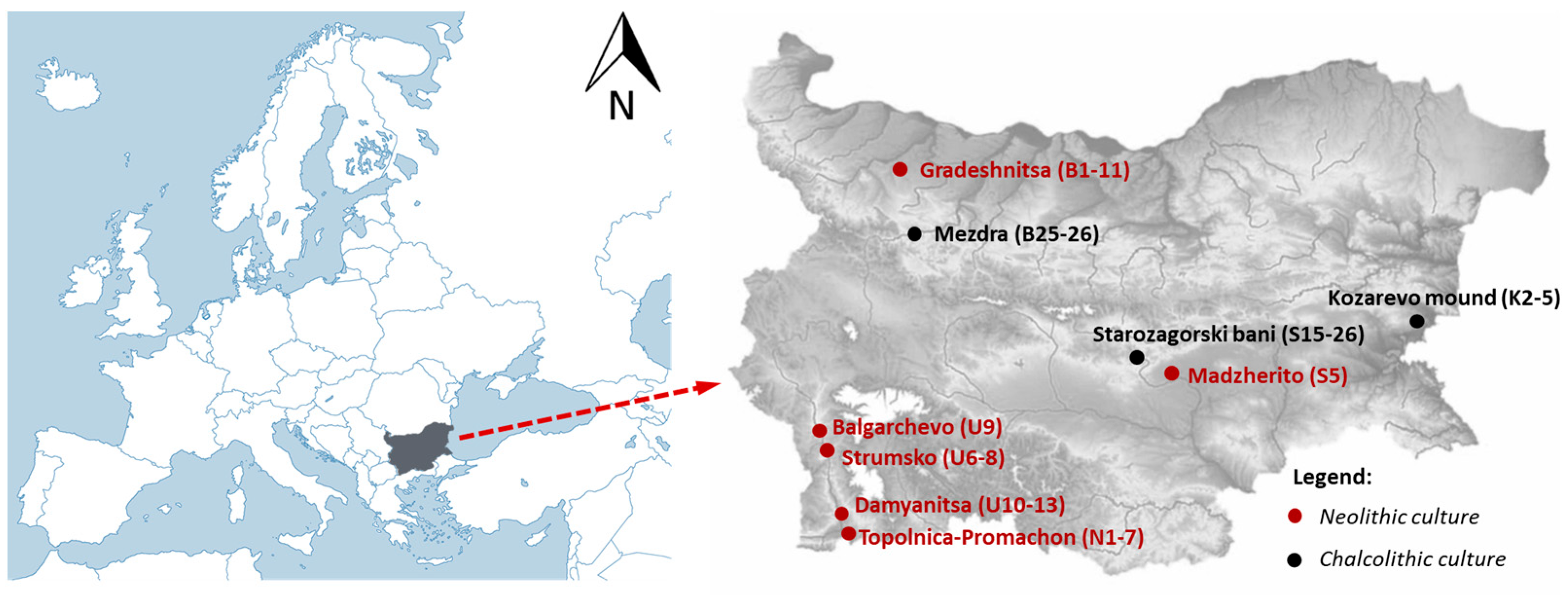
Figure 1. Map of Bulgaria with archeological sites where the analyzed pottery sherds were collected.
Minerals 15 00877 g001
Figure 2. Biplot of the first two principal components resulting from PCA of LIBS data of the red and dark decorations. The blue scales, right and top, refer to the loadings; the black scales, left and bottom, refer to the scores.
Minerals 15 00877 g002
Figure 3. Exemplary LIBS spectra comparing the decoration and the corresponding engobe of one of the dark-decorated sherds (sherd N4), highlighting two spectral regions that include barium lines (at 577.76 nm, 582.63 nm, 585.37 nm, 597.17 nm, 599.71 nm, and 606.31 nm) and manganese lines (at 601.35 nm, 601.66 nm, and 602.18 nm).
Minerals 15 00877 g003
Figure 4. Representative section of spectra from two selected sherds indicating differences in the titanium spectral lines (at 498.17 nm, 499.11 nm, 499.95 nm, 500.72 nm) intensities and comparing the presence of titanium in decorated areas with their corresponding engobes. Red-decorated sherd S15 (a) and dark-decorated sherd B5 (b).
Minerals 15 00877 g004
Figure 5. Representative ATR-FTIR spectra of selected samples illustrating (a) the presence of calcite (Cal), clay minerals, and hematite (Hem), which contribute to the red coloration of the paint; (b) characteristic features of hydromagnesite (Hmgs) and magnetite (Mag), which are associated with the dark coloration of the paint; (c) absorption bands of manganese oxides, which are also responsible for dark-colored paints; (d) spectral signatures of proteinaceous materials; and (e) bands attributed to lipids, suggesting the presence of oils or resins.
Minerals 15 00877 g005
Table 1. Synthesis of the pottery information with the corresponding photos (EN—Early Neolithic; LN—Late Neolithic; EC—Early Chalcolithic; LC—Late Chalcolithic). The type of decoration refers to the decoration technique. The marker in the photo represents 1 cm.
Sample PhotosSample IDType of DecorationEpochCultural AffiliationArcheological Site
Minerals 15 00877 i001U6paintLN/ECBalgarchevo IStrumsko
Minerals 15 00877 i002U7paintLN/ECBalgarchevo IStrumsko
Minerals 15 00877 i003U8paintLN/ECBalgarchevo IStrumsko
Minerals 15 00877 i004U9paintLN/ECBalgarchevo IBalgarchevo
Minerals 15 00877 i005U10paintLNTopolnica–AcropotamosDamyanitsa
Minerals 15 00877 i006U11paintLNTopolnica–AcropotamosDamyanitsa
Minerals 15 00877 i007U13paintLNTopolnica–AcropotamosDamyanitsa
Minerals 15 00877 i008N1paintLNTopolnica–AcropotamosTopolnica–Promachon
Minerals 15 00877 i009N2paintLNTopolnica–AcropotamosTopolnica–Promachon
Minerals 15 00877 i010N4paintLNTopolnica–AcropotamosTopolnica–Promachon
Minerals 15 00877 i011N5paintLNTopolnica–AcropotamosTopolnica–Promachon
Minerals 15 00877 i012N7paintLNTopolnica–AcropotamosTopolnica–Promachon
Minerals 15 00877 i013B1paintENGradeshnitsaGradeshnitsa, Malo Pole
Minerals 15 00877 i014B3paintENGradeshnitsaGradeshnitsa, Malo Pole
Minerals 15 00877 i015B4paintENGradeshnitsaGradeshnitsa, Malo Pole
Minerals 15 00877 i016B5paintENGradeshnitsaGradeshnitsa, Malo Pole
Minerals 15 00877 i017B6paintENGradeshnitsaGradeshnitsa, Malo Pole
Minerals 15 00877 i018B7paintENGradeshnitsaGradeshnitsa, Malo Pole
Minerals 15 00877 i019B8paintENGradeshnitsaGradeshnitsa, Malo Pole
Minerals 15 00877 i020B9paintENGradeshnitsaGradeshnitsa, Malo Pole
Minerals 15 00877 i021B10paintENGradeshnitsaGradeshnitsa, Malo Pole
Minerals 15 00877 i022B11paintENGradeshnitsaGradeshnitsa, Malo Pole
Minerals 15 00877 i023B25paintLCKrivodol–Sălcuţa–BubaniMezdra, Kaleto
Minerals 15 00877 i024B26paintLCKrivodol–Sălcuţa–BubaniMezdra, Kaleto
Minerals 15 00877 i025S5paintENKaranovo IMadzherito
Minerals 15 00877 i026S15Pseudo-incrustationLCKaranovo VIStarozagorski Bani
Minerals 15 00877 i027S16Pseudo-incrustationLCKaranovo VIStarozagorski Bani
Minerals 15 00877 i028S20Pseudo-incrustationLCKaranovo VIStarozagorski Bani
Minerals 15 00877 i029S21Pseudo-incrustationLCKaranovo VIStarozagorski Bani
Minerals 15 00877 i030S26Pseudo-incrustationLCKaranovo VIStarozagorski Bani
Minerals 15 00877 i031K2paintLC Kozarevo Mound
Minerals 15 00877 i032K3paintLC Kozarevo Mound
Minerals 15 00877 i033K4Pseudo-incrustationLC Kozarevo Mound
Minerals 15 00877 i034K5Pseudo-incrustationLC Kozarevo Mound
Table 2. Characteristic spectral lines employed for elemental identification in LIBS spectra. The Roman numerals I and II denote spectral lines emitted from neutral atoms and singly ionized atoms, respectively.
ElementWavelengths (nm)
Ca315.89 II, 317.93 II, 393.36 II, 396.85 II, 457.85 I, 458.14 I, 527.03 I *, 671.77 I
Si250.69 I *, 251.61 I, 252.41 I, 252.85 I, 288.16 I
Al308.22 I *, 309.27 I, 394.40 I, 396.15 I
Mg279.55 II, 280.27 II, 285.21 I, 382.93 I, 383.23 I, 516.73 I *
Na449.42 I *, 589.00 I, 589.59 I
K404.41 I *, 693.88 I
Fe271.90 I, 275.01 I, 293.69 I *, 302.06 I, 356.54 I, 357.01 I, 358.12 I, 374.55 I
Mn259.37 II, 403.08 I, 403.31 I, 403.45 I, 475.40 I *
Ti334.94 II, 336.12 II, 337.75 I *, 338.38 II, 498.17 I, 499.11 I, 499.95 I, 500.72 I
Sr407.77 II, 421.55 II *, 460.73 I
Ba455.40 II, 493.41 II *, 553.55 I
* Characteristic spectral lines employed to perform the statistical analysis with PCA.
Table 3. Mineral content of the decorations on the sherds with their corresponding IDs. (tr—traces).
Sample IDMineral Composition
U6quartz, magnetite, hematite, and hydromagnesite
U7quartz, magnetite, hematite, and protein (tr) (in brown) // quartz magnetite, hematite, and organic (tr) (in red)
U8quartz, magnetite, and hematite
U9quartz and hematite (brown) // quartz, hematite, and magnetite (red)
U10quartz, magnetite, and hematite
U11quartz, magnetite, gypsum (tr), and hydromagnesite
U13quartz, magnetite, and hematite
N1Magnetite and hematite
N2quartz, magnetite, calcite, and lipids (tr)
N4quartz, magnetite, hematite, and calcite
N5quartz, magnetite, and hematite
N7quartz and hematite
B1quartz, hematite, and magnetite
B3quartz and magnetite
B4quartz and manganese oxides
B5quartz, hematite, calcite, and organic (tr)
B6quartz, hematite, magnetite, manganese oxides, and calcite (tr)
B7quartz, hematite, manganese oxides, and protein (tr)
B8quartz, hematite, and carbonate (tr) (brown)
B9quartz, hematite, magnetite, and carbonate (tr) (brown) // quartz, hematite, carbonate (tr), and organic (tr) (red)
B10quartz, hematite, manganese oxides, carbonate (tr), and lipids (tr) (black) // quartz, hematite, carbonate (tr), and lipids (tr) (red)
B11quartz, hematite, magnetite, and calcite tr (brown) // quartz and hematite (red)
B25quartz, kaolinite, and hematite
B26calcite, hematite, kaolinite, and quartz (red) // calcite, hematite, kaolinite, and quartz (yellow)
S5quartz, hematite, and calcite
S15quartz, hematite, calcite (tr), and organic (tr)
S16quartz, hematite, magnetite, and calcite (tr)
S20quartz, hematite, and calcite (tr)
S21quartz, hematite, and calcite
S26quartz, hematite, calcite, and magnetite (tr)
K2quartz, kaolinite, and hematite
K3quartz and hematite
K4quartz, hematite, and magnetite
K5quartz and hematite
Disclaimer/Publisher’s Note: The statements, opinions and data contained in all publications are solely those of the individual author(s) and contributor(s) and not of MDPI and/or the editor(s). MDPI and/or the editor(s) disclaim responsibility for any injury to people or property resulting from any ideas, methods, instructions or products referred to in the content.