Article

Kinetic Control of Oxygenated Apatites: Dynamic Operation of a Pilot-Scale Precipitation Reactor for Bone-Mimetic Biomaterials

1
Laboratory of Biology and Health, University Hassan II of Casablanca, Casablanca 20360, Morocco
2
Laboratory of Process Engineering and Environment, University Hassan II of Casablanca, Casablanca 20360, Morocco
*
Author to whom correspondence should be addressed.
Minerals 2025, 15(7), 700; https://doi.org/10.3390/min15070700
Submission received: 27 May 2025 / Revised: 27 June 2025 / Accepted: 28 June 2025 / Published: 30 June 2025
(This article belongs to the Section Biomineralization and Biominerals)

Abstract

This study investigates the dynamic operation of a pilot-scale precipitation reactor designed to produce oxygenated phosphocalcium apatites with controlled composition and low crystallinity, closely mimicking the mineral phase of bone. Our approach is based on integrating kinetic monitoring and dynamic reactor control to direct the formation of apatites with tailored structural and chemical properties. Three synthesis routes were explored using CaCO3, Ca(NO3)2, and CaCl2 as calcium precursors, under optimized Ca/P molar ratios. The evolution of ionic concentrations (Ca2+, PO43−), peroxide and molecular oxygen incorporation, and carbonate content was monitored over a reaction time range of 2 min to 4 h. Characterization by X-ray diffraction (XRD), Fourier-transform infrared spectroscopy (FTIR), scanning electron microscopy (SEM), and chemical analysis revealed a time-dependent transformation of amorphous phases into poorly crystalline apatites with specific textures. After 60 min, the Ca/P atomic ratio stabilized at approximately 1.575, and the resulting apatites exhibited structural features comparable to those of human bone. This study highlights the influence of reactor operation time on precipitation kinetics and the properties of bioactive apatites in a scalable system. The results offer promising prospects for the large-scale production of bone-mimetic materials. However, the lack of biological validation remains a limitation. Future studies will assess the cytocompatibility and bioactivity of these materials to confirm their potential for biomedical applications.

1. Introduction

Phosphocalcium apatites are a widely studied class of bioceramics due to their close resemblance to the mineral phase of tooth and bone tissues [1,2,3,4,5,6,7,8]. Their chemical composition, crystallinity, and morphology directly influence their biological properties, including biocompatibility, resorption rate, and their capacity to bind or release therapeutic agents [9,10,11,12,13,14].
Particular attention has been given to the incorporation of oxygenated species (e.g., peroxide and molecular oxygen) into the apatite structure, given their potential for controlled oxygen release and enhanced regenerative or antiseptic effects [15,16,17,18,19,20,21,22,23,24,25,26,27,28,29,30,31,32,33]. These doped apatites offer promising avenues in bone and dental engineering.
Recently, oxygen-releasing systems such as hydrogels, polymeric scaffolds, and microbubbles have been widely investigated for enhancing tissue regeneration and local antimicrobial activity [34,35,36,37]. While these systems provide a rapid burst of oxygen, their release kinetics and long-term stability remain challenging. In this context, oxygen-doped apatites represent a promising alternative due to their inherent biocompatibility, mineral nature, and gradual degradation profile.
However, the synthesis of oxygen-rich apatites remains a challenge, especially when transitioning from laboratory to pilot or industrial scale. The precipitation process is greatly influenced by parameters such as temperature, pH, Ca/P molar ratio, and reaction time, all of which impact the incorporation and stability of oxygenated groups [15,16,17,18,19,20,21,22,23,24,25,26,27,28,29,30,31,32,33]. Although several works have addressed the role of synthesis parameters on a laboratory scale, few studies have investigated the kinetic evolution of these materials under dynamic operating conditions or the impact of reactor control strategies on their final structure and functionality [38,39,40].
Indeed, the precipitation duration, variation in reactive species concentrations in solution, and the nature of calcium precursors influence not only the chemical composition of the products formed but also their crystallinity, richness in functional groups, and microstructure [41,42,43,44,45,46,47].
In our previous works, we proposed a series of three studies dedicated to optimizing the synthesis conditions of oxygenated phosphocalcium apatites prepared from the reactive pairs CaCO3–H3PO4, Ca(NO3)2–H3PO4, and CaCl2–H3PO4. These investigations enabled us to determine the optimal conditions for obtaining stable oxygenated apatites with an atomic Ca/P ratio of 1.575 [21,23,30], a value known to influence not only crystallinity and stoichiometry but also dissolution behavior in physiological environments, which is crucial for bone remodeling and bioresorption processes.
Nevertheless, the relationship between precipitation kinetics, reactor operation, and the structural evolution of the resulting products has not been thoroughly explored.
The present study introduces a novel approach based on real-time kinetic monitoring and dynamic control of a pilot-scale precipitation reactor, aiming to guide the formation of oxygenated apatites with tailored structural and chemical properties. This strategy enables the precise tuning of operating time to modulate key characteristics including crystallinity, Ca/P ratio, and oxygenation level. The evolution of ionic concentrations (Ca2+, PO43−), peroxide decomposition, and crystallographic transformation are systematically studied as a function of reactor operation time.
This work provides a scalable and robust route for synthesizing functional oxygenated apatites. These materials are particularly suitable for bone regeneration and controlled antiseptic delivery, bridging the gap between bench-scale synthesis and the manufacture of clinically relevant biomaterials.

2. Materials and Methods

2.1. Chemicals

The syntheses were carried out using three calcium precursors: calcium carbonate (CaCO3, purity ≥ 99%, Merck, Darmstadt, Allemagne), calcium nitrate tetrahydrate (Ca(NO3)2.4H2O, Sigma-Aldrich), and calcium chloride dihydrate (CaCl2.2H2O, Prolabo, Fontenay-sous-Bois, Paris, France). Phosphoric acid (H3PO4, 84%, Merck, KGaA, Darmstadt, Allemagne) acted as the phosphorus source. Fresh hydrogen peroxide solution (H2O2, 30%, Sigma-Aldrich, St. Louis, MI, USA) was used as the oxidizing agent. All reagents used in this study were of analytical grade.

2.2. Synthesis Conditions

The syntheses were conducted using an aqueous precipitation method at a controlled temperature (40 ± 1 °C) and an adjusted pH, regulated with an ammonia solution (NH4OH, 25% purity, d = 0.92)
Table 1 summarizes the experimental conditions, including pH, Ca/P molar ratio, and reaction duration (D), used for each calcium precursor (CaCO3, Ca(NO3)2·4H2O, and CaCl2·2H2O). These parameters were the result of prior systematic optimization studies [21,23,30], aimed at maximizing peroxide incorporation and phase stability while maintaining good structural and chemical reproducibility with Ca/P = 1.575.
All syntheses were carried out in a 1 L pilot-scale glass reactor equipped with a mechanical stirrer, a thermostatic jacket, and a pH-stat probe enabling automatic and continuous pH adjustment (Figure 1). This system was designed according to a hydrodynamic scaling approach specifically adapted for precipitating media. This methodological choice ensures the reproducibility of operating conditions while enabling the investigation of the impact of reactor operating time on the precipitation kinetics and structural evolution of the solids.
The reactor operating time was modulated between 2 min and 4 h, with samples taken at regular intervals.
The apatites derived from the three calcium salts, CaCO3, Ca(NO3)2, and CaCl2, shall be reported sequentially: Ap.OxCaCO3, Ap.OxCa(NO3)2, and Ap.OxCaCl2.
All experiments were performed in triplicate, at least, to ensure the reproducibility of the results. The values reported correspond to the means of three replicate measurements. The low variability observed (<±0.01) justified the use of mean values without graphical error bars.

2.3. Precipitate Treatment

At each sampling time, the precipitate formed was immediately filtered using a filter press and then dried in a freeze-dryer. The dry samples were ground and stored in a desiccator until they were analyzed.

2.4. Characterization Methods

The filtrates were analyzed by chemical analysis, and the powders were examined using chemical analysis, infrared spectrometry, X-ray diffraction, and scanning electron microscopy.
Calcium content was determined by complexometric titration using EDTA (ΔCa = 0.005) [48], phosphorus was quantified by colorimetry using the ammonium molybdate–vanadate method (ΔP = 0.005) [49], molecular oxygen was estimated by acid attack of the powder followed by gas volume displacement measurement, using soda asbestos to adsorb the released (CO2 (Δ(%O2) = 0.02). A second measurement, performed without soda asbestos, allowed for the determination of the carbonate ion content by difference (Δ(%CO2) = 0.02) [17]. Peroxide ions were quantified by manganometric titration using KMnO4 under acidic conditions (Δ(%O22−) = 0.01) [50]. X-ray diffraction analysis was conducted using a SEIFERTXRD 3000 P diffractometer equipped with CuKα radiation (λ = 1.5406 Å), operated at 40 kV and 30 mA. Scans were performed over a 2θ range of 25–50° with a step size of 0.02° and a counting time of 2 s per step. Data processing was performed using WinXPOW software version 2.22 (GE Inspection Technologies, Germany). For infrared absorption analysis, 1 mg of the powdered sample was thoroughly mixed with 300 mg of KBr and then compressed into pellets under vacuum. The pellets were analyzed using a PerkinElmer Spectrum 1600 FTIR spectrometer, operated using Spectrum software, version 6.3.1 (PerkinElmer Inc., Waltham, MA, USA). Spectra were recorded in the range of 4000–400 cm−1 at a resolution of 4 cm−1 with 32 scans. Scanning electron microscopy (SEM) was performed with a JEOL JSM-6490 LV microscope, using JEOL software, version 1.0.1 (JEOL Ltd., Tokyo, Japan). Samples were coated with carbon to a thickness of approximately 10 nm using a sputter-coater. Observations were performed under high vacuum conditions with an accelerating voltage of 20 kV.
All analyses (XRD, FTIR, SEM) were performed on samples obtained from at least three independent experiments to ensure the reproducibility of the results.

3. Results

3.1. Study of the Physicochemical Characteristics of the Products Obtained as a Function of Reactor Operating Time

3.1.1. Study by Chemical Analysis

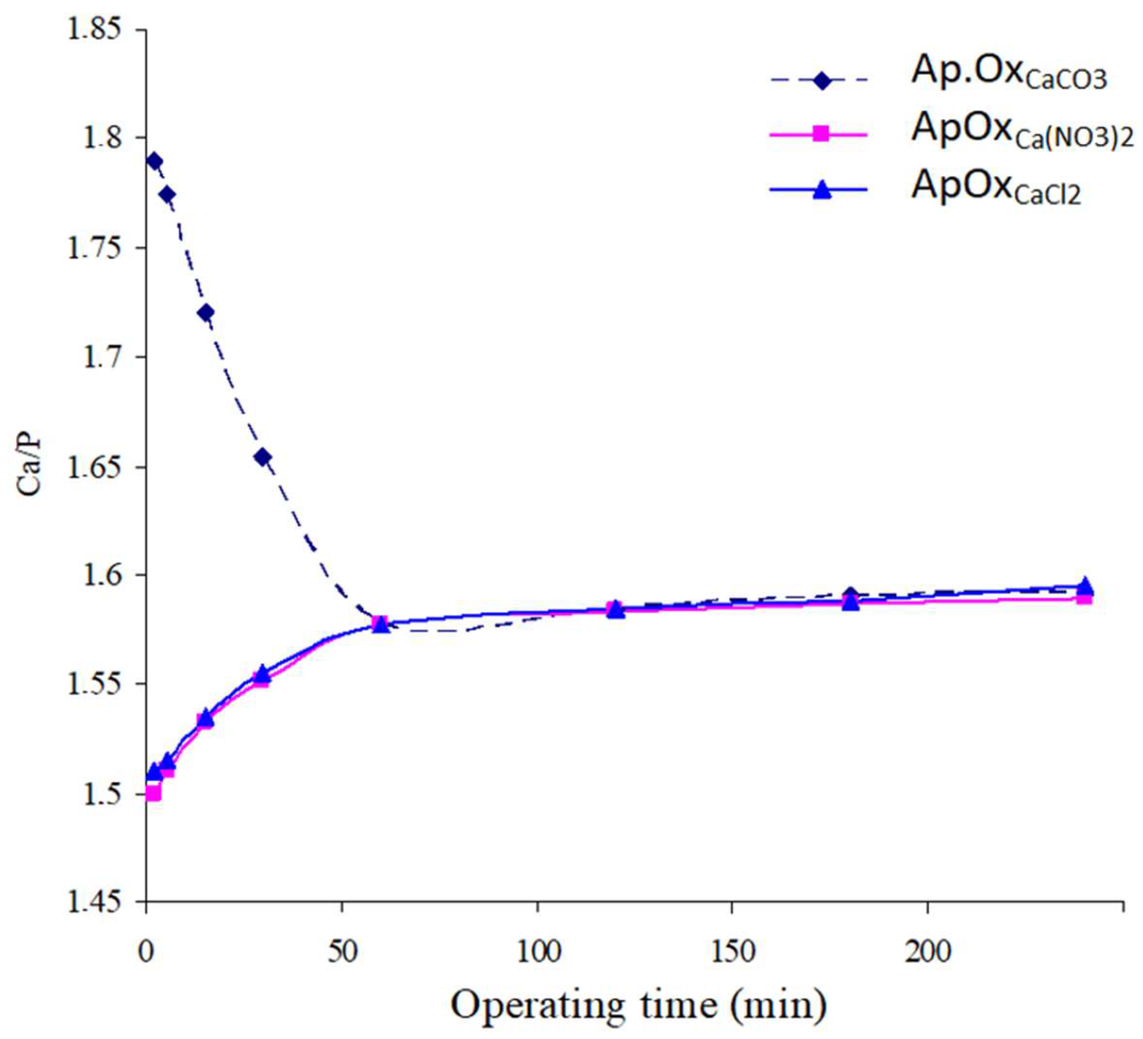
Variation of the Solids’ Ca/P Atomic Ratio as a Function of Time
The variations in the solids’ Ca/P atomic ratio as a function of reactor operating time for the three syntheses are shown in Figure 2. The following observations can be made:
  • In the case of Ap.OxCaCO3, the Ca/P atomic ratio decreases rapidly with increasing reactor operating time, stabilizing at approximately 1.578. The precipitate formed at the first moments of the reaction (2 min), with a ratio of 1.79, contains calcium carbonate. During the reactor operating time, the latter reacts with phosphorus to give an apatite with a ratio of 1.578.
  • In the case of Ap.OxCa(NO3)2 or Ap.OxCaCl2, the solids’ Ca/P atomic ratio increases continuously until it reaches a value of 1.577 after one hour. Beyond this time, the Ca/P ratio shows a slight increase.
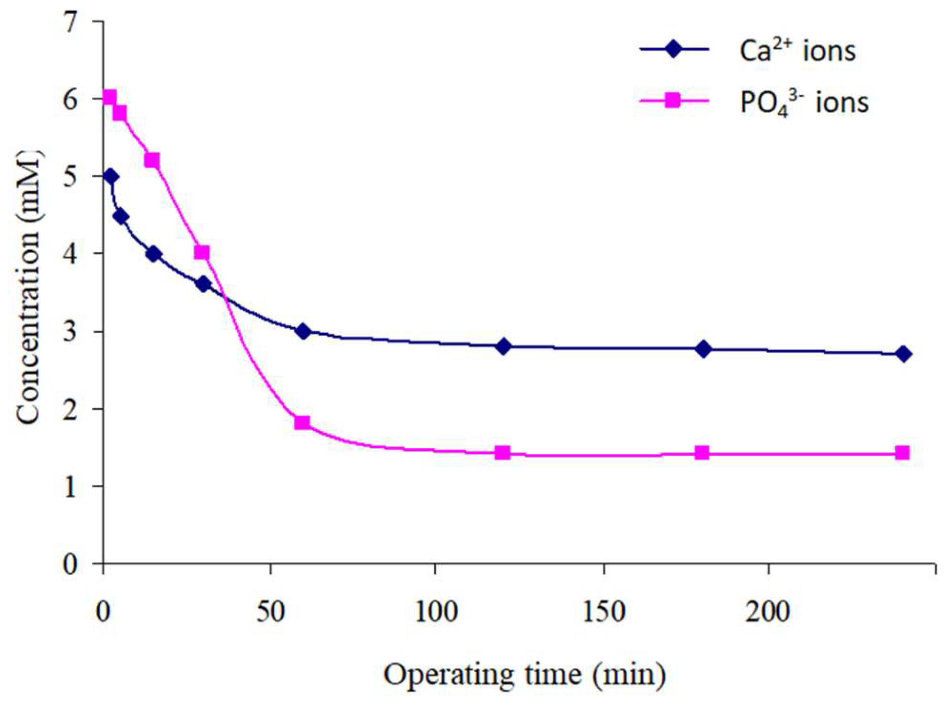
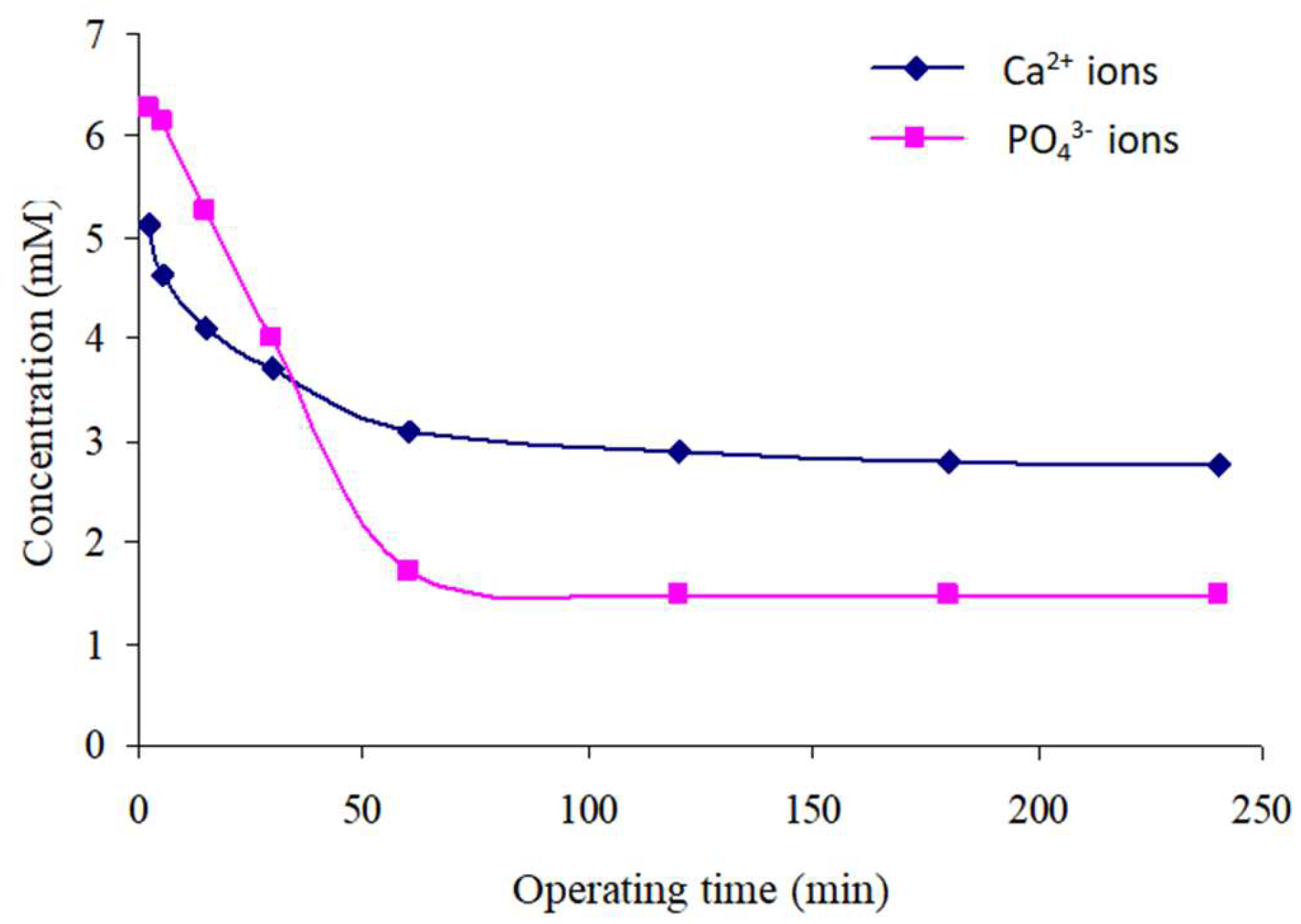
Variation in the Concentrations of Free Ca2+ and PO43− Ions in Solution over Time
Figure 3, Figure 4 and Figure 5 shows the changes in the free concentrations of Ca2+ and PO43− ions in solution over time.
The PO43− ion concentration in all three syntheses decreases rapidly to a nearly stable value (2.22 mM (Ap.OxCaCO3), 1.43 mM (Ap.OxCa(NO3)2), and 1.48 mM (Ap.OxCaCl2)) after approximately 30 min.
The same phenomenon occurs during the evolution of the Ca2+ ion concentration in solution while synthesizing calcium nitrate or chloride. However, in the case of synthesis from calcium carbonate, the evolution of the Ca2+ ion concentration is different:
  • The first stage represents the initial moments of precipitation and extends until approximately 2 min. We observe a sudden increase in Ca2+ ion concentrations, followed by a rapid decrease within the first 15 min.
  • The second stage, located beyond 15 min, experiences a continuous increase in Ca2+ ion concentration according to a logarithmic law. This concentration becomes almost stable at 14.6 mM after approximately 1 h of reactor functioning.
This fluctuation in the concentrations of Ca2+ ions in the solution during the reactor’s dynamic functioning is due to the dissolution rate of calcium salts in the solution. Calcium nitrate and calcium chloride dissolve rapidly, while calcium carbonate remains in suspension.
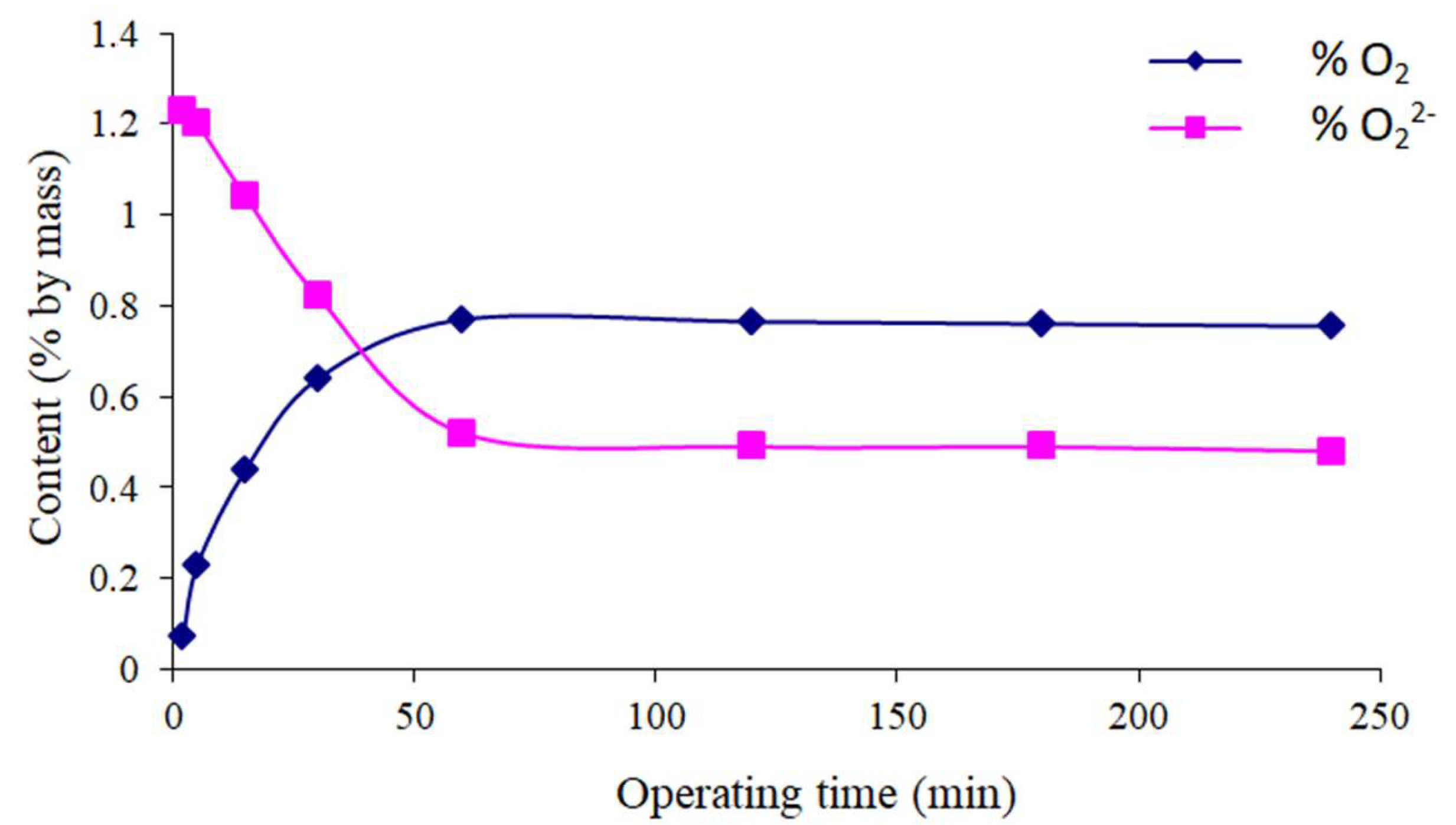
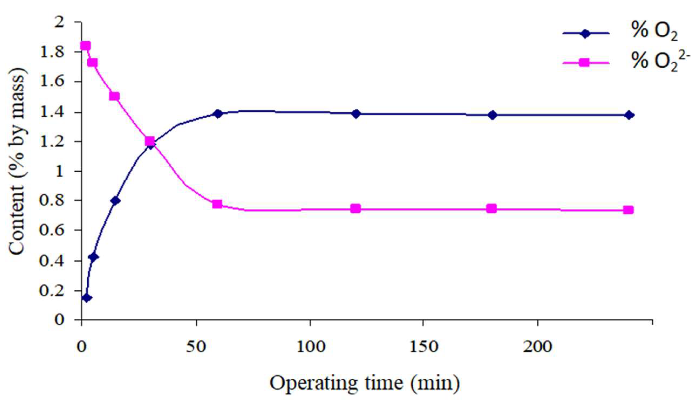
Variation of the Oxygenated Species Content of Solids as a Function of Time
The chemical analysis of the precipitates obtained (Figure 6, Figure 7 and Figure 8) shows that, initially, the precipitates contain numerous peroxide groups and little molecular oxygen. Over time, the quantity of peroxide groups decreases slightly while that of molecular oxygen increases. This mechanism causes an intra-crystalline decomposition of the peroxide groups in the apatitic structure and the insertion of additional peroxide groups from the solution [16].
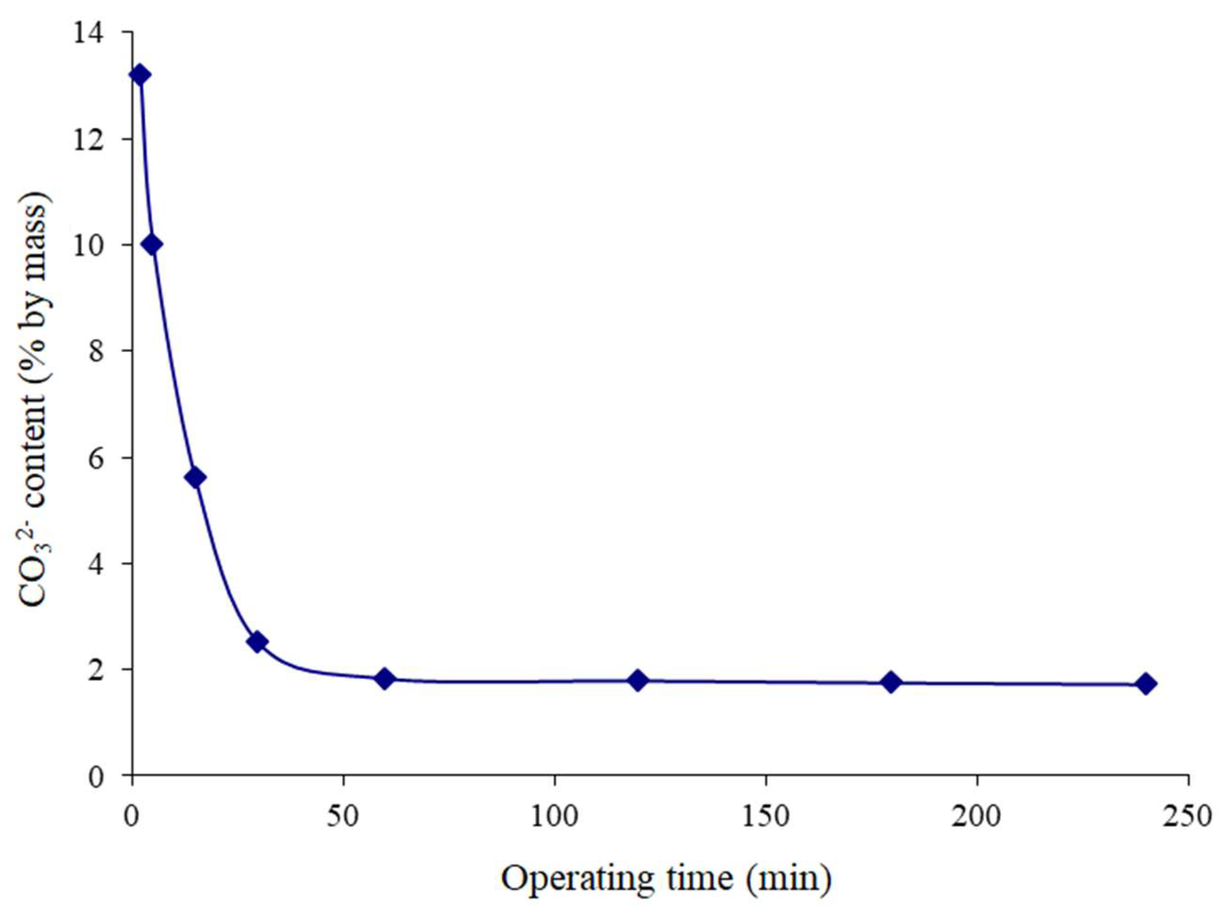
Variation in the Solid’s Carbonate Ion Content Prepared from Calcium Carbonate as a Function of Time
Figure 9 depicts the evolution of the carbonate ion content of the solid derived from calcium carbonate as a function of the reactor operating time. A sharp drop in carbonate ions is observed in the precipitates obtained during the first 30 min of reactor functioning. Indeed, the precipitate obtained after 2 min of reaction contains 13.2% carbonate. This confirms the presence of unreacted calcium carbonate mixed with the precipitates collected in the first minutes of reactor functioning.
After one hour of functioning, the precipitate contained only 1.83% carbonate. Beyond this time, the decrease in the carbonate ion content becomes slight.

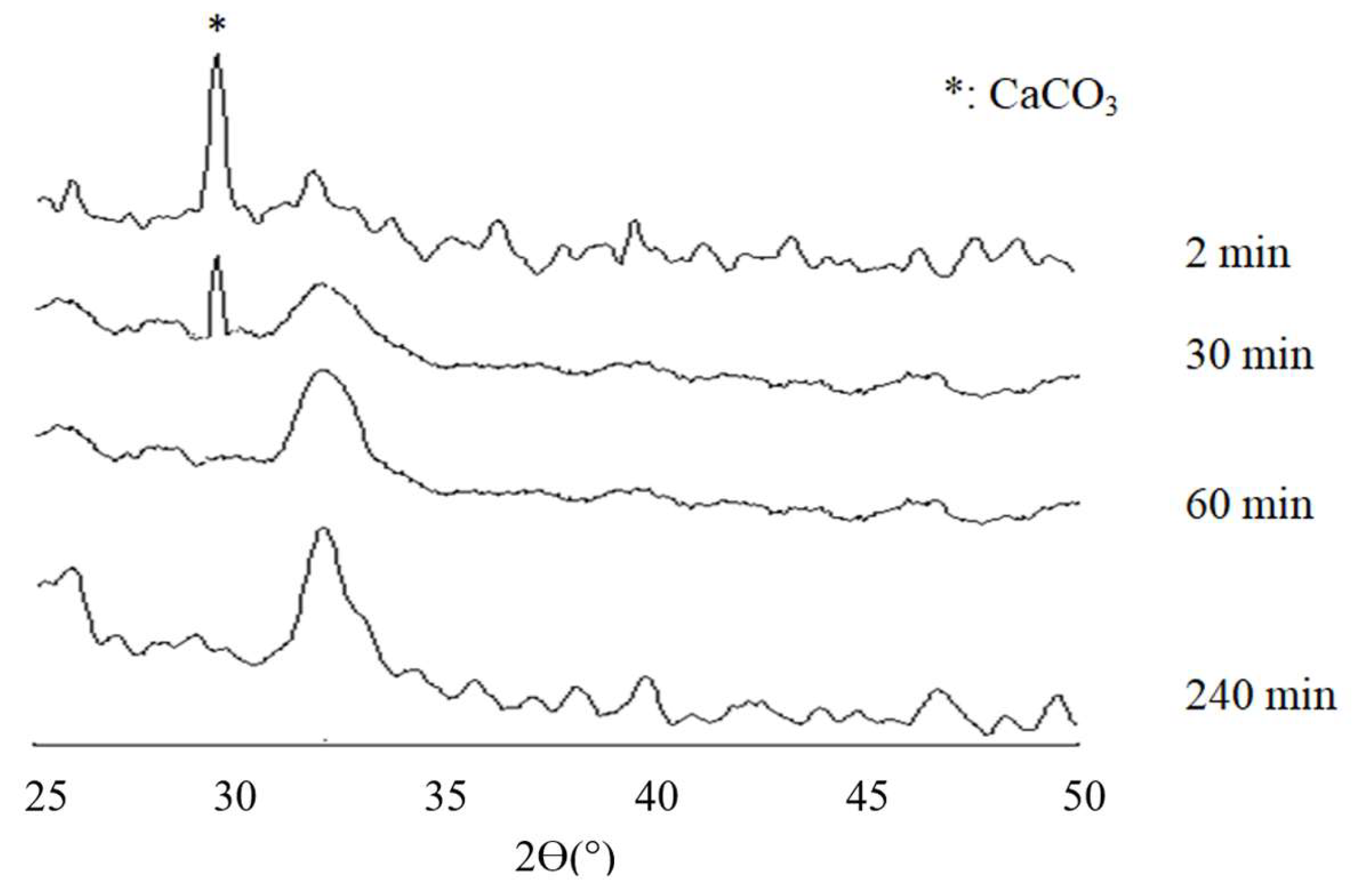
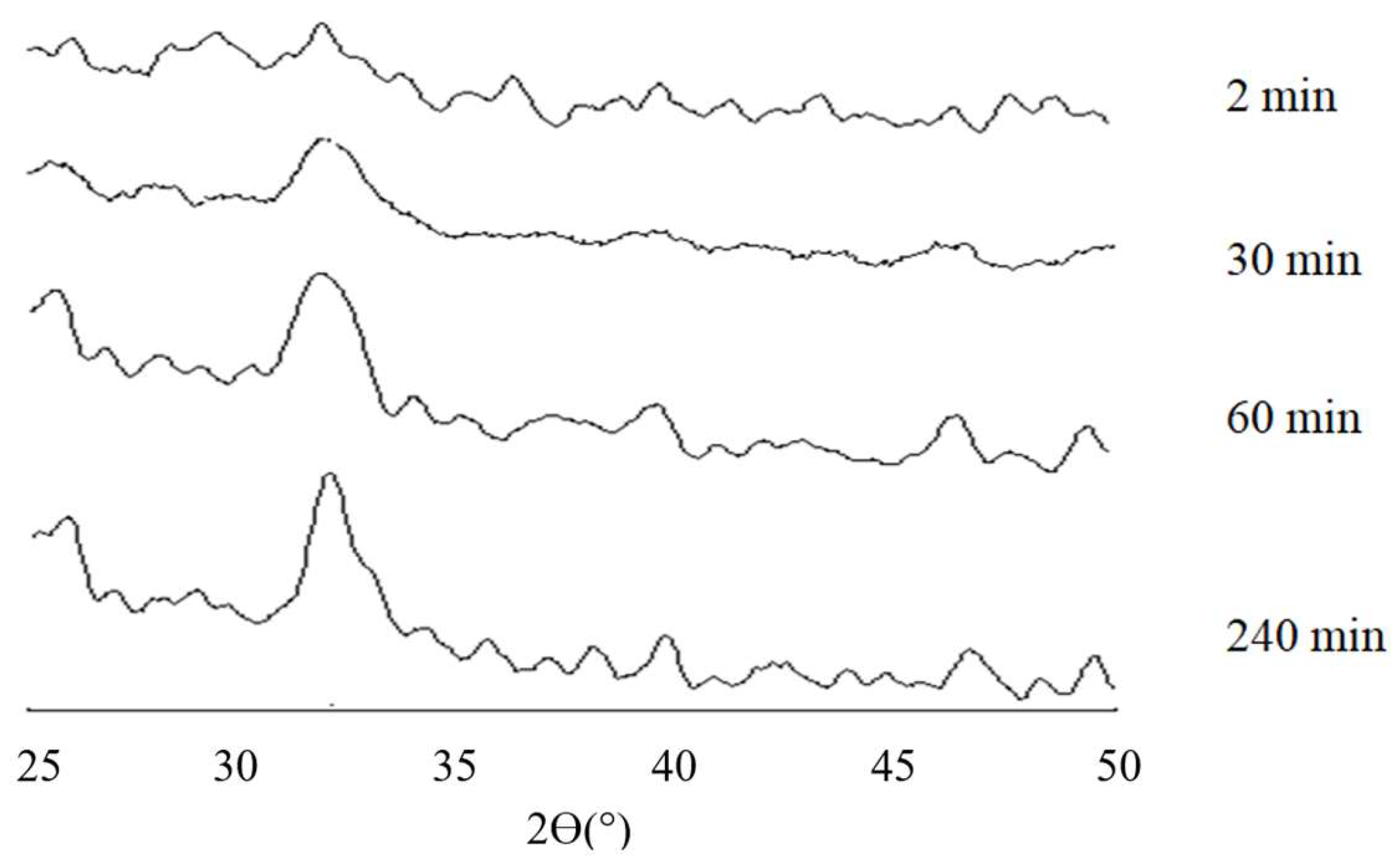
3.2. X-Ray Diffraction Analysis

The patterns of X-ray diffraction of the products obtained from calcium carbonate as a function of reactor operating time are displayed in Figure 10. The patterns of the products received in the initial phases of the reaction revealed the presence of an intense peak (located at 2θ = 29.45′) characteristic of calcium carbonate [51]. The intensity of this peak gradually decreases over time until it disappears after 1 h of reaction. This result is consistent with the findings of chemical analyses (Ca/P and % CO32−). Depending on the reactor’s operating time, these patterns also indicate the transformation of the precipitate from an amorphous to a poor crystalline state.
Due to the similar evolution of the products’ X-ray diffraction patterns derived from calcium nitrate and those prepared from calcium chloride, we have only presented those of Ap.OxCa(NO3)2 (Figure 11). These patterns show an amorphous state of the precipitates that gradually evolves, as a function of the operating time, to a poorly crystalline state similar to that of bone [52,53,54]. The presence of lines characteristic of other elements could not be detected.

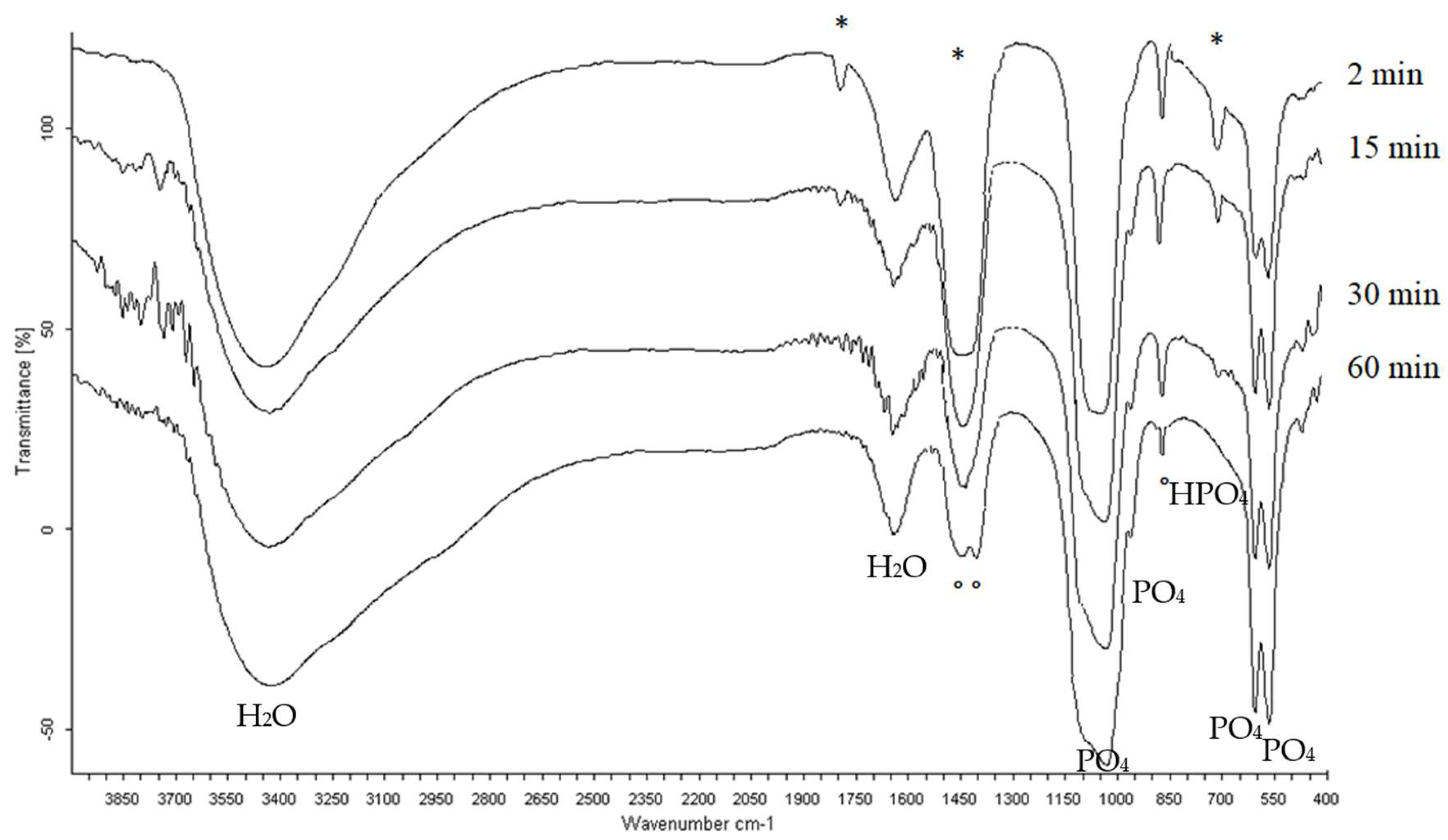
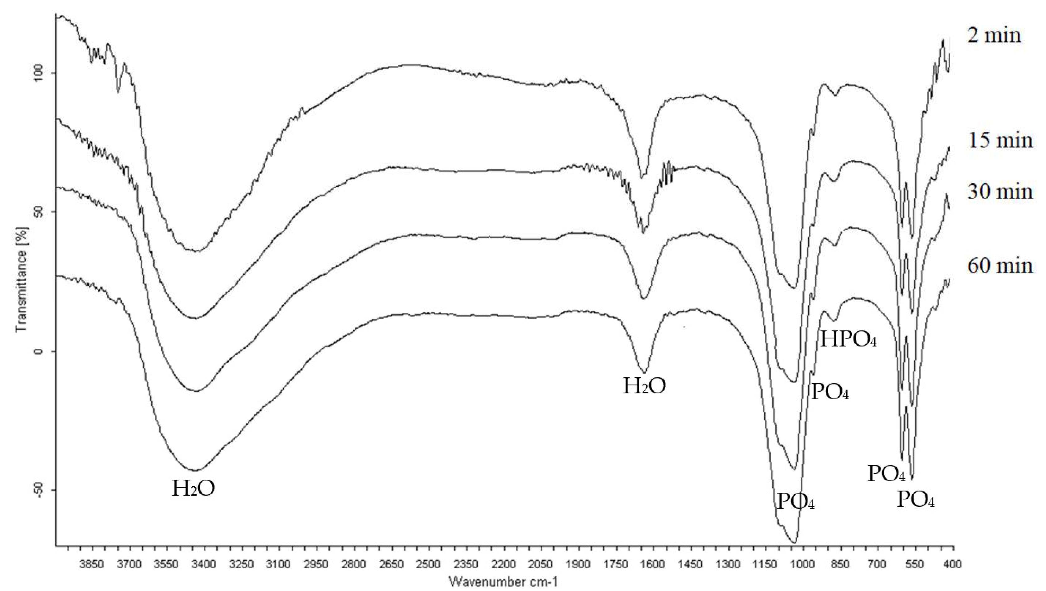
3.3. Infrared Spectrometry Analysis

The infrared absorption spectra of prepared products from calcium carbonate, obtained at different reactor operating times, are depicted in Figure 12. The infrared spectra of the precipitates formed at the first moments of the reaction show, in addition to the bands attributable to the ions (PO43−, HPO42−) characteristic of non-stoichiometric apatites, the existence of bands due to carbonate ions (714, 1480, and 1800 cm−1) characteristic of the presence of calcite [52,55] which has not yet reacted. Beyond 30 min of reactor functioning, the two bands at 714 and 1800 cm−1 disappear. The band located at 1480 cm−1 undergoes a splitting into two bands (1412 and 1462 cm−1) that are associated, according to Vignoles [56], with the carbonate ions situated in the B-type sites of the apatitic structure. This validates the findings of the X-ray diffraction and chemical analyses.
The infrared spectrum of the products obtained from calcium nitrate and calcium chloride evolve similarly during reactor functioning. This is why we have chosen to present only those of Ap.OxCa(NO3)2 (Figure 13). These spectra only reveal the characteristic bands of apatites from the first minutes of precipitation (2 min).

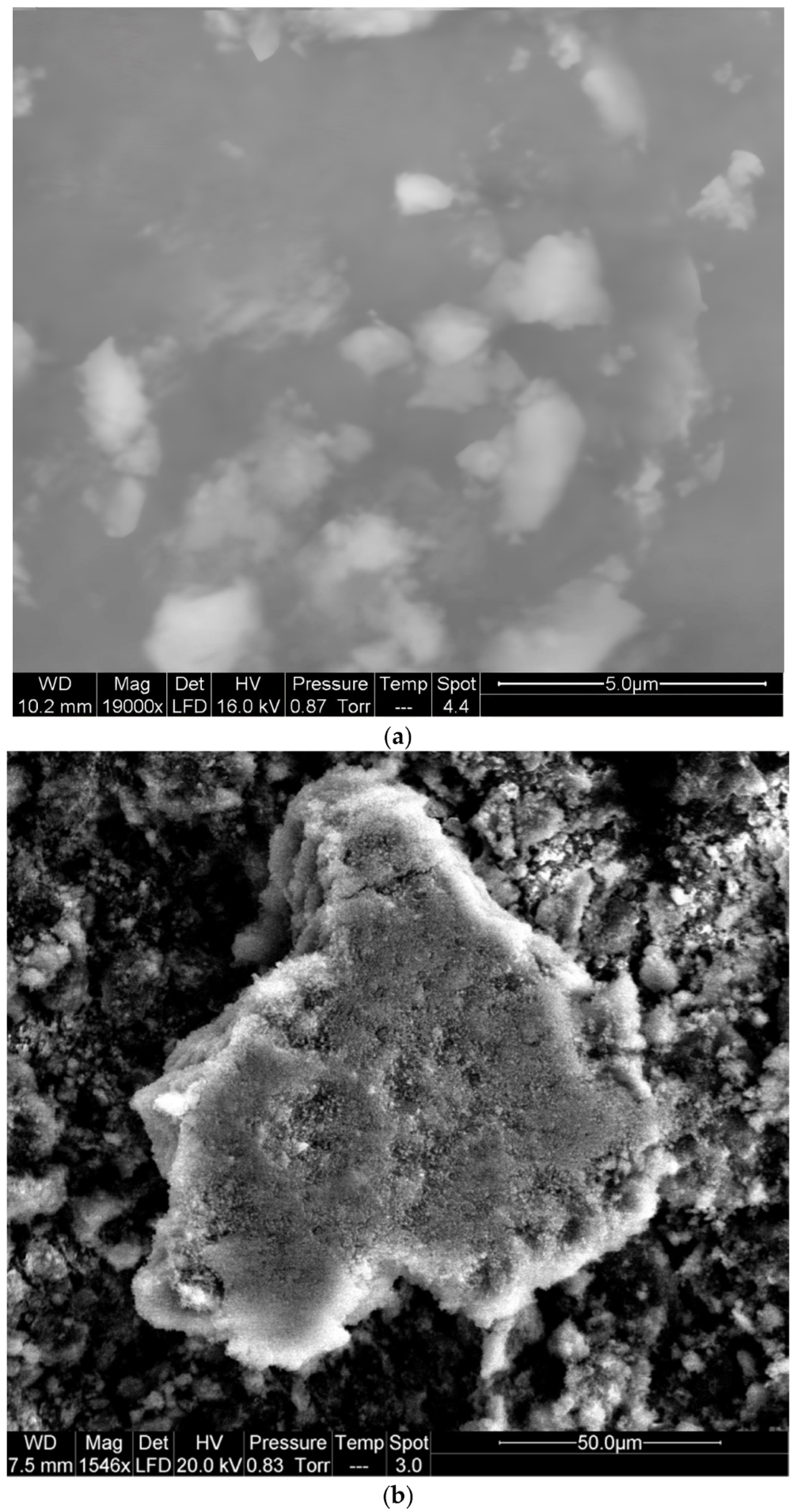
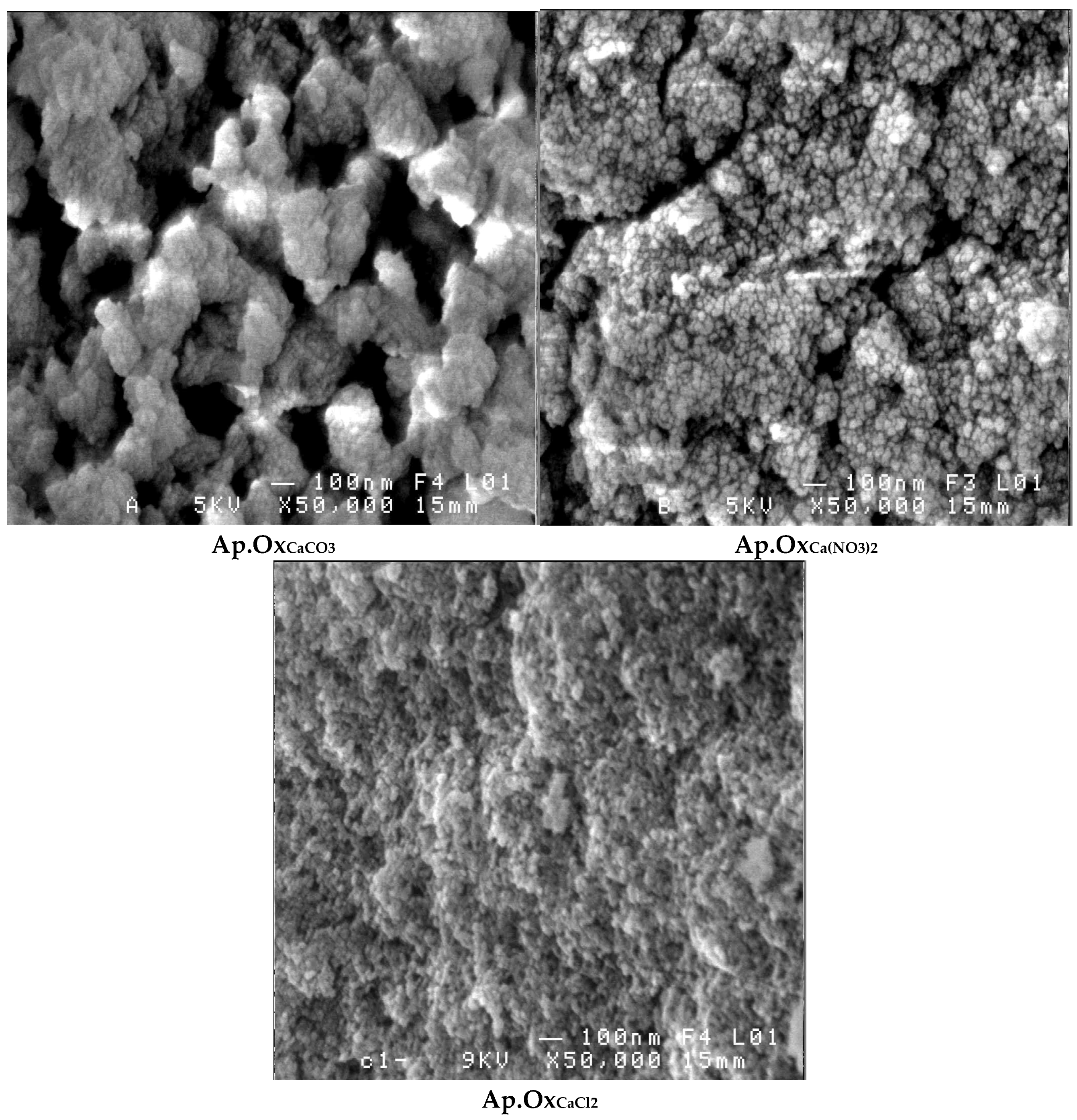
3.4. Scanning Electron Microscopy Analysis

Scanning electron microscopy of the precipitates obtained from the three syntheses of phosphocalcium oxygenated apatites over the reactor’s operating time reveals that the apatite phase forms through the transformation of the amorphous phase. Indeed, an amorphous phase is observed from the first minutes of reactor functioning (Figure 14a). After 5 min of functioning, this phase is no longer observable by electron microscopy, and the particles are arranged in dense clusters surrounded by less dense parts (Figure 14b).
These findings are not sufficient enough for us to conclude that these apatites are formed by growth on the surface of amorphous clusters, as reported by Eanes et al. [57], or by internal conversion of the amorphous phase, as reported by Heughebaert [52].
After four hours of reactor functioning, the scanning electron microscopy examination of the three apatites obtained showed that their structures differ (Figure 15); Ap.OxCaCO3 is in a porous form and Ap.OxCa(NO3)2 is less porous, while Ap.OxCaCl2 is dense.

4. Discussion

During the first moments of reactor functioning, supersaturation of soluble ionic species is established. This enables the precipitation of amorphous phosphate in the form of seeds, the nucleation step that is essential for the precipitation of the solid in solution [58,59,60].
When germs form in the initial stages of the reaction, the solid precipitates (Figure 1). It contains many peroxide groups and little molecular oxygen (Figure 5, Figure 6 and Figure 7). XRD and SEM analyses reveal that this product is amorphous (Figure 9, Figure 10, Figure 14 and Figure 15). During reactor operation, the amount of peroxide groups decreases while that of molecular oxygen increases, according to a mechanism of intra-crystalline decomposition of peroxide ions into molecular oxygen [16]. This mechanism has also been investigated in peroxide-substituted biomimetic apatites by Ana et al. (2022), who reported that peroxide groups can be stabilized in the apatitic lattice and gradually decomposed under mild physiological conditions without compromising biocompatibility [31].
Additionally, the Ca/P ratio approaches the value of 1.575, ultimately producing an apatite whose crystallinity is comparable to that of the mineral phase of natural bone (Figure 11 and Figure 12) [52,53,54].
The evolution of the apatites obtained from the amorphous state to the poorly crystalline state and not to the well-crystalline state, under the preparation conditions that we have predefined, is certainly due to the temperature (T = 40 °C) at which the reactor was maintained. Indeed, Heughebaert [52] demonstrated that the transformation of the amorphous phase into apatite occurred much more rapidly at higher synthesis temperatures. Boskey and Posner [61] reported that apatite precipitation is systematically preceded by the formation of an amorphous precursor phase. The conversion of this precursor into the apatitic phase was described as an autocatalytic process that is kinetically dependent on temperature and pH. Eanes et al. [57] also confirmed the existence of an amorphous precursor with a variable composition that evolves towards a well-defined crystalline phase, i.e., the OCP. The latter, being relatively labile, hydrolyzed to form well-crystallized stoichiometric phosphocalcium hydroxyapatite, with the transformation accelerating at higher temperatures.
These results confirm that controlling the reactor’s operating time serves as a direct lever to modulate the crystallinity, porosity, and oxygen incorporation. These parameters are critical for adjusting the dissolution rate, the release of active species, and ultimately the performance in bone regeneration.
Unlike most studies conducted at the laboratory scale, this work proposes a reproducible and scalable strategy. A kinetic-based approach ensures the stability of physicochemical properties within the pre-dimensioned reactor; this is a promising criterion for potential biomedical applications.
This methodology could be extended to other complex precipitating systems (such as doped hydroxyapatites or functionalized mesoporous materials) where kinetic–structural interactions play a crucial role. It also paves the way for the engineering of multifunctional materials with controlled release capabilities.
Furthermore, the biological relevance of oxygenated apatites remains a key challenge. The incorporation of peroxide ions raises concerns about their in vivo stability and potential cytotoxicity. However, recent studies have shown that such materials can release oxygen in a sustained and safe manner, offering antibacterial effects while maintaining cytocompatibility [31]. Although biological tests have not yet been conducted, the present findings suggest a promising clinical potential for the synthesized material. Future work will involve in vitro cytotoxicity and antibacterial assays to validate the biomedical applicability of these oxygenated apatites. Also, undoped hydroxyapatites and conventional reference biomaterials will be included as control groups to better contextualize the performance of the oxygenated apatites within comparative biological and physicochemical evaluations.

5. Conclusions

This study demonstrates the potential of dynamic and kinetically controlled reactor operation to guide the structuring of oxygen-rich phosphocalcium apatites.
The dynamic functioning of the reactor in the process we developed revealed that, after one hour, a phosphocalcium oxygenated apatite is generated from phosphoric acid and one of the following calcium salts: CaCO3, Ca(NO3)2, or CaCl2.
These apatites are oxygen-rich and exhibit a Ca/P ratio close to 1.575, suggesting potential compatibility with bone resorption rates and the gradual release of oxygen-containing species.
This study also showed that, beyond one hour, the oxygenated species and carbonate ion content of these apatites, in the case of carbonated apatite, remain almost unchanged. At the same time, their Ca/P ratio undergoes a slight increase.
Furthermore, these apatites exhibit a crystallinity comparable to that of human bone and display diverse morphological features, indicating promising use in the biomedical field. However, the current study lacks biological validation. Future research will address this by conducting in vitro cytotoxicity, antibacterial, and osteogenic assays to confirm biocompatibility and therapeutic relevance.

6. Future Work

  • Future investigations will focus on validating the functional performance of the synthesized oxygenated apatites under conditions relevant to biomedical applications. A key research direction involves evaluating the controlled release behavior of oxygenated species in simulated physiological media, to quantify release kinetics and assess the stability of the oxygenated groups over time [62,63]. In parallel, efforts will be made to enhance the physicochemical properties of the materials through targeted functionalization. This includes the incorporation of bioactive ions, such as strontium (Sr2+) [64], or natural macromolecules like collagen [65], to potentially improve their mechanical strength and bioactivity. Collagen, a major component of the extracellular matrix, is widely recognized for its role in supporting cell adhesion, proliferation, and osteogenic differentiation. Its integration into apatite-based materials has shown promising results in enhancing biomaterial–cell interactions and accelerating bone regeneration in various preclinical models [66,67]. Therefore, functionalizing the oxygenated apatites with collagen may significantly improve their relevance for use as bone scaffolds.
  • Another essential direction concerns the biological assessment of the materials, particularly their cytocompatibility, osteoconduction potential, and antiseptic performance [68,69]. These aspects will be investigated through in vitro assays on relevant cell lines and, where appropriate, in vivo studies on animal models to provide a comprehensive understanding of their biomedical relevance. Finally, the feasibility of scaling up the synthesis process will be examined, including the shaping of the materials into granules, scaffolds, or thin coatings [70]. This will require maintaining the structural and functional properties of the apatites during processing, thus paving the way for potential translational applications.

Author Contributions

Conceptualization: S.B. and H.C.; Investigation: S.B.; Methodology: S.B. and H.C.; Resources: K.D.; Validation: K.D. and H.C.; Writing—original draft: S.B.; Writing—review and editing: S.B. and M.B. All authors have read and agreed to the published version of the manuscript.

Funding

This research received no external funding.

Data Availability Statement

The original contributions presented in this study are included in the article. Further inquiries can be directed to the corresponding author.

Conflicts of Interest

The authors declare that they have no known competing financial interests or personal relationships that could have appeared to influence the work reported in this paper.

References

  1. LeGeros, R.Z. Calcium phosphates in oral biology and medicine. Monogr. Oral. Sci. 1991, 15, 1–201. [Google Scholar] [PubMed]
  2. Dorozhkin, S.V. Calcium Orthophosphates: Applications in Nature, Biology, and Medicine; Elsevier: Amsterdam, The Netherlands, 2012. [Google Scholar]
  3. Jalaludeen, A.M.; Ramakrishnan, R.; Gunasekaran, S.S.; Thajuddin, N.; Selvam, M.K.; Ali, B.M.S.; Dua, R.; Ramakrishnan, P.; Ramesh, M.D.; Vinayagam, S.; et al. Advancements in hydroxyapatite synthesis and surface modifications for emerging biomedical applications. Inorg. Chem. Commun. 2024, 170, 113414. [Google Scholar] [CrossRef]
  4. Fitriyana, D.F.; Ismail, R.; Bayuseno, A.P.; Siregar, J.P.; Cionita, T. Characterization of Hydroxyapatite Extracted from Crab Shell Using the Hydrothermal Method with Varying Holding Times. J. Renew. Mater. 2024, 12, 1145–1163. [Google Scholar] [CrossRef]
  5. Mysore, T.H.M.; Patil, A.Y.; Hegde, C.; Sudeept, M.A.; Kumar, R.; Soudagar, M.E.M.; Fattah, I.M.R. Apatite insights: From synthesis to biomedical applications. Eur. Polym. J. 2024, 209, 112842. [Google Scholar] [CrossRef]
  6. Lin, F.; Basem, A.; Khaddour, M.H.; Salahshour, S.; Li, W.; Sabetvand, R. Advancing mechanical and biological characteristics of polymer-ceramic nanocomposite scaffolds for sport injuries and bone tissue engineering: A comprehensive investigation applying finite element analysis and artificial neural network. Ceram. Int. 2025, 51, 12758–12773. [Google Scholar] [CrossRef]
  7. Maria, S.V.; Malaiappan, S. Biocompatibility and Periodontal Regenerative Potential of Hydroxyapatite Nanoparticles from Portunus Sanguinolentus Shells: A Crystallographic, Morphological, And Molecular Gene Expression Analysis. J. Dent. 2025, 157, 105762. [Google Scholar] [CrossRef]
  8. Otsuka, Y.; Rawal, A.; Koshy, P.; Ben-Nissan, B.; Kono, H.; Kikuchi, M. Structural transformations in mechanochemically-synthesized strontium-doped hydroxyapatite. Ceram. Int. 2025. In Press. [Google Scholar] [CrossRef]
  9. Bohner, M. Calcium orthophosphates in medicine: From ceramics to calcium phosphate cements. Injury 2000, 31 (Suppl. 4), 37–47. [Google Scholar] [CrossRef]
  10. Habraken, W.; Habibovic, P.; Epple, M.; Bohner, M. Calcium phosphates in biomedical applications: Materials for the future? Mater. Today. 2016, 19, 69–87. [Google Scholar] [CrossRef]
  11. Ielo, I.; Calabrese, G.; De Luca, G.; Conoci, S. Recent advances in hydroxyapatite-based biocomposites for bone tissue regeneration in orthopedics. Int. J. Mol. Sci. 2022, 23, 9721. [Google Scholar] [CrossRef]
  12. Zielińska, A.; Karczewski, J.; Eder, P.; Kolanowski, T.; Szalata, M.; Wielgus, K.; Szalata, M.; Kim, D.; Shin, S.R.; Słomski, R.; et al. Scaffolds for drug delivery and tissue engineering: The role of genetics. J. Control. Release 2023, 359, 207–223. [Google Scholar] [CrossRef] [PubMed]
  13. Kareem, R.O.; Bulut, N.; Kaygili, O. Hydroxyapatite biomaterials: A comprehensive review of their properties, structures, medical applications, and fabrication methods. J. Chem. Rev. 2024, 6, 1–26. [Google Scholar] [CrossRef]
  14. Santoro, A.; Voto, A.; Fortino, L.; Guida, R.; Laudisio, C.; Cillo, M.; D’Ursi, A.M. Bone Defect Treatment in Regenerative Medicine: Exploring Natural and Synthetic Bone Substitutes. Int. J. Mol. Sci. 2025, 26, 3085. [Google Scholar] [CrossRef]
  15. Simpson, D.R. Oxygen-rich apatite. Am. Mineral. 1969, 54, 560–562. [Google Scholar]
  16. Rey, C. Study of the Relationships Between Apatites and Molecular Compounds. Ph.D. Thesis, National Polytechnic Institute of Toulouse, Toulouse, France, 1984. (In French). [Google Scholar]
  17. Ledard, C.; Benque, E.; Lacout, J.L.; Rey, C. Bony or dental filling biomaterials and processes for preparation thereof. US Patent 5141561, 25 August 1992. [Google Scholar]
  18. Belouafa, S.; Chaair, H.; Digua, K.; Sallek, B.; Mountacer, H. New synthesis process of a carbonated apatite with antiseptic properties. Phosphorus Sulfur Silicon Relat. Elem. 2005, 180, 2679–2687. (In French) [Google Scholar] [CrossRef]
  19. Belouafa, S.; Chaair, H.; Digua, K.; Oudadesse, H.; Sallek, B.; Mountacer, H. Use of experimental designs for the modeling of the preparation of a calcium phosphate with antiseptic properties for biomedical applications. Phosphorus Sulfur Silicon Relat. Elem. 2006, 181, 337–349. (In French) [Google Scholar] [CrossRef]
  20. Belouafa, S.; Chaair, H.; Digua, K.; Sallek, B.; Essadani, A.; Oudadesse, H. Synthesis, characterization and thermal behaviour of a Phosphocalcic Oxygenated Apatite. J. Adv. Mater. Covina 2007, 139–142. [Google Scholar]
  21. Chaair, H.; Belouafa, S.; Digua, K.; Sallek, B.; Oudadesse, H.; Mouhir, L. Advanced statistical optimization of parameters of synthesis process of oxygenated carbonated apatite. Phosphorus Sulfur Silicon Relat. Elem. 2008, 183, 2752–2768. [Google Scholar] [CrossRef]
  22. Belouafa, S.; Chaair, H.; Loukili, H.; Digua, K.; Sallek, B. Characterization of antiseptic apatite powders prepared at biomimetics temperature and pH. Mater. Res. 2008, 11, 93–96. [Google Scholar] [CrossRef]
  23. Chroqui, W.; Akhiyat, I.; Belouafa, S.; Chaair, H.; Digua, K.; Benhayoune, H.; Sallek, B. Optimization of the synthesis of antiseptic-loaded phosphocalcic apatites prepared from calcium nitrate and phosphoric acid. Phosphorus Sulfur Silicon Relat. Elem. 2010, 185, 2220–2228. (In French) [Google Scholar] [CrossRef]
  24. Vandecandelaere, N.; Bosc, F.; Rey, C.; Drouet, C. Peroxide-doped apatites: Preparation and effect of synthesis parameters. Powder Technol. 2014, 255, 3–9. [Google Scholar] [CrossRef]
  25. Yahyaoui, R.; Azzaoui, K.; Lamhamdi, L.; Mejdoubi, E.; Elabed, S.; Hammouti, B. Preparation of oxygenated apatite from hydrolysis of cured brushite cement in aqueous medium. Der Pharm. Chem. 2014, 6, 133–138. [Google Scholar]
  26. Belouafa, S.; Faouzi, F.E.; Sallek, B.; Digua, K.; Zaoui, F.; Rifki, C.; Chaair, H. Study of the influence of synthesis parameters on the chemical composition of oxygenated carbonated apatites. Phosphorus Sulfur Silicon Relat. Elem. 2014, 189, 1346–1353. (In French) [Google Scholar] [CrossRef]
  27. Lamhamdi, A.; Azzaouia, K.; Mejdoubia, E.; Hammouti, B.; Elansaria, L.; Jabria, M.; Berrabah, M.B. Elbali. Modeling and optimization of the synthesis of oxygenated apatite by hydrolysis of dicalcium phosphate dihydrate (DCPD) using the Box-Behnken design. Der Pharm. Chem. 2015, 7, 46–52. [Google Scholar]
  28. Jerdioui, S.; Elansari, L.L.; Mejdoubi, E.; Azzaoui, K.; Lamhamdi, A.; Jodeh, S.; Hamed, O.; Tahani, A.; Zenkouar, M.; Zarrouk, A.; et al. Physicochemical study of the effect of maturation time on the chemical composition of an oxygenated apatite. J. Mater. 2017, 8, 1211–1219. [Google Scholar]
  29. Naanaai, L.; Azzaoui, K.; Lamhamdi, A.; Mejdoubi, E.; Lakrat, M.; Jodeh, S. Study and optimization of oxygenated apatite obtained by dissolution-reprecipitation of hydroxyapatite in a solution of hydrogen peroxide. Chem. Afr. 2020, 3, 227–235. [Google Scholar] [CrossRef]
  30. Belouafa, S.; Chaair, H.; Digua, K. Application of statistical experimental design and surface plot technique to optimize oxygenated apatite synthesis. Adv. Mater. Sci. Eng. 2020, 2020, 1–6. [Google Scholar] [CrossRef]
  31. Ana, I.D.; Lestari, A.; Lagarrigue, P.; Soulie, J.; Anggraeni, R.; Maube-Bosc, F.; Thouron, C.; Duployer, B.; Tenailleau, C.; Drouet, C. Safe-by-design antibacterial peroxide-substituted biomimetic apatites: Proof of concept in tropical dentistry. J. Funct. Biomater. 2022, 13, 144. [Google Scholar] [CrossRef]
  32. Jerdioui, S.; Elansari, L.L.; Bouammali, H.; Azzaoui, K.; Sabbahi, R.; Hammouti, B.; Mejdoubi, M. Effect of calcium/phosphorus ratio on the chemical and structural properties of oxygenated apatite synthesized by neutralization. Moroc. J. Chem. 2024, 12, 145–156. [Google Scholar] [CrossRef]
  33. Jerdioui, S.; Bouammalia, H.; Mejdoubi, E.; Touzani, R.; Azzaoui, K.; Hammouti, B.; Sabbahi, R.; Nandiyanto, A.B.D.; Elansari, L.L. Physico-chemical characteristics of Ca/P ratio on the composition and structure of oxygenated apatite. Commun. Sci. Technol. 2024, 9, 100–106. [Google Scholar] [CrossRef]
  34. Jiang, S.; Zheng, Y.; Xia, H.; Liu, Z.; Rao, S.; Wang, Y.; Sun, H.; Lu, X.; Xie, C. Oxygen-Releasing Hydrogels for Tissue Regeneration. Adv. NanoBiomed Res. 2024, 4, 2300133. [Google Scholar] [CrossRef]
  35. Zoneff, E.; Wang, Y.; Jackson, C.; Smith, O.; Duchi, S.; Onofrillo, C.; Farrugia, B.; Moulton, S.E.; Williams, R.; Parish, C.; et al. Controlled oxygen delivery to power tissue regeneration. Nat. Commun. 2024, 15, 4361. [Google Scholar] [CrossRef]
  36. Li, M.; Zhao, Z.; Yi, J. Biomaterials Designed to Modulate Reactive Oxygen Species for Enhanced Bone Regeneration in Diabetic Conditions. J. Funct. Biomater. 2024, 15, 220. [Google Scholar] [CrossRef] [PubMed]
  37. Bisazza, A.; Giustetto, P.; Rolfo, A.; Caniggia, I.; Balbis, S.; Guiot, C.; Cavalli, R. Microbubble-Mediated Oxygen Delivery to Hypoxic Tissues as a New Therapeutic Device. In Proceedings of the 2008 30th Annual International Conference of the IEEE Engineering in Medicine and Biology Society, Vancouver, BC, Canada, 20–25 August 2008; IEEE: New York, NY, USA, 2008. [Google Scholar]
  38. Sokolova, M.; Putnins, A.; Kreicbergs, I.; Locs, J. Scale-up of wet precipitation calcium phosphate synthesis. Key Eng. Mater. 2014, 604, 216–219. [Google Scholar] [CrossRef]
  39. Wojasin, M.; Latocha, J.; Liszewska, P.; Makowski, Ł.; Sobieszuk, P.; Ciach, T. Scaled-up 3D-printed reactor for precipitation of lecithin-modified hydroxyapatite nanoparticles. Ind. Eng. Chem. Res. 2021, 60, 12944–12955. [Google Scholar] [CrossRef]
  40. Kovrlija, I.; Menshikh, K.; Marsan, O.; Rey, C.; Combes, C.; Locs, J.; Loca, D. Exploring the formation kinetics of octacalcium phosphate from alpha-tricalcium phosphate: Synthesis scale-up, determination of transient phases, their morphology and biocompatibility. Biomolecules 2023, 13, 462. [Google Scholar] [CrossRef]
  41. Posner, A.S.; Betts, F. Synthetic amorphous calcium phosphate and its relation to bone mineral structure. Acc. Chem. Res. 1975, 8, 273–281. [Google Scholar] [CrossRef]
  42. Heughebaert, J.C.; Montel, G. Conversion of amorphous tricalcium phosphate into apatitic tricalcium phosphate. Calcif. Tissue Int. 1982, 34, S103–S108. [Google Scholar]
  43. LeGeros, R.Z. Calcium phosphate materials in restorative dentistry: A review. Adv. Dent. Res. 1988, 2, 164–180. [Google Scholar] [CrossRef]
  44. Lagno, F.; Rocha, S.D.F.; Katsarou, L.; Demopoulos, G.P. Supersaturation-controlled synthesis of dicalcium phosphate dihydrate and nanocrystalline calcium-deficient hydroxyapatite. Ind. Eng. Chem. Res. 2012, 51, 6605–6612. [Google Scholar] [CrossRef]
  45. Vandecandelaere, N.; Rey, C.; Drouet, C. Biomimetic apatite-based biomaterials: On the critical impact of synthesis and post-synthesis parameters. J. Mater. Sci. Mater. Med. 2012, 23, 2593–2606. [Google Scholar] [CrossRef]
  46. Lotsari, A.; Rajasekharan, A.K.; Halvarsson, M.; Andersson, M. Transformation of amorphous calcium phosphate to bone-like apatite. Nat. Commun. 2018, 9, 4170. [Google Scholar] [CrossRef]
  47. Nikolenko, M.V.; Vasylenko, K.V.; Myrhorodska, V.D.; Kostyniuk, A.; Likozar, B. Synthesis of calcium orthophosphates by chemical precipitation in aqueous solutions: The effect of the acidity, Ca/P molar ratio, and temperature on the phase composition and solubility of precipitates. Processes. 2020, 8, 1009. [Google Scholar] [CrossRef]
  48. Meyer, J.L.; Lee, K.E.; Bergert, J.H. The inhibition of calcium oxalate crystal growth by multidentate organic phosphonates: Effect of pH. Calcif. Tissue Res. 1977, 23, 83–86. [Google Scholar] [CrossRef]
  49. Gee, A.; Dietz, V.R. Determination of phosphate by differential spectrophotometry. Anal. Chem. 1953, 25, 1320–1324. [Google Scholar] [CrossRef]
  50. Trombe, J.C.; Montel, G. Some features of the incorporation of oxygen in different oxidation states in the apatitic lattice-I on the existence of calcium and strontium oxy apatites. J. Inorg. Nucl. Chem. 1978, 40, 15–21. [Google Scholar] [CrossRef]
  51. López-Lara, T.; Castaño, V.M. Time Dependency of CaO-Treated Expansive Soils. Electron. J. Geotech. Eng. 2001, 6, 1–9. [Google Scholar]
  52. Heughebaert, J.C. Contribution to the Study of the Evolution of Amorphous Precipitated Calcium Orthophosphates into Apatitic Orthophosphates. Ph.D. Thesis, National Polytechnic Institute of Toulouse, Toulouse, France, 1977. (In French). [Google Scholar]
  53. Assis, C.M.; Vercik, L.C.O.; Santos, M.L.; Fook, M.V.L. Comparison of crystallinity between natural hydroxyapatite and synthetic cp-Ti/HA coatings. Mat. Res. 2005, 8, 207–211. [Google Scholar] [CrossRef]
  54. Thamaraiselvi, T.V.; Prabakaran, K.; Rajeswari, S. Synthesis of Hydroxyapatite that Mimic Bone Minerology. Trends Biomater. Artif. Organs. 2006, 19, 81–83. [Google Scholar]
  55. Nadir, S.; Belainass, A.; Irhzo, A.; Lacout, J.L. Synthèse par neutralisation d’une hydroxyapatite pure à partir de l’acide orthophosphorique technique et de la calcite. Phosphorus Sulfur Silicon Relat. Elem. 1996, 112, 33–40. [Google Scholar] [CrossRef]
  56. Vignoles, M.; Bonel, G.; Holcomb, D.W.; Young, R.A. Influence of preparation conditions on the composition of type B carbonated hydroxyapatite and on the localization of the carbonate ions. Calcif. Tissue Int. 1988, 43, 33–40. [Google Scholar] [CrossRef]
  57. Eanes, E.; Gillesen, I.; Posner, A. Intermediate states in the precipitation of hydroxyapatite. Nature 1965, 208, 365–367. [Google Scholar] [CrossRef]
  58. Eanes, E.D.; Meyer, J.L. The maturation of crystalline calcium phosphates in aqueous suspensions at physiologic pH. Calcif. Tissue Res. 1977, 23, 259–269. [Google Scholar] [CrossRef]
  59. Tomson, M.B.; Nancollas, G.H. Mineralization kinetics: A contant composition approach. Science 1978, 200, 1059–1060. [Google Scholar] [CrossRef]
  60. Brown, W.E.; Schroeder, L.W.; Ferris, J.S. Interlayering of crystalline octacalcium phosphate and hydroxyapatite. J. Phys. Chem. 1979, 83, 1385–1388. [Google Scholar] [CrossRef]
  61. Boskey, A.L.; Posner, A.S. Formation of hydroxyapatite at low supersaturation. J. Phys. Chem. 1976, 80, 40–45. [Google Scholar] [CrossRef]
  62. Guan, Y.; Gao, N.; Niu, H.; Dang, Y.; Guan, J. Oxygen-release microspheres capable of releasing oxygen in response to environmental oxygen level to improve stem cell survival and tissue regeneration in ischemic hindlimbs. J. Control. Release. 2021, 331, 376–389. [Google Scholar] [CrossRef]
  63. Zeng, Y.; Yan, C.; Chen, G.; Chen, Z.; Wang, F. Advances in oxygen-releasing matrices for regenerative engineering applications. Med. Biol. Eng. Comput. 2025, 1–16. [Google Scholar] [CrossRef]
  64. Anand, A.; Sengupta, S.; Galusek, D.; Beltrán, A.M.; Galusková, D.; Boccaccini, A.R. A new approach to overcome cytotoxic effects of Cu by delivering dual therapeutic ions (Sr, Cu). J. Trace Elem. Med. Biol. 2025, 87, 127565. [Google Scholar] [CrossRef]
  65. Peng, T.; Maity, A.; Grande, D.A. Nanotechnologies; Biomaterials, and Scaffolds. In Regenerative Medicine in Sports and Orthopaedics: A New Approach; Springer Nature: Cham, Switzerland, 2025; pp. 435–462. [Google Scholar] [CrossRef]
  66. Feng, Y.; Shi, Y.; Tian, Y.; Yang, Y.; Wang, J.; Guo, H.; Banitaba, S.N.; Khademolqorani, S.; Li, J. The Collagen-Based Scaffolds for Bone Regeneration: A Journey through Electrospun Composites Integrated with Organic and Inorganic Additives. Processes 2023, 11, 2105. [Google Scholar] [CrossRef]
  67. Todd, E.A.; Mirsky, N.A.; Silva, B.L.G.; Shinde, A.R.; Arakelians, A.R.L.; Nayak, V.V.; Marcantonio, R.A.C.; Gupta, N.; Witek, L.; Coelho, P.G. Functional Scaffolds for Bone Tissue Regeneration: A Comprehensive Review of Materials, Methods, and Future Directions. J. Funct. Biomater. 2024, 15, 280. [Google Scholar] [CrossRef]
  68. Park, S.Y.; Yi, S.M.; On, S.W.; Che, S.A.; Lee, J.Y.; Yang, B.E. Evaluation of Low-Crystallinity Apatite as a Novel Synthetic Bone Graft Material: In Vivo and In Vitro Analysis. J. Dent. 2025, 154, 105597. [Google Scholar] [CrossRef]
  69. Khalil, W.A.; Alharbi, H. Comparative analysis of calcium dynamics in regular-set versus fast-set bioceramics: Impact on cytotoxicity and apatite formation. J. Endod. 2025, 51, 325–331. [Google Scholar] [CrossRef]
  70. Barui, S.; Hadagalli, K.; Mukherjee, S.; Roy, S.; Bhattacharjee, D.; Basu, B. Pilot-scale manufacturing of phase-pure and highly crystalline hydroxyapatite: Lessons learned and process protocols. Int. J. Appl. Ceram. Technol. 2022, 19, 762–772. [Google Scholar] [CrossRef]
Figure 1. Schematic representation of the experimental process used for the synthesis of oxygenated apatites. M: reservoir of phosphate solution; V: flow regulation valve; R: precipitation reactor containing calcium precursor (suspension or solution); P: pH-stat; F: filtration unit; L: lyophilizer; G: grinder.
Minerals 15 00700 g001
Figure 2. Variation of the Ca/P ratio in the synthesized products as a function of reactor operating time.
Minerals 15 00700 g002
Figure 3. Variation of the concentration of calcium and phosphate ions in the filtrate of Ap.OxCaCO3 as a function of the reactor operating time.
Minerals 15 00700 g003
Figure 4. Variation of the concentration of calcium and phosphate ions in the filtrate of Ap.OxCa(NO3)2 as a function of the reactor operating time.
Minerals 15 00700 g004
Figure 5. Variation of the concentration of calcium and phosphate ions in the filtrate of Ap.OxCaCl2 as a function of the reactor operating time.
Minerals 15 00700 g005
Figure 6. Variation of the oxygenated species content of Ap.OxCaCO3 as a function of the reactor operating time.
Minerals 15 00700 g006
Figure 7. Variation of the oxygenated species content of Ap.OxCa(NO3)2 as a function of the reactor operating time.
Minerals 15 00700 g007
Figure 8. Variation of the oxygenated species content of Ap.OxCaCl2 as a function of the reactor operating time.
Minerals 15 00700 g008
Figure 9. Variation of the carbonate ion content of Ap.OxCaCO3 as a function of the reactor operating time.
Minerals 15 00700 g009
Figure 10. Evolution of X-ray diffraction patterns of Ap.OxCaCO3 as a function of reactor operating time.
Minerals 15 00700 g010
Figure 11. Evolution of X-ray diffraction patterns of Ap.OxCa(NO3)2 as a function of reactor operating time.
Minerals 15 00700 g011
Figure 12. Evolution of the infrared spectra of Ap.OxCaCO3 as a function of the reactor operating time. *: CO32− from calcite, °: B-type CO32−.
Minerals 15 00700 g012
Figure 13. Evolution of the infrared spectra of Ap.OxCa(NO3)2 as a function of the reactor operating time.
Minerals 15 00700 g013
Figure 14. Evolution of the morphology of the precipitates obtained in the first minutes of reactor operation. (a) after 2 min, showing the initial nucleation of nanostructured clusters; (b) after 5 min, displaying the growth of aggregated, irregularly shaped agglomerates.
Minerals 15 00700 g014
Figure 15. SEM micrographs of the three products obtained after 4 h of reactor operation.
Minerals 15 00700 g015
Table 1. Optimal conditions for the syntheses of oxygenated phosphocalcic apatites [21,23,30].
Calcium SaltT (°C)D (h)pHCa/P
CaCO34017.381.647
Ca(NO3)24017.871.542
CaCl24017.871.513
Note: % H2O2 = 30%. Optimized experimental conditions for the synthesis of oxygenated phosphocalcium apatites using three different calcium precursors. These values were selected based on precursor-specific optimization for oxygen incorporation and structural stability [26,27,28].
Disclaimer/Publisher’s Note: The statements, opinions and data contained in all publications are solely those of the individual author(s) and contributor(s) and not of MDPI and/or the editor(s). MDPI and/or the editor(s) disclaim responsibility for any injury to people or property resulting from any ideas, methods, instructions or products referred to in the content.

