Article

Heat-Induced Mn2+ and Fe2+ Oxidation in Heterophyllosilicates: Kupletskite and Kupletskite-(Cs)

by
Elena S. Zhitova
1,*,
Andrey A. Zolotarev
1,2,
Rezeda M. Sheveleva
1,2,
Roman Yu. Shendrik
3,
Frank C. Hawthorne
4,
Anton A. Nuzhdaev
1,
Natalia S. Vlasenko
2,
Ekaterina V. Kaneva
3 and
Victor N. Yakovenchuk
5
1
Institute of Volcanology and Seismology, Russian Academy of Sciences, 683006 Petropavlovsk-Kamchatsky, Russia
2
St. Petersburg State University, 199034 St. Petersburg, Russia
3
Vinogradov Institute of Geochemistry, Russian Academy of Sciences, 664033 Irkutsk, Russia
4
Department of Geological Sciences, University of Manitoba, Winnipeg, MB R3T2N2, Canada
5
Geological Institute, Kola Science Center, Russian Academy of Sciences, 184209 Apatity, Russia
*
Author to whom correspondence should be addressed.
Minerals 2025, 15(6), 587; https://doi.org/10.3390/min15060587
Submission received: 22 April 2025 / Revised: 19 May 2025 / Accepted: 28 May 2025 / Published: 30 May 2025
(This article belongs to the Special Issue High-Pressure and High-Temperature Mineral Physics)

Abstract

The crystal–chemical behavior of two layered titanosilicate minerals with porous crystal structures, kupletskite, K2NaMn72+Ti2(Si4O12)2O2(OH)4F, and kupletskite-(Cs), Cs2NaMn72+Ti2(Si4O12)2O2(OH)4F, was investigated under high-temperature conditions using single-crystal and powder X-ray diffraction; infrared and optical absorption spectroscopy and electron-microprobe analysis. Both minerals undergo topotactic transformation to dehydroxylated and oxidized high-temperature (HT) modifications at temperature above 500 °C while maintaining the basic bond topology of the astrophyllite structure-type. The high-temperature structures show contraction of the unit-cell parameters similar to that of Fe2+-dominant astrophyllite, indicating that Mn2+ oxidizes along with Fe2+ in M(2)–M(4) sites. The oxidation of Mn2+ is confirmed by the increase of the Mn3+-related absorption (in optical spectra) that is inversely correlated with the intensity of O–H bands in the infrared spectra. The Fe,Mn-oxidation is also evident by the contraction of the M(2), M(3), and M(4)O6 octahedra. The M(1)–O bond length increases slightly, indicating a preference for mono- and divalent cations to occupy the M(1) site in the heated structure; this may be due to site-selective oxidation and/or migration of unoxidized cations (as previously shown for lobanovite) to this site. The role of extra framework A-site cations (K, Cs) in thermal expansion of these minerals is discussed.

1. Introduction

Kupletskite, K2NaMn2+7Ti2(Si4O12)2O2(OH)4F, and kupletskite-(Cs), Cs2NaMn2+7Ti2(Si4O12)2O2(OH)4F, are titanosilicates with layered structures, also known as layered titanosilicate micas in the terminology of N.V. Belov [1]. Their crystal structures consist of one brucite-type octahedral (O) layer sandwiched between two heteropolyhedral (H) layers. The H layer is constructed of zweier [T4O12]8− chains of TO4 tetrahedra [2] (T = Si, Al) and Dφ6 octahedra (D = Ti, Nb, and Zr; φ = O, F, and OH). These O and H layers form HOH blocks. The HOH titanosilicates are numerous and may form topologically different structures [3]. In recent mineral nomenclature, kupletskite and kupletskite-(Cs) are part of the kupletskite group and astrophyllite supergroup [4]. Kupletskite is the Mn-analogue of astrophyllite, K2NaFe2+7Ti2(Si4O12)2O2(OH)4F, and kupletskite-(Cs) is the Mn-Cs-analogue of astrophyllite; all three minerals have the astrophyllite structure type (Figure 1) [5]. In their crystal structures, two HOH blocks are linked by sharing the XPD site (mainly occupied by F). The O layer contains four sites, M(1)–(4), occupied by Mn, Fe, and Mg. The H layer contains a net of SiO4 groups and Tiφ6 octahedra, φ = 5O + 1F. The astrophyllite-type structure is porous (Figure 1), with cavities of two types: the first cavity is occupied by A-site cations (K or Cs) with minor H2O content, while the second cavity is filled with B-site cations, mainly Na. Cesium minerals are quite rare, and so kupletskite-(Cs) is of interest as a natural sink for Cs that is accommodated in large cavities of the structure.
In our previous work, Zhitova et al. [6,7,8] showed for the first time in titanosilicates (using astrophyllite-supergroup minerals) that simultaneous Fe oxidation and dehydroxylation (or deprotonation) processes happen at temperatures above 500 °C, resulting in the formation of high-temperature modifications. First, astrophyllite, K2NaFe2+7Ti2(Si4O12)2O2(OH)4F, experiences coupled Fe-oxidation dehydroxylation and defluorination, leading to the formation of the high-temperature modification K2Na(Fe3+,Mg)7Ti2(Si4O12)2O2O4O, which is stable from 550 to 750 °C [6]. The transformation is irreversible, and the high-temperature modification remains stable at room conditions [6]. The same behaviour is shown by bafertisite, Ba2Fe2+4Ti2(Si2O7)2O2(OH)2F2. However, in bafertisite, Fe2+ > OH apfu, and this precludes formation of the stoichiometric high-temperature oxidized modification [7]. The most novel results were found for lobanovite, K2Na(Fe2+4Mg2Na)Ti2(Si4O12)2O2(OH)4, in which oxidation–dehydroxylation reactions combined with migration of Fe and Mg cations over the M(1)–(4) sites in order to satisfy the valence-sum rule [9,10,11] at the apical oxygen of the TiO5 square pyramid [8].
Temperature-induced Fe oxidation has been reported for amphibole-, mica-, and tourmaline-supergroup minerals (Fe-rich phlogopite [12,13,14,15], illite [16], biotite [17], vermiculite [18]; tourmaline-supergroup minerals [19,20,21,22], and amphibole-supergroup minerals [23,24,25] and references therein. Recently, temperature-induced migration of M-site Fe2+ and Fe3+ has been observed for the amphibole-supergroup ofminerals comprising riebeckite and ◻[Na2][Fe2+3Fe3+2]Si8O22(OH)2, forming CR3+-disordered riebeckite with an atypical cation distribution [26]. High-temperature Raman spectra [27] show that electrons released by oxidation couple with the phonon spectrum to produce itinerant polarons. These drastically affect the magnitude [28] of the conductivity of the amphibole. In turn, the occupancy of the A-site affects the temperature at which the polarons form, and the resulting conductivity is very anisotropic [29]. Due to the effect of stress-driven alignment of amphiboles during plate motion, subducting metamorphic rocks can show strongly anisotropic conductivity on a large scale [30]. Thus, the Fe-oxidation process in minerals can affect large-scale Earth processes at elevated temperatures and pressures in the Earth [31].
Due to their porous crystal structure, these minerals are considered prototypes of ion exchangers, sorbents, and catalysts [32,33,34]. The experimental studies revealed the exchange of extra framework cations for other alkali metals and one-dimensional ion conductivity of alkali metals Na+, K+, Rb+, Cs+, Ag+, and Pb2+, as well as two-dimensional ion conductivity for Li+ ions within channels [35]. The presence of 5- and 6-coordinated Ti in the heteropolyhedral layers is promising in term of their catalytic properties [32,34].
This study characterizes the high-temperature behavior of two Mn minerals with an astrophyllite structure type that differs by A-site extra-framework cations: K for kupletskite and Cs for kupletskite-(Cs). Although the astrophyllite-supergroup minerals do not dominate the chemical compositions of the rocks in which they occur (as do amphiboles and micas), their high-temperature behavior may affect their host rocks at a smaller scale than is the case for the major rock-forming minerals.

2. Materials and Methods

2.1. Materials

The kupletskite studied in this work originates from the Khibiny alkaline complex (Kola peninsula, Russia) [36], and the sample is denoted K25. Kupletskite-(Cs) originates from Dara-i-Pioz Glacier (Alai Range, Tien Shan Mtn, Tajikistan) [37], and the sample is denoted CsK25.
The heat-treated (or annealed) modifications are denoted as K for kupletskite and CsK for kupletskite-(Cs) followed by annealing temperature. The high-temperature (HT) modifications of kupletskite and kupletskite-(Cs) were obtained by heating K25 and CsK25 in an oven with the following strategy: 30 min heating to T (°C), 30 min kept at that T (°C), followed by cooling to room temperature.
The main work was done on each mineral. However, as kupletskite-(Cs) is rare and the quantity for analytical study was limited, some experiments were done only for kupletskite.

2.2. Methods

2.2.1. Electron-Microprobe Analysis

Several crystals of kupletskite and kupletskite-(Cs) were mounted in epoxy blocks, polished, carbon-coated, and analyzed using the scanning electron microscope Hitachi S-3400N equipped with an AzTec analyzer Energy 350 (St. Petersburg State University, St. Petersburg, Russia). The operating conditions were energy-dispersive X-ray spectroscopy (EDS) mode at 20 kV, 1.5 nA, and a 5 µm spot size and 10 mm working distance. The standards are NaCl (Na), KCl (K), CaSO4 (Ca), Cs2O (Cs), PbO (Pb), MgO (Mg), MnO (Mn), FeO (Fe), Al2O3 (Al2O3), TiO2 (Ti), Nb2O5 (Nb), SiO2 (Si), and BaF2 (F).

2.2.2. High-Temperature In Situ X-Ray Diffraction

The thermal behavior of kupletskite and kupletskite-(Cs) was studied by in situ high-temperature X-ray diffraction (HTXRD) in the 25–950 °C temperature range in air with a Rigaku Ultima IV powder X-ray diffractometer (CuKα1+2 radiation, U = 40 kV, I = 30 mA, Bragg–Brentano geometry, PSD D-Tex Ultra) with Rigaku HT 1500 high-temperature attachment in air (St. Petersburg State University, St. Petersburg, Russia). A thin powder sample was deposited on a Pt sample holder (20 × 12 × 2 mm3) from a heptane suspension. The temperature step and the heating rate were 25 °C and 4°/min, respectively. The reversibility of the observed phase transformation was checked by re-recording the powder patterns of kupletskite and kupletskite-(Cs) heated above 500 °C and then cooled to room temperature.
The unit-cell parameters were refined by the Rietveld method (the data are provided in Tables S1 and S2, and Figure S1b) using Topas 4.2 [38], and the atom coordinates, site scattering, and isotropic-displacement parameters were kept fixed. Refinement of the unit-cell parameters was done in the temperature ranges 25–450 °C for kupletskite and kupletskite-(Cs) and 550–725 °C for their high-temperature modifications. The 475–525 °C range was excluded due to broadening of some reflections in the patterns, indicating the coexistence of kupletskite/kupletskite-(Cs) and their high-temperature modifications. Neutral scattering factors were used for all atoms. The background was modeled using a Chebyshev polynomial approximation of 12th order. The peak profile was described using the fundamental parameters approach. Refinement of preferred orientation parameters confirmed the presence of a significant preferred orientation along the [001] direction.
The main coefficients of the thermal-expansion tensor were determined using a second-order approximation of temperature dependencies for the unit-cell parameters (Tables S3 and S4) in the ranges 25–450 °C for kupletskite and kupletskite-(Cs) and 550–725 °C for their high-temperature modifications by the DTC program [39,40]. The DTC program was also used to determine the orientation of the principal axes of the thermal-expansion tensor with respect to the crystallographic axes. The thermal-expansion tensor was visualized using the TEV program [41].

2.2.3. Single-Crystal X-Ray Diffraction

Crystals of K25, K650, CsK25, and CsK650 were examined in air at room temperature using a single-crystal diffractometer Bruker SMART APEX operated at 50 kV and 40 mA, equipped with a CCD area-detector and graphite-monochromatized MoKα radiation, λ = 0.71073 Å (St. Petersburg State University, St. Petersburg, Russia). The data were collected and processed using the Bruker software APEX2 [42]; details of data collection are listed in Table 1. The intensity data were reduced and corrected for Lorentz, polarization, and background effects using the Bruker software APEX2 [42]. A semi-empirical absorption correction based upon the intensities of equivalent reflections was applied using SADABS [43]. The diffraction data obtained during single-crystal X-ray experiments were indexed in a standard triclinic unit cell (Table 1). The structures have been refined using SHELXL program package [43] within the Olex2 shell [44].

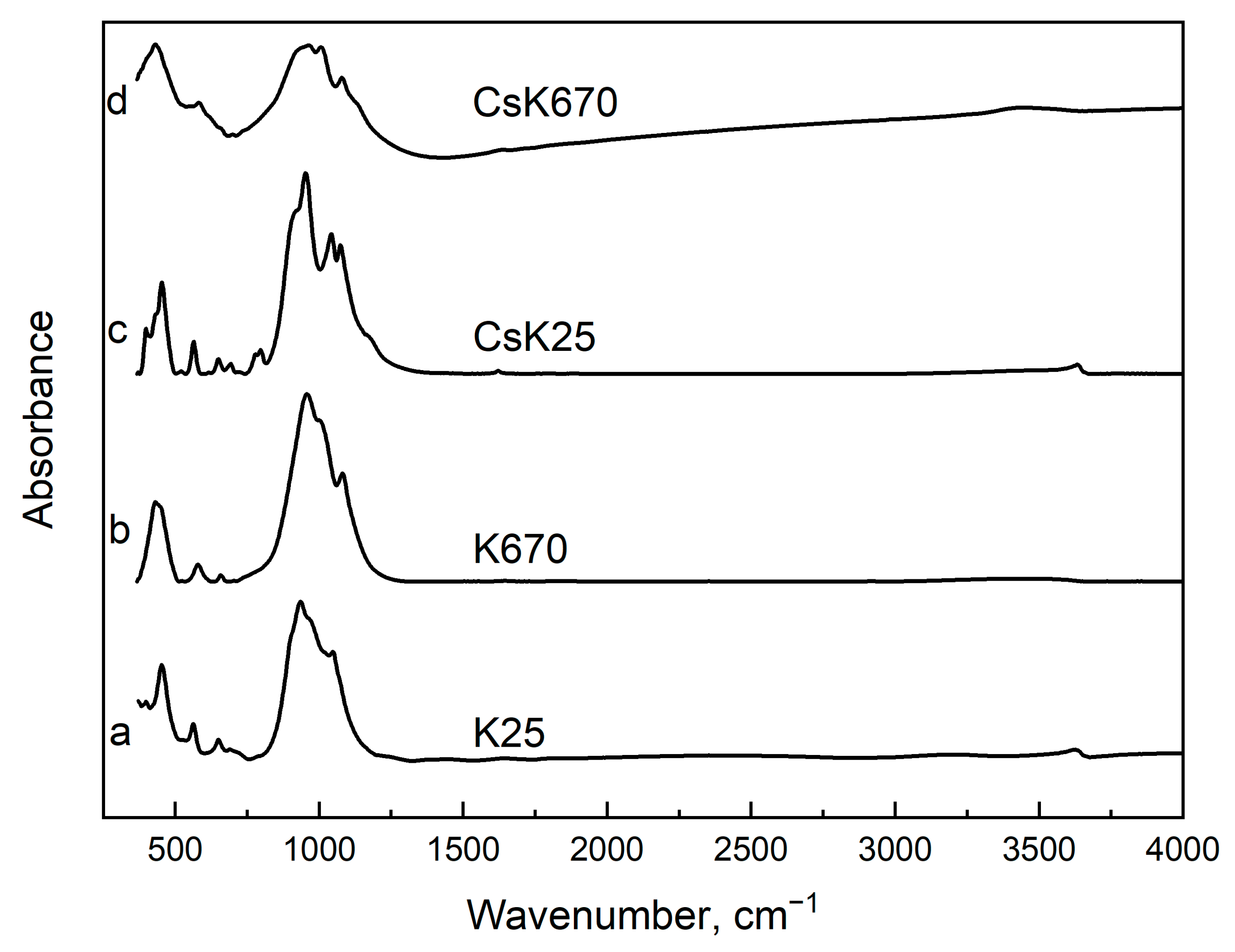
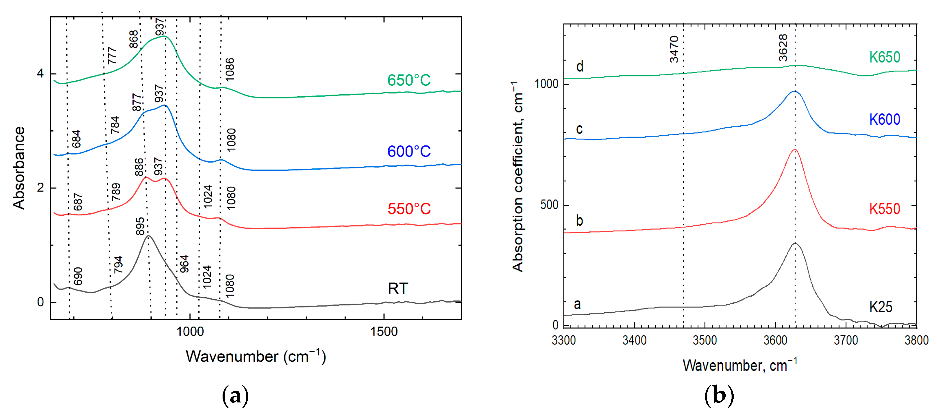
2.2.4. Infrared Spectroscopy

Infrared (IR) absorption spectra were measured using: (a) a Bruker Vertex IR spectrometer for K25, CsK25, K670, CsK670 (St. Petersburg State University, St. Petersburg, Russia), and (b) a Micran-3 infrared microscope and a Simex FT-801 spectrometer with a Ge-attenuated total reflection (ATR) module for K25, K550, K600, and K650 (these data are correlated with optical absorption spectroscopy) (Vinogradov Institute of Geochemistry, Irkutsk, Russia). The absorption spectra of the K25, K550, K600, and K650 with 0.02 mm thickness were measured in transmittance mode of the infrared microscope.

2.2.5. Optical Absorption Spectroscopy

Optical absorption spectra in the Ultraviolet/Visible/Near Infrared (UV/Vis/NIR) spectral region were recorded using a Perkin-Elmer Lambda 950 spectrophotometer and kupletskite plates with a thickness of 0.02–0.04 mm (Vinogradov Institute of Geochemistry, Irkutsk, Russia). The absorption spectra were recorded from the K25, K550, K600, and K650 samples.

3. Results

3.1. Chemical Composition

The mean chemical compositions of kupletskite and kupletskite-(Cs) are provided in Table 2. The empirical chemical formulae were calculated on the basis of Si + Al = 8. The analyses of chemical composition confirm that the starting material for high-temperature experiments was kupletskite and kupletskite-(Cs) (Table 2).

3.2. Thermal Evolution

The high-temperature behavior of kupletskite and kupletskite-(Cs) is distinct for two temperature regions: (a) 25–500 °C and (b) 500–775 °C. The latter region is characterized by a shift of reflections to higher 2-theta angles (Figure 2 and Figure S1a). At 800 °C, both minerals decompose. The in situ high-temperature powder X-ray diffraction patterns show the inheritance of the astrophyllite structural type by the high-temperature modifications (i.e., the preservation of the main structure topology on heating) and the reduction in the unit-cell parameters at a temperature above 500 °C (Figure 2 and Figure S1) due to the shift of the pattern to the high-angle region in the 2-theta axis.
The variation of the unit-cell parameters of kupletskite and kupletskite-(Cs) versus temperature is shown in Figure 3, and the data on astrophyllite [6] are provided for comparison. The 25–450 °C temperature range is characterized primarily by an increase of the unit-cell parameters (i.e., expansion), while the 525–775 °C temperature range shows more complex behavior with some parameters increasing and others decreasing (i.e., simultaneous expansion and contraction in different crystallographic directions) (Figure 3). The range 450–525 °C is not provided because of the presence of both initial and high-temperature modifications simultaneously and corresponding overlap of reflections.
The thermal expansion coefficients for kupletskite, kupletskite-(Cs), and their high-temperature modifications are provided in Table 3 (selected) and Tables S5 and S6 (full). The figures of thermal expansion are shown in Figure 4. The thermal behavior of both minerals and their high-temperature modifications is strongly anisotropic. For kupletskite, the maximum thermal expansion occurs within the plane of the layers and may correspond to their straightening (or decrease in corrugation). At the same time, for kupletskite-(Cs), the maximum thermal expansion is observed along the stacking direction. The high-temperature modification of kupletskite is characterized by contraction (owing to oxidation) in all directions with nearly equal coefficients within and between layers (Figure 4), similar to astrophyllite [6]. The thermal behavior of kupletskite-(Cs) is different and shows expansion with the maximal coefficient along the stacking direction (Figure 4).

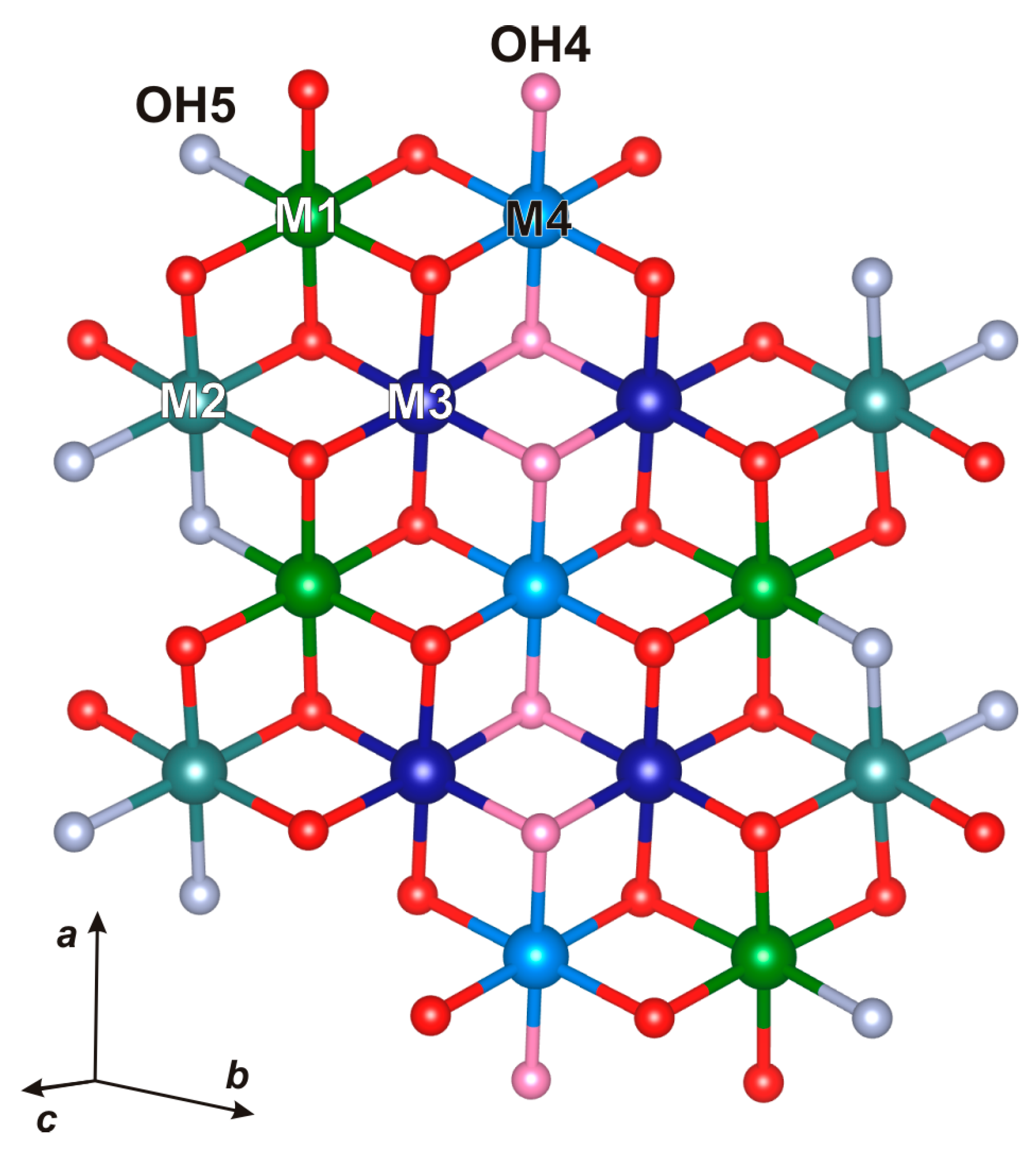
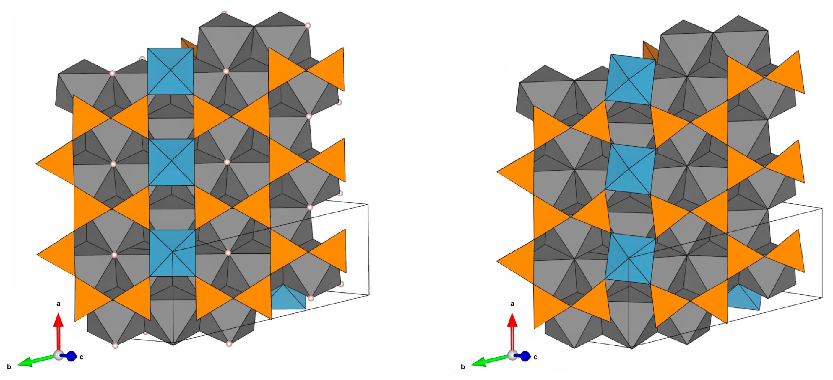
3.3. Crystal Structures

The crystal structures of kupletskite, kupletskite-(Cs), and their high-temperature modifications (K25, K650, CsK25, and CsK650) are triclinic and P-1 space-group symmetry. All crystal structures refined to good convergence indices (Table 1), and the structure models agree with previously reported data [5,45,46,47,48]. The atom coordinates, isotropic-displacement parameters, and site occupancies are provided in Table S7, while anisotropic-displacement parameters are provided in Table S8. Selected bond lengths are listed in Table S9. The X-ray diffraction data confirm the powder X-ray diffraction data: the high-temperature modifications retain the structure type of astrophyllite but are characterized by a reduction in the unit-cell parameters relative to initial kupletskite and kupletskite-(Cs) (Table 1).
The O layer incorporates four metal sites M(1)–(4) coordinated by O(1)–(7), two oxygen atoms of which OH(4) and OH(5) are protonated (Figure 5) in K25 and CsK25 and become deprotonated in the high-temperature modifications (K650, CsK650). The site-scattering values and bond lengths are provided in Table 4. The main structural changes affect the O sheet: the M(1)–O bond length increases slightly with reduction of site scattering, indicating Na-Li migration to the M(1) site and/or site-selective oxidation of transition metals. The site scattering also decreases at the M(2) site and may indicate partial migration of Na-Li. The site-scattering values fluctuate slightly at the M(3) and M(4) sites, which may be interpreted as an absence of significant change in their occupancies. The maximal contraction of M–O bond length occurs at the M(2) and M(4) sites (Figure 5); it is less pronounced for the M(3) site, indicating that most Fe2+,Mn2+ subject to oxidation occurs at the M(2) and M(4) sites and, to a lesser extent, at M(3).
The H layer has four symmetrically distinct silicate tetrahedra, T(1)–(4), that share corners with each other and with Dφ6 octahedra. The structural changes caused by oxidation–dexydroxylation also affect the H layer. The Dφ6 octahedra in kupletskite and kupletskite-(Cs) are distorted; the apical bond D–O2 (1.81–1.86 Å) is significantly shorter than DXPD (2.05–2.09 Å). In the high-temperature modifications of kupletskite and kupletskite-(Cs), the apical bonds of the Dφ6 octahedra are 1.90–1.96 Å for D–O2 and 1.99–2.00 Å for DXPD. The regulation of Dφ6 octahedra occurs due to a saturation of the O2 atom with additional valence units in the result of M-cations oxidation. In addition, there is rotation of the SiO4 tetrahedra and Dφ6 octahedra, resulting in a more distorted H layer (Figure 6).
The interlayer A site in kupletskite/kupletskite-(Cs) is split, forming the A1 and A2 sites ~0.8–0.9 Å apart. The interlayer also contains the B site occupied predominantly by Na and low-occupied oxygen (O17w site) coordinating A2. No significant changes in the geometry of the A- and B-polyhedra were detected.

3.4. Dexydroxylation

The dehydroxylation process is clearly tracked by the reduction of O–H intensities in the principal OH-stretching region of the infrared spectra in the wavenumber regions 3800–3000 cm−1 (principal O–H stretching) for high-temperature modifications relative to the unheated crystals (Figure 7).
Another set of ATR FTIR spectra were recorded for K25, K550, K600, and K650 from the single grain placed by the cleavage plane to the beam (E || {001}) of the same material as optical absorption spectra in order to monitor the heat-induced changes (Figure 8). The variations observed between the spectra in Figure 7 and Figure 8 are attributed to the polarization effect that is characteristic of ATR spectroscopy (Figure 8). Specifically, only the vibrational bands that were active when the incident beam is oriented parallel to {001} of the sample were detected. In contrast, all IR-active bands were captured in the powder IR absorption spectra presented in Figure 7. The peak assignment was made using [49,50]. The infrared spectra recorded from K25 exhibit the following bands: 1075, 1024, 964 (shoulder), 895, 794, and 690 cm−1. The bands at 1075 and 1024 cm−1 are assigned to the Si–O stretching vibrations in Si–O–Si chains. The bands at 964, 895, and 794 cm−1 correspond to the stretching vibrations of apical Si–O bonds. The band at 690 cm−1 is assigned to the bending vibration of O–Si–O. Upon the heating of kupletskite, the infrared spectra change bands at 895, 789, and 690 cm−1 (K25), shift to 886, 789, and 687 cm−1 (K550), and the intensity of shoulder at 964 cm−1 (K25) decreases with the appearance of a new band at 937 cm−1 (K550). Heating to higher temperatures (K600, K650) leads to further shift of the bands to 877, 784, and 684 cm−1 (K600) and 868, 784 cm−1 and disappearance of the 690–680 cm−1 band. These changes occur synchronously with increase of intensity for the band at 937 cm−1 (Figure 8). The intensity of 3628 cm−1, attributed to O–H stretching, decreases in K600 and disappears in K650.

3.5. The Mn Oxidation

The optical absorption spectrum of the initial kupletskite (K25) exhibits an absorption edge at 380 nm, which can be attributed to d-d transitions of Mn2+ ions (Figure 9). The sharp structure corresponding to these transitions is not resolved due to the strong exchange interaction between Mn2+ ions.
The optical absorption spectra of K600 show increased absorption in the 400–600 nm spectral region. Analysis of the difference spectrum (Figure 10) shows a UV band at approximately 400 nm that is attributed to the ligand-to-metal charge transfer (LMCT) from Mn3+ to O2− and from Fe3+ to O2− [51]. The bands at 430 and 505 nm correspond to spin-allowed d-d transitions in Mn3+ ions [51,52,53]. The temperature dependencies of the Mn3+-related absorption and the O–H absorption band at 3628 cm−1 are inversely correlated (Figure 10), suggesting that oxidation of Mn ions from the divalent to the trivalent state is accompanied by dehydroxylation.

4. Discussion

Structural studies of two manganese heterophyllosilicates: kupletskite and kupletskite-(Cs) using powder and single-crystal X-ray diffraction show a discontinuous reduction in the unit-cell parameters at temperatures of about 500 °C and above. By analogy with the high-temperature behavior of astrophyllite [6], bafertisite [7], and lobanovite [8], it has been suggested that this transformation occurs due to dehydroxylation and iron oxidation. The reduction of O–H-related bands is evident from infrared spectra. The contraction of the unit-cell volume is due to the shortening of M–O bond lengths, since Fe3+–O bonds are shorter than the Fe2+–O bonds [54]. If only Fe oxidizes, then the degree of the unit-cell contraction should correlate with the content of Fe2+. However, as can be seen from Table 5, the unit-cell contraction is practically identical for kupletskite, kupletskite-(Cs), and astrophyllite, despite different Fe2+ content. For astrophyllite, we know that 4–5 apfu of Fe2+ undergo oxidation, among which 4 are compensated by 4(OH)- groups, and the 5th by partial defluorination (F- → O2−) [6]. For the samples studied here, the Fe2+ apfu [2.22 apfu for kupletskite and 1.78 apfu for kupletskite-(Cs)] is much less. However, the degree of unit-cell volume contraction as a result of annealing is identical for kupletskite, kupletskite-(Cs), and astrophyllite. The identical degree of volume contraction and reduction of metal–oxygen bond lengths in minerals with different Fe contents indicates that additional mechanism(s) of oxidation must occur during dehydroxylation. The change in Mn oxidation state obtained by optical absorption spectroscopy (Figure 9) is inversely correlated with the intensity of the O–H band in the infrared spectra, indicating the synchronicity of these processes (Figure 10).
The following structural criteria have been calculated for each polyhedra: the (polyhedral) volume; bond-angle variance; distortion index; and quadratic elongation (Table 5). From the results in Table 5, it is apparent that the main structural changes upon heating affect the O layer for both kupletskite and kupletskite-(Cs). The largest compression and distortion occur for the M(2)O6 octahedron, significant changes with similar distortion indices occur for the M(3)O6, M(4)O6 octahedra, and the smallest distortions occur for the M(1)O6 octahedron, which confirms our proposal that the non-oxidizing elements are concentrated at the M(1) site. The distortions of the polyhedra of the H layer are less significant and are most likely caused by the need to satisfy the valence-sum requirements in the structures of the high-temperature modifications. We did not record any significant changes in the bond geometry and/or position of the interlayer cations at the A and B sites (Table 5). However, the thermal expansion figure for the HT modification of kupletskite-(Cs) is very different from those of kupletskite and astrophyllite, since in the temperature range of 500–800 °C, there is a volume expansion (not contraction) caused by expansion of the structure along the direction of layer stacking. If we carefully examine the evolution of the unit-cell parameters with temperature (Figure 3), the interlayer distance d00n clearly reflects this difference: for astrophyllite and kupletskite, the interlayer distance of the high-temperature modification is smaller than that of the initial structures; for kupletskite-(Cs), d00n does not fall below the initial room temperature value. It seems reasonable to suggest that this indicates the role of Cs ions in the stabilization of thermal expansion of the astrophyllite structural type, as Cs is larger than K [55], and its presence in the channels leads to a limitation on the minimum possible separation of the O layers.
Table 5. Geometrical parameters derived from the crystal structures of kupletskite, kupletskite-(Cs), and their high-temperature modifications.
ParameterSampleM(1)M(2)M(3)M(4)
toct, ÅK2.49 → 2.382.47 → 2.292.47 → 2.322.42 → 2.31
CsK2.49 → 2.362.48 → 2.282.47 → 2.302.42 → 2.29
Bond angle variance, °K59.3 → 93.746.5 → 94.641.9 → 88.039.1 → 37.3
CsK59.7 → 94.747.1 → 101.045.1 → 90.743.7 → 44.18
Voctahedra, Å3K13.88 → 13.9413.44 → 11.6413.23 → 12.1712.72 → 11.22
CsK13.69 → 13.5313.50 → 11.5213.28 → 12.0012.78 → 11.2
Distortion indexK0.007 → 0.0170.026 → 0.0720.017 → 0.0290.014 → 0.041
CsK0.006 → 0.0180.027 → 0.0790.019 → 0.0310.015 → 0.042
Quadratic elongationK1.0179 → 1.02991.0152 → 1.03821.0132 → 1.02801.0123 → 1.0156
CsK1.0180 → 1.03041.0155 → 1.04371.0144 → 1.02921.0138 → 1.0179
T(1)T(2)T(3)T(4)
Vtetrahedra, Å3K2.19 → 2.172.21 → 2.172.24 → 2.202.18 → 2.15
CsK2.17 → 2.152.21 → 2.152.20 → 2.192.17 → 2.14
Distortion indexK0.008 → 0.0110.006→ 0.0040.008 → 0.0050.007 → 0.010
CsK0.009 → 0.0120.011 → 0.0030.009 → 0.0060.009 → 0.009
Quadratic elongationK1.0021 → 1.00181.0013 → 1.00151.0018 → 1.00301.0021 → 1.0015
CsK1.0019 → 1.00181.0018→ 1.00221.0024 → 1.00401.0019 → 1.0015
DAB
Vpolyhedra, Å3K9.78 → 9.6872.8 → 69.834.52 → 33.18
CsK9.89 → 9.8874.8 → 72.535.65 → 34.33
Distortion indexK0.026 → 0.0110.091 → 0.820.013 → 0.060
CsK0.018 → 0.0080.043 → 0.0490.010 → 0.063
Quadratic elongationK1.0179 → 1.0102 1.1500 → 1.1510
CsK1.0097 → 1.0049 1.1688 → 1.1770
toct—octahedra thickness; Vpolyhedra—polyhedral volume, calculated by the Vesta program [56]; Distortion index calculated by the Vesta program [56]; Quadratic elongation calculated by the Vesta program [56].
We have shown for the first time, using heterophyllosilicates as an example, that temperature-induced oxidation of Mn occurs along with Fe, coupling with dehydroxylation of the O layer. Comparison of iron- and manganese-dominant minerals shows the structural identity of the oxidation reactions of these two elements. Considering the widespread occurrence of iron-oxidation reactions in rock-forming minerals, amphiboles, tourmalines, micas, and clays, one can assume similar behavior for their manganese-dominant analogues. By analogy with the oxidation reactions of Fe, it seems reasonable to propose that the oxidation of Mn can be implied in Earth processes at elevated temperatures and pressures, albeit on a more local scale than Fe due to the restricted distribution of Mn-rich rocks. The presence of dehydroxylated minerals (or their varieties) with trivalent manganese may also indicate heating to a temperature above 500 °C in an oxygen environment. The results obtained may also have applications in materials science, since these minerals have a porous structure and are capable of incorporating monovalent and divalent cations, including Cs and transition d-elements.

Supplementary Materials

The following supporting information can be downloaded at: https://www.mdpi.com/article/10.3390/min15060587/s1, Figure S1. (a) X-ray diffraction patterns for kupletskite in the temperature range from 25 to 950 °C: (I) 25–475 °C kupletskite, (II) 500–775 °C high-temperature modification of kupletskite, (III) 800–950 °C products of kupletskite decomposition. (b) X-ray diffraction pattern for kupletskite measured at 200 °C with fitting of the Rietveld refinement. Table S1. Unit-cell parameters refined for kupletskite at different temperatures; Table S2. Unit-cell parameters refined for kupletskite-(Cs) at different temperatures; Table S3. Coefficients of equations used for approximation of unit-cell parameters of kupletskite and its HT modification; Table S4. Coefficients of equations used for approximation of unit-cell parameters of kupletskite-(Cs) and its HT modification; Table S5. The main characteristics of thermal expansion/contraction for kupletskite (angles are given in degrees, coefficients are × 106, °C−1); Table S6. The main characteristics of thermal expansion/contraction for kupletskite-(Cs) (angles are given in degrees, coefficients are × 106, °C−1); Table S7. Atom coordinates, equivalent isotropic-displacement parameters (Å2), site occupancies for kupletskite (K25), kupletskite-(Cs) (CsK25) and their high-temperature modifications (K650 and CsK650); Table S8. Anisotropic-displacement parameters (Å2) for kupletskite (K25), kupletskite-(Cs) (CsK25) and their high-temperature modifications (K650 and CsK650); Table S9. Selected bond-distances (Å) for for kupletskite (K25), kupletskite-(Cs) (CsK25) and their high-temperature modifications (K650 and CsK650). The crystal structure data for kupletskite (K25), kupletskite-(Cs) (CsK25) and their high-temperature modifications (K650 and CsK650) are available as CIF–files from the CCDC/FIZ Karlsruhe database as CSD # 2442952 (K25), 2442953 (K650), 2442954 (Cs650) and 2442955 (Cs25) at https://www.ccdc.cam.ac.uk accessed on the 15 May 2025.

Author Contributions

Conceptualization, E.S.Z. and A.A.Z.; methodology, E.S.Z., A.A.Z., R.Y.S. and F.C.H.; software, E.S.Z., A.A.Z., R.M.S., R.Y.S., F.C.H., A.A.N. and N.S.V.; validation, E.S.Z., A.A.Z., R.M.S., R.Y.S., F.C.H., A.A.N., E.V.K. and V.N.Y.; formal analysis, E.S.Z., A.A.Z., R.M.S., R.Y.S., A.A.N. and N.S.V.; investigation, E.S.Z., A.A.Z., R.M.S., R.Y.S., F.C.H., A.A.N., N.S.V., E.V.K. and V.N.Y.; resources, E.S.Z., A.A.Z. and V.N.Y.; writing—original draft preparation, E.S.Z., A.A.Z., R.M.S., R.Y.S. and F.C.H.; writing—review and editing, E.S.Z., A.A.Z., R.M.S., R.Y.S., F.C.H., A.A.N., N.S.V., E.V.K. and V.N.Y.; visualization, E.S.Z., R.M.S. and R.Y.S.; supervision, E.S.Z., A.A.Z. and F.C.H.; project administration, E.S.Z.; funding acquisition, E.S.Z. All authors have read and agreed to the published version of the manuscript.

Funding

This research was supported by the Russian Science Foundation (project no. 22-77-10036 for E.S.Z., A.A.Z., R.M.S., A.A.N. and E.V.K.).

Data Availability Statement

The original contributions presented in this study are included in the article/Supplementary Material. Further inquiries can be directed to the corresponding author(s).

Acknowledgments

The infrared and UV/Vis absorption spectra were measured at the Center of Isotope and Geochemical Research for Collective Use (A. P. Vinogradov Institute of Geochemistry of the Siberian Branch of the Russian Academy of Sciences), which is acknowledged. St. Petersburg State University Resource Centres “X-ray diffraction research methods” and “Geomodel” are also acknowledged for the equipment access. We thank the reviewers for their constructive comments and the editors for processing the manuscript. Technical support of the St. Petersburg State University Resource Centres “X-ray diffraction research methods” and “Geomodel” is carried out within the framework of SPbSU, grants No. 125021702335-5 and No. 124032000029-9, for both Resource Centres, respectively. FCH was supported by a Discovery Grant from the Natural Sciences and Engineering Research Council of Canada. The field work was carried out by V.N.Y. as part of the FMEZ-2024-0008 project. We would like to thank the reviewers for their constructive work.

Conflicts of Interest

The authors declare no conflicts of interest.

References

  1. Belov, N.V. Essays on Structural Mineralogy; Nauka: Moscow, Russia, 1976; pp. 1–344. (In Russian) [Google Scholar]
  2. Liebau, F. Structural Chemistry of Silicates: Structure, Bonding and Classification; Springer: Berlin, Germany, 1985. [Google Scholar]
  3. Ferraris, G. Modular structures–the paradigmatic case of the heterophyllosilicates. Z. Kristallogr. Cryst. 2008, 223, 76–84. [Google Scholar] [CrossRef]
  4. Sokolova, E.; Cámara, F.; Hawthorne, F.C.; Ciriotti, M.E. The astrophyllite supergroup: Nomenclature and classification. Mineral. Mag. 2017, 81, 143–153. [Google Scholar] [CrossRef]
  5. Piilonen, P.C.; Lalonde, A.E.; McDonald, A.M.; Gault, R.A.; Larsen, A.O. Insights into astrophyllite-group minerals. I. Nomenclature, composition and development of a standardized general formula. Can. Mineral. 2003, 41, 1–26. [Google Scholar] [CrossRef]
  6. Zhitova, E.S.; Krivovichev, S.V.; Hawthorne, F.C.; Krzhizhanovskaya, M.G.; Zolotarev, A.A.; Abdu, Y.A.; Yakovenchuk, V.N.; Pakhomovsky, Y.A.; Goncharov, A.G. High-temperature behaviour of astrophyllite, K2NaFe72+Ti2(Si4O12)2O2(OH)4F: A combined X-ray diffraction and Mössbauer spectroscopic study. Phys. Chem. Miner. 2017, 44, 595–613. [Google Scholar] [CrossRef]
  7. Zhitova, E.S.; Zolotarev, A.A.; Krivovichev, S.V.; Goncharov, A.G.; Gabdrakhmanova, F.A.; Vladykin, N.V.; Krzhizhanovskaya, M.G.; Shilovskikh, V.V.; Vlasenko, N.S.; Zolotarev, A.A. Temperature-induced iron oxidation in bafertisite Ba2Fe42+Ti2(Si2O7)2O2(OH)2F2: X-ray diffraction and Mössbauer spectroscopy study. Hyperfine Interact. 2017, 238, 1–12. [Google Scholar] [CrossRef]
  8. Zhitova, E.S.; Zolotarev, A.A.; Hawthorne, F.C.; Krivovichev, S.V.; Yakovenchuk, V.N.; Goncharov, A.G. High-temperature Fe oxidation coupled with redistribution of framework cations in lobanovite, K2Na(Fe2+4Mg2Na)Ti2(Si4O12)2O2(OH)4—The first titanosilicate case. Acta Crystallogr. 2019, 75, 578–590. [Google Scholar] [CrossRef]
  9. Brown, I.D. The Chemical Bond in Inorganic Chemistry. The Bond Valence Model, 2nd ed.; Oxford University Press: Oxford, UK, 2016; pp. 1–344. [Google Scholar]
  10. Hawthorne, F.C. A bond-topological approach to theoretical mineralogy: Crystal structure, chemical composition and chemical reactions. Phys. Chem. Miner. 2012, 39, 841–874. [Google Scholar] [CrossRef]
  11. Hawthorne, F.C. Toward theoretical mineralogy: A bond-topological approach. Am. Mineral. 2015, 100, 696–713. [Google Scholar] [CrossRef]
  12. Russell, R.L.; Guggenheim, S. Crystal structures of near-endmember phlogopite at high temperatures and heat-treated Fe-rich phlogopite: The influence of the O, OH, F site. Can. Mineral. 1999, 37, 711–729. [Google Scholar]
  13. Chon, C.-M.; Lee, C.-K.; Song, Y.; Kim, S.A. Structural changes and oxidation of ferroan phlogopite with increasing temperature: In situ neutron powder diffraction and Fourier transform infrared spectroscopy. Phys. Chem. Miner. 2006, 33, 289–299. [Google Scholar] [CrossRef]
  14. Ventruti, G.; Zema, M.; Scordari, F.; Pedrazzi, G. Thermal behavior of a Ti-rich phlogopite from Mt. Vulture (Potenza, Italy): An in situ X-ray single-crystal diffraction study. Am. Mineral. 2008, 93, 632–643. [Google Scholar] [CrossRef]
  15. Zema, M.; Ventruti, G.; Lacalamita, M.; Scordari, F. Kinetics of Fe-oxidation/deprotonation process in Fe-rich phlogopite under isothermal conditions. Am. Mineral. 2010, 95, 1458–1466. [Google Scholar] [CrossRef]
  16. Murad, E.; Wagner, U. The thermal behaviuor of an Fe-rich illite. Clay Miner. 1996, 31, 45–52. [Google Scholar] [CrossRef]
  17. Güttler, B.; Niemann, W.; Redfern, S.A.T. EXAFS and XANES spectroscopy study of the oxidation and deprotonation of biotite. Mineral. Mag. 1989, 53, 591–602. [Google Scholar] [CrossRef]
  18. Veith, J.A.; Jackson, M.L. Iron oxidation and reduction effects on structural hydroxyl and layer charge in aqueous suspensions of micaceous vermiculites. Clays Clay Miner. 1974, 22, 345–353. [Google Scholar] [CrossRef]
  19. Korovushkin, V.V.; Kuzmin, V.; Belov, V.F. Mossbauer studies of structural features in tourmaline of various genesis. Phys. Chem. Miner. 1979, 4, 209–220. [Google Scholar] [CrossRef]
  20. Ferrow, E.A.; Annersten, H.; Gunawardane, R.P. Mössbauer effect study on the mixed valence state of iron in tourmaline. Mineral. Mag. 1988, 52, 221–228. [Google Scholar] [CrossRef]
  21. Bačik, P.; Ozdin, D.; Miglierini, M.; Kardošova, P.; Pentra, M.; Haloda, J. Crystallochemical effects of heat treatment on Fedominant tourmalines from Dolni Bory (Czech Republic) and Vlachovo(Slovakia). Phys. Chem. Miner. 2011, 38, 599–611. [Google Scholar] [CrossRef]
  22. Filip, J.; Bosi, F.; Novák, M.; Skogby, H.; Tuček, J.; Čuda, J.; Wildner, M. Iron redox reactions in the tourmaline structure: Hightemperature treatment of Fe3+-rich schorl. Geochim. Cosmochim. Acta 2012, 86, 239–256. [Google Scholar] [CrossRef]
  23. Oberti, R.; Della Ventura, G.; Dyar, M.D. Combining structure refinement and spectroscopies: Hints and warnings for more efficient tolls to decipher the mechanism of deprotonation in amphiboles. Period. Mineral. 2015, ECMS 2015, 131–132. [Google Scholar]
  24. Della Ventura, G. FTIR spectroscopy at HT: Applications and problems. Period. Mineral. 2015, ECMS 2015, 7–8. [Google Scholar]
  25. Susta, U.; Della Ventura, G.; Bellatreccia, F.; Hawthorne, F.C.; Oberti, R. HT-FTIR spectroscopy of riebeckite. Period. Mineral. 2015, ECMS 2015, 167–168. [Google Scholar]
  26. Della Ventura, G.; Redhammer, G.J.; Galdenzi, F.; Ventruti, G.; Susta, U.; Oberti, R.; Radica, F.; Marcelli, A. Oxidation or cation re-arrangement? Distinct behavior of riebeckite at high temperature. Am. Mineral. 2023, 108, 59–69. [Google Scholar] [CrossRef]
  27. Mihailova, B.; Della Ventura, G.; Waeselmann, N.; Bernardini, S.; Xu, W.; Marcelli, A. Polarons in rock-forming minerals: Physical implications. Condens. Matter 2022, 7, 68. [Google Scholar] [CrossRef]
  28. Della Ventura, G.; Galdenzi, F.; Marcelli, A.; Cibin, G.; Oberti, R.; Hawthorne, F.C.; Bernardini, S.; Mihailova, B. In situ simultaneous Fe K-edge XAS spectroscopy and resistivity measurements of riebeckite: Implications for anomalous electrical conductivity in subduction zones. Geochemistry 2023, 84, 126037. [Google Scholar] [CrossRef]
  29. Bernardini, S.; Della Ventura, G.; Schlüter, J.; Hawthorne, F.C.; Mihailova, B. The effect of A-site cations on charge-carrier mobility in Fe-rich amphiboles. Am. Mineral. 2024, 109, 1545–1553. [Google Scholar] [CrossRef]
  30. Bernardini, S.; Della Ventura, G.; Hawthorne, F.C.; Marcelli, A.; Salvini, F.; Mihailova, B. The effect of anisotropic electrical conductivity of amphiboles on geophysical anomalies observed in subduction zones. Sci. Rep. 2025; accepted. [Google Scholar] [CrossRef]
  31. Della Ventura, G.; Bernardini, S.; Redhammer, J.G.; Galdenzi, F.; Radica, F.; Marcelli, A.; Hawthorne, F.C.; Oberti, R.; Mihailova, B. The oxidation of iron in amphiboles at high temperatures: A review and implications for large-scale Earth processes. Rend. Lincei Sci. Fis. Nat. 2024, 35, 893–906. [Google Scholar] [CrossRef]
  32. Ferraris, G. Heterophyllosilicates, a potential source of nanolayers for materials science. In Minerals as Advanced Materials I.; Krivovichev, S.V., Ed.; Springer: Berlin/Heidelberg, Germany, 2008; pp. 157–163. [Google Scholar]
  33. Ferraris, G.; Merlino, S. Micro- and Mesoporous Mineral Phases; Mineralogical Society of America: Washington, DC, USA, 2005; Volume 57, pp. 1–448. [Google Scholar]
  34. Lin, Z.; Paz, F.A.A.; Rocha, J. Layered titanosilicates. In Layered Mineral Structures and Their Application in Advanced Technologies; Brigatti, M.F., Mottana, A., Eds.; Mineralogical Society of Great Britain and Ireland: London, UK, 2011; Volume 11. [Google Scholar] [CrossRef]
  35. Aksenov, S.M.; Yamnova, N.A.; Chukanov, N.V.; Kabanova, N.A.; Kobeleva, E.A.; Deyneko, D.V.; Krivovichev, S.V. Theoretical analysis of cation-migration paths in microporous heterophyllosilicates with astrophyllite and veblenite type structures. J. Struct. Chem. 2022, 63, 293–301. [Google Scholar] [CrossRef]
  36. Yakovenchuk, V.; Ivanyuk, G.; Pakhomovsky, Y.; Men’shikov, Y. Khibiny; Laplandia Minerals: Apatity, Russia, 2005. [Google Scholar]
  37. Yefimov, A.F.; Dusmatov, V.D.; Ganzeyev, A.A.; Katayeva, Z.T. Cesium kupletskite, a new mineral. Dokl. Akad. Nauk. SSSR 1971, 197, 140–143. [Google Scholar]
  38. Bruker AXS. Topas V4.2: General Profile and Structure Analysis Software for Powder Diffraction Data; Bruker AXS: Karlsruhe, Germany, 2009. [Google Scholar]
  39. Belousov, R.; Filatov, S. Algorithm for calculating the thermal expansion tensor and constructing the thermal expansion diagram for crystals. Glass Phys. Chem. 2007, 33, 271–275. [Google Scholar] [CrossRef]
  40. Bubnova, R.S.; Firsova, V.A.; Filatov, S.K. Software for determining the thermal expansion tensor and the graphic representation of its characteristic surface (theta to tensor-TTT). Glass Phys. Chem. 2013, 39, 347–350. [Google Scholar] [CrossRef]
  41. Langreiter, T.; Kahlenberg, V. TEV—A program for the determination of the thermal expansion tensor from diffraction data. Crystals 2015, 5, 143–153. [Google Scholar] [CrossRef]
  42. Bruker AXS. APEX2, Version 2014.11-0; Bruker AXS: Madison, WI, USA, 2014. [Google Scholar]
  43. Sheldrick, G.M. Crystal structure solution with ShelXT. Acta Crystallogr. 2015, A71, 3–8. [Google Scholar]
  44. Dolomanov, O.V.; Bourhis, L.J.; Gildea, R.J.; Howard, J.A.K.; Puschmann, H. OLEX2: A complete structure solution, refinement and analisis program. Appl. Crystallogr. 2009, 42, 339–341. [Google Scholar] [CrossRef]
  45. Cámara, F.; Sokolova, E.; Abdu, Y.; Hawthorne, F.C. The crystal structures of niobophyllite, kupletskite-(Cs) and Sn-rich astrophyllite: Revisions to the crystal chemistry of the astrophyllite-group minerals. Can. Mineral. 2010, 48, 1–16. [Google Scholar] [CrossRef]
  46. Woodrow, P.J. The crystal structure of astrophyllite. Acta Crystallogr. 1967, 22, 673–678. [Google Scholar] [CrossRef]
  47. Piilonen, P.C.; McDonald, A.M.; Lalonde, A.E. Insights into astrophyllite-group minerals. II. Crystal chemistry. Can. Mineral. 2003, 41, 27–54. [Google Scholar] [CrossRef]
  48. Sokolova, E. Further developments in the structure topology of the astrophyllite-group minerals. Mineral. Mag. 2012, 76, 863–882. [Google Scholar] [CrossRef]
  49. Nakamoto, K. Infrared and Raman Spectra of Inorganic and Coordination Compounds, Part B: Applications in Coordination, Organometallic, and Bioinorganic Chemistry; John Wiley & Sons: Hoboken, NJ, USA, 2009. [Google Scholar]
  50. Sokolova, E.; Day, M.C.; Hawthorne, F.C.; Kasatkin, A.V.; Downs, R.T.; Horváth, L.; Pfenninger-Horváth, E. Laverovite, K2NaMn7Zr2(Si4O12)2O2(OH)4F, a new astrophyllite-supergroup mineral from Mont Saint-Hilaire, Québec, Canada. Can. Mineral. 2019, 57, 201–213. [Google Scholar] [CrossRef]
  51. Hålenius, U.; Bosi, F. Color of Mn-bearing gahnite: A first example of electronic transitions in heterovalent exchange coupled IVMn2+VIMn3+ pairs in minerals. Am. Mineral. 2014, 99, 261–266. [Google Scholar] [CrossRef]
  52. Czaja, M.; Lisiecki, R.; Chrobak, A.; Sitko, R.; Mazurak, Z. The absorption-and luminescence spectra of Mn3+ in beryl and vesuvianite. Phys. Chem. Miner. 2018, 45, 475–488. [Google Scholar] [CrossRef]
  53. Fridrichová, J.; Bačík, P.; Ertl, A.; Wildner, M.; Dekan, J.; Miglierini, M. Jahn-Teller distortion of Mn3+-occupied octahedra in red beryl from Utah indicated by optical spectroscopy. J. Mol. Struct. 2018, 1152, 79–86. [Google Scholar] [CrossRef]
  54. Gagné, O.C.; Hawthorne, F.C. Bond-length distributions for ions bonded to oxygen: Results for the transition metals and quantification of the factors underlying bond-length variation in inorganic solids. IUCrJ 2020, 7, 581–629. [Google Scholar] [CrossRef]
  55. Hawthorne, F.C.; Gagné, O.C. New ion radii for oxides and oxysalts, fluorides, chlorides and nitrides. Acta Crystallogr. 2024, B80, 326–339. [Google Scholar] [CrossRef]
  56. Momma, K.; Izumi, F. VESTA 3 for three-dimensional visualization of crystal, volumetric and morphology data. J. Appl. Crystallogr. 2011, 44, 1272–1276. [Google Scholar] [CrossRef]
Figure 1. The astrophyllite structure type is of alternating octahedral and heteropolyhedral layers.
Minerals 15 00587 g001
Figure 2. The evolution of the powder X-ray diffraction pattern of kupletskite/kupletskite-(Cs) with temperature.
Minerals 15 00587 g002
Figure 3. Temperature dependencies of the unit-cell parameters and d001-spacings for kupletskite and kupletskite-(Cs) compared to astrophyllite [6] (ESDs fall within the limits of the symbols).
Minerals 15 00587 g003
Figure 4. (a) The crystal structure of kupletskite/kupletskite-(Cs) (the unit cell is shown by the thin black lines) and figures of thermal expansion for kupletskite at T = 300 °C (b), high-temperature modification of kupletskite at T = 650 °C (c), kupletskite-(Cs) at T = 300 °C (d), and high-temperature modification of kupletskite-(Cs) at T = 650 °C (e).
Minerals 15 00587 g004
Figure 5. The bond topology of the O layer in the crystal structure of kupletskite, kupletskite-(Cs), and astrophyllite; the M(1)–M(4) sites are shown by different colors. The protonated oxygen atoms are designated as OH(4) (pink) and OH(5) (grey).
Minerals 15 00587 g005
Figure 6. The topology of the H layer in the crystal structure of kupletskite/kupletskite-(Cs) (left) and their high-temperature modification (right). Comparison of the images shows a change in the rotation of the Dφ6 octahedra and distortions in the geometry of the T4O12 chains.
Minerals 15 00587 g006
Figure 7. Infrared spectra of (a) kupletskite and (b) its high-temperature modification; (c) kupletskite-(Cs) and (d) its high-temperature modification.
Minerals 15 00587 g007
Figure 8. The ATR infrared spectra (E || {001}) of kupletskite (K25) and its high-temperature modifications (K550, K600, and K650) in the 1700–600 cm−1 (a) and 3800–3300 cm−1 (b) ranges.
Minerals 15 00587 g008
Figure 9. Optical absorption spectra of kupletskite, K25 (curve 1), and its high-temperature modifications K550 (curve 2) and K600 (curve 3). In the inset, the difference spectrum of the spectra recorded from K25 and K550 is provided.
Minerals 15 00587 g009
Figure 10. Temperature dependences of Mn3+ absorption (1) and intensity of the O–H-related band at 3628 cm−1 (2).
Minerals 15 00587 g010
Table 1. Crystallographic data, data collection, and refinement parameters for kupletskite (K25), kupletskite-(Cs) (CsK25), and their high-temperature modifications (K650 and CsK650).
SampleK25K650CsK25CsK650
Crystal data
Crystal systemTriclinicTriclinicTriclinicTriclinic
Space groupP-1P-1P-1P-1
Unit-cell dimensions
a, b, c (Å),
α, β, γ (°)
5.3976(2)
11.9431(7)
11.7092(6)
113.066(5)
94.702(4)
103.086(4)
5.3233(5)
11.8826(14)
11.5362(12)
112.756(10)
93.699(8)
104.462(9)
5.3904(10)
11.946(2)
11.799(2)
113.135(5)
94.573(6)
103.115(17)
5.312(2)
11.832(4)
11.739(5)
112.996(10)
93.587(10)
104.39(4)
Unit-cell volume (Å3)664.06(6)640.90(13)668.2(2)647.3(5)
Z1111
Calculated density (g/cm−3)3.2743.3543.6353.726
Absorption coefficient (μ/mm−1)4.9924.4816.2046.244
Data collection
DiffractometerBruker APEX II
Temperature (K)293 K
RadiationMoKα
2θ range (°)3.852–72.83.892–72.883.864–59.993.832–59.986
h, k, l ranges−8 ≤ h ≤ 8,
−19 ≤ k ≤ 19,
−18 ≤ l ≤ 19
−8 ≤ h ≤ 8,
−19 ≤ k ≤ 19,
−18 ≤ l ≤ 18
−7 ≤ h ≤ 7,
−16 ≤ k ≤ 16,
−14 ≤ l ≤ 16
−7 ≤ h ≤ 7,
−16 ≤ k ≤ 16,
−16 ≤ l ≤ 16
F(000)634.0625.0693.0686.0
Total reflections collected12,05011,358868612,060
Unique reflections (Rint)6044
(0.0325)
5837
(0.0303)
3591
(0.0518)
3766
(0.0476)
Unique reflections F > 4σ(F)4768453126672831
Structure refinement
Refinement methodFull-matrix least-squares on F2
Data/restrains/parameters6044/2/2535837/0/2453591/2/2563766/0/251
R1 [F > 4σ(F)],
wR2 [F > 4σ(F)]
0.0433,
0.1119
0.0416,
0.1002
0.0323,
0.0715
0.0476,
0.1108
R1 all, wR2 all0.0590,
0.1232
0.0611,
0.1110
0.0657,
0.0904
0.0697,
0.1229
Goodness-of-fit on F21.0511.0371.0611.044
Largest diff. peak and hole (ēÅ−3)1.29/−1.811.57/−1.371.72/−1.451.29/−1.52
Table 2. Chemical composition of kupletskite and kupletskite-(Cs).
MineralKupletskiteKupletskite-(Cs)ConstituentKupletskiteKupletskite-(Cs)
n2227Constituent
Constituent, wt. %averagerangeaveragerangeapfu, calculated as Si + Al = 8
Na2O2.772.64–2.932.111.76–2.83Na1.151.00
K2O6.226.03–6.380.730.61–0.95K1.700.23
CaO1.431.27–1.590.730.56–0.92Ca0.330.19
Cs2O--14.4014.25–15.20Cs-1.50
PbO--0.370–1.09Pb-0.02
MgO1.421.29–1.550.040–0.25Mg0.450.01
MnO18.6217.05–20.1617.7917.41–18.37Mn3.373.68
FeO (1)11.8214.23–17.208.7410.54–11.72Fe2+2.111.78
Fe2O34.152.57Fe3+0.670.47
Li2O (2)--0.74-Li-0.73
Al2O30.810.64–1.190.120–0.48Al0.200.03
TiO211.1810.88–11.486.965.83–8.04Ti1.801.28
Nb2O52.121.83–2.236.355.17–8.56Nb0.200.70
SiO236.4735.80–37.8932.6232.11–33.26Si7.807.97
F1.190.68–1.630.840.65–1.19F0.800.65
H2O (3)2.67-2.35-OH3.803.83
O (3)0.50-0.58-O0.200.52
2F = O−0.50-−0.35-
Total100.87 99.60
(1) Fe is divided into Fe2+ and Fe3+ as Fe2+:Fe3+~3:1; (2) Li2O is calculated as sum of octahedral cations equal to 7 apfu; Li2O has been previously detected in kupletskite-(Cs) as 0.46 [37] and 0.69 [45] wt.%; (3) OH/O ratio is calculated based on charge balance; n—number of analyses.
Table 3. The main characteristics of thermal expansion/contraction for kupletskite, kupletskite-(Cs) at T = 300 °C and their high-temperature modifications at T = 650 °C (angles are provided in degrees, coefficients are ×106, °C−1).
T, °Cα11α22α3311a22b33cαaαbαcαααβαγαV
Kupletskite
3004.311.56.724.912.2204.9(4)11.2(4)6.7(3)−0.01(9)0.6(2)0.2(2)22.5(6)
Kupletskite-(Cs)
300−3.41.617.14733.926.7−1(1)0.1(7)13.3(7)−3.7(4)−2.0(4)2.7(4)15(2)
High-temperature modification of kupletskite
650−39.2−19.9−11.827.335.422.3−34(2)−23(1)−15(2)5(2)−9(2)−6(1)−71(3)
High-temperature modification of kupletskite-(Cs)
6503.2−6.710.326.910.839.52(1)−6(2)5.5(6)−4.1(4)0.6(9)−3(1)7(2)
Table 4. Selected site-scattering values and bond lengths for kupletskite, kupletskite-(Cs), and their high-temperature modifications.
SampleKupletskiteHT KupletskiteKupletskite-(Cs)HT Kupletskite-(Cs)AstrophylliteHT Astrophyllite
V, Å3664.1640.9668.2647.3655.5633.0
ΔV/V, %−3.5−3.1−3.4
Octahedra M(1)O6
ē M(1)23.0521.1021.7519.2823.6921.14
Δē M(1)−2.0−2.5−2.6
<M(1)–O>, Å2.2032.2192.1932.1972.1762.185
Δ<M(1)–O>, Å+0.016+0.004+0.009
Octahedra M(2)O6
ēM(2)25.0023.8325.0024.6125.0623.01
ΔēM(2)−1.2−0.4−2.0
<M(2)–O>, Å2.1742.0912.1792.0892.1562.102
Δ<M(2)–O>, Å−0.083−0.090−0.054
Octahedra M(3)O6
ēM(3)24.6124.4823.8324.6124.1824.28
ΔēM(3)−0.1+0.8+0.1
<M(3)–O>, Å2.1622.1172.1672.1082.1442.097
Δ<M(3)–O>, Å−0.045−0.059−0.047
Octahedra M(4)O6
ēM(4)23.3124.0925.0024.2223.5124.15
ΔēM(4)+0.8−0.8+0.6
<M(4)–O>, Å2.1332.0472.1382.0492.1282.059
Δ<M(4)–O>, Å−0.086−0.089−0.069
Octahedra Dφ6
D–O(2), Å1.8081.9021.8551.9601.8111.952
DXPD, Å2.0882.0022.0511.9932.1001.982
<D–φ>, Å1.9581.9461.9591.9641.9581.945
Tetrahedra TO4
<T1–O>, Å1.6231.6201.6181.6141.6151.607
<T2–O>, Å1.6291.6181.6271.6131.6251.620
<T3–O>, Å1.6371.6271.6271.6251.6321.624
<T4–O>, Å1.6201.6141.6181.6111.6141.606
Extraframework sites A, B
<A1–φ>, Å3.2833.2253.3363.3183.2983.243
<A2–φ>, Å3.2993.2263.3693.345--
<B–φ>, Å2.6212.5952.6482.6292.6152.548
ReferenceThis workThis work[6]
V—unit-cell volume; ΔV—the change in the unit-cell volume after annealing; ē—number of electrons calculated by site occupancy; Δē—the change in that parameter after annealing; <M(1)–O>—the average bond lengths; Δ<M(1)–O>—the change in bond length after annealing.
Disclaimer/Publisher’s Note: The statements, opinions and data contained in all publications are solely those of the individual author(s) and contributor(s) and not of MDPI and/or the editor(s). MDPI and/or the editor(s) disclaim responsibility for any injury to people or property resulting from any ideas, methods, instructions or products referred to in the content.

