Article

Siderite Decomposition Kinetics—Influence of Time, Temperature, and Isomorphous Impurities

by
Mariola Kądziołka-Gaweł
1,*,
Zdzisław Adamczyk
2,
Dariusz Łukowiec
3,
Joanna Klimontko
1,
Marcin Wojtyniak
4 and
Jacek Nowak
2
1
Institute of Physics, University of Silesia, 75 Pułku Piechoty 1, 41-500 Chorzów, Poland
2
Faculty of Mining, Safety Engineering and Industrial Automation, Silesian University of Technology, Akademicka 2A, 44-100 Gliwice, Poland
3
Materials Research Laboratory, Faculty of Mechanical Engineering, Silesian University of Technology, Konarskiego 18A St., 44-100 Gliwice, Poland
4
Institute of Physics, Silesian University of Technology, Konarskiego 22B, 44-100 Gliwice, Poland
*
Author to whom correspondence should be addressed.
Minerals 2025, 15(4), 428; https://doi.org/10.3390/min15040428
Submission received: 10 March 2025 / Revised: 10 April 2025 / Accepted: 17 April 2025 / Published: 19 April 2025

Abstract

:
Siderite (FeCO3) is an iron-bearing carbonate mineral that is the most abundant sedimentary iron formation on Earth. The influence of time, temperature, and isomorphous impurities on the kinetics of siderite decomposition in air was studied using Mössbauer spectroscopy, X-ray diffraction, X-ray fluorescence spectroscopy, and the Transmission Electron Microscopy method. Two rock siderite samples were used for investigations, one containing a significant amount of magnesium. The siderite decomposition process begins at a temperature of 300 °C. In a sample containing practically no magnesium, complete decomposition occurs at a temperature of 450 °C, and in a sample with magnesium at a temperature of 500 °C. The annealing time does not affect the width of the temperature range where siderite decomposition occurs. Still, it affects the degree of siderite decomposition: the longer the annealing time, the greater the amount of siderite decomposition. The decomposition products of siderite annealed in air are iron oxides. In the sample containing practically no Mg, this oxide was mainly hematite, and in the sample containing magnesium, it was magnesioferrite. Iron oxides formed directly in siderite decomposition are poorly crystalline, and we can treat them as iron oxide nanoparticles. They maintain this form in a wide temperature range, especially in a magnesium-containing sample. The presence of this element significantly slows down the process of magnesioferrite crystallization.

1. Introduction

Siderite (FeCO3) is an iron-bearing carbonate common in many diverse sedimentary settings, particularly in marine and lacustrine environments. This iron carbonate is an antiferromagnetic mineral with a Neel temperature of 38 K, which is paramagnetic at an ordinary temperature [1]. Siderite crystallizes in the trigonal crystal system and is rhombohedral in shape, typically with curved and striated faces [2]. The theoretical Fe content in this mineral is 48.27 wt.%, but natural siderites often contain significant Mg, Ca, and Mn substitutions for Fe in the lattice, and pure siderite is seldom found [3]. Thermal decomposition and oxidation of the mineral siderite are topics of interest because of both their commercial applications and scientific questions regarding the progression of products. For example, siderite has attracted considerable attention as this mineral is related to CO2 capture [4,5] or natural protection against corrosion [6,7]. Knowledge of the process mentioned above is used, among others, in paleomagnetic studies where products of this process can potentially carry a chemical remanent magnetization [2]. Siderite is also used as a clean reductant for the suspension magnetization roasting process [8], and the thermal decomposition of the mineral siderite is a topic of interest because of the possibility of strongly magnetic phase formation without reducing agents during the iron production process using siderite ore [9,10].
As is commonly known [9,10,11], the final product of siderite decomposition in an oxidizing atmosphere is generally hematite. However, siderite’s phase transformation and microstructure are highly complex and related not only to the atmosphere in which thermal decomposition occurs but also to temperature or heating rate [12,13]. Additionally, some impurities like Mn and Mg exist in natural siderite as isomorphism states with iron and influenced decomposition [1,14]. For these reasons, the properties of iron carbonate still need to be investigated, as many questions regarding its high-temperature stability and the influence of its composition on this process remain open. For these reasons, in our previous work [15], we investigated the thermal decomposition mechanism of siderite in oxidizing O2 and reducing CO2 atmosphere, and a phase analysis of iron-bearing minerals formed during these processes was presented. The type of atmosphere will significantly influence the siderite decomposition process and the type of decomposition products. We showed that magnetite, hematite, and maghemite are products of siderite decomposition after annealing in the oxygen atmosphere up to a temperature of 500 °C. In contrast, hematite is the main component of the sample above this temperature, and magnetite is the main product of siderite decomposition in the CO2 atmosphere, but hematite, maghemite, wüstite, and olivine were also present in the sample. Moreover, the decomposition process of siderite is shifted toward higher temperatures for the sample with the highest magnesium content.
This study investigates siderite’s thermal decomposition, its decomposition products in the air atmosphere, and the influence of annealing time, temperature, and isomorphous impurities in the siderite structure on the process mentioned. Knowledge of these processes can help, for example, better understand siderite’s pyrolysis behavior, which may have implications for exploiting Fe-bearing carbonates as a source of iron ore. In contrast to our previous work [15], where the sample was annealed at a specific temperature for three hours, we investigated the effect of different heating times on siderite decomposition. Moreover, in this work, the process of siderite decomposition is carried out on the same sample in the entire temperature range, so we also investigate the temperature changes that the decomposition products of this iron carbonate undergo. In the previous work, the raw sample of siderite was annealed at a given temperature and atmosphere, so we obtained information on the degree of siderite decomposition and the products formed from this process at this specific annealing temperature. The Fe-bearing phase’s transformation and identification were characterized using Mössbauer spectroscopy. This method uses the Fe nucleus to probe its local surroundings and is one of the best methods for identifying iron and its properties in solids. The Mössbauer investigation was supported by X-ray diffraction, X-ray fluorescence spectroscopy, and the Transmission Electron Microscopy method.

2. Materials and Methods

The studied siderites come from the upper parts of the Ruda coal seams, in the eastern part of the Chwałowice Trough, in the Upper Silesian Coal Basin. A detailed description of the investigated materials has been described in our previous work [15]. The research was carried out on two rock samples taken from different seams. These samples were designated Sd1 and Sd3. All samples were crushed and then ground in a ball mill. After that, powdered samples with a grain size of less than 0.05 mm were obtained. The samples were annealed in a muffle furnace at the following temperatures: 300 °C, 350 °C, 400 °C, 450 °C, 500 °C, 550 °C, 600 °C, 700 °C, 800 °C, and 900 °C. The annealing was carried out in an air atmosphere for half, one, and two hours. The entire heating process from 300 °C to 900 °C was carried out on the same sample. This means that the sample was heated at 300 °C for, for example, half an hour, cooled to room temperature, and the necessary measurements were taken. Then, this sample was heated at 350 °C, cooled to room temperature, and appropriate measurements were taken. And so on, up to 900 °C. Identifying phases formed due to siderite decomposition is more precise when the measurement is performed at room temperature and in stable conditions. Such a heat treatment method may slightly affect the contribution of phases formed. However, as the results show, the heating time does not affect the type of phases formed.

2.1. X-Ray Fluorescence

The X-ray fluorescence (XRF) method with a ZSX Primus II Rigaku spectrometer (RIGAKU, Tokyo, Japan) determined the chemical composition of the initial siderite samples. The spectrometer, equipped with the 4 kW, 60 kV Rh anode and wavelength dispersion detection system, allowed for analyzing the elements from Be to U. No external standards were necessary. Only the internal standards coupled with the fundamental parameters were implemented. The samples for the analysis were prepared in the form of pressed tablets. According to the manufacturer, the global uncertainty in determining the content of individual elements is less than 1%.

2.2. X-Ray Powder Diffraction

Minerals present in the investigated initial siderite samples and after annealing them at 400 °C, 700 °C, and 900 °C were determined using the X-ray powder diffraction (XRD) method. A high-resolution PANalytical Empyrean diffractometer (Malvern Panalytical, Malvern, UK) equipped with a PIXcel detector was used with Cu Kα radiation (40 kV, 30 mA). Rietveld refinement was performed in a licensed X’Pert High Score Plus with a PDF-4 crystallography database. The percentages of individual components in initial samples were determined using the Rietveld method.

2.3. Transmission Electron Microscopy

The images of the structure and morphology of selected siderite samples were recorded using a Titan 80-300 (FEI) S/TEM (FEI, Hillsboro, OR, USA) microscope at 300 kV. The analysis of Transmission Electron Microscopy (TEM) and especially HRTEM images provides information about the changing size of the nanoparticles forming different materials and their crystalline structure.

2.4. Mössbauer Spectroscopy

The 57Fe Mössbauer transmission spectra were recorded at room temperature using an Integrated Mössbauer Spectroscopy Measurement System (designed by Wacław Musiał and Jacek Marzec, Krakow, Poland) and a linear arrangement of RITVERC source 57Co:Rh, a multichannel analyzer, an absorber, and a detector. A gas proportional counter LND 45431 was used as a gamma-ray detector. The spectrometer velocity scale was calibrated at room temperature with a 25 μm thick α-Fe foil. All Mössbauer measurements were carried out on powdered samples. The Mössbauer spectra were evaluated by least-square fitting of the lines using the MossWinn4.0i program. Some parts of the spectra were fitted with a hyperfine magnetic field distribution. The model and implementation are based on the Voigt-based fitting method [16].

3. Results

3.1. X-Ray Fluorescence

Table 1 presents the contents of the elements in the investigated siderite sample whose concentration in the sample was higher than 0.05 wt.%. The chemical composition was dominated by C, O, and Fe, which is obvious due to the presence of siderite in the samples. It should be noted that the amount of Fe in sample Sd1 is higher by ~10 wt.% compared to sample Sd3. On the other hand, sample Sd3 contains large amounts of Mg, which in sample Sd1 is practically absent. The occurrence of Mn is also noteworthy, which, together with Ca and Mg, may isomorphously replace Fe in the siderite structure. The samples also contain small amounts of Si and Al, which, in the presence of K, may indicate the occurrence of small admixtures of aluminosilicates in the samples.

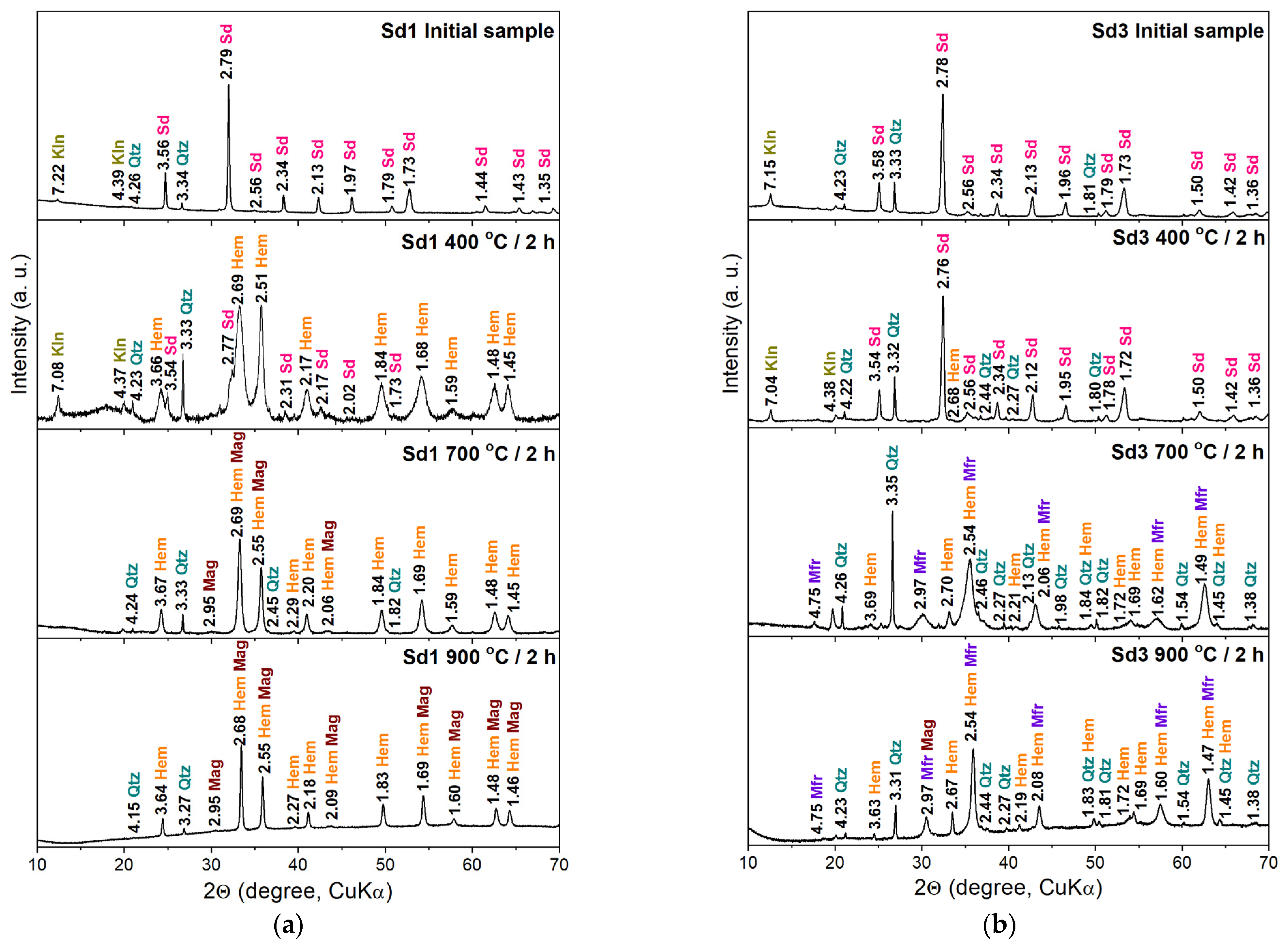
3.2. X-Ray Powder Diffraction

X-ray diffraction measurements indicate (Figure 1, Table 2) that initial samples Sd1 and Sd3 contain mainly siderite (FeCO3). The concentration of this iron carbonate is higher than 94% vol. for the Sd1 sample and 84% vol. for the Sd3 sample. Small amounts of accompanying minerals from the silicate and aluminosilicate groups, such as quartz (SiO2), illite (K0.65Al2.0[Al0.65Si3.35O10](OH)2), and kaolinite (Al2Si2O5(OH)4) are also detected, especially in the Sd3 sample. The minerals identified here do not contain elements such as Mg, Ca, or Mn, which are detected in the XRF measurement results (Table 1) and occur mainly in the Sd3 sample. For this reason, these elements probably doped the minerals in the samples, especially siderite.
Siderite is still present in both samples after annealing them at 400 °C (Figure 1, Table 2). However, its concentration decreases with increasing annealing time. Moreover, the siderite concentration in Sd3 is almost three times higher than in the Sd1 sample after annealing at 400 °C (Table 2). Sample Sd1 also contains significant amounts of hematite (α-Fe2O3), practically absent in sample Sd3.
The Sd1 sample annealed at 700 °C contains mainly hematite (more than 90% vol.), and small contributions of quartz and magnetite (Fe3O4) are also detected. Conversely, hematite constitutes only a small part of the Sd3 sample annealed at this temperature, and the diffraction pattern indicates the presence of lines related to magnesioferrite (MgFe2O4). Magnesioferrite is a magnesium iron oxide mineral, a member of the magnetite series of spinels. The appearance of this spinel is due to the large amount of Mg in this sample. It should also be noted that the broadened peaks associated with magnetite in sample Sd1 and magnesioferrite in sample Sd3 (Figure 1) indicate the nanometric size of the little crystallites.
Diffractograms of both siderite samples annealed at 900 °C indicate mainly hematite in sample Sd1 and magnesioferrite in sample Sd3. The diffraction lines associated with magnetite (Sd1 sample) and magnesioferrite (Sd3 sample) are much narrower than those obtained for samples annealed at lower temperatures, suggesting a crystallization process of these oxides. Additionally, the lines connected with magnetite are visible on the XRD pattern of the Sd3 sample. It can be assumed that magnetite forms solid solutions with magnesioferrite in this sample.

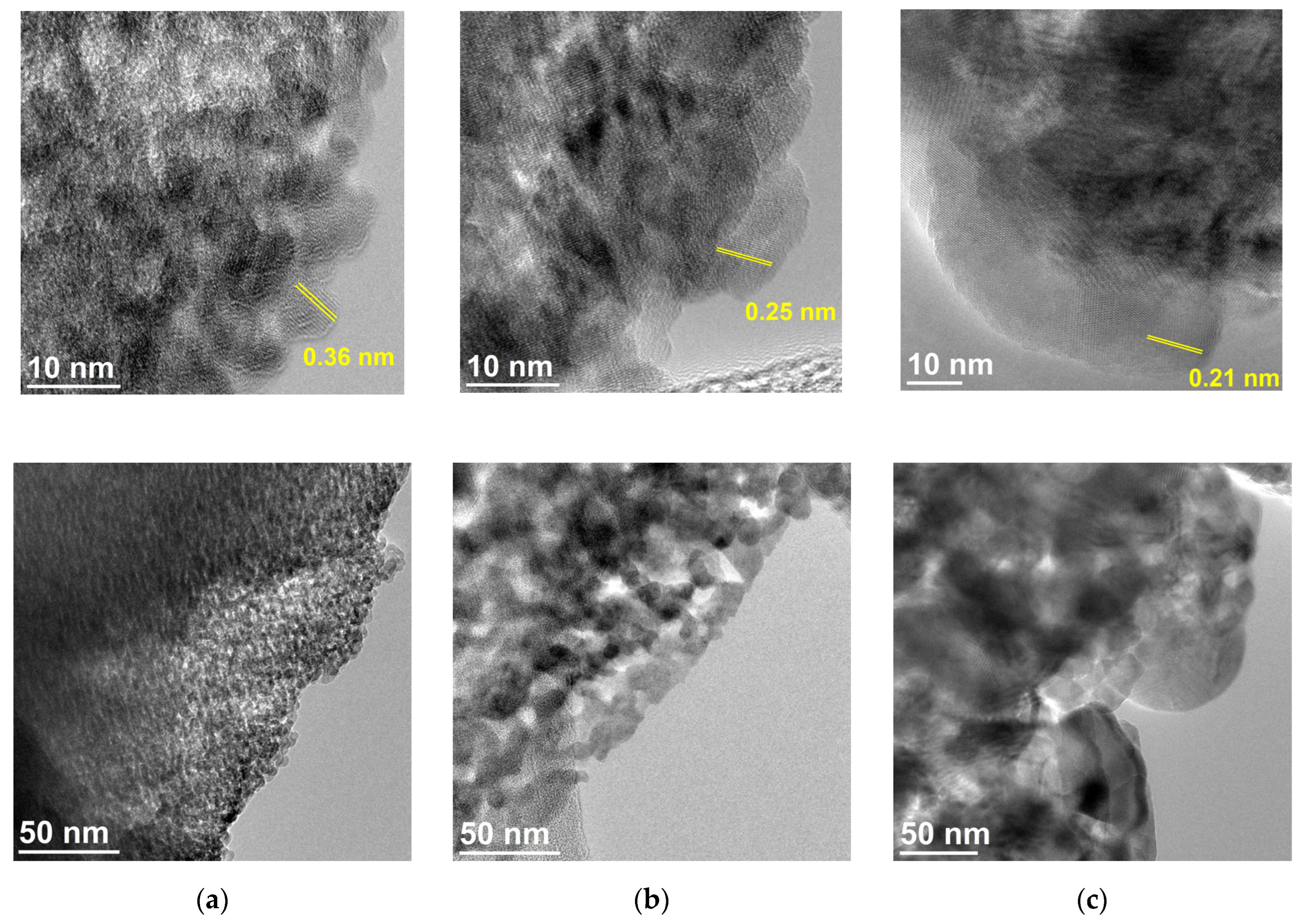
3.3. Transmission Electron Microscopy

Figure 2 shows the structure and morphology of the Sd3 siderite sample annealed at 400 °C, 700 °C, and 900 °C for two hours obtained using Transmission Electron Microscopy (TEM), including high-resolution mode (HRTEM). In the case of the Sd3 sample annealed at 400 °C, semi-spherical magnetite nanoparticles of about 5–8 nm in size were observed with clearly visible crystal planes with interplanar distances of about 0.36 nm corresponding to (012) planes of hexagonal siderite (Figure 2a). As the annealing temperature increases, a distinct increase in nanoparticle size is evident for forming materials. For the Sd3 sample annealed at 700 °C, the nanoparticle size corresponds to 15–20 nm with planes of distances ~0.25 nm that can be attributed to (113) planes of face-centered cubic (fcc) magnesioferrite or (113) planes of (fcc) magnetite (Figure 2b). In contrast, for the sample annealed at 900 °C, the size of the nanoparticles reaches about 50–100 nm, where the measured lattice distances of 0.21 nm can be indexed as (004) planes of (fcc) magnesioferrite or (004) planes of (fcc) magnetite (Figure 2c).

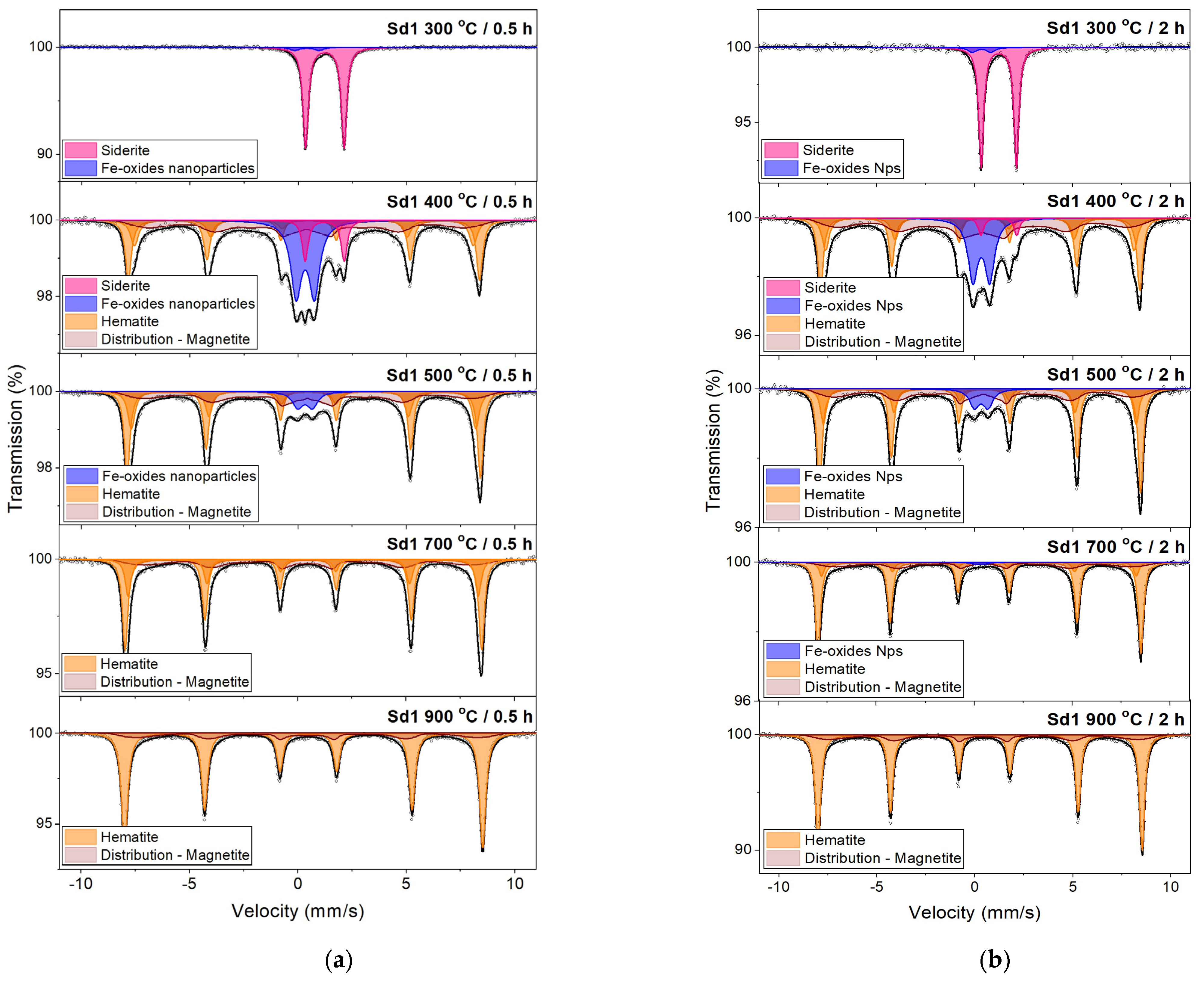
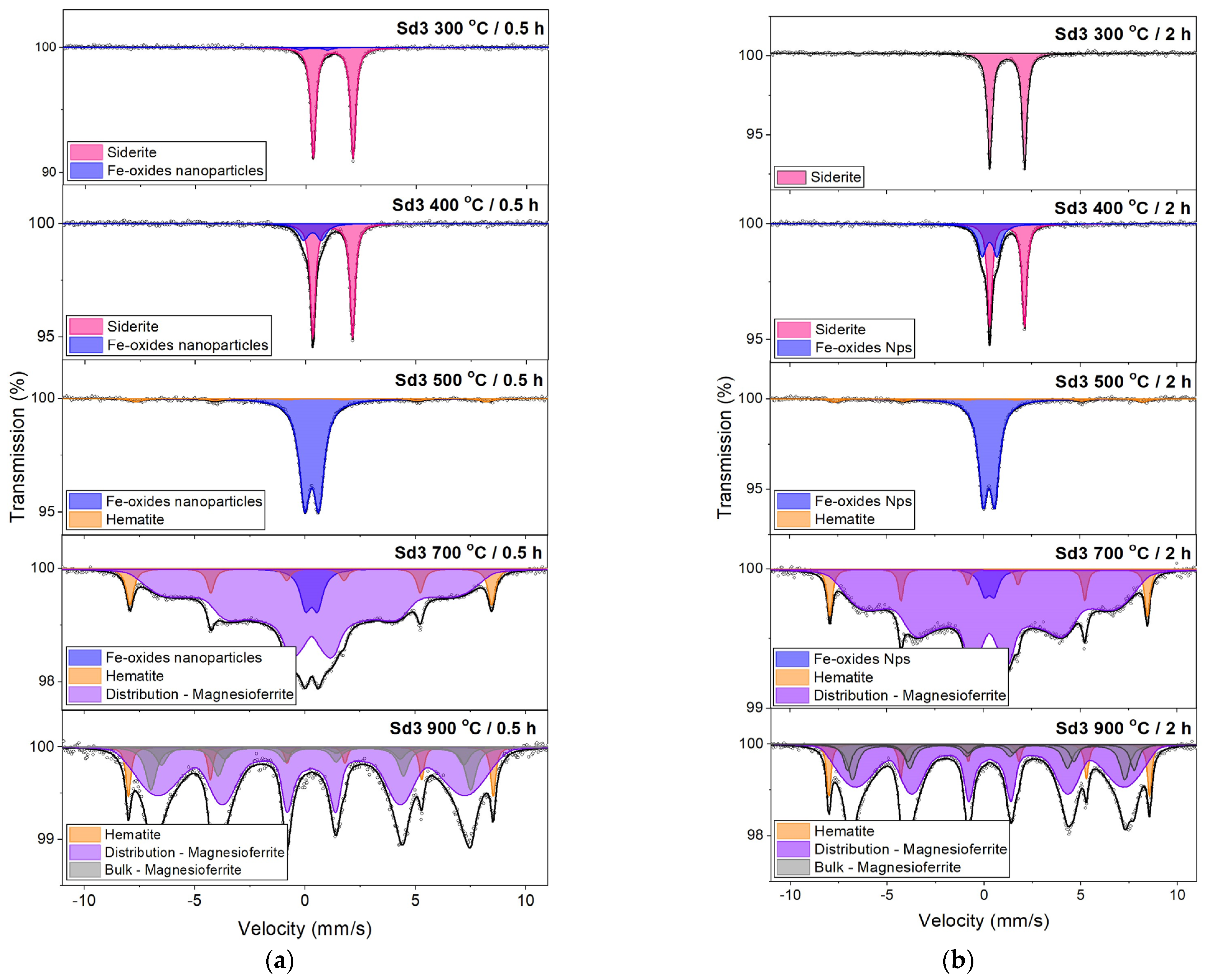
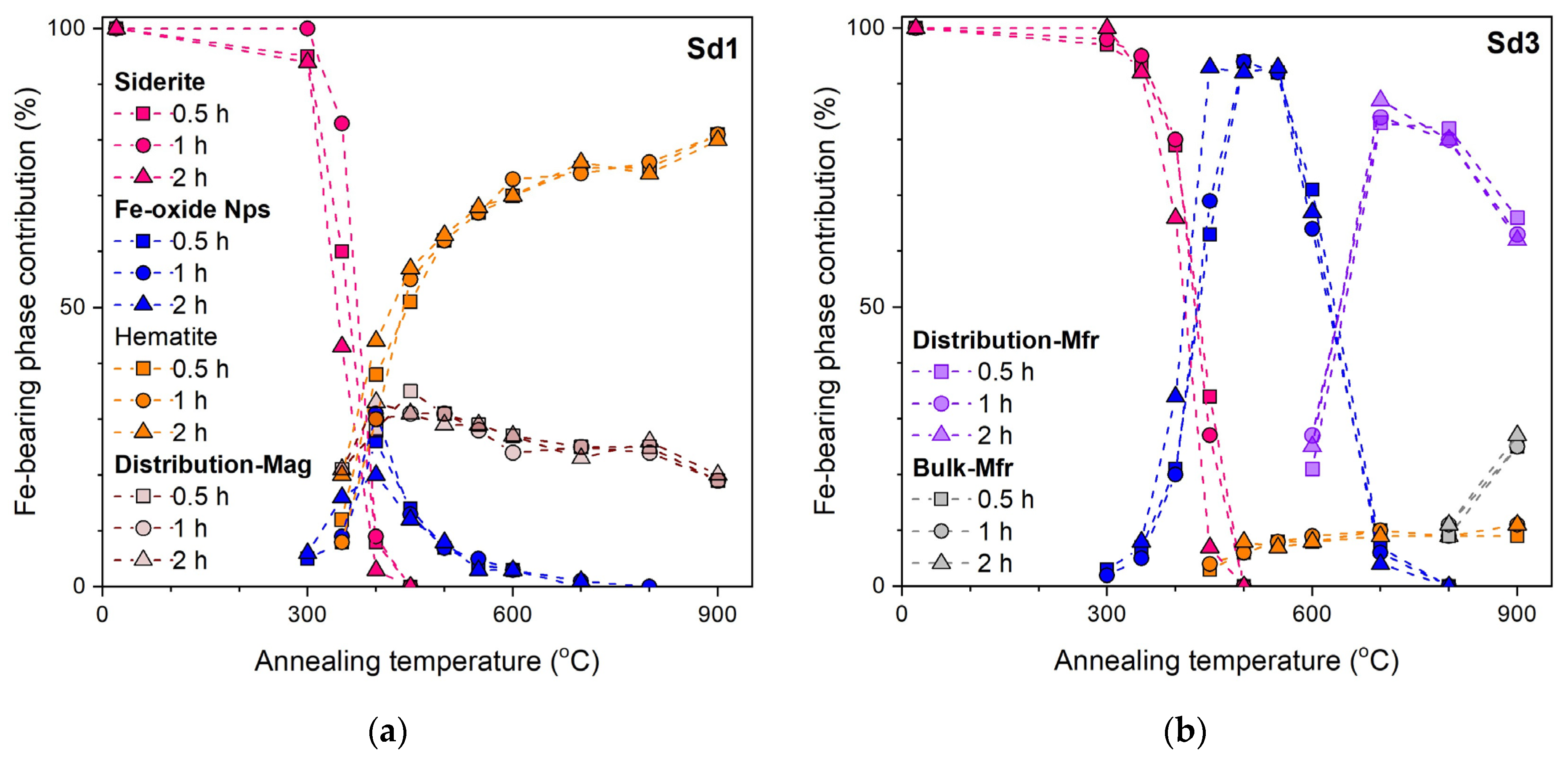
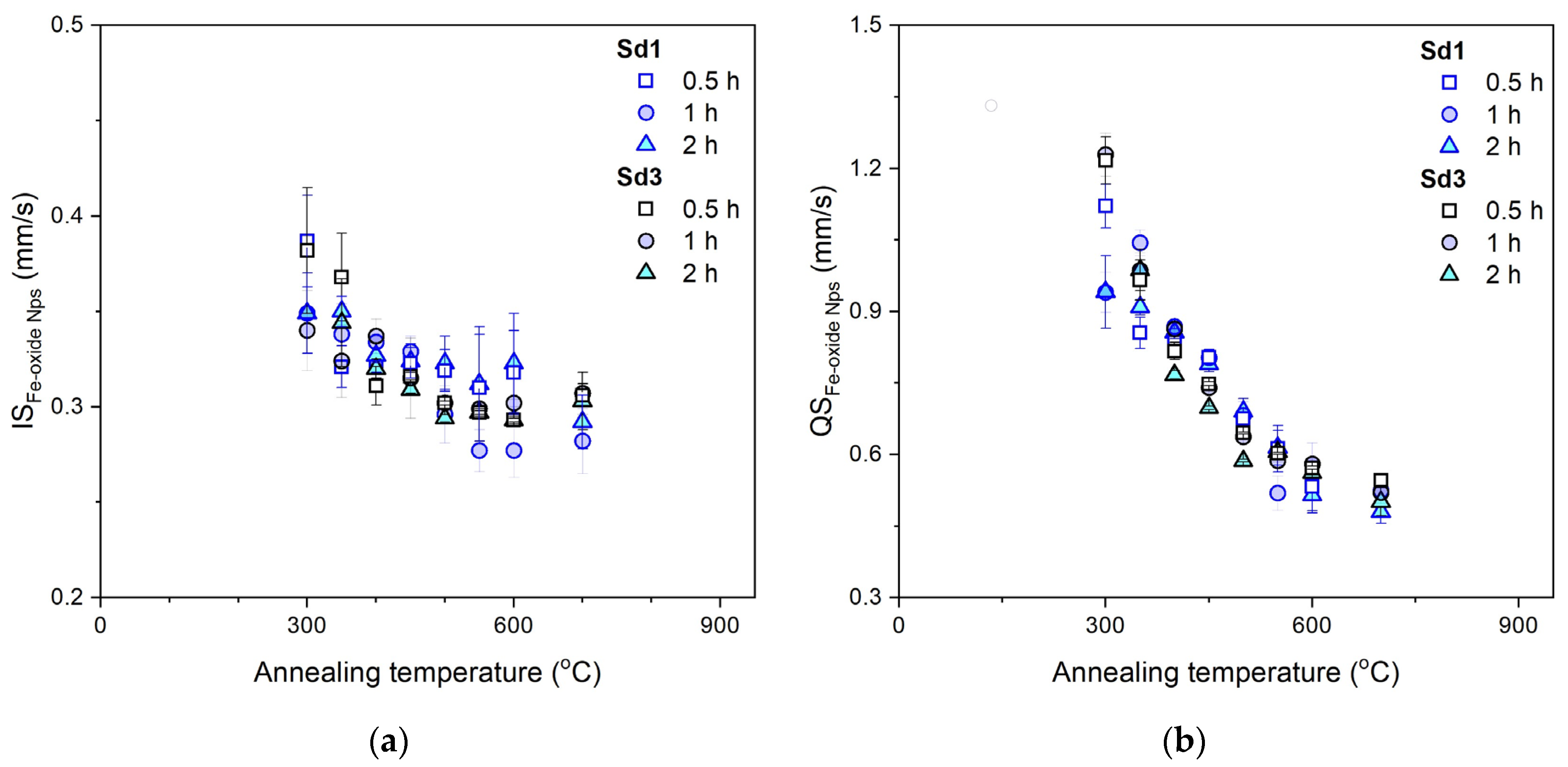
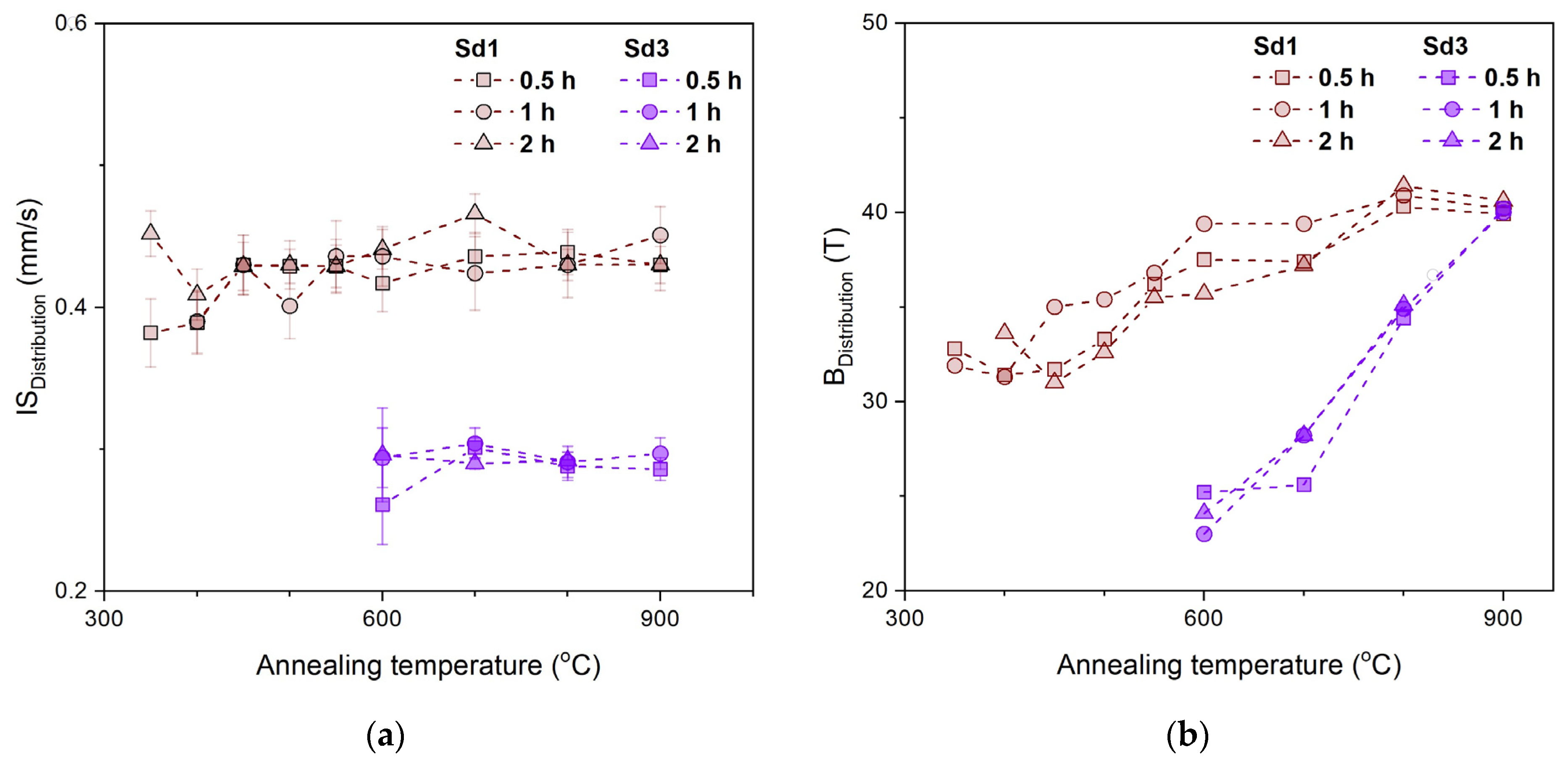
3.4. Mössbauer Spectroscopy

The Mössbauer spectra of initial siderite samples Sd1 and Sd3 were characteristic of a ferrous quadrupole doublet with isomer shift values IS = 1.23(1) mm/s, 1.24(1) mm/s and quadrupole splitting QS = 1.80(1) mm/s, 1.81(1) mm/s, respectively for Sd1 and Sd3 sample. These values agree well with the literature data [17,18] and indicate the presence of Fe2+ ions in the siderite structure. The absence of other components in the Mössbauer spectra indicates that siderite is the only iron-bearing mineral in the studied samples.
The selected Mössbauer spectra of siderite samples Sd1 and Sd 3 annealed at different temperatures and times are presented in Figure 3 and Figure 4. The contribution of individual components present on the Mössbauer spectra depending on the annealing temperature and annealing time are shown in Figure 5. The doublet characteristic of siderite is visible on the Mössbauer spectra of the Sd1 sample up to a temperature of 450 °C, and in the case of the Sd3 sample up to a temperature of 500 °C. However, its contribution to the spectrum decreases as the annealing temperature increases, and the longer the annealing time, the more significant the amount of siderite decomposition (Figure 5).
The ferric quadrupole doublet is the first component indicating the decomposition of siderite, which appears in the Mössbauer spectra of samples annealed already at a temperature of 300 °C (Figure 3 and Figure 4) in both samples, Sd1 and Sd3. The hyperfine parameters of this doublet versus annealing temperature and annealing time are presented in Figure 6. As the annealing temperature increases, its contribution to the spectrum increases, reaching a maximum of 400 °C for the Sd1 sample and 550 °C for Sd3 (Figure 5). The following components visible in the spectra of annealed samples, whose contribution increases as the contribution of the quadrupole doublet decreases, are related to iron oxides (Figure 3 and Figure 4).
Hematite is the main iron oxide appearing in the Mössbauer spectra of the Sd1 sample, and its contribution in the spectrum increases as the contribution of the ferric doublet decreases (Figure 5). The decomposition of siderite in the presence of air and the formation of hematite is illustrated by the reaction: 2FeCO3 + ½O2 → Fe2O3 + 2CO2 [17]. As we know, the room temperature Mössbauer spectrum of hematite is a characteristic sextet with an isomer shift of 0.37 mm/s, a quadrupole shift of −0.11 mm/s, and a hyperfine magnetic field of approximately 51 T [19]. Here, instead of one sextet representing Fe2O3, two are visible, differing in field values from ~1 T to ~2 T. This second sextet is probably the result of the substitution of Fe by other elements, such as Al, which reduces the value of the hyperfine field [20]. The appearance of the second sextet associated with hematite may also result from the fact that this oxide is formed continuously over a wide range of temperatures in siderite decomposition, and some of it is still poorly crystalline. This hypothesis may be confirmed by seeing one sextet associated with hematite at high temperatures instead of two. It is also possible that the two sextets result from hematite formation by different reactions. Above 400 °C, the amount of hematite in the Sd1 sample increases while the concentration of magnetite and ferric doublet decreases (Figure 5a). Therefore, we can assume the reaction proposed in the literature [21]: 4Fe3O4 + O2→ 6Fe2O3. We also do not rule out that both hypotheses are correct, and hematite is formed in two different reactions, but other elements, such as Al, may also occupy the Fe positions.
The Mössbauer spectra of the annealed Sd3 sample differ significantly from those obtained for the Sd1 sample. As the annealing temperature increases, the contribution of the ferric doublet decreases, and a sextet associated with hematite appears, but its contribution does not exceed 11%. The distribution of hyperfine fields adjusts a significant part of the spectrum. The obtained values of the isomer shift and hyperfine field of this distribution for various temperatures and annealing times are presented in Figure 7. The X-ray diffraction and Transmission Electron Microscopy measurements indicate that this part of the spectrum will be associated with poor crystalline magnesioferrite (sample Sd3) and magnetite (sample Sd1). A reaction in which magnetite is formed during siderite decomposition in the presence of air is illustrated by the reaction: 3FeCO3 + ½O2 → Fe3O4 + 3CO2 [21,22]. Moreover, the average isomer shift values obtained for this distribution (Figure 7a) indicate that in sample Sd1, the distribution is associated with magnetite [23], and the lower IS values obtained for the distribution in sample Sd3 characterize magnesioferrite [24]. Higher values of the hyperfine magnetic field in the Sd1 sample than those obtained for Sd3 (Figure 7b) may also indicate a greater degree of order in this phase. Above a temperature of 700 °C, the crystallization process of magnesioferrite begins. The Mössbauer spectra of the Sd3 sample show clear sextets associated with this iron oxide. Although the results of X-ray diffraction measurements indicate magnetite, the obtained value of the isomer shifts of these sextets indicate magnesioferrite or magnesiomagnetite, at least in the initial phase of crystallization.

4. Discussion

Two rock siderite samples were annealed at temperatures ranging from 300 °C to 900 °C and for different annealing times (half, one, and two hours). X-ray diffraction results show that sample Sd1 contains approximately 94% vol. siderite, and sample Sd3 contains about 84% vol. of this iron-bearing carbonate (Table 2). The samples studied also contain small amounts of minerals such as quartz, illite, and kaolinite. X-ray fluorescence analysis shows that these rock siderite samples contain mainly elements included in the abovementioned minerals, except magnesium, which is particularly abundant (~4 wt.%) in sample Sd3 (Table 1). This element is not a component of any mentioned minerals, meaning it mainly substitutes Fe in siderite in the Sd3 sample.
The siderite decomposition process started at a temperature of 300 °C in both samples and it ended at 450 °C in the Sd1 sample, and in the Sd3 sample at 500 °C. The longer the annealing time, the more significant the amount of siderite decomposition (Figure 6). The component in the Mössbauer spectra that signals the beginning of the siderite decomposition process is the ferric quadrupole doublet. This doublet appears in the Mössbauer spectra of both samples, Sd1 and Sd3. As the annealing temperature increases, the contribution of this doublet in the spectrum also increases, reaching a maximum at 400 °C for the Sd1 sample and 500 °C for Sd3, in other words, at the temperatures at which the largest amount of siderite decomposed in studied samples. The following components visible in the Mössbauer spectra of annealed samples, whose contribution increases as the contribution of the ferric quadrupole doublet decreases, are related to iron oxides (Figure 3 and Figure 4) like hematite and magnetite. The results of X-ray diffraction also indicate the presence of these iron oxides. Therefore, we associate this doublet with these iron oxides’ superparamagnetic nanoparticles (Nps). Also, the hyperfine parameters of this component (Figure 6) indicate its origin [25,26,27,28]. The isomer shift (IS) and the quadrupole splitting (QS) of both samples in annealing temperatures from 300 °C to 700 °C were in the range of 0.38–0.28 mm/s and 1.22–0.48 mm/s, respectively. The large QS values should be attributed to the large electric field gradient for the atoms in the surface coating of particles compared to the core of that particle. For a large particle relative to the outer shell, the contribution is small, and the measured quadrupole splitting will also be small. As the annealing temperature increases, the QS values decrease, which indicates an increase in the particle diameter, as evidenced by a decrease in the contribution of ferric quadrupole doublet in the Mössbauer spectrum. It means that the crystallization process of Fe-oxides begins. However, it would be necessary to explain the quadrupole splitting occurring in this doublet, which is evident for hematite, while it should be explained for magnetite. In hematite, a quadrupole shift occurs in the magnetically ordered state, and quadrupole splitting in the paramagnetic state because of the canting angle of the spins with respect to the electric field gradient axis. On the other hand, magnetite crystallizes in a regular structure, and for the paramagnetic state or nanocrystallites, we would expect to observe a single line and not a doublet. However, if we consider the fact that the magnetite produced during siderite decomposition is a poorly crystalline phase and assume small particle size, then we can explain the presence of a doublet as representing a remarkable part of Fe atoms located in the region of the nanoparticle surface, and because of broken cubic symmetry, these atoms are exposed to a nonzero electric field gradient [29]. The X-ray diffraction analysis results show that above a temperature of 700 °C in the Sd1 sample, we mainly have hematite and small amounts of magnetite. In sample Sd3, the opposite is observed; we observe small amounts of hematite and mainly magnesioferrite. The occurrence of magnesioferrite instead of magnetite in the Sd3 sample is due to the large amount of magnesium in this sample. In the Mössbauer spectra, both magnetite and magnesioferrite are represented by hyperfine field distribution, indicating these phases’ poor crystallinity. Broad diffraction lines and Transmission Electron Microscopy results also indicate this. The crystallization process begins only above 700 °C. Moreover, in the Sd3 sample containing much more Mg than the Sd1 sample, the crystallization processes of Fe-oxides proceed much slower, which indicates a practically unchanged contribution of nanoparticles of these oxides in a wide range of heating temperatures from 450 °C to 550 °C. Magnesium stabilizes the structure of magnesioferrite, and its oxidation to hematite is practically not observed up to a temperature of 900 °C. The fact that the substitution of Mg or Mn ions in the Fe position stabilizes the structure of magnesioferrite has been observed [12,28].
According to the literature [17,30,31,32] and as already indicated in the introduction, the main product of decomposition of siderite heated in air atmosphere is hematite. The research results presented here show that this is instead the case for pure siderite. Isomorphic impurities occurring in the siderite structure strongly influence the siderite decomposition process and the type of decomposition products.

5. Conclusions

The influence of annealing time, temperature, and isomorphous impurities on siderite decomposition kinetics was studied using Mössbauer spectroscopy, X-ray diffraction, the X-ray fluorescence method, and Transmission Electron Microscopy. Two rock siderite samples contained siderite as the main phase and small amounts of additional phases such as quartz, illite, and kaolinite. One sample also contained a significant amount of magnesium, which probably substitutes for Fe in the siderite structure. The samples were heated in air for half, one, and two hours at temperatures ranging from 300 °C to 900 °C. The following conclusions have been obtained:
(1)
The siderite decomposition process begins at a temperature of 300 °C. In the sample containing a small amount of magnesium (<1 wt.%), complete decomposition takes place at a temperature of 450 °C, and in a sample with a larger amount of this element (~4 wt.%) at a temperature of 500 °C.
(2)
The annealing time does not significantly affect the width of the temperature range where siderite decomposition occurs. Still, it does affect the degree of siderite decomposition: the longer the heating time, the greater the amount of siderite decomposition. The dynamics of the processes undergone by the sample annealed for half an hour and an hour are similar. However, in the sample annealed for two hours, the degree of siderite decomposition is much greater than shorter annealing times.
(3)
The decomposition products of siderite annealed in air are iron oxides. In the sample without Mg, the oxide was mainly hematite, which confirms the above-mentioned literature data, and magnesioferrite in the sample containing Mg. The consequence of substituting Mg in the Fe position is the stabilization of the structure of magnesioferrite, and its oxidation to hematite was not observed even at 900 °C.
(4)
Iron oxides formed directly in siderite decomposition are poorly crystalline, and we can treat them as nanoparticles. They maintain this form in a wide temperature range, especially in a magnesium-containing sample. The presence of this element significantly slows down the process of magnesioferrite crystallization. Mg2+ replaces Fe2+ in magnetite to form the magnesioferrite, whilst simultaneously stabilizing the crystal structure as a spinel. Due to the increase in the amount of magnesium or magnesioferrite in a sample, the transformation of hematite to magnetite would be reduced.
In our summary, it is also necessary to refer and compare the results obtained here to those presented in the previous work [15] on the thermal decomposition of siderite and characterization of the decomposition products under O2 and CO2 atmospheres. At the outset, it should be noted that the process of siderite decomposition, especially the type of products formed because of this process, largely depends on the heat treatment method. The results of both works show, however, that the process of siderite decomposition begins slightly above a temperature of 300 °C; this is the result of studies using Mössbauer spectroscopy. The results of this method also show that in the initial phase of siderite decomposition, iron oxide nanoparticles are formed, and only at higher temperatures do they begin to crystallize. Methods such as X-ray diffraction or thermogravimetric analysis (TGA) and differential scanning calorimetry (DSC) indicate a slightly higher temperature of siderite decomposition, namely around 350 °C [33], which results from their sensitivity. The results of the previous work suggested that the Mg present in the sample may affect the temperature range in which siderite decomposition occurs and the type of its products. The results of the present work confirmed these assumptions.

Author Contributions

M.K.-G. carried out the conceptualization, designed the methodology, and performed the measurements/investigations, and validation. She also prepared the original draft, reviewed, edited the manuscript, and secured the resources; investigation, M.K.-G., M.W., J.K. and D.Ł.; formal analysis, M.K.-G., Z.A. and D.Ł.; samples preparation, J.N. All authors have read and agreed to the published version of the manuscript.

Funding

This research received no external funding.

Data Availability Statement

The data may be made available by the authors upon individual request.

Acknowledgments

This research was supported by the Research Excellence Initiative project of the University of Silesia in Katowice.

Conflicts of Interest

The authors declare no conflicts of interest.

References

  1. Isambert, A.; Valet, J.-P.; Gloter, A.; Guyot, F. Stable Mn-magnetite derived from Mn-siderite by heating in air. J. Geophys. Res. 2003, 108, 2283. [Google Scholar] [CrossRef]
  2. Ponomar, V.P.; Dudchenko, N.O.; Brik, A.B. Phase transformations of siderite ore by the thermomagnetic analysis data. J. Magn. Magn. Mater. 2017, 423, 373–378. [Google Scholar] [CrossRef]
  3. Zhua, X.; Hana, Y.; Suna, Y.; Gaoa, P.; Li, Y. Thermal Decomposition of Siderite Ore in Different Flowing Atmospheres: Phase Transformation and Magnetism. Miner. Process. Extr. Metall. Rev. 2022, 44, 201–208. [Google Scholar] [CrossRef]
  4. Kelektsoglou, K. Carbon capture and storage: A review of mineral storage of CO2 in Greece. Sustainability 2018, 10, 4400. [Google Scholar] [CrossRef]
  5. Mendoza, M.; Santos, A.; López, E.; Drozd, V.; Durygin, A.; Chen, J.; Saxena, S. Iron oxides as efficient sorbents for CO2 capture. J. Mater. Res. Technol. 2019, 8, 2944–2956. [Google Scholar] [CrossRef]
  6. Fosbol, P.L.; Thomsen, K.; Stenby, E.H. Review and recommended thermodynamic properties of FeCO3. Corros. Eng. Sci. Technol. 2010, 45, 115–135. [Google Scholar] [CrossRef]
  7. Santoso, R.; Rahmawati, S.; Gadesa, A.; Wahyuningrum, D. Understanding Passive Layer Formation for Further Corrosion Management in Gas Production Pipes. J. Phys. Conf. Ser. 2017, 877, 012062. [Google Scholar] [CrossRef]
  8. Zhang, Q.; Sun, Y.; Han, Y.; Li, Y.; Gao, P. Reaction behavior and non-isothermal kinetics of suspension magnetization roasting of limonite and siderite. Int. J. Miner. Metall. Mater. 2023, 30, 824–833. [Google Scholar] [CrossRef]
  9. Zhu, D.; Luo, Y.; Pan, J.; Zhou, X. Reaction Mechanism of Siderite Lump in Coal-Based Direct Reduction. High Temp. Mater. Proc. 2016, 35, 185–194. [Google Scholar]
  10. Zhang, X.; Han, Y.; Li, Y.; Sun, Y. Effect of Heating Rate on Pyrolysis Behavior and Kinetic Characteristics of Siderite. Minerals 2017, 7, 211. [Google Scholar] [CrossRef]
  11. Zhang, Z.; Liu, H.; Lu, P.; Chen, T.; Ma, W. Nanostructured α-Fe2O3 derived from siderite as an effective Hg(II) adsorbent: Performance and mechanism. Appl. Geochem. 2018, 96, 92–99. [Google Scholar] [CrossRef]
  12. Dubrawski, J.V. Thermal decomposition of some siderite-magnesite minerals using DSC. J. Therm. Anal. 1991, 37, 1213–1221. [Google Scholar] [CrossRef]
  13. Jagtap, S.B.; Pande, A.R.; Gokarn, A.N. Kinetics of thermal decomposition of siderite: Effect of particle size. Int. J. Miner. Process. 1992, 36, 113–124. [Google Scholar] [CrossRef]
  14. Goldin, D.M.; Kulikova, G.V. On the dissociation mechanism of carbonates and their isomorphous mixture. J. Therm. Anal. Calorim. 1984, 29, 139–145. [Google Scholar] [CrossRef]
  15. Kądziołka-Gaweł, M.; Nowak, J.; Szubka, M.; Klimontko, J.; Wojtyniak, M. Thermal Decomposition of Siderite and Characterization of the Decomposition Products under O2 and CO2 Atmospheres. Minerals 2023, 13, 1066. [Google Scholar] [CrossRef]
  16. Rancourt, D.; Ping, J. Voigt-based methods for arbitrary-shape static hyperfine parameter distributions in Mössbauer spectroscopy. Nucl. Instrum. Meth. B 1991, 58, 85–97. [Google Scholar] [CrossRef]
  17. Dhupe, A.P.; Gokarn, A.N. Studies in the thermal decomposition of natural siderites in the presence of air. Int. J. Miner. Process. 1990, 28, 209–220. [Google Scholar] [CrossRef]
  18. Ristić, M.; Krehula, S.; Reissner, M.; Musić, S. 57Fe Mössbauer, XRD, FT-IR, FE SEM Analyses of Natural Goethite, Hematite and Siderite. Croat. Chem. Acta 2017, 90, 499–507. Available online: https://hrcak.srce.hr/190802 (accessed on 17 December 2017). [CrossRef]
  19. Bodker, F.; Hansen, M.F.; Bender Koch, C.; Lefmann, K.; Morup, S. Magnetic properties of hematite nanoparticles. Phys. Rev. B Condens. Matter 2000, 61, 6826–6838. [Google Scholar] [CrossRef]
  20. De Grave, E.; Bowen, L.; Amarasiriwardena, D.; Vandenberghe, R. 57Fe Mössbauer effect study of highly substituted aluminum hematites: Determination of the magnetic hyperfine field distributions. J. Magn. Magn. Mater. 1988, 72, 129–140. [Google Scholar] [CrossRef]
  21. Chen, C.; Han, Y.; Zhang, Y.; Liu, Y.; Liu, Y. Efficient Utilization of Siderite- and Hematite-Mixed Ore by Suspension Magnetization Roasting: A Pilot-Scale Study. Sustainability 2022, 14, 10353. [Google Scholar] [CrossRef]
  22. Koziol, A. Experimental determination of siderite stability and application to Martian Meteorite ALH84001. Am. Min. 2004, 89, 294–300. [Google Scholar] [CrossRef]
  23. Halmar, N.; Hammer, M.; Netland, H.; Perkins, N.; Streyle, T.; Thomasson, C.; Vanhoever, L. Characterization of Iron Oxide Samples Using Mössbauer Spectroscopy and Hysteresis Loops. J. Undergrad. Rep. Phys. 2023, 33, 100010. [Google Scholar] [CrossRef]
  24. Lee, S.; Kim, S.; Kim, C. Superexchange Interactions in MgFe2O4. J. Korean Phys. Soc. 2006, 48, 583–588. [Google Scholar]
  25. Wu, W.; Lin, X.; Duan, H.; Wang, J. Mössbauer And High-Temperature Magnetic Properties of Different Size Fe3O4 Nanostructure. Int. J. Mod. Phys. B 2012, 26, 1250073. [Google Scholar] [CrossRef]
  26. Gervits, N.; Gippius, A.; Tkachev, A.; Demikhov, E.; Starchikov, S.; Lyubutin, I.; Vasiliev, A.; Chekhonin, V.; Abakumov, M.; Semkina, M.; et al. Magnetic properties of biofunctionalized iron oxide nanoparticles as magnetic resonance imaging contrast agents. Beilstein J. Nanotechnol. 2019, 10, 1964–1972. [Google Scholar] [CrossRef]
  27. Klekotka, U.; Winska, E.; Satuła, D.; Kalska-Szostko, B. Mössbauer Studies of Surface Modified Magnetite Particles. Acta Phys. Pol. A 2018, 134, 1003–1006. [Google Scholar] [CrossRef]
  28. Chen, Y.-B.; Du, Y.; Guo, Y.-F.; Guo, X.-M. Quantitative Investigation of MgO, Al2O3 and SiO2 Effects on Solid-State Formation of Secondary Hematite in Sintering Process of Iron Ore Fines. Minerals 2022, 12, 282. [Google Scholar] [CrossRef]
  29. Satuła, D.; Kalska-Szostko, B.; Szymańskia, K.; Dobrzyńskia, L.; Kozubowski, J. Microstructure and Magnetic Properties of Iron Oxide Nanoparticles Prepared by Wet Chemical Method. Acta Phys. Pol. A 2008, 114, 1615–1621. [Google Scholar] [CrossRef]
  30. Gallagher, P.; Warne, S. Thermomagnetometry and thermal decomposition of siderite. Thermochim. Acta 1981, 43, 253–267. [Google Scholar] [CrossRef]
  31. Musić, S.; Popović, S. Mössbauer spectroscopic and X-ray diffraction study of the thermal decomposition of natural siderite and goethite. J. Radioanal. Nucl. Chem. 1987, 111, 27–41. [Google Scholar] [CrossRef]
  32. Zhu, Y.; Banerjee, S.; Gill, J.; Williams, Q. Rock magnetic properties related to thermal treatment of siderite: Behavior and interpretation. J. Geophys. Res. 2000, 105, 783–794. [Google Scholar] [CrossRef]
  33. Garcia, S.; Rosenbauer, R.J.; Palandri, J.; Maroto-Valer, M.M. Sequestration of non-pure carbon dioxide streams in iron oxyhydroxide-containing saline repositories. Int. J. Greenh. Gas Control 2012, 7, 89–97. [Google Scholar] [CrossRef]
Figure 1. XRD patterns of the samples Sd1 (a) and Sd3 (b) annealed at different temperatures in the air atmosphere for two hours. The interplanar spacing values (given in angstroms) are shown in the diffractograms. Sd—siderite, Kln—kaolinite, Mag—magnetite, Mfr—magnesioferrite, Hem—hematite, Qtz—quartz.
Minerals 15 00428 g001
Figure 2. The TEM and HRTEM images of Sd3 sample with measured lattice spacing (yellow markers) annealed at 400 °C (a), 700 °C (b), and 900 °C (c) for two hours.
Minerals 15 00428 g002
Figure 3. Room temperature Mössbauer spectra for Sd1 sample annealed at selected temperatures for half (a) and two (b) hours. The experimental points, fitting curves, and spectral components corresponding to iron sites in different phases (colored lines) are presented.
Minerals 15 00428 g003
Figure 4. Room temperature Mössbauer spectra for Sd3 sample annealed at selected temperatures for half (a) and two (b) hours. The experimental points, fitting curves, and spectral components corresponding to iron sites in different phases (colored lines) are presented.
Minerals 15 00428 g004
Figure 5. The relative contribution of Fe-bearing phases present in samples Sd1 (a) and Sd3 (b) depending on the annealing temperature and time determined from the analysis of Mössbauer spectra; Nps—nanoparticles, Mag—magnetite, Mfr—magnesioferrite. The dashed lines indicate the trends of the data and act as a guide to the eye.
Minerals 15 00428 g005
Figure 6. Isomer shift IS (a) and quadrupole splitting QS (b) values versus annealing temperature and annealing time obtained for ferric doublet representing Fe-oxide nanoparticles in Sd1 and Sd3 sample.
Minerals 15 00428 g006
Figure 7. Isomer shift IS (a) and hyperfine magnetic field B (b) values versus annealing temperature and annealing time obtained for hyperfine magnetic field distribution representing poorly crystalline magnetite in the Sd1 sample and magnesioferrite in the Sd3 sample.
Minerals 15 00428 g007
Table 1. Element content in the investigated siderites Sd1 and Sd3 obtained using the X-ray fluorescence method.
SampleElement Concentration (wt.%)
COMgAlSiPKCaTiMnFeSum
Sd16.1748.600.802.634.580.170.320.860.090.6034.9699.78
Sd36.2650.803.934.066.930.070.520.920.130.3725.5399.52
Table 2. Mineral composition of siderite samples Sd1 and Sd3 annealed at 400 °C, 700 °C, and 900 °C for half, one, and two hours based on XRD analyses by the Rietveld method in % vol.
TemperatureInitial
Sd1
400 °C700 °C900 °C
Component0.5 h1 h2 h0.5 h1 h2 h0.5 h1 h2 h
Siderite94282720------
QuartzTrace1196232111
Illite1---------
Kaolinite5414------
Hematite-576370939597908156
Magnetite----52181843
TemperatureInitial
Sd3
400 °C700 °C900 °C
Component0.5 h1 h2 h0.5 h1 h2 h0.5 h1 h2 h
Siderite84818075------
Quartz78910121312131313
Illite3---------
Kaolinite69911------
Hematite-1121165131011
Magnetite----15109102022
Magnesioferrite-112627174645754
Disclaimer/Publisher’s Note: The statements, opinions and data contained in all publications are solely those of the individual author(s) and contributor(s) and not of MDPI and/or the editor(s). MDPI and/or the editor(s) disclaim responsibility for any injury to people or property resulting from any ideas, methods, instructions or products referred to in the content.

