


From Source to Sink: U-Pb Geochronology and Lithochemistry Unraveling the Missing Link Between Mesoarchean Anatexis and Magmatism in the Carajás Province, Brazil

 , ,
, ,
Abstract
1. Introduction
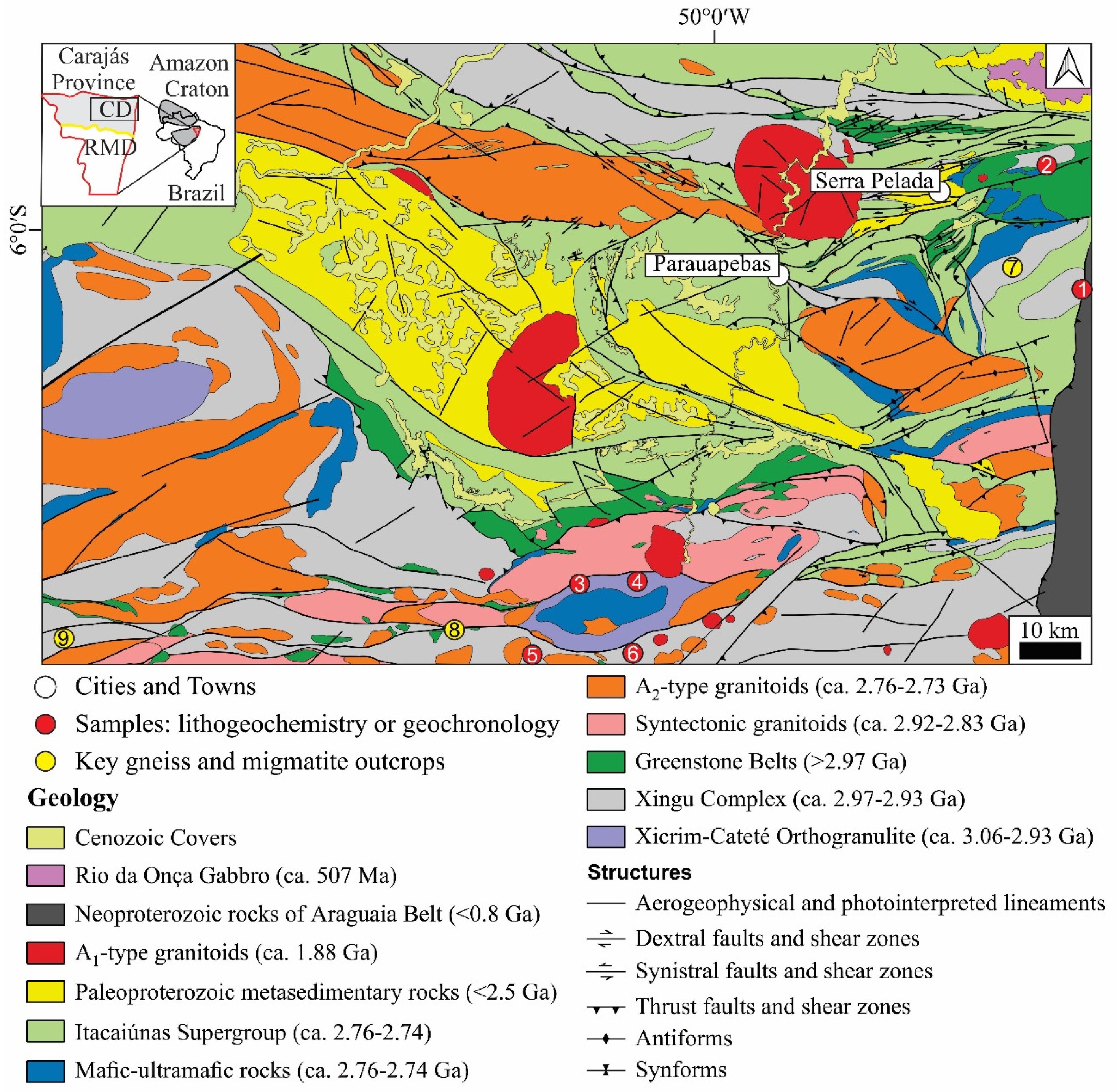
2. Geological Setting of the Carajás Province
3. Analytical Procedures
3.1. Geochemistry
3.2. U-Pb Geochronology
4. Morphology, Constituent Parts, and Petrography of the ca. 2.86 Ga Migmatites of the Carajás Domain
4.1. Migmatite Morphologies
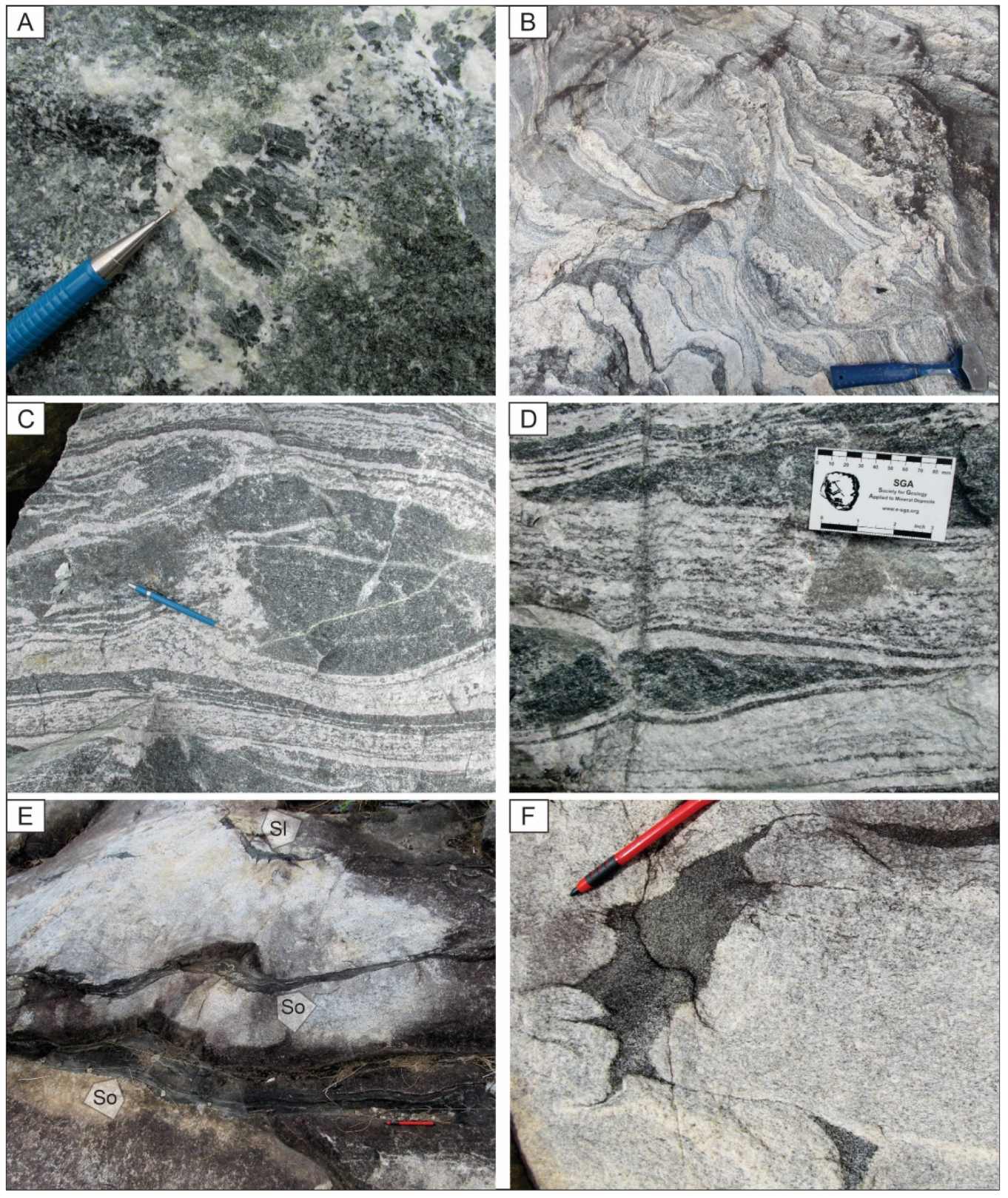
4.2. Constituent Parts of the Migmatites
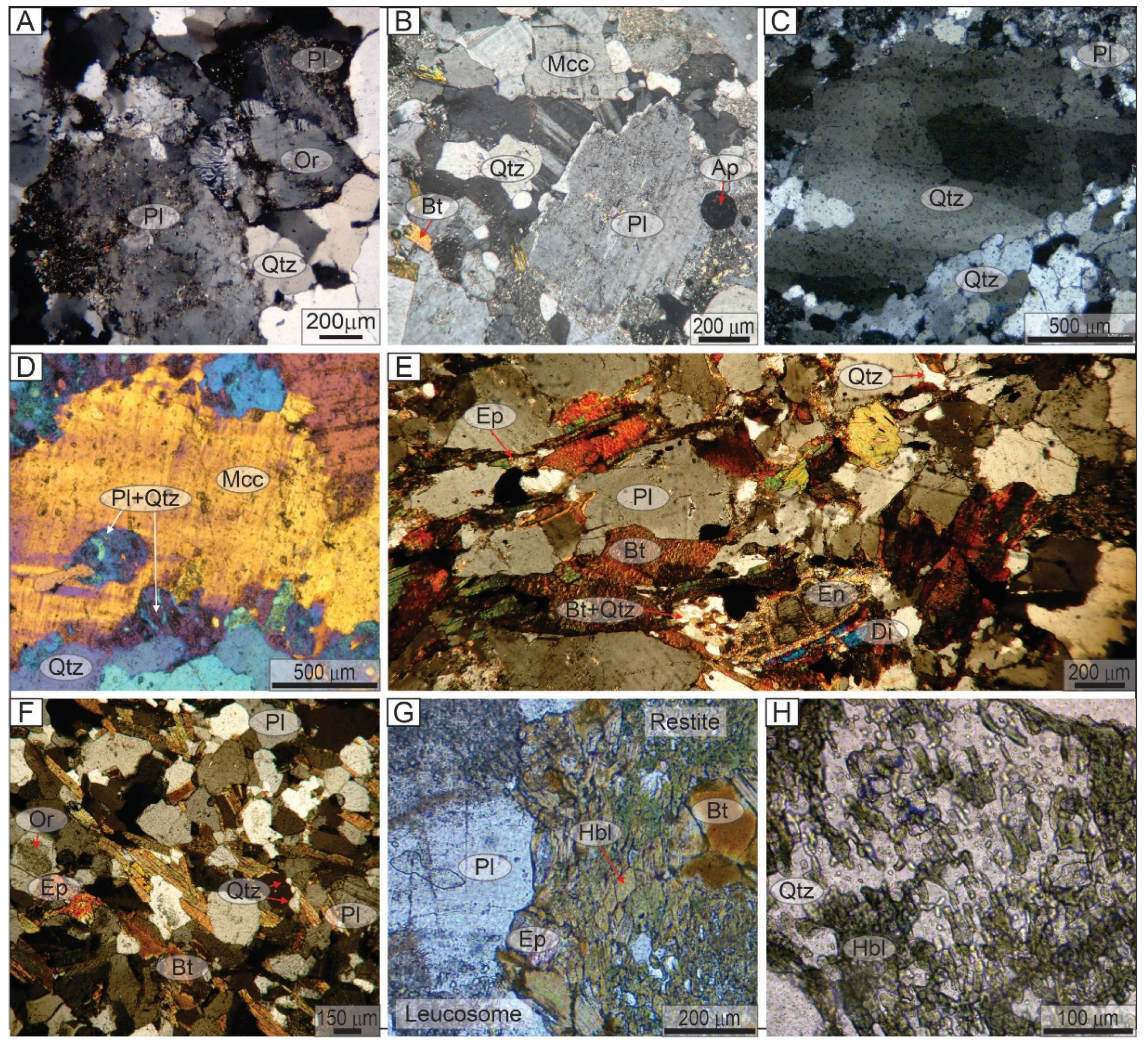
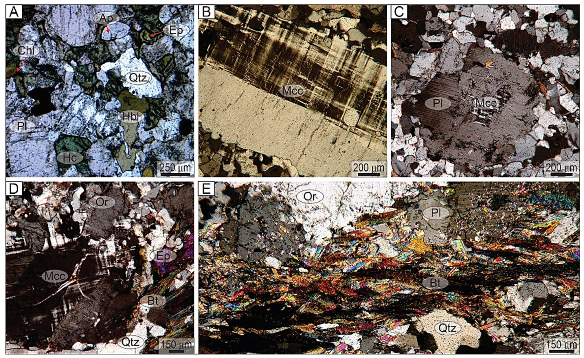
4.2.1. Paleosome

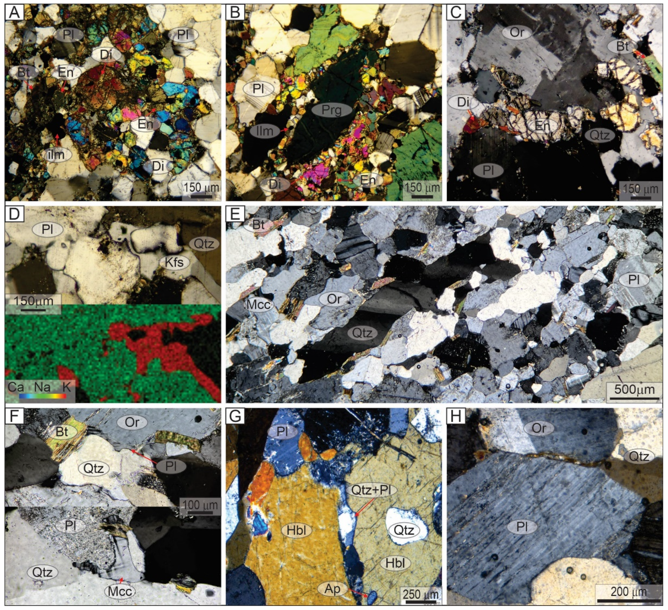
4.2.2. Neosome
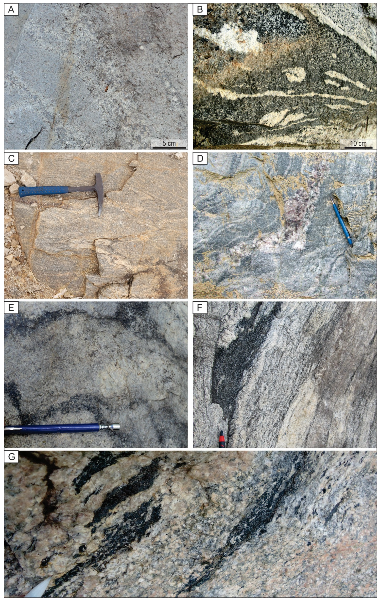
5. Mesoarchean Syntectonic Granitoids

6. Geochemical Characteristics of the Leucosome in the Migmatites of the Xicrim-Cateté Orthogranulite and Xingu Complex
6.1. Major, Minor, and Trace Element Compositions
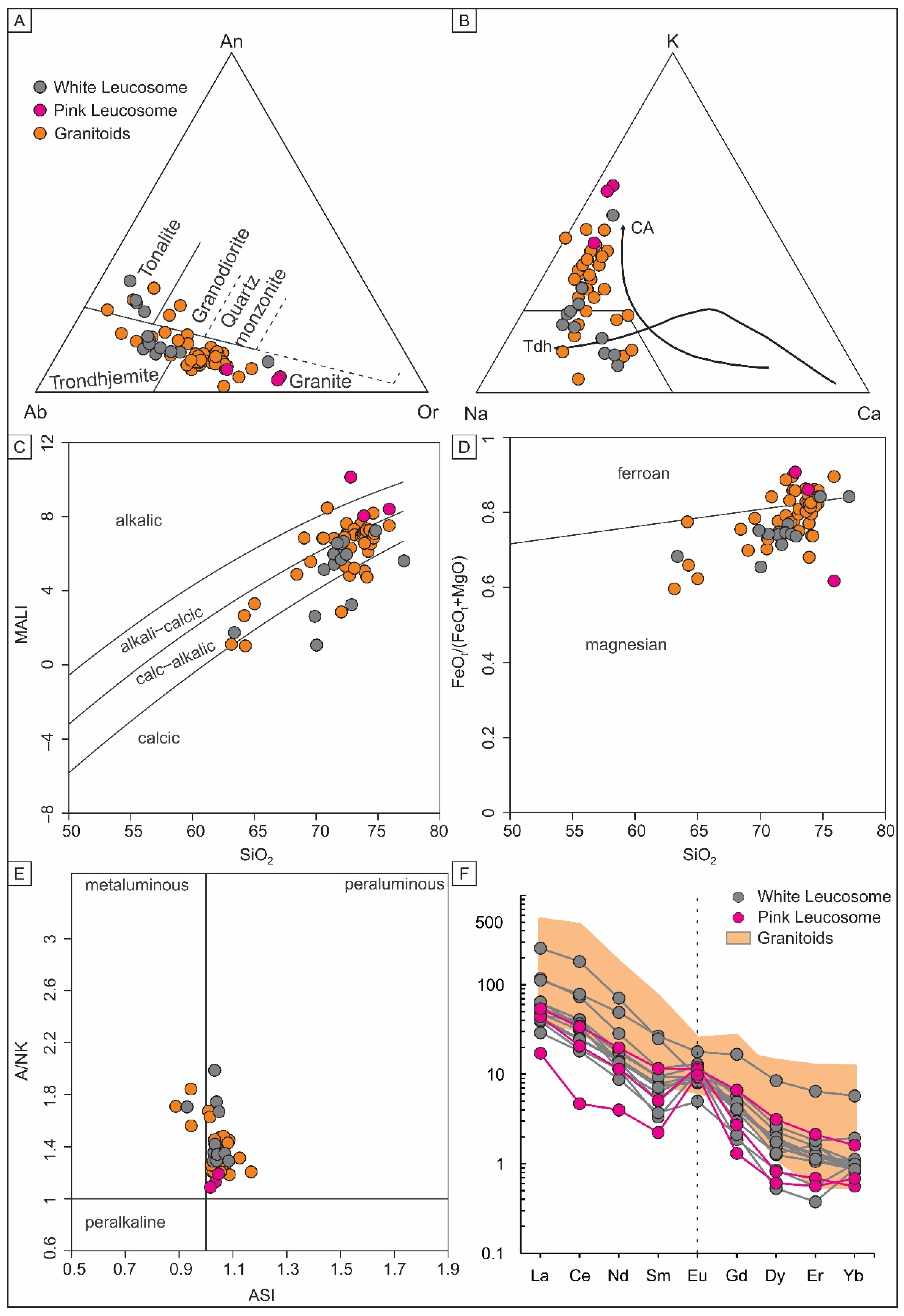
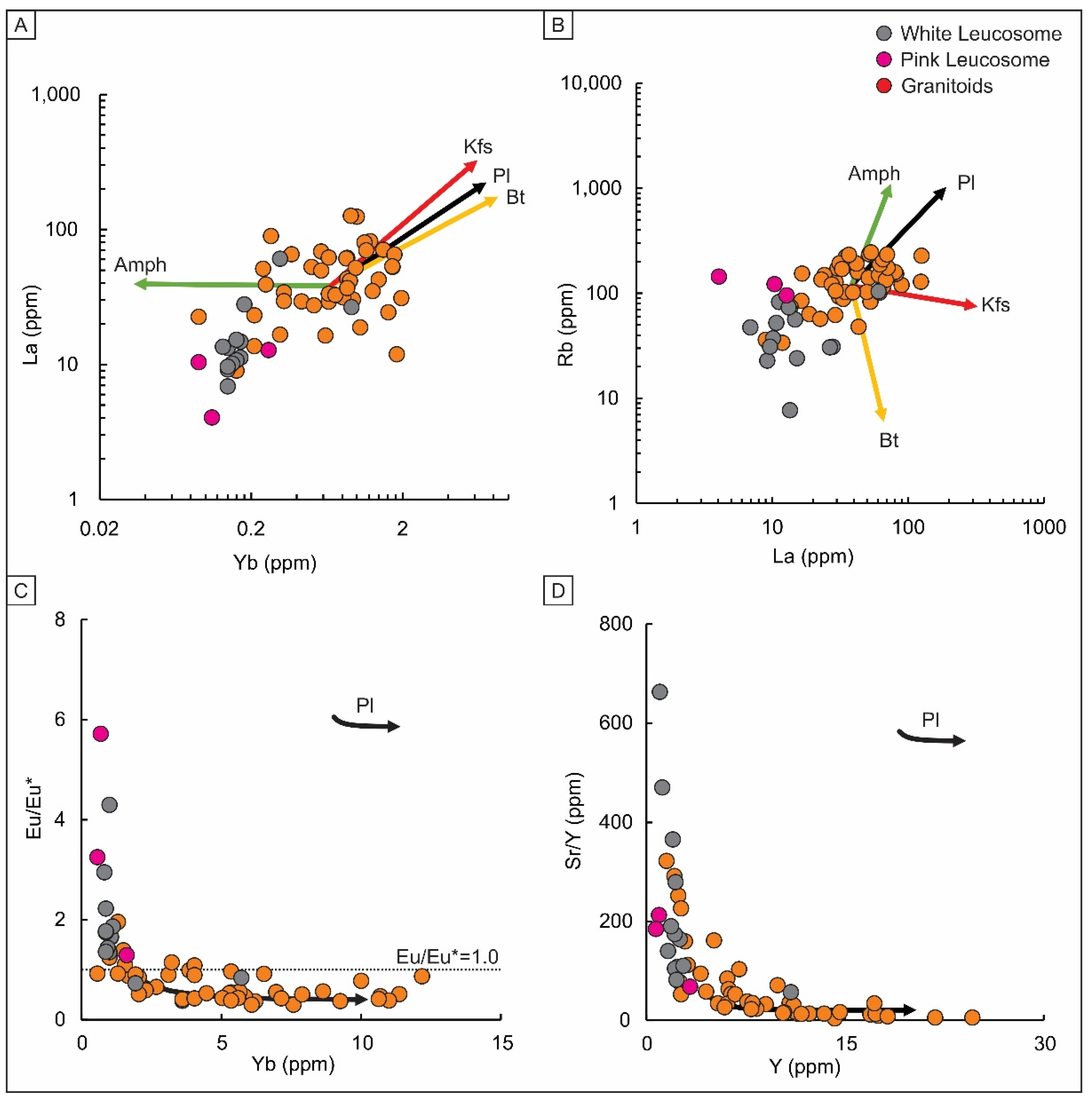
6.2. Binary Vector Diagrams
7. LA-ICP-MS U-Pb Zircon Geochronology
7.1. Hercynite Metadiorite (Sample 1H4)


7.2. White Leucosome (Sample 3A1)
7.3. White Leucosome (Sample 1G1)
7.4. Pink Leucosome (Sample 1J1)
8. Discussion
8.1. Mechanisms of the Partial Melting in the Xicrim-Cateté Orthogranulite and Xingu Complex in the Late Mesoarchean
8.2. The Amplitude of the Late Mesoarchean Anatetic Event and the Relationship with Syntectonic Granitoids
8.3. Geodynamic Implications for the Crustal Evolution and Metallogenesis of the Carajás Province
9. Conclusions
Supplementary Materials
Author Contributions
Funding
Data Availability Statement
Acknowledgments
Conflicts of Interest
References
- Brown, M. Granites, Migmatites and Residual Granulites: Relationships and Processes. In Working with Migmatites; Mineralogical Association of Canada: Quebec City, QC, Canada, 2008; pp. 97–144. [Google Scholar]
- Sawyer, E.W.; Cesare, B.; Brown, M. When the Continental Crust Melts. Elements 2011, 7, 229–234. [Google Scholar] [CrossRef]
- Brown, C.R.; Yakymchuk, C.; Brown, M.; Fanning, C.M.; Korhonen, F.J.; Piccoli, P.M.; Siddoway, C.S. From Source to Sink: Petrogenesis of Cretaceous Anatectic Granites from the Fosdick Migmatite–Granite Complex, West Antarctica. J. Petrol. 2016, 57, 1241–1278. [Google Scholar] [CrossRef]
- Korhonen, F.J.; Brown, M.; Grove, M.; Siddoway, C.S.; Baxter, E.F.; Inglis, J.D. Separating Metamorphic Events in the Fosdick Migmatite–Granite Complex, West Antarctica. J. Metamorph. Geol. 2012, 30, 165–192. [Google Scholar] [CrossRef]
- Martini, A.; de Bitencourt, M.F.; Weinberg, R.F.; De Toni, G.B.; Lauro, V.S.N. From Migmatite to Magma—Crustal Melting and Generation of Granite in the Camboriú Complex, South Brazil. Lithos 2019, 340–341, 270–286. [Google Scholar] [CrossRef]
- Sawyer, E.W. Disequilibrium Melting and the Rate of Melt-Residuum Separation During Migmatization of Mafic Rocks from the Grenville Front, Quebec. J. Petrol. 1991, 32, 701–738. [Google Scholar] [CrossRef]
- Brown, M. Crustal Melting and Melt Extraction, Ascent and Emplacement in Orogens: Mechanisms and Consequences. J. Geol. Soc. Lond. 2007, 164, 709–730. [Google Scholar] [CrossRef]
- Solar, G.S.; Brown, M. Petrogenesis of Migmatites in Maine, USA: Possible Source of Peraluminous Leucogranite in Plutons? J. Petrol. 2001, 42, 789–823. [Google Scholar] [CrossRef]
- Delinardo da Silva, M.A.; Monteiro, L.V.S.; Santos, T.J.S.; Moreto, C.P.N.; Sousa, S.D.; Faustinoni, J.M.; Melo, G.H.C.; Xavier, R.P.; Toledo, B.A.M. Mesoarchean Migmatites of the Carajás Province: From Intra-Arc Melting to Collision. Lithos 2021, 388–389, 106078. [Google Scholar] [CrossRef]
- Machado, N.; Lindenmayer, Z.; Krogh, T.E.; Lindenmayer, D. U-Pb Geochronology of Archean Magmatism and Basement Reactivation in the Carajás Area, Amazon Shield, Brazil. Precambrian Res. 1991, 49, 329–354. [Google Scholar] [CrossRef]
- Pidgeon, R.T.; Macambira, M.J.B.; Lafon, J.-M. Th–U–Pb Isotopic Systems and Internal Structures of Complex Zircons from an Enderbite from the Pium Complex, Carajás Province, Brazil: Evidence for the Ages of Granulite Facies Metamorphism and the Protolith of the Enderbite. Chem. Geol. 2000, 166, 159–171. [Google Scholar] [CrossRef]
- Marangoanha, B.; de Oliveira, D.C.; Dall’Agnol, R. The Archean Granulite-Enderbite Complex of the Northern Carajás Province, Amazonian Craton (Brazil): Origin and Implications for Crustal Growth and Cratonization. Lithos 2019, 350–351, 105275. [Google Scholar] [CrossRef]
- Feio, G.R.L.; Dall’Agnol, R.; Dantas, E.L.; Macambira, M.J.B.; Santos, J.O.S.; Althoff, F.J.; Soares, J.E.B. Archean Granitoid Magmatism in the Canaã Dos Carajás Area: Implications for Crustal Evolution of the Carajás Province, Amazonian Craton, Brazil. Precambrian Res. 2013, 227, 157–185. [Google Scholar] [CrossRef]
- Silva, L.R.; Oliveira, D.C.; Santos, M.N.S. Diversity, Origin and Tectonic Significance of the Mesoarchean Granitoids of Ourilândia Do Norte, Carajás Province (Brazil). J. S. Am. Earth Sci. 2018, 82, 33–61. [Google Scholar] [CrossRef]
- Silva, L.R.; de Oliveira, D.C.; do Nascimento, A.C.; Lamarão, C.N.; de Arimatéia Costa de Almeida, J. The Mesoarchean Plutonic Complex from the Carajás Province, Amazonian Craton: Petrogenesis, Zircon U–Pb SHRIMP Geochronology and Tectonic Implications. Lithos 2022, 432–433, 106901. [Google Scholar] [CrossRef]
- Leite-Santos, P.J.; do Nascimento, A.C.; De Oliveira, D.C.; da Silva, L.R.; Gabriel, E.O. Granodiorito Pantanal, Granodiorito Nova Canadá e Granito Velha Canadá: Dados Isotópicos De U–Pb–Hf–Nd–Sr e Implicações Para Os Processos de Crescimento e Retrabalhamento Crustal. In Proceedings of the 17 Simpósio de Geologia da Amazônia, Santarém, Brazil, 23–25 October 2023; Sociedade Brasileira de Geologia: Santarém, Brazil, 2023; pp. 377–382. [Google Scholar]
- da Silva, L.R.; de Oliveira, D.C.; Galarza, M.A.; do Nascimento, A.C.; Marangoanha, B.; Marques, G.T. Zircon U–Pb–Hf Isotope and Geochemical Constraints on the Petrogenesis and Tectonic Setting of Mesoarchean Granitoids from the Carajás Province, Amazonian Craton, Brazil. Precambrian Res. 2023, 398, 107204. [Google Scholar] [CrossRef]
- do Nascimento, A.C.; de Oliveira, D.C.; Gabriel, E.O.; Leite-Santos, P.J. Geology, Geochemistry and Zircon SHRIMP U–Pb Geochronology of Mesoarchean High-Mg Granitoids: Constraints on Petrogenesis, Emplacement Timing and Deformation of the Água Limpa Suite in the Carajás Province, SE Amazonian Craton. J. Geol. Soc. Lond. 2024, 182, jgs2024-098. [Google Scholar] [CrossRef]
- Pinheiro, R.V.L.; Holdsworth, R.E. Evolução Tectonoestratigráfica Dos Sistemas Transcorrentes Carajás e Cinzeiro, Cinturão Itacaiúnas, Na Borda Leste Do Cráton Amazônico, Pará. Rev. Bras. Geoci. 2000, 30, 597–606. [Google Scholar] [CrossRef]
- Araújo, O.J.B.; Maia, R.G.N. Programa Levantamento Geológicos Básicos do Brasil, Folha Serra dos Carajás (SB.22-Z-A), Estado do Pará: Texto Explicativo; Serviço Geológico do Brasil: Recife, Brazil, 1991. [Google Scholar]
- Grandjean da Costa, F.; Araújo dos Santos, P.; Corrêa de Oliveira Serafim, I.C.; Lima Costa, I.S.; Roopnarain, S. From Mesoarchean Drips to Modern–Style Tectonics in the Carajás Province, Amazonian Craton. J. S. Am. Earth Sci. 2020, 104, 102817. [Google Scholar] [CrossRef]
- Teixeira, N.A.; Campos, L.D.; de Paula, R.R.; Lacasse, C.M.; Ganade, C.E.; Monteiro, C.F.; Lopes, L.B.L.; Oliveira, C.G. de Carajás Mineral Province—Example of Metallogeny of a Rift above a Cratonic Lithospheric Keel. J. S. Am. Earth Sci. 2021, 108, 103091. [Google Scholar] [CrossRef]
- Santos, J.O.S. Geotectônica Dos Escudos Das Guianas e Brasil-Central. In Geologia, Tectônica e Recursos Minerais do Brasil; Bizzi, L.A., Schobbenhaus, C., Viddotti, R.M., Gonçalves, J.H., Eds.; Serviço Geológico do Brasil: Brasilia, Brazil, 2003; pp. 169–222. [Google Scholar]
- Vasquez, M.L.; Rosa-Costa, L.T. Geologia e Recursos Minerais do Estado do Pará: Texto Explicativo do Mapa Geológico e Recursos Minerais do Estado do Pará; Serviço Geológico do Brasil: Belém, Brazil, 2008. [Google Scholar]
- Moreto, C.P.N.; Monteiro, L.V.S.; Xavier, R.P.; Amaral, W.S.; dos Santos, T.J.S.; Juliani, C.; de Souza Filho, C.R. Mesoarchean (3.0 and 2.86 Ga) Host Rocks of the Iron Oxide–Cu–Au Bacaba Deposit, Carajás Mineral Province: U–Pb Geochronology and Metallogenetic Implications. Miner. Depos. 2011, 46, 789–811. [Google Scholar] [CrossRef]
- Souza, Z.S.; Potrel, A.; Lafon, J.-M.; Althoff, F.J.; Martins Pimentel, M.; Dall’Agnol, R.; de Oliveira, C.G. Nd, Pb and Sr Isotopes in the Identidade Belt, an Archaean Greenstone Belt of the Rio Maria Region (Carajás Province, Brazil): Implications for the Archaean Geodynamic Evolution of the Amazonian Craton. Precambrian Res. 2001, 109, 293–315. [Google Scholar] [CrossRef]
- Oliveira, M.A.; Dall’Agnol, R.; Althoff, F.J.; da Silva Leite, A.A. Mesoarchean Sanukitoid Rocks of the Rio Maria Granite-Greenstone Terrane, Amazonian Craton, Brazil. J. S. Am. Earth Sci. 2009, 27, 146–160. [Google Scholar] [CrossRef]
- de Almeida, J.A.C.; Dall’Agnol, R.; de Oliveira, M.A.; Macambira, M.J.B.; Pimentel, M.M.; Rämö, O.T.; Guimarães, F.V.; Leite, A.A.d.S. Zircon Geochronology, Geochemistry and Origin of the TTG Suites of the Rio Maria Granite-Greenstone Terrane: Implications for the Growth of the Archean Crust of the Carajás Province, Brazil. Precambrian Res. 2011, 187, 201–221. [Google Scholar] [CrossRef]
- Feio, G.R.L.; Dall’Agnol, R.; Dantas, E.L.; Macambira, M.J.B.; Gomes, A.C.B.; Sardinha, A.S.; Oliveira, D.C.; Santos, R.D.; Santos, P.A. Geochemistry, Geochronology, and Origin of the Neoarchean Planalto Granite Suite, Carajás, Amazonian Craton: A-Type or Hydrated Charnockitic Granites? Lithos 2012, 151, 57–73. [Google Scholar] [CrossRef]
- Monteiro, L.V.S.; Xavier, R.P.; de Souza Filho, C.R.; Moreto, C.P.N. Metalogênese Da Província Carajás. In Metalogênese das Províncias Minerais Brasileiras; da Silva, M.G., Rocha Neto, M.B., Jost, H., Kuyumjian, R.M., Eds.; Serviço Geológico do Brasil: Belo Horizonte, Brazil, 2014; pp. 43–92. [Google Scholar]
- DOCEGEO Revisão Litoestratigráfica da Província Mineral de Carajás. Província Carajás: Litoestratigrafia e Principais Depósitos Minerais; do Doce, C.V.R., Ed.; Sociedade Brasileira de Geologia: Belém, Brazil, 1988; pp. 11–54. [Google Scholar]
- Barros, C.E.D.M.; Macambira, M.J.B.; Barbey, P.; Scheller, T. Dados Isotópicos Pb-Pb Em Zircão (Evaporação) e Sm-Nd Do Complexo Granítico Estrela, Província Mineral De Carajás, Brasil: Implicações Petrológicas e Tectônicas. Rev. Bras. Geociências 2004, 34, 531–538. [Google Scholar] [CrossRef]
- Sousa, S.D.; Monteiro, L.V.S.; de Oliveira, D.C.; Silva, M.A.D.; Moreto, C.P.N. O Greenstone Belts Sapucaia Na Região de Água Azul Do Norte, Província Mineral de Carajás: Contexto Geológico e Caracterização Petrográfica. In Contribuições a Geologia da Amazônia; Gorayeb, P.S.d.S., de Lima, A.M.M., Eds.; Sociedade Brasileira de Geologia: Belém, Brazil, 2015; Volume 9, pp. 317–338. [Google Scholar]
- Costa, U.A.P.; Paula, R.R.; Silva, D.P.B.; Barbosa, J.P.O.; Silva, C.M.G.; Tavares, F.M.; Oliveira, J.K.M.; Justo, A.P. Programa Geologia do Brasil—PGB. Mapa de Integração Geológico-Geofísica da Província Carajás. Escala 1:250.000; Serviço Geológico do Brasil: Belém, Brazil, 2016. [Google Scholar]
- De Avelar, V.G.; Lafon, J.-M.; Correia, F.C., Jr.; Macambira, E.M.B. O Magmatismo Arqueano Da Região de Tucumã-Província Mineral de Carajás: Novos Resultados Geocronológicos. Rev. Bras. Geociências 1999, 29, 453–460. [Google Scholar] [CrossRef]
- Moreto, C.P.N.; Monteiro, L.V.S.; Xavier, R.P.; Creaser, R.A.; DuFrane, S.A.; Tassinari, C.C.G.; Sato, K.; Kemp, A.I.S.; Amaral, W.S. Neoarchean and Paleoproterozoic Iron Oxide-Copper-Gold Events at the Sossego Deposit, Carajás Province, Brazil: Re-Os and U-Pb Geochronological Evidence. Econ. Geol. 2015, 110, 809–835. [Google Scholar] [CrossRef]
- Martins, P.L.G.; Toledo, C.L.B.; Silva, A.M.; Chemale, F.; Santos, J.O.S.; Assis, L.M. Neoarchean Magmatism in the Southeastern Amazonian Craton, Brazil: Petrography, Geochemistry and Tectonic Significance of Basalts from the Carajás Basin. Precambrian Res. 2017, 302, 340–357. [Google Scholar] [CrossRef]
- Siepierski, L.; Ferreira Filho, C.F. Magmatic Structure and Petrology of the Vermelho Complex, Carajás Mineral Province, Brazil: Evidence for Magmatic Processes at the Lower Portion of a Mafic-Ultramafic Intrusion. J. S. Am. Earth Sci. 2020, 102, 102700. [Google Scholar] [CrossRef]
- Rossignol, C.; Antonio, P.Y.J.; Narduzzi, F.; Rego, E.S.; Teixeira, L.; de Souza, R.A.; Ávila, J.N.; Silva, M.A.L.; Lana, C.; Trindade, R.I.F.; et al. Unraveling One Billion Years of Geological Evolution of the Southeastern Amazonia Craton from Detrital Zircon Analyses. Geosci. Front. 2022, 13, 101202. [Google Scholar] [CrossRef]
- Pereira, M.A.M.; Moreto, C.P.N.; Dellinardo-Silva, M.A.; Melo, G.H.C.; Correa, A.I.C.M.; Sanches, J.M.; Silva, A.D.F.; Soares, M.A.M.; Costa, L.C.C.; Araújo, J. Microstructures as a Tectonic Guide to Unravel Structural Controls on Cu-Au Mineralization at the Neoarchean Pantera IOCG Deposit, Carajás Domain. In Proceedings of the Anais do XI Simpósio Brasileiro de Exploração Mineral, Ouro Preto, Brazil, 19–22 May 2024. [Google Scholar]
- Tavares, F.M.; Trouw, R.A.J.; da Silva, C.M.G.; Justo, A.P.; Oliveira, J.K.M. The Multistage Tectonic Evolution of the Northeastern Carajás Province, Amazonian Craton, Brazil: Revealing Complex Structural Patterns. J. S. Am. Earth Sci. 2018, 88, 238–252. [Google Scholar] [CrossRef]
- Vasquez, M.L.; Macambira, M.J.B.; Armstrong, R.A. Zircon Geochronology of Granitoids from the Western Bacajá Domain, Southeastern Amazonian Craton, Brazil: Neoarchean to Orosirian Evolution. Precambrian Res. 2008, 161, 279–302. [Google Scholar] [CrossRef]
- Dall’Agnol, R.; Teixeira, N.P.; Rämö, O.T.; Moura, C.A.V.; Macambira, M.J.B.; de Oliveira, D.C. Petrogenesis of the Paleoproterozoic Rapakivi A-Type Granites of the Archean Carajás Metallogenic Province, Brazil. Lithos 2005, 80, 101–129. [Google Scholar] [CrossRef]
- Bordalo, R.A.; dos Santos, T.J.S.; Dantas, E.L. Structural Evolution and U/Pb Zircon Age of the Xambioá Gneiss Dome, Contributions to the Araguaia Fold Belt Tectonic History. J. S. Am. Earth Sci. 2020, 104, 102753. [Google Scholar] [CrossRef]
- Teixeira, W.; Hamilton, M.A.; Girardi, V.A.V.; Faleiros, F.M.; Ernst, R.E. U-Pb Baddeleyite Ages of Key Dyke Swarms in the Amazonian Craton (Carajás/Rio Maria and Rio Apa Areas): Tectonic Implications for Events at 1880, 1110 Ma, 535 Ma and 200 Ma. Precambrian Res. 2019, 329, 138–155. [Google Scholar] [CrossRef]
- Carneiro, L.S.; Moreto, C.P.N.; Monteiro, L.V.S.; Delinardo-Silva, M.A.; Melo, G.H.C.; Xavier, R.P.; Pozocco, E.; Carvalho, J.A.; Schoenherr, I.; Oliveira, G.D.; et al. The 2.86 Ga Hades and Hades Nordeste Copper Deposits, SW Carajás: Implications for the Mesoarchean Deformation-Mineralization Processes. In Proceedings of the 17 Simpósio de Geologia da Amazônia, Santarém, Brazil, 23–25 October 2023; Sociedade Brasileira de Geologia: Santarém, Brazil, 2023. [Google Scholar]
- Melo, G.H.C.; Monteiro, L.V.S.; Xavier, R.P.; Moreto, C.P.N.; Arquaz, R.M.; Silva, M.A.D. Evolution of the Igarapé Bahia Cu-Au Deposit, Carajás Province (Brazil): Early Syngenetic Chalcopyrite Overprinted by IOCG Mineralization. Ore Geol. Rev. 2019, 111, 102993. [Google Scholar] [CrossRef]
- Xavier, R.P.; Soares Monteiro, L.V.; Moreto, C.P.N.; Pestilho, A.L.S.; Coelho de Melo, G.H.; Delinardo da Silva, M.A.; Aires, B.; Ribeiro, C.; Freitas e Silva, F.H. The Iron Oxide Copper-Gold Systems of the Carajás Mineral Province, Brazil. In Geology and Genesis of Major Copper Deposits and Districts of the World: A Tribute to Richard H. Sillitoe; Society of Economic Geologists: Littleton, CO, USA, 2012; Volume 16, pp. 433–454. [Google Scholar]
- Vendemiatto, M.A.; Enzweiler, J. Routine Control of Accuracy in Silicate Rock Analysis by X-Ray Fluorescence Spectrometry. Geostand. Newsl. 2001, 25, 283–291. [Google Scholar] [CrossRef]
- Navarro, M.S.; Andrade, S.; Ulbrich, H.; Gomes, C.B.; Girardi, V.A.V. The Direct Determination of Rare Earth Elements in Basaltic and Related Rocks Using ICP-MS: Testing the Efficiency of Microwave Oven Sample Decomposition Procedures. Geostand. Geoanalyt. Res. 2008, 32, 167–180. [Google Scholar] [CrossRef]
- Janoušek, V.; Farrow, C.M.; Erban, V. Interpretation of Whole-Rock Geochemical Data in Igneous Geochemistry: Introducing Geochemical Data Toolkit (GCDkit). J. Petrol. 2006, 47, 1255–1259. [Google Scholar] [CrossRef]
- Brophy, J.G.; Ota, T.; Kunihro, T.; Tsujimori, T.; Nakamura, E. In Situ Ion-Microprobe Determination of Trace Element Partition Coefficients for Hornblende, Plagioclase, Orthopyroxene, and Apatite in Equilibrium with Natural Rhyolitic Glass, Little Glass Mountain Rhyolite, California. Am. Mineral. 2011, 96, 1838–1850. [Google Scholar] [CrossRef]
- Ewart, A.; Griffin, W.L. Application of Proton-Microprobe Data to Trace-Element Partitioning in Volcanic Rocks. Chem. Geol. 1994, 117, 251–284. [Google Scholar] [CrossRef]
- Nash, W.P.; Crecraft, H.R. Partition Coefficients for Trace Elements in Silicic Magmas. Geochim. Cosmochim. Acta 1985, 49, 2309–2322. [Google Scholar] [CrossRef]
- Vermeesch, P. IsoplotR: A Free and Open Toolbox for Geochronology. Geosci. Front. 2018, 9, 1479–1493. [Google Scholar] [CrossRef]
- Mehnert, K.R. Migmatites and the Origin of Granitic Rocks; Elsevier Publishing Company: Amsterdam, The Netherlands, 1968. [Google Scholar]
- Sawyer, E.W. Atlas of Migmatites; Canadian Science Publishing: Ottawa, ON, Canada, 2008; ISBN 9780660197883. [Google Scholar]
- Holness, M.B. Decoding Migmatite Microstructures. In Working with Migmatites; Mineralogical Association of Canada: Quebec City, QC, Canada, 2008; pp. 57–76. [Google Scholar]
- Holness, M.B.; Sawyer, E.W. On the Pseudomorphing of Melt-Filled Pores During the Crystallization of Migmatites. J. Petrol. 2008, 49, 1343–1363. [Google Scholar] [CrossRef]
- Vernon, R.H. Microstructures of Melt-Bearing Regional Metamorphic Rocks. In Origin and Evolution of Precambrian High-Grade Gneiss Terranes, with Special Emphasis on the Limpopo Complex of Southern Africa; Geological Society of America: Boulder, CO, USA, 2011; Volume 207, pp. 1–11. ISBN 9780813712079. [Google Scholar]
- Warr, L.N. IMA–CNMNC Approved Mineral Symbols. Mineral. Mag. 2021, 85, 291–320. [Google Scholar] [CrossRef]
- McDonough, W.F.; Sun, S.S. The Composition of the Earth. Chem. Geol. 1995, 120, 223–253. [Google Scholar] [CrossRef]
- O’Connor, J.T. A Classification for Quartz-Rich Igneous Rocks Based on Feldspar Ratios. Geol. Surv. Prof. Pap. 1965, 525, 79–84. [Google Scholar]
- Frost, B.R.; Barnes, C.G.; Collins, W.J.; Arculus, R.J.; Ellis, D.J.; Frost, C.D. A Geochemical Classification for Granitic Rocks. J. Petrol. 2001, 42, 2033–2048. [Google Scholar] [CrossRef]
- Shand, S.J. Eruptive Rocks: Their Genesis, Composition, Classification and Their Relation to Ore Deposits, with a Chapter on Meteorites, 2nd ed.; Thomas Murby and Co.: London, UK, 1943. [Google Scholar]
- Ashworth, J.R.; Johannes, W.; Grant, J.A.; Olsen, S.N.; McLellan, E.L.; Tracy, R.J.; Barr, D.; Touret, J. Migmatites; Ashworth, J.R., Ed.; Springer: Boston, MA, USA, 1985; ISBN 978-1-4612-9438-2. [Google Scholar]
- Lappin, A.R.; Hollister, L.S. Partial Melting in the Central Gneiss Complex near Prince Rupert, British Columbia. Am. J. Sci. 1980, 280, 518–545. [Google Scholar] [CrossRef]
- Lee, Y.; Cho, M. Fluid-Present Disequilibrium Melting in Neoarchean Arc-Related Migmatites of Daeijak Island, Western Gyeonggi Massif, Korea. Lithos 2013, 179, 249–262. [Google Scholar] [CrossRef]
- Mogk, D.W. Ductile Shearing and Migmatization at Mid-crustal Levels in an Archaean High-grade Gneiss Belt, Northern Gallatin Range, Montana, USA. J. Metamorph. Geol. 1992, 10, 427–438. [Google Scholar] [CrossRef]
- Sawyer, E.W. Migmatites Formed by Water-Fluxed Partial Melting of a Leucogranodiorite Protolith: Microstructures in the Residual Rocks and Source of the Fluid. Lithos 2010, 116, 273–286. [Google Scholar] [CrossRef]
- Watkins, J.M.; Clemens, J.D.; Treloar, P.J. Archaean TTGs as Sources of Younger Granitic Magmas: Melting of Sodic Metatonalites at 0.6–1.2 GPa. In Contributions to Mineralogy and Petrology; Springer: Berlin/Heidelberg, Germany, 2007; Volume 154, pp. 91–110. [Google Scholar] [CrossRef]
- Weinberg, R.F.; Hasalová, P. Water-Fluxed Melting of the Continental Crust: A Review. Lithos 2015, 212–215, 158–188. [Google Scholar] [CrossRef]
- Siepierski, L.; Ferreira Filho, C.F. Spinifex-Textured Komatiites in the South Border of the Carajas Ridge, Selva Greenstone Belt, Carajás Province, Brazil. J. S. Am. Earth Sci. 2016, 66, 41–55. [Google Scholar] [CrossRef]
- Goldfarb, R.J.; Baker, T.; Dubé, B.; Groves, D.I.; Hart, C.J.R.; Gosselin, P. Distribution, Character, and Genesis of Gold Deposits in Metamorphic Terran. In One Hundredth Anniversary Volume; Society of Economic Geologists: Littleton, CO, USA, 2005; pp. 407–450. [Google Scholar]
- do Nascimento, A.C.; de Oliveira, D.C.; da Silva, L.R.; Lamarão, C.N. Mineral Thermobarometry and Its Implications for Petrological Constraints on Mesoarchean Granitoids from the Carajás Province, Amazonian Craton (Brazil). J. S. Am. Earth Sci. 2021, 109, 103271. [Google Scholar] [CrossRef]
- do Nascimento, A.C.; de Oliveira, D.C.; Gabriel, E.O.; Marangoanha, B.; Ribeiro da Silva, L.; da Aleixo, E.C. Mineral Chemistry of the Água Limpa Suite: Insights into Petrological Constraints and Magma Ascent Rate of Mesoarchean Sanukitoids from the Sapucaia Terrane (Carajás Province, Southeastern Amazonian Craton, Brazil). J. S. Am. Earth Sci. 2023, 132, 104683. [Google Scholar] [CrossRef]
- Klimm, K.; Holtz, F.; Johannes, W.; King, P.L. Fractionation of Metaluminous A-Type Granites: An Experimental Study of the Wangrah Suite, Lachlan Fold Belt, Australia. Precambrian Res. 2003, 124, 327–341. [Google Scholar] [CrossRef]
- Spencer, C.J.; Kirkland, C.L.; Taylor, R.J.M. Strategies towards Statistically Robust Interpretations of in Situ U-Pb Zircon Geochronology. Geosci. Front. 2016, 7, 581–589. [Google Scholar] [CrossRef]
- Goodwin, L.B.; Tikoff, B. Competency Contrast, Kinematics, and the Development of Foliations and Lineations in the Crust. J. Struct. Geol. 2002, 24, 1065–1085. [Google Scholar] [CrossRef]
- Slagstad, T.; Jamieson, R.A.; Culshaw, N.G. Formation, Crystallization, and Migration of Melt in the Mid-Orogenic Crust: Muskoka Domain Migmatites, Grenville Province, Ontario. J. Petrol. 2005, 46, 893–919. [Google Scholar] [CrossRef]
- Guernina, S.; Sawyer, E.W. Large-scale Melt-depletion in Granulite Terranes: An Example from the Archean Ashuanipi Subprovince of Quebec. J. Metamorph. Geol. 2003, 21, 181–201. [Google Scholar] [CrossRef]
- Hawkesworth, C.; Cawood, P.A.; Dhuime, B.; Kemp, T. Tectonic Processes and the Evolution of the Continental Crust. J. Geol. Soc. Lond. 2024, 181, jgs2024-027. [Google Scholar] [CrossRef]
- Van Kranendonk, M.J.; Collins, W.J.; Hickman, A.H.; Powley, M.J. Critical Tests of Vertical vs. Horizontal Tectonic Models for the Archaean East Pilbara Granite–Greenstone Terrane, Pilbara Craton, Western Australia. Precambrian Res. 2004, 131, 173–211. [Google Scholar] [CrossRef]
- Lamont, T.N.; Searle, M.P.; Waters, D.J.; Roberts, N.M.W.; Palin, R.M.; Smye, A.; Dyck, B.; Gopon, P.; Weller, O.M.; St-Onge, M.R. Compressional Origin of the Naxos Metamorphic Core Complex, Greece: Structure, Petrography, and Thermobarometry. Bull. Geol. Soc. Am. 2020, 132, 149–197. [Google Scholar] [CrossRef]
- Kusky, T.; Windley, B.F.; Polat, A.; Wang, L.; Ning, W.; Zhong, Y. Archean Dome-and-Basin Style Structures Form during Growth and Death of Intraoceanic and Continental Margin Arcs in Accretionary Orogens. Earth-Sci. Rev. 2021, 220, 103725. [Google Scholar] [CrossRef]
- Arndt, N. Formation and Evolution of the Continental Crust. Geochem. Perspect. 2013, 2, 405–533. [Google Scholar] [CrossRef]
- Condie, K.C. Tectonic Settings. In Earth as an Evolving Planetary System; Elsevier: Amsterdam, The Netherlands, 2016; pp. 43–88. ISBN 9780128036891. [Google Scholar]
- Touret, J.L.R.; Santosh, M.; Huizenga, J.M. Composition and Evolution of the Continental Crust: Retrospect and Prospect. Geosci. Front. 2022, 13, 101428. [Google Scholar] [CrossRef]
- Skirrow, R.G. Iron Oxide Copper-Gold (IOCG) Deposits—A Review (Part 1): Settings, Mineralogy, Ore Geochemistry and Classification. Ore Geol. Rev. 2022, 140, 104569. [Google Scholar] [CrossRef]
- Groves, D.I.; Bierlein, F.P.; Meinert, L.D.; Hitzman, M.W. Iron Oxide Copper-Gold (IOCG) Deposits through Earth History: Implications for Origin, Lithospheric Setting, and Distinction from Other Epigenetic Iron Oxide Deposits. Econ. Geol. 2010, 105, 641–654. [Google Scholar] [CrossRef]
- Skirrow, R.G.; van der Wielen, S.E.; Champion, D.C.; Czarnota, K.; Thiel, S. Lithospheric Architecture and Mantle Metasomatism Linked to Iron Oxide Cu-Au Ore Formation: Multidisciplinary Evidence from the Olympic Dam Region, South Australia. Geochem. Geophys. Geosystems 2018, 19, 2673–2705. [Google Scholar] [CrossRef]
- McPhie, J.; DellaPasqua, F.; Allen, S.R.; Lackie, M.A. Extreme Effusive Eruptions: Palaeoflow Data on an Extensive Felsic Lava in the Mesoproterozoic Gawler Range Volcanics. J. Volcanol. Geotherm. Res. 2008, 172, 148–161. [Google Scholar] [CrossRef]
- Jagodzinski, E.A.; Reid, A.J.; Crowley, J.L.; Wade, C.E.; Curtis, S. Precise Zircon U-Pb Dating of the Mesoproterozoic Gawler Large Igneous Province, South Australia. Results Geochem. 2023, 10, 100020. [Google Scholar] [CrossRef]

| Rock Type | White Leucosome: Xicrim-Cateté Orthogranulite | White Leucosome: Xingu Complex | Pink Leucosome: Xingu Complex | |||||||||||||
|---|---|---|---|---|---|---|---|---|---|---|---|---|---|---|---|---|
| Sample | SM36L1 | SM36L2 | SM41L | SM39N | 1G1 | 1G2 | 1G3 | 1G4 | 1G5 | 1G6 | 1G7 | XS24L | XS37L | 1J1 | 1J2 | 1J3 |
| Oxides (wt%) | ||||||||||||||||
| SiO2 | 69.90 | 70.04 | 72.86 | 63.38 | 71.45 | 72.21 | 71.42 | 72.03 | 70.64 | 71.71 | 72.46 | 77.10 | 74.80 | 75.90 | 73.85 | 72.79 |
| TiO2 | 0.21 | 0.19 | 0.04 | 0.50 | 0.18 | 0.11 | 0.18 | 0.19 | 0.22 | 0.16 | 0.17 | 0.07 | 0.10 | 0.06 | 0.07 | 0.04 |
| Al2O3 | 16.80 | 16.55 | 15.88 | 16.58 | 15.50 | 15.09 | 15.44 | 15.35 | 15.67 | 15.32 | 15.09 | 13.50 | 13.65 | 12.69 | 14.27 | 14.54 |
| FeOt | 1.97 | 1.65 | 0.67 | 4.56 | 1.42 | 0.93 | 1.34 | 1.32 | 1.50 | 1.15 | 1.12 | 0.96 | 1.28 | 0.29 | 0.68 | 0.49 |
| MnO | 0.02 | 0.03 | 0.01 | 0.06 | 0.02 | 0.02 | 0.03 | 0.02 | 0.02 | 0.02 | 0.02 | 0.01 | 0.01 | 0.01 | 0.01 | 0.01 |
| MgO | 0.65 | 0.87 | 0.24 | 2.12 | 0.48 | 0.28 | 0.47 | 0.45 | 0.52 | 0.46 | 0.39 | 0.18 | 0.24 | 0.18 | 0.11 | 0.05 |
| CaO | 3.74 | 4.29 | 3.14 | 4.65 | 2.21 | 1.71 | 1.86 | 2.07 | 2.38 | 1.66 | 1.87 | 1.46 | 1.15 | 0.60 | 1.03 | 0.55 |
| Na2O | 4.90 | 4.51 | 4.63 | 4.98 | 5.13 | 4.71 | 5.38 | 5.20 | 5.14 | 5.26 | 4.93 | 4.19 | 2.65 | 2.62 | 3.84 | 3.19 |
| K2O | 1.45 | 0.84 | 1.75 | 1.41 | 2.50 | 3.66 | 2.46 | 2.55 | 2.38 | 2.96 | 2.90 | 2.88 | 5.74 | 6.38 | 5.23 | 7.49 |
| P2O5 | 0.11 | 0.04 | 0.03 | 0.14 | 0.07 | 0.05 | 0.07 | 0.07 | 0.08 | 0.06 | 0.07 | 0.02 | 0.03 | 0.02 | 0.09 | 0.02 |
| LOI | 0.58 | 0.60 | 0.40 | 0.90 | 0.67 | 0.55 | 0.67 | 0.69 | 0.73 | 0.72 | 0.57 | 0.22 | 0.31 | 0.50 | 0.41 | 0.39 |
| Total | 100.74 | 99.84 | 99.75 | 99.77 | 99.80 | 99.40 | 99.50 | 100.10 | 99.50 | 99.60 | 99.70 | 101.08 | 100.41 | 99.30 | 99.70 | 99.60 |
| Trace elements (ppm) | ||||||||||||||||
| Zr | 75.0 | 34.3 | 67.3 | 118.8 | 115.4 | 82.1 | 98.7 | 93.7 | 106.7 | 93.9 | 97.3 | 62.0 | 176.0 | 33.8 | 51.8 | 74.6 |
| Zn | 34.0 | 11.0 | 9.0 | 46.0 | 28.5 | 22.1 | 28.6 | 27.0 | 26.0 | 24.7 | 23.2 | 26.0 | 23.0 | 18.1 | 13.6 | 10.4 |
| Rb | 30.9 | 7.7 | 24.1 | 30.5 | 56.4 | 82.6 | 52.3 | 37.4 | 22.9 | 73.3 | 31.0 | 47.3 | 104.5 | 122.0 | 95.4 | 144.0 |
| Sr | 731 | 615 | 564 | 625 | 414 | 372 | 255 | 225 | 186 | 354 | 227 | 663 | 308 | 198 | 223 | 129 |
| Y | 2.00 | 2.20 | 1.20 | 10.90 | 2.52 | 2.13 | 2.36 | 2.15 | 2.26 | 1.86 | 1.62 | 1.00 | 2.80 | 0.93 | 3.28 | 0.70 |
| Nb | 2.00 | 1.40 | 0.40 | 3.80 | 2.71 | 1.64 | 2.28 | 2.34 | 4.08 | 1.59 | 1.80 | 1.00 | 2.20 | 2.48 | 1.08 | 0.54 |
| Ta | 0.10 | n.d | n.d | 0.10 | 0.06 | 0.06 | 0.14 | 0.08 | 0.07 | 0.07 | 0.14 | 0.10 | 0.30 | 0.25 | 0.09 | 0.08 |
| Ba | 1010 | 529 | 1432 | 548 | 744 | 961 | 698 | 691 | 855 | 747 | 1032 | 2620 | 2360 | 1954 | 1469 | 1435 |
| La | 27.80 | 13.50 | 15.20 | 26.60 | 14.75 | 11.25 | 10.73 | 10.13 | 9.21 | 13.23 | 9.65 | 6.90 | 60.70 | 10.41 | 12.81 | 4.05 |
| Ce | 45.20 | 20.10 | 21.20 | 48.20 | 25.05 | 19.09 | 24.73 | 15.42 | 11.57 | 22.44 | 14.92 | 11.10 | 111.50 | 12.67 | 20.81 | 2.87 |
| Pr | 4.27 | 1.91 | 1.78 | 5.81 | 2.57 | 2.06 | 1.86 | 1.92 | 1.56 | 2.43 | 1.89 | 1.20 | 10.65 | 1.73 | 2.66 | 0.58 |
| Nd | 13.00 | 6.20 | 5.20 | 22.30 | 8.36 | 6.75 | 6.26 | 6.40 | 5.18 | 7.91 | 6.45 | 4.00 | 32.30 | 5.21 | 9.00 | 1.82 |
| Sm | 1.72 | 0.74 | 0.49 | 3.96 | 1.38 | 1.14 | 1.10 | 1.10 | 0.91 | 1.38 | 1.05 | 0.55 | 3.69 | 0.75 | 1.71 | 0.33 |
| Eu | 0.73 | 0.64 | 0.60 | 1.00 | 0.68 | 0.55 | 0.45 | 0.46 | 0.62 | 0.52 | 0.52 | 0.28 | 0.53 | 0.68 | 0.64 | 0.55 |
| Gd | 0.82 | 0.59 | 0.37 | 3.31 | 1.14 | 0.89 | 0.93 | 0.86 | 0.79 | 0.98 | 0.78 | 0.42 | 1.32 | 0.54 | 1.32 | 0.26 |
| Tb | 0.07 | 0.07 | 0.03 | 0.39 | 0.12 | 0.10 | 0.09 | 0.09 | 0.09 | 0.10 | 0.08 | 0.02 | 0.17 | 0.05 | 0.16 | 0.03 |
| Dy | 0.43 | 0.32 | 0.21 | 2.08 | 0.56 | 0.43 | 0.43 | 0.45 | 0.48 | 0.38 | 0.31 | 0.13 | 0.65 | 0.20 | 0.77 | 0.15 |
| Ho | 0.07 | 0.07 | 0.03 | 0.37 | 0.09 | 0.08 | 0.07 | 0.07 | 0.08 | 0.07 | 0.06 | 0.02 | 0.11 | 0.04 | 0.13 | 0.03 |
| Er | 0.18 | 0.26 | 0.09 | 1.03 | 0.25 | 0.20 | 0.21 | 0.20 | 0.20 | 0.18 | 0.17 | 0.06 | 0.29 | 0.11 | 0.34 | 0.09 |
| Tm | 0.04 | 0.03 | 0.02 | 0.14 | 0.03 | 0.03 | 0.03 | 0.02 | 0.02 | 0.02 | 0.02 | 0.02 | 0.06 | 0.01 | 0.04 | 0.01 |
| Yb | 0.18 | 0.13 | 0.16 | 0.92 | 0.17 | 0.17 | 0.16 | 0.15 | 0.14 | 0.14 | 0.14 | 0.14 | 0.31 | 0.09 | 0.26 | 0.11 |
| Lu | 0.02 | 0.03 | 0.01 | 0.12 | 0.03 | 0.03 | 0.03 | 0.03 | 0.03 | 0.03 | 0.02 | 0.02 | 0.04 | 0.02 | 0.04 | 0.02 |
| Hf | 1.90 | 0.70 | 2.30 | 3.10 | 3.25 | 2.59 | 2.82 | 2.72 | 3.03 | 2.79 | 2.94 | 2.00 | 5.20 | 1.16 | 1.74 | 4.37 |
| Pb | 11.00 | 0.90 | 1.20 | 4.10 | 11.33 | 14.71 | 8.57 | 11.40 | 9.84 | 11.25 | 10.06 | 20.00 | 17.00 | 14.65 | 9.53 | 14.44 |
| Th | 2.15 | 0.30 | 0.40 | 1.30 | 1.65 | 2.10 | 4.04 | 2.33 | 0.55 | 3.33 | 2.13 | 2.19 | 18.80 | 3.75 | 3.74 | 2.28 |
| U | n.d | n.d | 0.20 | n.d | 0.37 | 0.78 | 1.06 | 0.50 | 0.40 | 0.83 | 0.64 | 0.94 | 1.29 | 1.27 | 0.45 | 3.18 |
| Oxide and trace element ratios | ||||||||||||||||
| Na2O/K2O | 3.38 | 5.37 | 2.65 | 3.53 | 2.05 | 1.29 | 2.19 | 2.04 | 2.16 | 1.78 | 1.70 | 1.45 | 0.46 | 0.41 | 0.73 | 0.43 |
| ASI | 1.02 | 1.03 | 1.04 | 0.91 | 1.02 | 1.02 | 1.04 | 1.02 | 1.02 | 1.03 | 1.03 | 1.07 | 1.08 | 1.03 | 1.03 | 1.01 |
| Fe-index | 0.75 | 0.65 | 0.74 | 0.68 | 0.75 | 0.77 | 0.74 | 0.75 | 0.74 | 0.71 | 0.74 | 0.84 | 0.84 | 0.62 | 0.86 | 0.91 |
| MALI | 2.61 | 1.06 | 3.24 | 1.74 | 5.42 | 6.66 | 5.98 | 5.68 | 5.14 | 6.56 | 5.96 | 5.61 | 7.24 | 8.4 | 8.04 | 10.13 |
| LaN/YbN | 104.92 | 70.55 | 64.54 | 19.64 | 58.94 | 44.96 | 45.56 | 45.88 | 44.69 | 64.20 | 46.82 | 33.48 | 133.02 | 78.58 | 33.47 | 25.01 |
| Eu/Eu* | 1.87 | 2.95 | 4.30 | 0.84 | 1.65 | 1.66 | 1.36 | 1.44 | 2.23 | 1.36 | 1.75 | 1.78 | 0.73 | 3.26 | 1.30 | 5.72 |
| Sr/Y | 366 | 279 | 470 | 57 | 164 | 174 | 108 | 105 | 82 | 190 | 140 | 663 | 110 | 213 | 68 | 185 |
| Normative feldspar proportions | ||||||||||||||||
| Or | 8.57 | 4.96 | 10.34 | 8.33 | 14.77 | 21.63 | 14.54 | 15.07 | 14.07 | 17.49 | 17.14 | 17.02 | 33.92 | 37.70 | 30.91 | 44.26 |
| Ab | 41.46 | 38.16 | 39.18 | 42.14 | 43.41 | 39.86 | 45.52 | 44.00 | 43.49 | 44.51 | 41.72 | 35.46 | 22.42 | 22.17 | 32.49 | 26.99 |
| An | 17.84 | 21.02 | 15.38 | 18.72 | 10.51 | 8.16 | 8.77 | 9.81 | 11.29 | 7.84 | 8.82 | 7.11 | 5.51 | 2.85 | 4.52 | 2.60 |
| Spots | Comm. 206 (%) | U (ppm) | Th (ppm) | Th/U | 207Pb/235U | 1σ | 206Pb/238U | 1σ | 207Pb/206Pb Age (Ma) | 2σ (Ma) | % Conc. |
|---|---|---|---|---|---|---|---|---|---|---|---|
| Hercynite Metadiorite (Sample 1H4) | |||||||||||
| 1H4_1 | 0.02 | 189.1 | 137.8 | 0.73 | 16.17 | 0.23 | 0.564 | 0.008 | 2883 | 11 | 100.1 |
| 1H4_2 | 0.02 | 337.0 | 138.9 | 0.41 | 15.81 | 0.25 | 0.559 | 0.011 | 2864 | 15 | 99.8 |
| 1H4_3 | 0.03 | 189.2 | 136.1 | 0.72 | 16.04 | 0.20 | 0.564 | 0.007 | 2860 | 10 | 100.9 |
| 1H4_4 | 0.01 | 397.0 | 150.0 | 0.38 | 15.35 | 0.22 | 0.551 | 0.008 | 2842 | 11 | 99.3 |
| 1H4_5 | 0.02 | 338.7 | 218.3 | 0.64 | 16.27 | 0.20 | 0.569 | 0.008 | 2870 | 9 | 101.0 |
| 1H4_6 | 0.02 | 349.0 | 220.3 | 0.63 | 16.10 | 0.19 | 0.568 | 0.007 | 2855 | 10 | 101.5 |
| 1H4_7 | 0.02 | 285.1 | 230.1 | 0.81 | 16.04 | 0.22 | 0.560 | 0.008 | 2869 | 11 | 99.7 |
| 1H4_8 | 0.02 | 284.0 | 204.0 | 0.72 | 15.65 | 0.22 | 0.560 | 0.009 | 2823 | 12 | 101.5 |
| 1H4_9 | 0.03 | 223.7 | 139.3 | 0.62 | 16.45 | 0.23 | 0.567 | 0.009 | 2893 | 12 | 99.9 |
| 1H4_10 | 0.03 | 220.8 | 142.2 | 0.64 | 16.05 | 0.23 | 0.561 | 0.009 | 2875 | 12 | 99.7 |
| 1H4_11 | 0.02 | 261.7 | 204.6 | 0.78 | 15.65 | 0.21 | 0.553 | 0.009 | 2869 | 12 | 98.7 |
| 1H4_12 | 0.01 | 492.0 | 261.8 | 0.53 | 16.45 | 0.29 | 0.571 | 0.010 | 2899 | 14 | 100.2 |
| White Leucosome (Sample 3A1) | |||||||||||
| 3A1_1 | 0.09 | 75.6 | 354.5 | 4.69 | 15.68 | 0.32 | 0.556 | 0.012 | 2866 | 18 | 99.1 |
| 3A1_2 | 0.08 | 79.2 | 346.5 | 4.38 | 15.78 | 0.27 | 0.561 | 0.010 | 2851 | 15 | 100.7 |
| 3A1_3 | 0.06 | 116.0 | 715.0 | 6.16 | 15.73 | 0.29 | 0.558 | 0.012 | 2871 | 18 | 99.2 |
| 3A1_4 | 0.04 | 136.2 | 246.8 | 1.81 | 15.64 | 0.28 | 0.558 | 0.011 | 2847 | 16 | 100.2 |
| 3A1_5 | 0.14 | 45.3 | 181.8 | 4.01 | 15.86 | 0.31 | 0.557 | 0.011 | 2873 | 18 | 99.4 |
| 3A1_6 | 0.11 | 58.1 | 240.3 | 4.14 | 15.82 | 0.25 | 0.560 | 0.010 | 2860 | 16 | 99.9 |
| 3A1_7 | 0.16 | 39.4 | 180.8 | 4.59 | 15.80 | 0.28 | 0.557 | 0.011 | 2881 | 17 | 98.6 |
| White Leucosome (Sample 1G1) | |||||||||||
| 1G1_1 | 0.01 | 302.0 | 308.0 | 1.02 | 15.52 | 0.23 | 0.558 | 0.009 | 2842 | 11 | 100.6 |
| 1G1_2 | 0.01 | 284.2 | 231.8 | 0.82 | 15.86 | 0.23 | 0.557 | 0.009 | 2878 | 10 | 99.3 |
| 1G1_3 | 0.02 | 210.6 | 198.4 | 0.94 | 15.39 | 0.16 | 0.555 | 0.007 | 2857 | 8 | 99.5 |
| 1G1_4 | 0.01 | 335.7 | 337.1 | 1.00 | 15.71 | 0.22 | 0.560 | 0.009 | 2854 | 10 | 100.6 |
| 1G1_5 | 0.02 | 268.7 | 173.9 | 0.65 | 15.23 | 0.21 | 0.547 | 0.008 | 2822 | 10 | 99.7 |
| 1G1_6 | 0.02 | 217.1 | 200.4 | 0.92 | 15.69 | 0.17 | 0.560 | 0.007 | 2856 | 8 | 100.2 |
| 1G1_7 | 0.02 | 218.4 | 171.0 | 0.78 | 15.47 | 0.23 | 0.552 | 0.009 | 2855 | 11 | 98.9 |
| 1G1_8 | 0.02 | 250.4 | 226.8 | 0.91 | 15.69 | 0.18 | 0.556 | 0.007 | 2854 | 9 | 100.0 |
| 1G1_9 | 0.01 | 302.8 | 244.8 | 0.81 | 15.22 | 0.20 | 0.545 | 0.008 | 2845 | 11 | 98.7 |
| 1G1_10 | 0.02 | 279.5 | 274.2 | 0.98 | 15.69 | 0.15 | 0.557 | 0.006 | 2859 | 8 | 99.7 |
| 1G1_11 | 0.02 | 197.8 | 197.7 | 1.00 | 15.73 | 0.18 | 0.561 | 0.007 | 2851 | 9 | 100.5 |
| 1G1_12 | 0.02 | 232.8 | 194.5 | 0.84 | 15.65 | 0.17 | 0.554 | 0.007 | 2857 | 9 | 99.5 |
| 1G1_13 | 0.03 | 153.2 | 132.7 | 0.87 | 15.71 | 0.18 | 0.557 | 0.008 | 2863 | 10 | 99.5 |
| 1G1_14 | 0.02 | 287.4 | 257.2 | 0.89 | 15.62 | 0.26 | 0.560 | 0.010 | 2847 | 11 | 100.6 |
| 1G1_15 | 0.02 | 270.4 | 205.1 | 0.76 | 16.51 | 0.24 | 0.568 | 0.009 | 2899 | 11 | 99.9 |
| 1G1_16 | 0.02 | 229.9 | 215.8 | 0.94 | 15.93 | 0.23 | 0.565 | 0.009 | 2856 | 11 | 101.2 |
| 1G1_17 | 0.01 | 335.0 | 278.1 | 0.83 | 15.65 | 0.14 | 0.559 | 0.006 | 2851 | 8 | 100.4 |
| 1G1_18 | 0.01 | 373.0 | 149.5 | 0.40 | 15.60 | 0.18 | 0.558 | 0.007 | 2845 | 9 | 100.7 |
| Pink Leucosome (Sample 1J1) | |||||||||||
| 1J1_1 | 0.02 | 287.5 | 212.2 | 0.74 | 15.46 | 0.28 | 0.542 | 0.012 | 2875 | 14 | 97.0 |
| 1J1_2 | 0.04 | 139.4 | 89.6 | 0.64 | 15.96 | 0.22 | 0.568 | 0.009 | 2833 | 14 | 102.3 |
| 1J1_3 | 0.03 | 186.4 | 119.3 | 0.64 | 16.17 | 0.23 | 0.567 | 0.009 | 2885 | 12 | 100.4 |
| 1J1_4 | 0.02 | 304.7 | 156.8 | 0.51 | 15.21 | 0.29 | 0.537 | 0.011 | 2860 | 16 | 96.9 |
| 1J1_5 | 0.01 | 512.0 | 137.1 | 0.27 | 16.02 | 0.21 | 0.566 | 0.008 | 2861 | 10 | 100.9 |
| 1J1_6 | 0.03 | 192.7 | 121.7 | 0.63 | 15.94 | 0.21 | 0.560 | 0.008 | 2862 | 11 | 100.1 |
| 1J1_7 | 0.02 | 328.0 | 186.9 | 0.57 | 16.10 | 0.20 | 0.566 | 0.008 | 2868 | 10 | 100.7 |
| 1J1_8 | 0.02 | 387.0 | 162.5 | 0.42 | 16.02 | 0.25 | 0.565 | 0.010 | 2864 | 12 | 100.5 |
| 1J1_9 | 0.03 | 218.6 | 90.0 | 0.41 | 16.01 | 0.21 | 0.561 | 0.009 | 2867 | 11 | 100.1 |
| 1J1_10 | 0.02 | 307.0 | 123.9 | 0.40 | 15.43 | 0.25 | 0.555 | 0.010 | 2836 | 13 | 100.2 |
| 1J1_11 | 0.03 | 179.2 | 124.6 | 0.70 | 15.96 | 0.22 | 0.560 | 0.008 | 2854 | 11 | 100.3 |
| 1J1_12 | 0.04 | 152.5 | 96.6 | 0.63 | 15.24 | 0.20 | 0.542 | 0.008 | 2846 | 12 | 98.1 |
Disclaimer/Publisher’s Note: The statements, opinions and data contained in all publications are solely those of the individual author(s) and contributor(s) and not of MDPI and/or the editor(s). MDPI and/or the editor(s) disclaim responsibility for any injury to people or property resulting from any ideas, methods, instructions or products referred to in the content. |
© 2025 by the authors. Licensee MDPI, Basel, Switzerland. This article is an open access article distributed under the terms and conditions of the Creative Commons Attribution (CC BY) license (https://creativecommons.org/licenses/by/4.0/).
Share and Cite
Delinardo-Silva, M.A.; Monteiro, L.V.S.; Moreto, C.P.N.; Faustinoni, J.; Santos, T.J.S.; Sousa, S.D.; Xavier, R.P. From Source to Sink: U-Pb Geochronology and Lithochemistry Unraveling the Missing Link Between Mesoarchean Anatexis and Magmatism in the Carajás Province, Brazil. Minerals 2025, 15, 265. https://doi.org/10.3390/min15030265
Delinardo-Silva MA, Monteiro LVS, Moreto CPN, Faustinoni J, Santos TJS, Sousa SD, Xavier RP. From Source to Sink: U-Pb Geochronology and Lithochemistry Unraveling the Missing Link Between Mesoarchean Anatexis and Magmatism in the Carajás Province, Brazil. Minerals. 2025; 15(3):265. https://doi.org/10.3390/min15030265
Chicago/Turabian StyleDelinardo-Silva, Marco Antônio, Lena Virgínia Soares Monteiro, Carolina Penteado Natividade Moreto, Jackeline Faustinoni, Ticiano José Saraiva Santos, Soraya Damasceno Sousa, and Roberto Perez Xavier. 2025. "From Source to Sink: U-Pb Geochronology and Lithochemistry Unraveling the Missing Link Between Mesoarchean Anatexis and Magmatism in the Carajás Province, Brazil" Minerals 15, no. 3: 265. https://doi.org/10.3390/min15030265
APA StyleDelinardo-Silva, M. A., Monteiro, L. V. S., Moreto, C. P. N., Faustinoni, J., Santos, T. J. S., Sousa, S. D., & Xavier, R. P. (2025). From Source to Sink: U-Pb Geochronology and Lithochemistry Unraveling the Missing Link Between Mesoarchean Anatexis and Magmatism in the Carajás Province, Brazil. Minerals, 15(3), 265. https://doi.org/10.3390/min15030265