Review

Confusion between Carbonate Apatite and Biological Apatite (Carbonated Hydroxyapatite) in Bone and Teeth

1
Department of Histology, Nihon University School of Dentistry at Matsudo, Chiba 271-8587, Japan
2
Department of Removable Prosthodontics and Geriatric Oral Health, Nihon University School of Dentistry at Matsudo, Chiba 271-8587, Japan
3
Department of Radiology, Nihon University School of Dentistry at Matsudo, Chiba 271-8587, Japan
*
Author to whom correspondence should be addressed.
Minerals 2022, 12(2), 170; https://doi.org/10.3390/min12020170
Submission received: 25 December 2021 / Revised: 20 January 2022 / Accepted: 24 January 2022 / Published: 28 January 2022
(This article belongs to the Special Issue Bone and Teeth Mineral Properties in Mammals)

Abstract

:
Biological apatite in enamel, dentin, cementum, and bone is highly individualized hydroxyapatite with high tissue dependency. Often, standard and average textbook values for biological apatite do not apply to actual subjects, and the reported results of analyses differ among investigators. In particular, the term biological apatite is often confusingly and incorrectly used to describe carbonate apatite. The purpose of this review is to prevent further confusion. We believe that apatite should be well understood across disciplines and the terminology clearly defined. According to a definition by the International Mineralogical Association’s Commission on New Minerals Nomenclature and Classification, biological apatite formed by living organisms is a type of hydroxyapatite. More specifically, it is carbonated hydroxyapatite, which is quite different from frequently misnamed carbonate apatite. We hope that this definition will be widely adopted to remove confusion around the naming of apatite in many research and applied fields.

1. Introduction

In the fields of anatomy, histology, physiology, biochemistry, and many oral histology textbooks, the inorganic component of tooth enamel, dentin, and bone is described as apatite. However, this is not a precise description. Apatite is a group name, and even though some histology and oral histology textbooks describe the inorganic component of enamel and bone as hydroxyapatite, which is slightly more advanced, this is still not precise. Hydroxyapatite, a member of the apatite group, also describes a wide range of isomorphic ion substitution phenomena in apatite that result in a range of compositions within a solid-solution series. For instance, in the OH–F series, hydroxyapatite includes all combinations in the composition range of Ca10(PO4)6(OH)2 to Ca10(PO4)6(OH, F)2.
In some textbooks and papers, as a more precise description, the inorganic component of enamel and bone is described as biological apatite, although this does not accurately characterize inorganic components in individual tissues. Biological apatite in enamel and biological apatite in bone are quite different in terms of inorganic components, crystal size, and crystallinity. Biological apatite is produced by an organism and differs from mineral apatite and synthetic apatite in many ways. In addition, biological apatite is diverse and differs from one tissue to another (tissue dependence), and from a broader perspective, from one animal to another.
Since its discovery, apatite has been called named differently in various parts of the world due to its variable color, transparency, shape, size, and chemical composition. Many writers have stated that the word apatite comes from the Greek, απατα’ειv, which means “to deceive even the mineralogist”; however, even the etymological origin of the word contains some errors. Notwithstanding, there is a history of many different terms being used for apatite in various fields due to its wide range of continuous solid-solution properties.
Apatite has been studied and used in various fields such as biology, anatomy and histology, mineralogy, biomaterials, analytical chemistry, pharmaceuticals, agriculture, and industrial mining. However, the name apatite has become confused from the origin of its etymology and has been used as a different name in the many research and industrial fields in which it is applied; therefore, the use of this term requires attention. Here, to prevent ongoing confusion, we review the historical use of the term apatite and emphasize the need for a correct understanding across disciplines and a clear definition when using the term.

2. Background

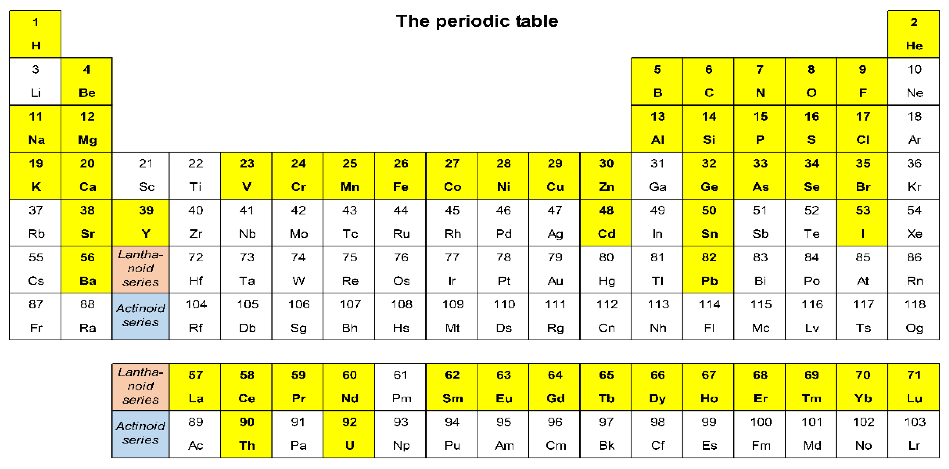
2.1. Definition of a Mineral

The definition of mineral differs between disciplines. In the socioeconomic field, it means valuable materials extracted from the earth, such as coal, oil, sand and gravel, iron ore, other mined commodities, and groundwater. In the medical and dental sciences, the term mineral refers to a variety of internal and external compounds or elements found in living organisms. A more restrictive definition is applied by the earth sciences, where mineral refers to a naturally occurring crystalline solid with a definite but not necessarily constant chemical composition [1]. However, this definition is not always agreed upon, even among mineralogists. The most widely used definition is Nickel’s [2]: “A mineral is an element or compound that is usually crystalline and formed as a result of geological processes.” It has often been discussed whether geological processes include fossilization and whether crystalline includes amorphous material. An example of fossilization is silicified wood, which comes from ancient trees that have been replaced by silica, and an example of an amorphous material is amber, which is fossilized ancient resin.
Mineralogy uses hierarchies of classification and systematization terms from species to phylum as biological classification, and the description of each mineral species is the most fundamental. The history of mineralogy includes various proposals for how to describe mineral properties; however, currently, the standards of the Commission on New Minerals, Nomenclature and Classification (CNMNC) of the International Union of Mineralogical Sciences are widely used [3]. Apart from the seven classical items; that is, (i) Mohs hardness, (ii) luster, (iii) color, (iv) streak, (v) density and specific gravity, (vi) cleavage, and (vii) fracture, the crystal chemistry of minerals (i.e., composition and crystallographic properties) is an essential requirement of the standards. This is due to the widespread use of modern X-ray crystallographic methods that allow for the analysis of crystallographic properties with more precision. X-ray diffraction (XRD) can capture compositional changes as changes in crystal lattice parameters.

2.2. Apatite Group in the Classic Standard Mineralogy Textbook, Dana’s System of Mineralogy

The first edition of Dana’s System of Mineralogy, which became a worldwide standard for mineralogy, has been revised and expanded numerous times. However, since the third edition, it was reorganized into a classification based on chemical composition. Since then, it has been revised so frequently that it is difficult to find another book in the entire history of scientific literature that rivals it because no other book has maintained such a complete bibliographic reference for more than half a century [4]. The publication still boasts the most reliable and comprehensive collection of mineral data. In the eighth edition, more than 3700 minerals are described by page 1872 [5]. According to the crystallographic database of the International Mineralogical Association (IMA), RRUFF™, which collects recently published data, there were 5011 mineral species as of June 2015 [6]. The CNMNC presents a mineral classification table based on chemical composition and crystal structure called the Nickel–Strunz mineral classification. Apatite is recorded as belonging to Nickel–Strunz Class 08: phosphates, arsenates, vanadates, and it is described as 08 BN 05 apatite-(CaOH), apatite-(CaCl), and apatite-(CaF) [7]. In Dana [5], published in 1997, apatite is described as being in the phosphate mineral group, a large group that includes many mineral species, and the silicate group, which makes up most of the earth’s crust. Dana also describes the various forms of apatite found worldwide and their alternative names.

2.3. Apatite Group and Solid-Solution Series

Solid solutions include intrusion solid solutions and substitutional solid solutions. The former occurs when other atoms or vacancies enter the space between atomic lattices and are known in metals and semiconductors. Apatite is part of a substitutional solid solution, in which atoms of approximately the same size replace the original atoms making up the crystal lattice. The general composition of apatite is expressed as A(5XO4)3Zq, and various elements can be substituted for A, X, and Z. White and Zhili [8] reported on the crystal structures of [A(1)2][A(2)3](BO4)3X apatite and collated and reviewed the related compounds [A(1)2][A(2)3](BO5)3X and [A(1)2][A(2)3](BO3)3X. They presented crystallographic data for as many as 84 species belonging to the apatite series. Pasero et al. [9] proposed a single unified view for the “nomenclature of the apatite supergroup minerals,” which was approved by the CNMNC. Hughes and Rakovar [10] published a paper, titled “Chemically Diverse: Apatite and Apatite Supergroup Minerals,” that presented the diversity of the variations in the chemical composition of apatite. Pan and Fleet [11] investigated minerals included in the apatite group and synthetic apatite and summarized their elemental substitutions (Figure 1), including 53 elements, about half the 109 elements in the periodic table, that can be substituted in the apatite crystal structure.
As mentioned, apatite constitutes a wide range of isomorphic ionic substitution series and continuous solid-solution series. The nomenclature of minerals in the solid-solution series has been discussed by Pasero et al. [9] and Hughes and Rakovar [10], but the CNMNC approved Nickel’s [2] nomenclature for mineralogy, including the following explanation for the solid-solution series of apatite: “For the purpose of nomenclature, a complete solid-solution series without structural order of the ions defining the end members are arbitrarily divided at 50 mol%, and the two portions are given different names, with each name applying to the compositional range from the end member to the 50% mark. For the sake of brevity, this will be called the “50% rule”. By analogy, the 50% rule applied to members of the ternary solid-solution series implies that mineral names should be given only the three end members; each name should apply to the compositional range from the end member to the nearest right bisectors of the sides of the composition triangle [as shown in Figure 2 in the current study]. For example, in the apatite series, Ca5(PO4)3(F, OH, and Cl), the apices of the compositional triangle can be represented by F, OH, and Cl, respectively, making A = Fluorapatite, B = Hydroxylapatite, and C = Chlorapatite.” It is essential to note that in the solid-solution series, the name of the end component is used to represent the region. Hydroxyapatite, which is also the name of the end component, is used even if half the amount of F or Cl is contained in the OH position.

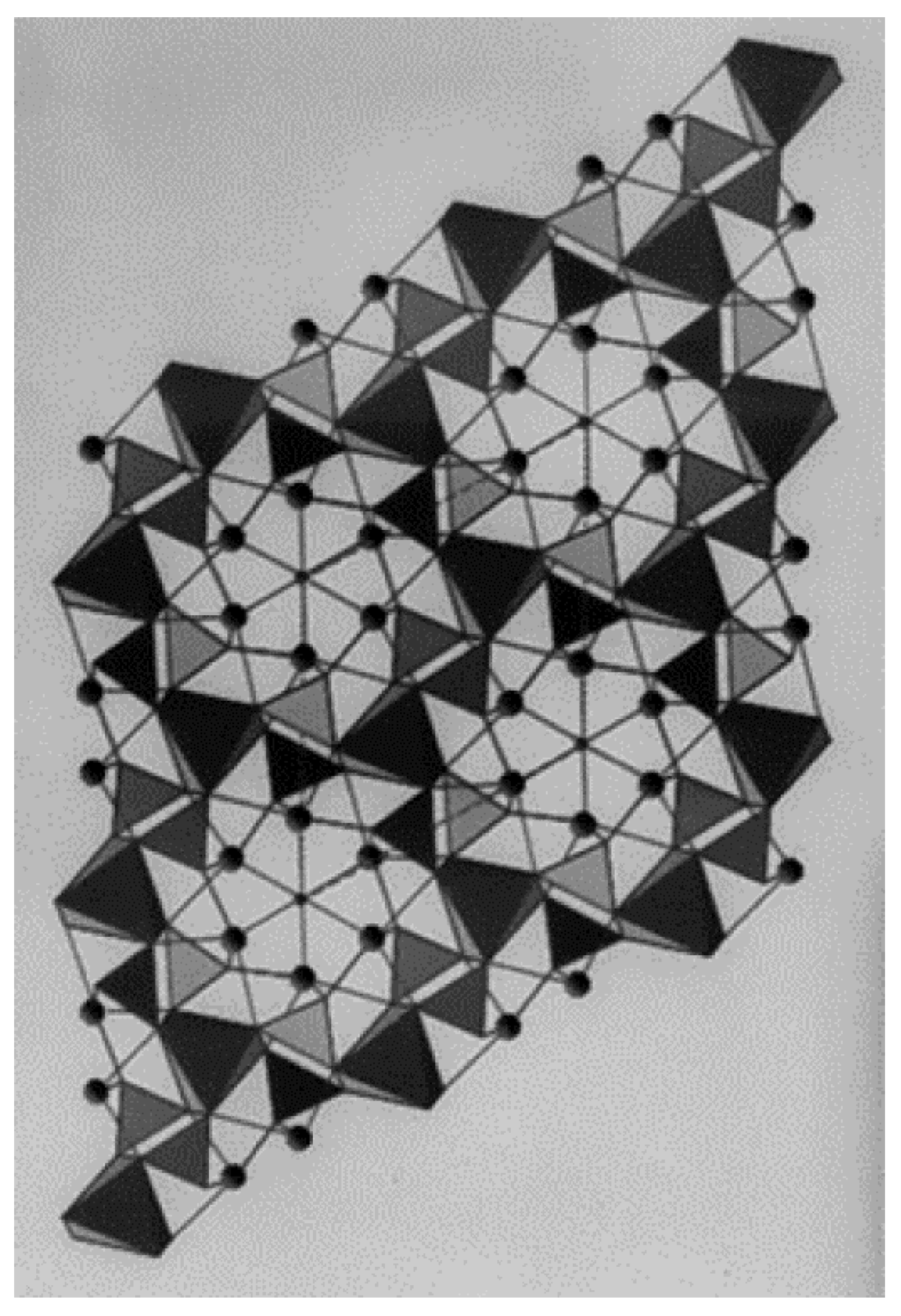
2.4. Crystal Structure of Apatite

In 1930, Mehmel [12] and Náray-Szabó [13] independently published the results of a crystal structure analysis of fluorapatite. This provided a way forward amidst the boiling debate on isomorphic ion substitution and the solid-solution series of apatite. These studies also represent the beginning of subsequent crystallographic studies of apatite. Since then, many crystallographic studies have been undertaken on apatite series structures [14,15,16]. The precise crystal structure of hydroxyapatite was analyzed by Kay et al. [17], and their diagram of the atomic arrangement of hydroxyapatite became the basis for subsequent studies (Figure 3).

3. Biological Apatite

3.1. Initial Research on Biological Apatite

Minerals produced by living organisms are called biominerals, and Lowenstam and Weiner [18] summarized their research on them in their paper “On Biomineralization”, based on a careful review of published papers. They proposed two main categories for biomineralization based on its formative mode: (i) biologically induced mineralization (BIM) and (ii) biologically controlled mineralization (BCM). BIM, a nonobjective formation, is a byproduct of biological activity, such as calcareous or siliceous stromatolites and dental calculi. BCM is the purposeful formation of physiological phenomena by organisms, such as shells, bones, and teeth. Most biological apatite is BCM, but as an exception, some guano-derived phosphate minerals collected in the southern islands, for example, Torishima in Japan, were formed by the reaction of bird droppings with limestone.

3.2. Distribution of Biological Apatite

Lowenstam and Weiner [18], reviewing the distribution of biological apatite, noted that it occurred not only in the phylum Chordata, but also in the Annelida, Micrococci, and Omnibacteria phyla. The classification here is based on the original work. In 1931, Hendricks et al. [19] introduced the pioneering work of DeJong and Roseberry, Hastings, and Morse, who reported that bone was carbonate apatite, Ca10CO3(PO4)6∙H2O according to the results of their X-ray experiments. They used the Debye–Shearer method, a basic XRD method, after immersing the bone samples in naphtha or exposing them to steam for 225 h to remove any organic matter that could have altered the original crystals. Incidentally, Shearer, who invented the Debye–Shearer method, collected XRD images of tooth enamel and dentin in 1936 and discussed the orientation characteristics of the enamel crystals [20]. The many studies related to biological apatite have been summarized in several reviews and monographs, including Driessens [21], LeGeros [22], Elliott [14], Sakae et al. [23], and Sakae et al. [24].

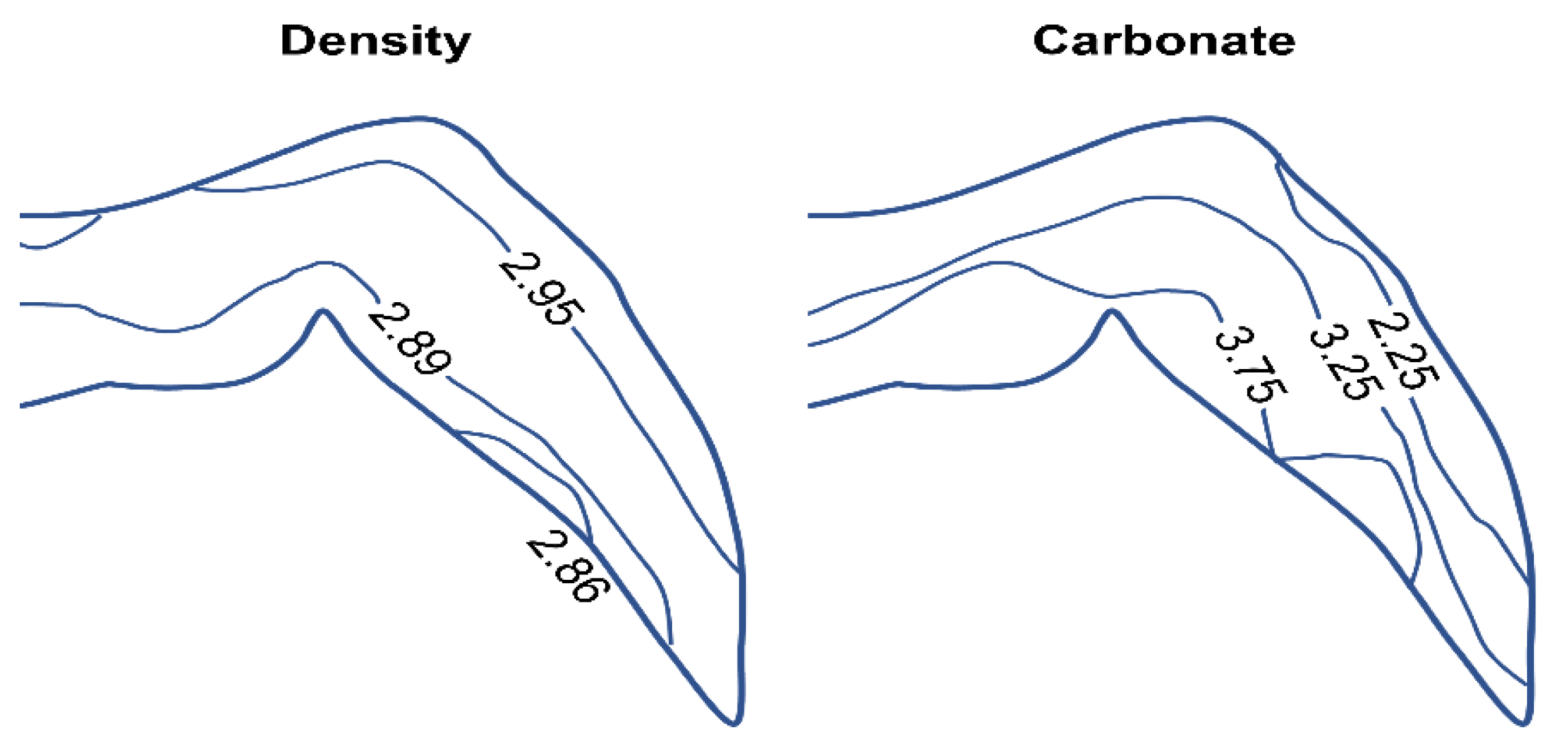
3.3. Indeterminate Composition of Biological Apatite and Differences among Animal Species, Tissues, and Body Sites

The chemical components of enamel, dentin, and bone are difficult to analyze because they are unique to hard tissues. In addition, there are wide variations and individual differences inherent in biological species, and the analyzed values published to date have never been standardized. The morphology of calcium phosphate and its crystal phases in organisms, including bones, have been debated for a long time. Of the significant number of reported analyses, the characteristic features are that it is nonstoichiometric, the Ca to P ratio (Ca:P) is not constant, and carbonate ions are present in relatively large amounts [24,25,26,27].
The existence of calcium carbonate separate from calcium phosphate (apatite) has been claimed for a long time. In 1940, Eisenberger et al. [28] chemically examined the composition of bone and concluded that bone is not carbonate + apatite, thereby rejecting previously published reports. There is still a debate on the two-component or multicomponent theory, and the one-component theory, in which carbonate ions are contained in apatite. The mainstream opinion was that the carbonate ions could not enter into the crystal structure of hydroxyapatite because their charge and molecular size were too different from OH ions. However, LeGeros [29] proved for the first time that carbonate ions could be substituted into the crystal structure of hydroxyapatite.
The typical values of carbonic acid in enamel, dentin, and bone are shown in Table 1. It is interesting to note the amount of carbonic acid contained in the products; for example, 3.5 wt% in enamel and 6.0 wt% in bone. The distribution and bias of the specific gravity and carbonic acid in enamel are shown in Figure 4.
While conventional XRD cannot detect differences between individual bone samples, a recent development, which can analyze microregions <100 µm, has advanced the study of bone apatite microregions. In addition to the micro XRD analysis of bone by Suwa et al. [32] and various analyses of bone by Nakada et al. [33], it is becoming possible to detect differences in even smaller areas, such as hard tissue, using synchrotron radiation [34,35,36,37,38]. The range of hardness (H) and Young’s modulus (E) observed over an individual tooth was far greater than previously reported. The mechanical properties of the enamel differed from the lingual to the buccal side of the molar, and correlated with the chemical compositions [39]. Talal et al. [40] summarized the compositions of enamel, dentin, and bone published in the literature and other sources, as shown in Table 2. Unfortunately, there are no descriptions in the referenced literature, and most bioapatite researchers would not accept such a compositional configuration. Vallet-Regí [41] described bioapatite, which makes up bone, as “calcium phosphate nanocrystals in bone, formed … in the spaces left between the collagen fibers that exhibit the particular feature of being monodispersed and nanometer-sized platelets of carbonate-hydroxyl-apatite. There is no other mineral phase present, and the crystallographic axis c of these crystals is arranged in parallel to the collagen fibers and the largest dimension of the platelet.”

3.4. Problem of Carbonate Ions in the Crystal Structure of Biological Apatite

In his book Apatite, McConnell [42] devoted an entire chapter to carbonate apatites to discuss the problem of apatite and carbonate ions. He described this as follows: “Except for some very obscure hypotheses and the related calculations based on analyses of bone, all other evidences for Ca5(PO4)3CO3 are based upon the premise that the volume is greater, and that this expansion will permit incorporation of the large CO3 group within the structure. Some have also supposed that a dimension as large as 9.518 kX comprises evidence [43,44,45] for 2F → CO3. Unfortunately, the proponents of this hypothesis do not supply adequate information on their preparations. For example, the material reported by Elliott [44], a sample of which was carefully examined by LeGeros et al. [45], produced at least five diffraction lines (within 2θ from 28° to 35°) that are not attributable to the apatite structure [42]. Thus, Elliott’s material is a mixture of two or more phases rather than a single apatitic phase.”
Cacciotti [46] described cationic and anionic substitutions in hydroxyapatite based on a careful survey of the literature as follows: “B-type carbonated hydroxyapatites are characterized by the following features: a decrease in a-axial length accompanied by an increase in c-axial length, changes in crystallite size and in the amount of crystallographic microstrains, optical birefringence and mechanical reinforcement of the bone, and incremented solubility.”

3.5. Crystal Structure Analysis of Enamel Apatite

X-ray crystal structure analysis is usually performed using single crystals. However, single crystals are rarely obtained from biological samples. Therefore, precise crystal structure analyses of the crystalline components of bones and teeth cannot be performed. However, the need for crystal structure analysis has increased in various fields where it is not easy to obtain single crystals. The Rietveld method of crystal structure analysis using powder samples, which was previously considered to be theoretical, is now being used in research [47]. In a breakthrough study, Young and Mackie [48] analyzed the crystal structure of human enamel apatite using the Rietveld method applied to powder samples. However, the samples they used were separated by specific gravity (>2.95) and various changes are observed in enamel, not only human enamel, including individual and regional differences in its specific gravity [49,50]. Therefore, their data only applied to that sample and should never have been generalized as the standard. Since then, crystal structure analysis of biological apatite by the Rietveld method or neutron diffraction has been performed [51,52], and these precise crystal structure analyses have continued to reveal diverse variations of biological apatite.

3.6. Carbonate Ions in Biological Apatite Are B-Type

In the crystal structure, the substitution of CO2 for the hydroxyl group OH along the c-axis is called A-type substitution, and substitution for the phosphate group PO3 is called B-type substitution These substitutions can be observed by X-ray crystallography as changes in the length of the a-axis and the c-axis of the unit cell or as changes in the position and intensity of the absorption bands by Fourier transform infrared absorption spectroscopy [45]. Vallet-Regí and Navarrete [41] presented diagrams for A-type and B-type substituted carbonate ions in the apatite crystal structure, and they compared the crystal lattice size and crystallinity of A-type and B-type hydroxyapatite. In 1991, LeGeros [22] reported that biological apatite is B-type carbonated hydroxyapatite and A-type apatite did not exist. In 2008, Frank-Kamenetskaya [53] found that tooth enamel is B-type, but apatite in dentin, phytolith, and dental calculus is A–B mixed type (B > A), and kidney stones may be both A–B mixed and B-type. It is still controversial whether the biological carbonate-containing apatite in bones and teeth is A- or B-type, although it has been recently reported that the substitution of both rather than one occurs in some biological apatites [46,54,55,56,57,58,59]. These papers are examples of how researchers from different fields compare their perspectives on carbonate ions in apatite.

3.7. Bioapatite Is Hydroxyapatite Containing Carbonic Acid

If we follow the nomenclature of the mineralogical solid-solution series, a carbonate must be more than half substituted at the OH and PO3 position to be called a carbonate apatite. The carbonate apatite described by Ishikawa et al. [60] in 2019 was B-type carbonated hydroxyapatite, as described by Ishikawa et al. [61] in 2010. As mentioned in the previous section, carbonated biological apatite is included in the generic name hydroxyapatite. Therefore, biological apatite should be called carbonated hydroxyapatite. If the apatite in bone is not carbonate-containing hydroxyapatite but carbonate apatite (according to the definition, carbonate ions occupy more than half the phosphoric acid positions), its Ca:P ratio should be much higher than 3.33, far from the hydroxyapatite Ca:P of 1.67. However, such a value has never been reported and the bone composition derived from the most recent instrumental analysis by Euw et al. [62] in 2019 was Ca7.5(PO4)2.8(HPO4)2.6(CO3)0.6(OH)0.2, which is a Ca:P of 1.39. They noted that this composition was applied only for the sheep bones experimented.
A more commonly used the typical bone chemical composition value is Ca8.31.7 (PO4)4.3 (HPO4 or CO3)1.7 (OH or 1/2 CO3)0.31.7, where □ indicates a deficiency, with a Ca:P of 1.93–1.38 (LeGeros et al. [63]). As shown above, the chemical compositions of biological apatite widely vary among the tissues, individual tooth, and individuals.
It is often the case that different names are used in different fields. Researchers involved with apatite, even those outside the field of mineralogy, are always interested in the origin of the name and always introduce the fact that apatite comes from the word deceive. However, as mentioned here, several papers have shown that this origin of the name is wrong. Furthermore, the rules defined in “Mineral Apatite Solid Solution [2]” have rarely been cited.

4. Discussion

The standard and average values of biological apatite described in textbooks do not apply to actual subjects, and reported analytical values differ among researchers. To prevent further confusion, it is necessary to correctly understand the term apatite across disciplinary boundaries and clearly define it when using it. In particular, recently, biological apatite has been confused with carbonate apatite, so it is necessary to recognize that carbonate apatite and carbonated hydroxyapatite produced by biological organisms are quite different. This review presents the nomenclature of the solid apatite solution presented by the mineralogical community that initially discovered and studied apatite. According to this nomenclature, biological apatite is carbonated hydroxyapatite, and carbonate apatite is completely different. We hope that this definition will bring clarity and eliminate confusion.

Author Contributions

T.K. (Tetsuro Kono) and T.S. conceptualized this manuscript. Investigation of references was done by T.K. (Tetsuro Kono), T.S., and H.N. Writing—original draft was done by T.K. (Tetsuro Kono) and T.S. Writing—review and editing was done by T.K. (Takashi Kaneda) and H.O. All authors reviewed the manuscript. H.O. provided supervision and final approval for the manuscript. All authors have read and agreed to the published version of the manuscript.

Funding

This work was supported by Grants-in-Aid for Scientific Research <KAKENHI> (grant number 18K09624) from the Japan Society for the Promotion of Science.

Informed Consent Statement

Not applicable.

Data Availability Statement

Data is contained within the article.

Acknowledgments

We would like to express our sincere gratitude to the Department of Histology laboratory members for their warm guidance and encouragement.

Conflicts of Interest

The authors declare no conflict of interest.

References

  1. Nesse, W. Introduction to mineralogy. Geol. Mag. 2002, 139, 491–492. [Google Scholar]
  2. Nickel, E.H. The definition of a mineral. Can. Mineral. 1995, 33, 689–690. [Google Scholar]
  3. International Mineralogical Association: The Commission on New Minerals, Nomenclature and Classification (CNMNC). Available online: https://www.ima-mineralogy.org/CNMNC_Strategy.htm (accessed on 12 November 2021).
  4. JW., J. A system of mineralogy. Nature 1892, 46, 217–218. [Google Scholar]
  5. Dana, J.D.; Dana, E.S.; Gaines, R.V.; Dana, J.D. Dana’s New Mineralogy: The System of Mineralogy of James Dwight Dana and Edward Salisbury Dana, Entirely Rewritten and Greatly Enlarged, 8th ed.; Gaines, R.V., Skinner, H.C.W., Foord, E.E., Mason, B., Eds.; Wiley-Interscience: New York, NY, USA, 1997; pp. 697–970. [Google Scholar]
  6. Definition & Classification of Minerals. Available online: http://earthresources.sakura.ne.jp/er/Min_D%26C.html (accessed on 12 November 2021).
  7. Minerals Arranged by the Nickel-Strunz (Ver10) Classification System. Available online: http://www.webmineral.com/strunz.shtml (accessed on 12 November 2021).
  8. White, T.J.; Zhili, D. Structural derivation and crystal chemistry of apatites. Acta Crystallogr. B 2003, 59, 1–16. [Google Scholar] [CrossRef] [PubMed]
  9. Pasero, M.; Kampf, A.R.; Ferraris, C.; Pekov, I.V.; Rakovan, J.; White, T.J. Nomenclature of the apatite supergroup minerals. Eur. J. Mineral. 2010, 22, 163–179. [Google Scholar] [CrossRef]
  10. Hughes, J.M.; Rakovan, J.F. Structurally robust, chemically diverse: Apatite and apatite supergroup minerals. Elements 2015, 11, 165–170. [Google Scholar] [CrossRef]
  11. Pan, Y.; Fleet, M.E. Compositions of the apatite-group minerals: Substitution mechanisms and controlling factors. Rev. Mineral. Geochem. 2002, 48, 13–49. [Google Scholar] [CrossRef]
  12. Mehmel, M. Über die Struktur des Apatits. Zeits Kristal 1930, 75, 323–331. [Google Scholar] [CrossRef]
  13. Náray-Szabó, S. Ein auf der Kristallstruktur basierendes Silicatsystem. Z. Physik Chem. Ab 1930, 9, 356–377. [Google Scholar] [CrossRef]
  14. Elliott, J.C. Structure and Chemistry of the Apatites and Other Calcium Orthophosphates, 1st ed.; Elsevier: Amsterdam, The Netherlands, 1994; pp. 111–117. [Google Scholar]
  15. Hughes, J.M.; Rakovan, J. The crystal structure of apatite, Ca5(PO4)3(F, OH, Cl). Rev. Mineral. Geochem. 2002, 48, 1–12. [Google Scholar] [CrossRef]
  16. Combes, C.; Cazalbou, S.; Rey, C. Apatite biominerals. Minerals 2016, 6, 34. [Google Scholar] [CrossRef] [Green Version]
  17. Kay, M.I.; Young, R.A.; Posner, A.S. Crystal structure of hydroxyapatite. Nature 1964, 204, 1050–1052. [Google Scholar] [CrossRef] [PubMed]
  18. Lowenstam, H.A.; Weiner, S. On Biomineralization, 1st ed.; Oxford University Press: New York, NY, USA, 1989; pp. 135–188. [Google Scholar]
  19. Hendrick, S.B.; Hill, W.L.; Jacob, K.D.; Jefferson, M.E. Structural characteristics of apatite-like substances and composition of phosphate rock and bone as determined from microscopical and X-ray diffraction examinations. Ind. Eng. Chem. 1931, 23, 1412–1418. [Google Scholar] [CrossRef]
  20. Shearer, G. The X-ray microscope. Br. J. Radiol. 1936, 9, 30–37. [Google Scholar] [CrossRef]
  21. Driessens, F.C.M. Mineral aspects of sentistry in monographs. In Oral Science, 1st ed.; Myers, H.M., Ed.; Karger: New York, NY, USA, 1982; Volume 10, pp. 53–57. [Google Scholar]
  22. LeGeros, R.Z. Calcium phosphates in oral biology and medicine. In Monographs in Oral Science, 1st ed.; Howard, M.M., Ed.; Karger: Basel, Switzerland, 1991; Volume 15, pp. 108–129. [Google Scholar]
  23. Sakae, T.; Suzuki, K.; Kozawa, Y. A Short review of studies on chemical and physical properties of enamel crystallites. In Tooth Enamel Microstructure, 1st ed.; Koenigswald, W.V., Sander, P.M., Eds.; Bookfield: Rotterdam, The Netherlands, 1997; Volume 1, pp. 31–39. [Google Scholar]
  24. Sakae, T.; Nakada, H.; LeGeros, J.P. Historical review of biological apatite crystallography. J. Hard. Tissue Biol. 2015, 24, 111–122. [Google Scholar] [CrossRef] [Green Version]
  25. Rowles, S.L. Chemistry of the mineral phase of dentin. In Structural and Chemical Organization of Teeth, 1st ed.; Miles, A.E.W., Ed.; Academic Press: New York, NY, USA, 1967; pp. 201–246. [Google Scholar]
  26. Broudevold, T.; Söremark, R. Chemistry of the mineral phase of enamel. In Structural and Chemical Organization of Teeth, 1st ed.; Miles, A.E.W., Ed.; Academic Press: New York, NY, USA, 1967; Volume 18, pp. 247–277. [Google Scholar]
  27. Foley, B.; Greiner, M.; McGlynn, G.; Schmahl, W.W. Anatomical variation of human bone bioapatite crystallography. Crystals 2020, 10, 859. [Google Scholar] [CrossRef]
  28. Eisenberger, S.; Lehrman, A.; Turner, W.D. The basic calcium phosphates and related systems. Some theoretical and practical aspects. Chem. Rev. 1940, 26, 257–296. [Google Scholar] [CrossRef]
  29. LeGeros, R.Z. Effect of carbonate on the lattice parameters of apatite. Nature 1965, 206, 403–404. [Google Scholar] [CrossRef] [PubMed]
  30. LeGeros, R.Z.; LeGeros, J.P. Phosphate minerals in human tissues. In Phosphate Minerals, 1st ed.; Nriagu, J.O., Moore, P.B., Eds.; Springer: Berlin/Heidelberg, Germany, 1984; Volume 12, pp. 351–385. [Google Scholar]
  31. Weatherell, J.A.; Robinson, C.; Hallsworth, A.S. Variations in the chemical composition of human enamel. J. Dent. Res. 1974, 53, 180–192. [Google Scholar] [CrossRef] [PubMed]
  32. Suwa, T.; Sakae, T.; Nakada, H.; LeGeros, R.Z.; Kobayash, K. Variation in composition of bone surrounding implants. Key Eng. Mater. 2006, 309−311, 19–22. [Google Scholar] [CrossRef]
  33. Nakada, H.; Numata, Y.; Sakae, T.; Kimura-Suda, H.; Tanimoto, Y.; Saeki, H.; Teranishi, M.; Kato, T.; LeGeros, R.Z. Changes in bone quality associated with the mineralization of new bone formed around implants-using XPS, polarized microscopy, and FTIR imaging. J. Hard. Tissue Biol. 2010, 19, 101–110. [Google Scholar] [CrossRef] [Green Version]
  34. Simmons, L.M.; Al-Jawad, M.; Kilcoyne, S.H.; Wood, D.J. Distribution of enamel crystallite orientation through an entire tooth crown studied using synchrotron X-ray diffraction. Eur. J. Oral Sci. 2011, 119, 19–24. [Google Scholar] [CrossRef] [PubMed]
  35. Simmons, L.M.; Montgomery, J.; Beaumont, J.; Davis, G.R.; Al-Jawad, M. Mapping the spatial and temporal progression of human dental enamel biomineralization using synchrotron X-ray diffraction. Arch. Oral Biol. 2013, 58, 1726–1734. [Google Scholar] [CrossRef]
  36. Besnard, C.; Harper, R.A.; Moxham, T.E.J.; James, J.D.; Storm, M.; Salvati, E.; Landini, G.; Shelton, R.M.; Korsunsky, A.M. 3D analysis of enamel demineralization in human dental caries using a high-resolution, large field of view synchrotron X-ray micro-computed tomography. Mater. Today 2021, 27, 102418. [Google Scholar]
  37. Dowsett, M.; Wiesinger, R.; Adriaens, M. X-ray diffraction. In Spectroscopy, Diffraction and Tomography in Art and Heritage Science, 1st ed.; Adriaens, M., Dowsett, M., Eds.; Elsevier: Amsterdam, The Netherlands, 2021; Volume 6, pp. 161–207. [Google Scholar]
  38. Eimar, H.; Ghadimi, E.; Marelli, B.; Vali, H.; Nazhat, S.N.; Amin, W.M.; Torres, J.; Ciobanu, O.; Albuquerque, R.F.J.; Tamimi, F. Regulation of enamel hardness by its crystallographic dimensions. Acta Biomater. 2012, 8, 3400–3410. [Google Scholar] [CrossRef]
  39. Cuy, J.L.; Mann, A.B.; Livi, K.J.; Teaford, M.F.; Weihs, T.P. Nanoindentation mapping of the mechanical properties of human molar tooth enamel. Arch. Oral Biol. 2002, 47, 281–291. [Google Scholar] [CrossRef]
  40. Talal, A.; Hamid, S.K.; Khan, M.; Khan, A.S. Structure of biological apatite: Bone and tooth. In Handbook of Ionic Substituted Hydroxyapatites, 1st ed.; Khan, A.S., Chaudhry, A.A., Eds.; Woodhead Publishing: Cambridge, UK, 2020; Volume 1, pp. 1–19. [Google Scholar]
  41. Vallet-Regi, M.; Navarrete, D.A. Chapter 1: Biological apatites in bone and teeth. In Nanoceramics in Clinical Use: From Materials to Applications, 2nd ed.; O’Brien, P., Ed.; Royal Society of Chemistry: Cambridge, UK, 2015; Volume 1, pp. 1–29. [Google Scholar]
  42. McConnell, D. Apatite-Its Crystal Chemistry, Mineralogy, Utilization, and Geologic and Biologic Occurrences, 1st ed.; Springer: Wien, Austria, 1973; pp. 56–80. [Google Scholar]
  43. Vasil’eva, Z.V. On the role of manganese in apatites. Mem. All·Union Mineral Soc. 1958, 87, 455–468. (In Russian) [Google Scholar]
  44. Elliott, J.C. Recent progress in the chemistry, crystal chemistry and structure of the apatites. Calcif. Tissue Res. 1969, 3, 293–370. [Google Scholar] [CrossRef]
  45. LeGeros, R.Z.; Trautz, O.R.; Klein, E.; LeGeros, J.P. Two types of carbonate substitution in the apatite structure. Experientia 1969, 25, 5–7. [Google Scholar] [CrossRef]
  46. Cacciotti, I. Cationic and anionic substitutions in hydroxyapatite. In Handbook of Bioceramics and Biocomposites, 1st ed.; Antoniac, I., Ed.; Springer: Cham, Switzerland, 2016; Volume 1, pp. 145–211. [Google Scholar]
  47. Kim, Y.; Izumi, F. Structure refinements with a new version of the Rietveld-refinement program RIETAN. J. Ceram. Soc. Jpn. 1994, 102, 401–404. [Google Scholar] [CrossRef] [Green Version]
  48. Young, R.A.; Mackie, P.E. Crystallography of human tooth enamel: Initial structure refinement. Mater. Res. Bull. 1980, 15, 17–29. [Google Scholar] [CrossRef]
  49. Weatherell, J.A.; Widmann, S.M.; Hamm, S.M. Sampling of enamel particles by means of strong acids for density measurements. Arch. Oral Biol. 1996, 11, 107–111. [Google Scholar] [CrossRef]
  50. Nozaki, Y.; Sakae, T. Estimation of the site occupancy ratio of carbonate ions in the apatite lattice and the water content ratio among the density fractions of human tooth enamel. Nihon Univ. J. Oral Sci. 2001, 27, 73–79. [Google Scholar]
  51. Ouladdiaf, B.; Rodriguez-Carvajal, J.; Goutaudier, C.; Ouladdiaf, S.; Grosgogeat, B.; Pradelle, N.; Colon, P. Crystal structure of human tooth enamel studied by neutron diffraction. Mater. Res. Express 2015, 2, 025401. [Google Scholar] [CrossRef]
  52. DeRocher, K.A.; Smeets, P.J.M.; Goodge, B.H.; Zachman, M.J.; Balachandran, P.V.; Stegbauer, L.; Cohen, M.J.; Gordon, L.M.; Rondinelli, J.M.; Kourkoutis, L.F.; et al. Chemical gradients in human enamel crystallites. Nature 2020, 583, 66–71. [Google Scholar] [CrossRef] [PubMed]
  53. Frank-Kamenetskaya, O.V. Structure, chemistry and synthesis of carbonate apatites-the main components of dental and bone tissues. In Minerals as Advanced Materials I, 1st ed.; Sergey, V.K., Ed.; Springer: Berlin, Germany, 2008; Volume 1, pp. 241–252. [Google Scholar]
  54. Rey, C.; Collins, B.; Goehl, T.; Dickson, I.R.; Glimcher, M.J. The carbonate environment in bone mineral: A resolution-enhanced Fourier transform infrared spectroscopy study. Calcif. Tissue Int. 1989, 45, 157–164. [Google Scholar] [CrossRef] [PubMed]
  55. Leventouri, T.; Antonakos, A.; Kyriacou, A.; Venturelli, R.; Liarokapis, E.; Perdikatsis, V. Crystal structure studies of human dental apatite as a function of age. Int. J. Biomater. 2009, 2009, 698547. [Google Scholar] [CrossRef] [PubMed] [Green Version]
  56. Spizzirri, P.G.; Cochrane, N.J.; Prawer, S.; Reynolds, E.C. A comparative study of carbonate determination in human teeth using Raman spectroscopy. Caries Res. 2012, 46, 353–360. [Google Scholar] [CrossRef] [PubMed]
  57. Grunenwald, A.; Keyser, C.; Sautereau, A.M.; Crubézy, E.; Ludes, B.; Drouet, C. Revisiting carbonate quantification in apatite (bio) minerals: A validated FTIR methodology. J. Archaeol. Sci. 2014, 49, 134–141. [Google Scholar] [CrossRef] [Green Version]
  58. Madupalli, H.; Pavan, B.; Tecklenburg, M.M.J. Carbonate substitution in the mineral component of bone: Discriminating the structural changes, simultaneously imposed by carbonate in A and B sites of apatite. J. Solid. State Chem. 2017, 255, 27–35. [Google Scholar] [CrossRef]
  59. Siddiqi, S.A.; Azhar, U. 6-Carbonate substituted hydroxyapatite. In Woodhead Publishing Series in Biomaterials Handbook of Ionic Substituted Hydroxyapatites, 1st ed.; Khan, A.S., Chaudhry, A.A., Eds.; Woodhead Publishing: Cambridge, UK, 2020; Volume 1, pp. 149–173. [Google Scholar]
  60. Ishikawa, K. Carbonate apatite bone replacement: Learn from the bone. J. Ceram. Soc. Jpn. 2019, 127, 595–601. [Google Scholar] [CrossRef] [Green Version]
  61. Ishikawa, K.; Matsuya, S.; Lin, X.; Lei, Z.; Yuasa, T.; Miyamoto, Y. Fabrication of low crystalline B-type carbonate apatite block from low crystalline calcite block. J. Ceram. Soc. Jpn. 2010, 118, 341–344. [Google Scholar] [CrossRef] [Green Version]
  62. Euw, V.S.; Wang, Y.; Laurent, G.; Drouet, C.; Babonneauet, F.; Nassif, N.; Azaïs, T. Bone mineral: New insights into its chemical composition. Sci. Rep. 2019, 9, 8456. [Google Scholar]
  63. Legros, R.; Balmain, N.; Bonel, G. Age-related changes in mineral of rat and bovine cortical bone. Calcif. Tissue Int. 1987, 41, 137–144. [Google Scholar] [CrossRef] [PubMed]
Figure 1. Periodic table of the elements. The elements present in apatite supergroup minerals in the range of ppm to several tenths percent of total weight. The relevant elements are shown in yellow. This figure was prepared originally based on the data from Pan and Fleet [11].
Minerals 12 00170 g001
Figure 2. Diagrammatic representation of a complete ternary solid-solution series. A, B, and C represent the three compositional fields, each of which merits a mineral name. Considering apatite as an example, if the three vertices (A, B, and C) are the end components fluorapatite, hydroxyapatite, and chlorapatite, respectively, then by the 50% rule, compositions in the region of A, B, and C are called fluorapatite, hydroxyapatite, and chlorapatite, respectively. In other words, in the OH–F system, the components of Ca10(PO4)6(OH)2 to Ca10(PO4)6(OH, F) are called hydroxyapatite.
Minerals 12 00170 g002
Figure 3. Atomic arrangement of hydroxyapatite after Kay et al. [17]. The figure shows a c-axis projection of the atomic configuration of hexagonal hydroxyapatite projected into the a-b plane. The tetrahedron in the figure is made by PO4, and the black circle represents Ca. The atomic coordinates are based on Kay et al. [17]. The figure was created using Crystal Maker (4.1.4).
Minerals 12 00170 g003
Figure 4. Composition of specific weight and carbonate in enamel of human molars. Comparison between enamel density and variation in carbonate concentration (%) in the same section of enamel. This figure was prepared originally based on the data of Weatherell et al. (1974) [31].
Minerals 12 00170 g004
Table 1. Typical composition of human enamel, dentin, and bone. This table was created based on the values at LeGeros et al. [30].
Composition wt%EnamelDentinBone
Calcium, Ca2+36.527.524.5
Phosphorus, as P17.713.011.5
(Ca/P) by weight2.062.122.33
(Ca/P) molar1.581.621.80
Sodium, Na+0.300.600.70
Potassium, K+0.080.050.02
Magnesium, Mg2+0.340.810.55
Carbonate, CO32−3.55.6.0
Fluoride, F0.010.020.02
Chloride, Cl0.300.010.10
Ignition products (950 °C)Apatite + β-TCPApatite + β-TCPApatite + CaO
Table 2. Relative proportions of enamel and dentin constituents. This table was created based on the values at Talal et al. [40].
ConstituentsEnamel %Dentin %
Ca10 (PO4)6 (OH)1.59 (CO)0.15 (Cl)0.1(F)0.0170-
Ca9Mg(HPO4) (PO4)61047
Ca8.5 Na1.5(PO4)4.5(CO3)2.52020
Ca9(PO4)4.5(CO3)1.5(OH)1.5-30
Ca8(citrate) (PO4)4.5H2O-3
Publisher’s Note: MDPI stays neutral with regard to jurisdictional claims in published maps and institutional affiliations.

