Article

Gibbsite Crystallinity and Morphology in Ferralsols and Bauxites

by
Arthur Stefanelli Gasparini
*,
Maurício Paulo Ferreira Fontes
,
Anderson Almeida Pacheco
and
João Carlos Ker
Soil Department, Federal University of Viçosa, Viçosa 36570-900, Minas Gerais, Brazil
*
Author to whom correspondence should be addressed.
Minerals 2022, 12(11), 1441; https://doi.org/10.3390/min12111441
Submission received: 22 September 2022 / Revised: 15 October 2022 / Accepted: 24 October 2022 / Published: 14 November 2022

Abstract

:
Gibbsite is a common mineral, present in several soil classes around the globe, especially in the tropical belt. It is also a key mineral in bauxite, the main natural source of aluminum. The occurrence of gibbsite in the soil greatly influences its physical and chemical characteristics. However, little is known about the important structural properties of this mineral, which determines how these characteristics will be expressed. In this work, we investigated the variation in the morphology and crystallinity of naturally occurring gibbsites from soils and bauxites. The crystallinity of the gibbsite was assessed using full width at half maximum (FWHM), mean crystallite dimensions (MCD), and deoxydylation temperature (DT) measurements. The results of these analyses were convergent, indicating a better crystallinity for gibbsites from the evaluated Amazon soils and bauxites, and worse crystallinity for the remaining soil samples. SEM analysis was used to assess the gibbsite morphology, and X-ray diffraction data were used to propose an orientation index for this mineral. The results indicated that there is little to no isomorphic substitution of Al by Fe in natural gibbsites, there is a prevalence of larger crystals, with a more laminar morphology, with orientation capability for the gibbsites of Amazon soils and bauxites, and smaller crystals tending to spherical shape for the other samples. These results suggest that natural gibbsite crystals of good crystallinity have large crystals with a laminar shape, while crystals of lower crystallinity present a smaller size and approximately an isodiametric shape.

1. Introduction

Among the oxides, oxyhydroxides, and hydroxides collectively known as aluminum oxides, gibbsite (Al(OH)3) is the most commonly found in soils. Its importance in the physical and chemical characteristics of this environment is remarkable. The presence of gibbsite in the soil is related to several characteristics of economic and environmental interest, such as the greater stability of soil aggregates [1,2], the almost irreversible phosphorus adsorption [3,4,5], and the flocculation of soil particles [6].
Gibbsite is a very common mineral in soils and can be particularly abundant in highly developed soils in tropical regions, especially in Ferralsols [7,8]. It can also be found in subtropical and temperate regions, and high amounts of gibbsite may be found under temperate climates in deeper soil horizons exposed to high leaching rates [9,10,11]. Additionally, it is a major component in bauxites, the world’s main source of aluminum, especially for those formed in areas characterized by a tropical monsoon climate, i.e., a hot rainy climate with alternating dry periods [12]. Brazil is home to the fourth-largest bauxite reserves in the world, all of which are lateritic in origin [13]. The abundance of gibbsite in these soil and bauxite environments can be explained by the mechanism of genesis of this mineral, which is often associated with the more advanced stages of weathering, in which the silica leaching has progressed in such a way as to prevent the neoformation of phyllosilicates [14,15,16]. In this context, gibbsite can form either directly from the weathering of primary aluminum-containing minerals, such as plagioclases, or through the long-term progressive desilication of aluminosilicates, which leads to the buildup of alumina that precipitates as gibbsite [9,14,17].
The growth of gibbsite crystals in a controlled acid solution occurs by the addition of aluminum hydroxide units, which are preferably attached to the edges of the crystal, and, less frequently, to the upper and lower surfaces favoring the growth of this mineral along the basal plane [18,19]. The more prominent growth on the X and Y-axis, to the detriment of the growth on the Z-axis, is attributed to the strong Al-OH-Al bond within the octahedral sheet, in contrast to the comparatively weak hydrogen bonds developed between the layers [20]. As a result, gibbsite crystals typically have a tabular pseudohexagonal habit and perfect cleavage in the basal plane, derived from their layered structure [21]. Despite this tendency, electron microscopy analysis has shown that gibbsite crystals can have very variable shapes, both in synthetic and natural crystals [22,23,24]. Since the gibbsite’s electrical charges tend to form mostly at its edges, its morphology is a feature of great interest [14].
Structurally, gibbsite is monoclinic, with unit cell dimensions: a = 0.8677 nm, b = 0.5074 nm, c = 0.9728 nm, and β = 94.54° [25,26]. Its structure consists of double sheets of hydroxyl, with Al ions occupying two-thirds of the octahedral interstices. Each double sheet is organized so that the OH ions of adjacent layers are located directly opposite each other, in an AB-BA-AB sequence [25]. These layers are oriented parallel to the plane (001) and linked together by hydrogen bonds [27]. Each aluminum atom in the gibbsite structure is coordinated with six oxygen atoms in the form of distorted octahedra, which share edges to form pseudohexagonal rings arranged parallel to the Z-axis [26].
The genesis of clay-sized minerals in soils often results in the formation of structural defects in these components. These defects, together with the crystal size, determine the crystallinity of a given mineral [28], a property that describes how well-formed the crystals of this mineral are. Crystallinity affects, among other attributes, the specific surface area and chemical reactivity of minerals, making it an important property of these soil components. The adsorption capability of key elements such as phosphorus is impacted by these characteristics. Because of this, low crystallinity Fe and Al oxides are regarded as some of the main components responsible for the strong P adsorption in highly developed soils [3,4,5]. The variation in the crystallinity of minerals such as kaolinite is already relatively well known, having been studied by several researchers for more than half a century [29]; however, for gibbsite, there is a lack of knowledge about the variations of this property, as well as about which factors lead to the formation of more or less crystalline gibbsites in the soil, which motivated the execution of this work. In this context, we investigated the crystallinity and morphology of gibbsites from the fine fraction of soils and bauxites to better understand the variations in the properties of this mineral. Our hypothesis is that gibbsites from different environments would present distinct crystallinity and morphology.

2. Materials and Methods

The study was conducted using samples from nine Ferralsols (samples 1–9), and four bauxites (samples 10–13). These samples were collected in locations of varying latitudes, as indicated in Figure 1, to allow for the evaluation of materials developed under various pedogenetic conditions. All Ferralsols studied had their surface (A) and subsurface (B) horizons sampled. The only exceptions were samples 4 and 5 (in which only the subsurface horizon was sampled) and 9 (soil over bauxite, in which only the surface horizon was sampled). The bauxite samples were ground in a ball mill, allowing them to be analyzed using the same procedures as the soil samples.
Mineralogical analyses were performed on a clay-sized fraction (Ø ≤ 2 μm) of the soil and bauxite samples, which were separated from the other fractions by differential sedimentation. To minimize iron oxide interference, the fraction was subjected to a selective dissolution procedure of iron oxides using the citrate-dithionite method [30].
The X-ray diffraction analyses were performed on a PAnalytical X’Pert Pro PW 3040/60 multifunctional diffractometer with cobalt tube (CoKα radiation, λ =1.79026 Å), a potential 40 kV generator, and a current generator of 40 mA. The samples were prepared using the random powder mount (non-oriented fine fraction) and the oriented suspension mount (oriented fine fraction) techniques [31]. The samples of the iron-removed non-oriented fine fraction were prepared with the addition of 10% M/M of metallic silicon as an internal standard and investigated from 3 to 60 °2θ with a scanning rate of 0.0492 °2θ.s−1. The interplanar distances for the reflections (002) and (110) of the gibbsite were corrected using the internal standard. The oriented raw and iron-removed fine fraction samples were investigated from 3 to 70°20′ °2θ, with a reading speed of 0.1194 °2θ.s−1.
Gibbsite crystallinity was evaluated by analyzing X-ray patterns and comparing the dehydroxylation temperatures of this mineral between samples. The X-ray patterns were analyzed by evaluating the full width at half maximum (FWHM) and the mean crystallite dimensions (MCD) of gibbsite’s (002), (021), (313), and (31-4) reflections, using the diffraction data obtained with the non-oriented fine fraction. The criteria used to select these peaks were: (i) the relatively small overlap between these peaks with those of other common minerals (including those from gibbsite itself), (ii) their relatively high intensity in the X-ray patterns, and (iii) the good diversity of atomic planes analyzed by their evaluation. The mean crystallite dimensions were calculated using the Scherrer equation: Dhkl (nm) = (K. λ. 57.3)/(FWHMc. cosθ). In this equation, K represents a constant (0.9); λ the wavelength of the equipment; 57.3 is the degree conversion factor for radians; FWHMc represents the difference between the FWHMhkl and the FWHMstandard; θ represents the Bragg angle.
The intensity ratio between the gibbsite’s reflections (002) and (110) was also investigated. To quantify this ratio, the following equation is proposed:
I110/002 = (I(110)-BI)/(I(002)-BI)
where I110/002 represents the intensity ratio between the reflections (110) and (002); I(110) and I(002) correspond to the intensities of the reflections (110) and (002), respectively; BI represents the baseline intensity.
The proportion of kaolinite and gibbsite in the fine iron-removed fraction of selected samples was evaluated using thermogravimetric analysis (DTG-60H Shimadzu, with simultaneous reading for thermogravimetric (TGA) and differential thermal analysis (DTA)). Simultaneously, the gibbsite dehydroxylation temperatures were evaluated using differential thermal analysis. It used 10 ± 0.5 mg of fine fraction per sample and the equipment worked with a circulating atmosphere of nitrogen gas, using a constant flow of 50 mL min−1, and a slow heating rate of 5 °C min−1.
The fine iron-removed fractions of selected samples were examined by scanning electron microscopy [32]. The samples were adhered in stubs using double-sided carbon tape, covered with gold, and examined in an SEM JEOL-JSM-6010LA, with 4 nm resolution and magnification from 8X to 300,000X, equipped with a micro-probe for chemical analysis by EDS with 133 eV resolution.

3. Results

3.1. Mineralogical Characterization

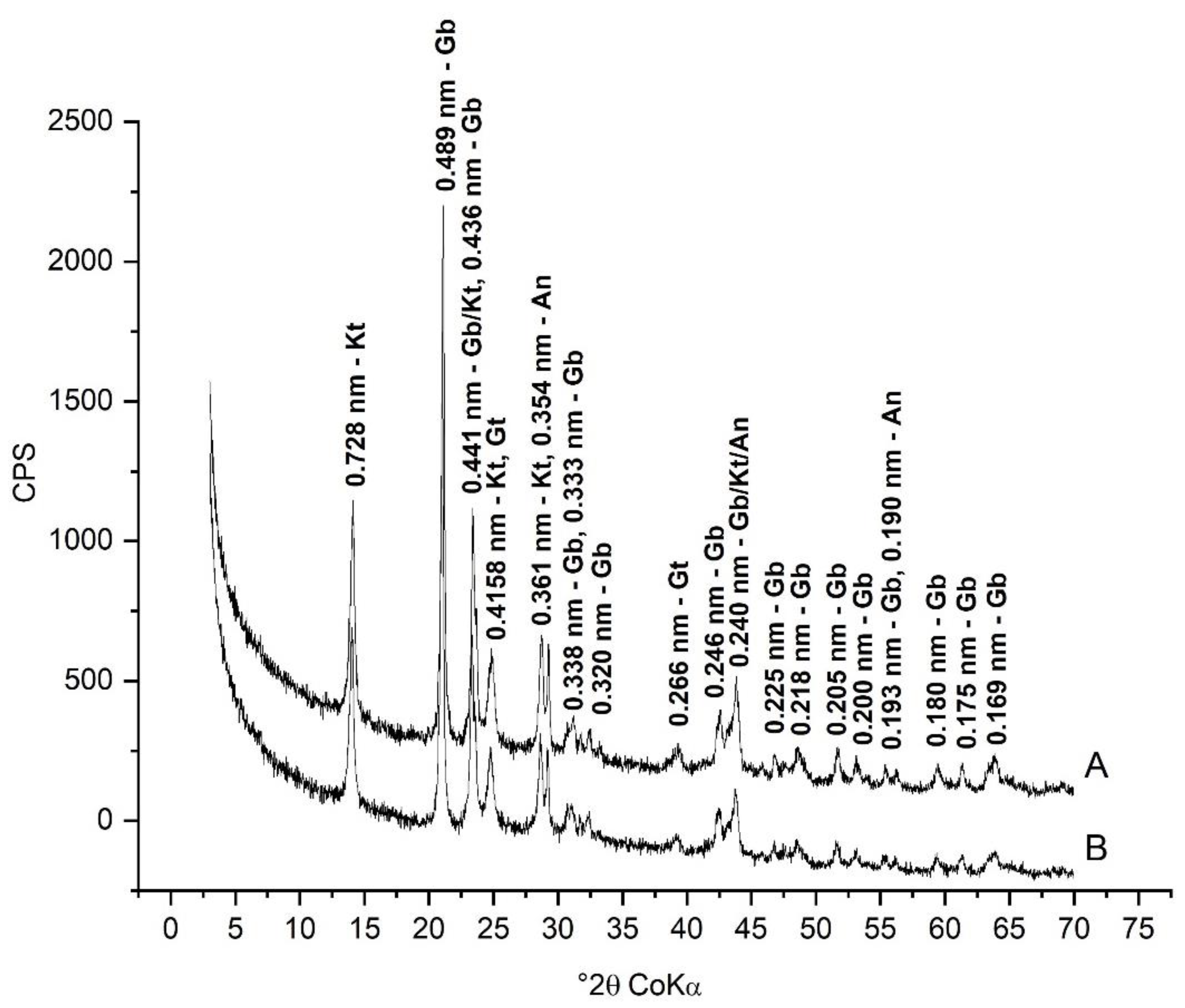
The mineralogical composition of the soils, represented by sample 3, proved to be, in general, quite typical of highly developed soils, as shown in Figure 2. The composition of bauxites showed a predominance of minerals characteristic of tropical bauxites, as can be observed in Figure 3. The minerals identified in the fine fraction of all the evaluated samples are shown in Table 1. It was observed in this fraction the predominance of gibbsite and kaolinite, with the frequent occurrence of goethite, hematite, and anatase. Maghemite and 2:1 minerals were also identified in the analyzed soils and bauxites. However, the participation of these components proved to be quite limited and restricted to some samples.
The gibbsite and kaolinite contents in the fine iron-removed fraction of representative samples, calculated using the mass loss with the thermogravimetric analysis, are shown in Table 2. The results of this analysis show the predominance of gibbsite over kaolinite in all samples, with the exceptions of soils 4 and 5. The gibbsite contents in the samples of the fine iron-removed fraction varied from 39.46 dag kg−1 in sample 4, up to 73.40 dag kg−1 in sample 11. Table 2 also shows the temperatures of the endothermic peak associated with the gibbsite dehydroxylation reaction for the evaluated samples. The dehydroxylation temperature (DT) of this mineral varied from 253.33 °C in sample 4, to 269.52 °C in sample 11, showing a maximum difference of 16.19 °C between these peaks.

3.2. Crystallographic Characterization

The crystallographic indexes evaluated by XRD are shown in Table 3. The interplanar distances observed for the gibbsite at the reflexions (002) and (110) varied little between samples. The highest d002-values were observed in samples 2B, 7A, 8B, 12, and 13 (0.4846 nm), and the lowest in samples 2A, 4B, and 5B (0.4838 nm). For d110, the highest value was observed in sample 2B (0.4380 nm), and the lowest was in sample 6A (0.4367 nm).
The lowest FWHM values were observed in Amazon soil samples (6, 7, and 8), and in the samples from bauxites and soil over bauxite. The highest values were found in soil samples from the Southeast and Midwest regions of Brazil (1, 2, 3, and 4). Among the peaks analyzed, the peak (002) showed the lowest FWHM values, varying from 0.1643 °2θ in sample 13 (bauxite) to 0.2442 °2θ in the subsurface horizon of sample 1.
The highest MCD values (Table 3) for all peaks analyzed were observed in samples of Amazon soils, bauxites, and in soil over bauxite. These MCDs were considerably higher than those of samples from the Southeast and Midwest of Brazil, especially for the reflection (002). For this peak, the first group of samples had average MCDs of 542.94 nm, and the second, of 145.75 nm.
Different types of results were observed with the analysis of the ratio between the intensities of reflections (110) and (002) of the gibbsite. In the X-ray patterns of the non-oriented fine fraction, the range of results was relatively small, varying from 0.360 in sample 13, to 0.539 in sample 4, as shown in Table 3; however, in samples of the oriented fine fraction, the differences in the values of the ratio I110/002 were considerably pronounced, ranging from 0.558 in sample 3B to 0.024 in samples 7B and 13.

3.3. Gibbsite Morphology

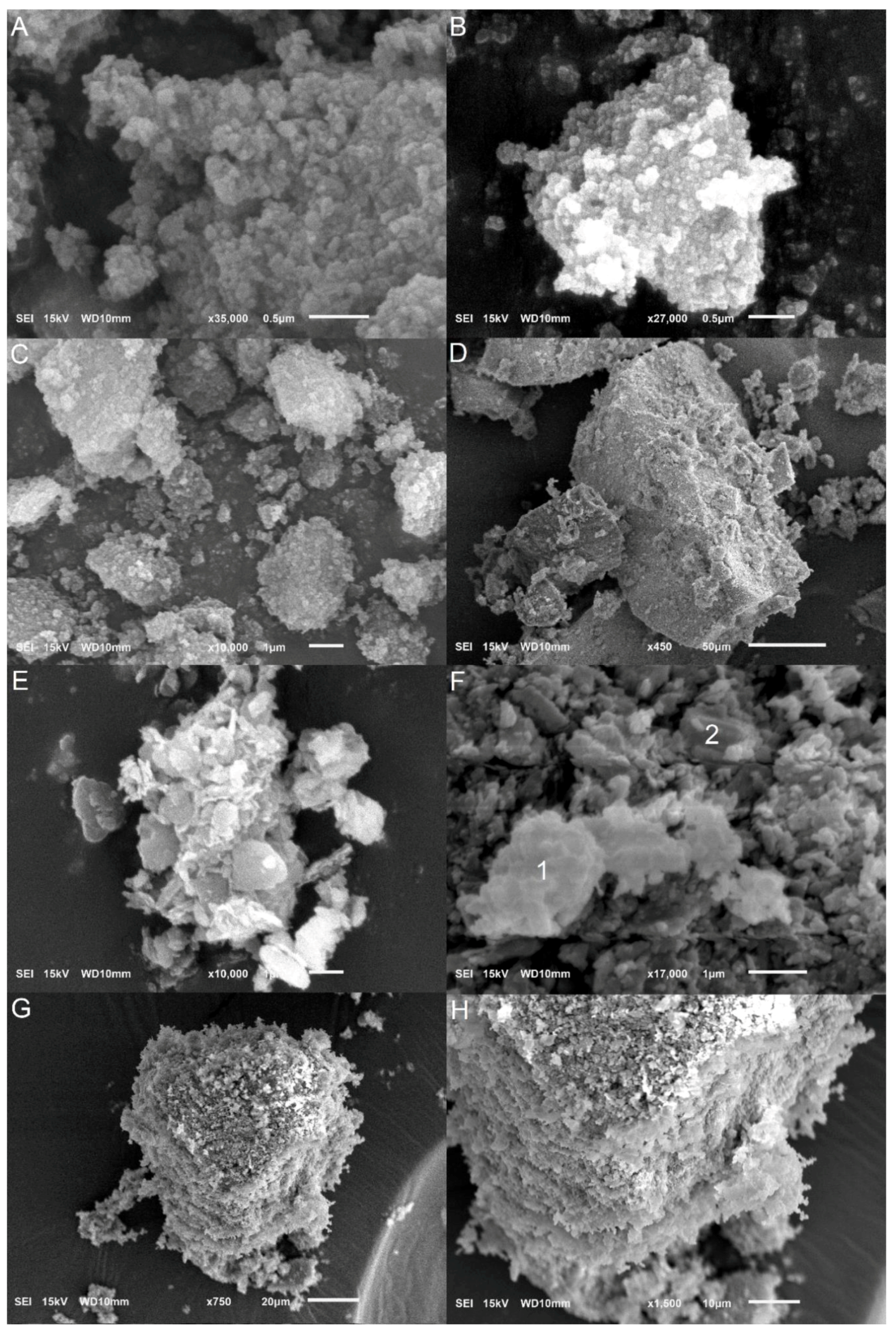
The micrographs obtained by scanning electron microscopy (SEM) are shown in Figure 4. It was possible to observe by this analysis, aided by the evaluation of the chemical composition of the minerals by EDS, a predominance of kaolinite and gibbsite in the fine fraction of the analyzed iron-removed samples.
In both soils and bauxites, the crystals of these minerals had a predominantly laminar/placoid morphology. Most gibbsite crystals, identified by the clear predominance of Al over Si by EDS, presented a mostly laminar/placoid and slightly hexagonal morphology, with larger dimensions in the X and Y directions, and smaller in the Z direction (Figure 4E); however, in sample 3, there was a predominance of gibbsite crystals with an apparent isodimensional shape (Figure 4A,B). Although less prevalent, prism-shaped gibbsites were also found in the studied samples (Figure 4F, point 2). The fine fraction crystals were predominantly organized into two forms of aggregation. In the first one, which can be seen in Figure 4G,H, the crystals were organized into layers oriented parallel to each other, following the face-to-face or edge-to-edge arrangement of the laminar minerals. The second type of aggregates, which are depicted in Figure 4C,D, had fairly smooth surfaces, with no discernible pattern of agglomeration.

4. Discussion

4.1. Gibbsite Characterization

The fine fraction of the analyzed soils and bauxites exhibited a remarkable presence of gibbsite, a fact observed both by the intensity of the reflections of this mineral in the X-ray patterns and by its quantification by thermogravimetry. The reflections (002) and (110) of the gibbsite were detected in all samples evaluated, with interplanar distances associated with both reflections varying slightly between samples. It was observed that the maximum variation of the interplanar distance in these two directions (0.0008 nm for reflection (002), and 0.0013 nm for reflection (110)) between the samples can be considered quite small, especially when compared, for example, to that of soil and bauxite goethites. These goethites, which frequently present isomorphic substitutions, exhibit interplanar distance variations as large as 0.0070 nm, as well as different crystallinities [7,33,34,35,36]. The octahedral sheet of kaolinites, similar in structure and composition to gibbsite, also frequently presents isomorphic substitutions, especially by iron. These substitutions seem to lead to increases in the interplanar distances and are often associated with kaolinites of lower crystallinity [37,38,39]. Similar results, indicating a small variation in the interplanar distance of gibbsite for these two peaks, were also observed by Ghidin et al. [38], in Ferralsols in the southern region of Brazil. This small variation in the d values for the analyzed peaks indicates that the distances between the successive atomic planes of the gibbsite do not differ much between its crystals present in the different soils and bauxites studied. This characteristic strongly suggests that there is no significant replacement of Al in the gibbsite structure by elements of different ionic radii, indicating that isomorphic substitution has little influence on the variation in the crystallinity of this mineral in soils and bauxites. It is worth noting that the isomorphic substitution between Al and Fe does not appear to be reciprocal. While Al replaces Fe in all major iron oxides commonly found in highly developed soils and bauxites, Fe does not seem to replace Al in gibbsites from both soils and bauxites, despite the fact that these Fe and Al oxides are often formed or altered in the same environment [40].

4.2. Gibbsite Crystallinity

In general, samples from the Southeast and Midwest soils showed higher values of FWHM, while samples of Amazon soils and bauxites showed lower values. This trend was maintained in all analyzed gibbsite reflections, such as for reflection (021), whose average for the first group of samples was 0.61 °2θ, while an average of 0.49 °2 was observed for the second one.
The MCD values followed an inverse trend, as expected, and were higher on average than those found in the literature for soils in Brazil’s Southeast-South regions. [17,22,41,42]. The MCD values for the reflection (002) of gibbsite presented by those authors were similar to those observed in this study for the Southeast/Midwest region soils (samples 1, 2, 3, and 4) but far lower than the observed values for Amazon soils and bauxites.
The ratio between the intensities of the reflections (110) and (002) (ratio I110/002) was investigated in light of the observation of a sharp increase in the intensity of the gibbsite reflection (002), as related to the reflection (110), in oriented fine fraction X-ray patterns, when compared with the ones obtained by the powder method. As shown in Table 3, this pattern was not consistent across the analyzed samples and produced a much narrower range of results in the non-oriented samples. In addition to detecting greater differences between the values of the I110/002 ratio in the samples of the oriented fine fraction, it was also possible to observe the presence of two distinct groups of results. The first group consists of samples 1, 2, 3, 4, and 5, with values of I110/002 above 0.4 (similar to those obtained for samples of the fine non-oriented fraction), and the second group is composed of the remaining samples (Amazon soils, soil over bauxite, and bauxites), with I110/002 ratios below 0.15. These findings suggest that the preferential orientation of the fine fraction minerals affects the disposition of gibbsite crystals in samples submitted to XRD and that this phenomenon can occur more or less sharply depending on the sample analyzed. Therefore, the morphological differences between the studied gibbsites were sufficiently pronounced to allow the preferential orientation of their crystals in some samples but not in others.
In addition to presenting higher I110/002 ratios, the samples of the first group (1, 2, 3, 4, and 5) differed from the other samples in other crystallographic and morphological characteristics. The gibbsite of these Ferralsols showed higher FWHM values and lower MCD for all the peaks evaluated, and their dehydroxylation temperatures were below the average of the evaluated samples. For convenience, therefore, the samples in this study were separated into two groups: the samples with gibbsite of high I110/002 ratio (1, 2, 3, 4, and 5), and the samples with gibbsite of low I110/002 ratio (samples from Amazon soils, soil over bauxite, and bauxites).
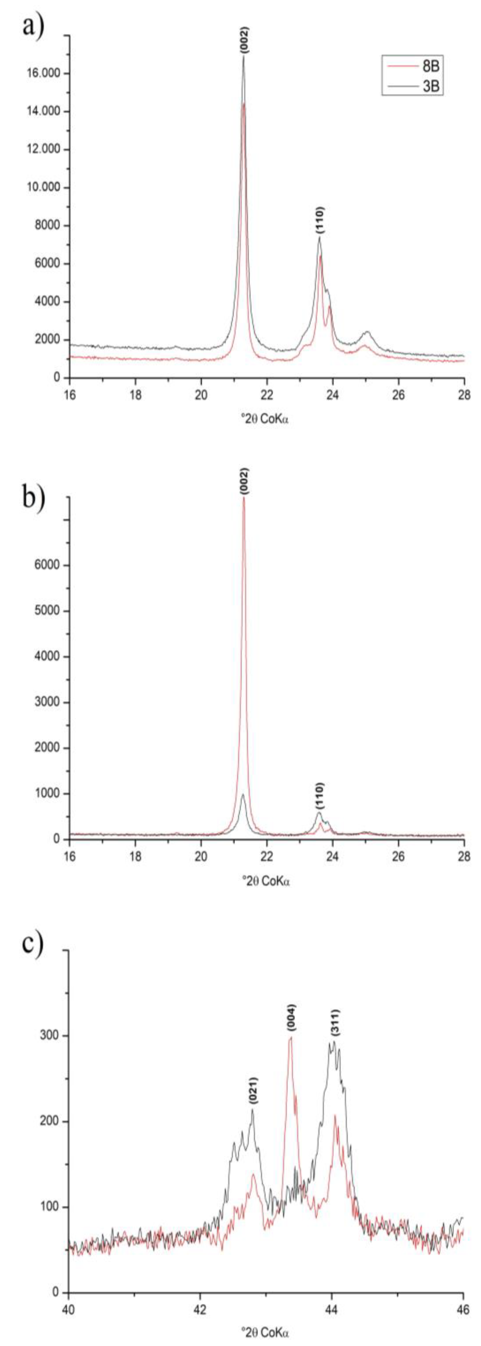
In Figure 5, it is possible to observe that in the diffractograms obtained from the non-oriented fine fraction (a), the intensity of the reflections (002) and (110) of the gibbsite was quite similar between samples 3B and 8B (high and low I110/002 ratio, respectively). However, in samples of the oriented fine fraction (b), the intensity of the gibbsite reflection (002) of sample 8B increased in comparison to the same reflection of sample 3B, whereas the opposite was observed for the reflection (110).
These findings suggest that the gibbsite in these samples has a different orientation capacity. For gibbsites prone to orientating themselves (samples from 6 to 13, represented in this example by sample 8B), the values of the I110/002 ratio suggest a tendency for preferential alignment of these crystals in a position in which the basal planes of the gibbsite are perpendicular to the orientation plane of the fine fraction minerals submitted to XRD. This orientation trend favored an increase in the intensity in the peak family (00ℓ), and a decrease in the intensity of the peaks outside this family of planes. This argument is reinforced by the increase in the reflection intensity (004) of gibbsite (I/I1 = 20) in relation to the other nearby reflections, such as (021) and (311) (both with I/I1 of 25), in the samples of lower I110/002 ratios in X-ray diffraction patterns of the oriented fine fraction, as shown in Figure 5c. The magnification effect of the reflections of the basal plane is quite typical in common phyllosilicates in the soil, such as kaolinites, given their laminar shape; however, this effect has not yet been observed in soil gibbsites, despite the crystalline habit of this mineral favoring the formation of thin platelets, with the upper and lower planes parallel to the basal plane [18,19]. The absence of this effect in several of the analyzed samples suggests that the predominant morphology of this mineral in the soil is highly variable.
In the X-ray diffraction patterns of the non-oriented fine fraction, the differences in the I110/002 ratios between the samples analyzed were considerably smaller. In these X-ray diffraction patterns, the I110/002 ratios seem to have suffered more interference from kaolinite, since the reflection (110) of gibbsite coincides with the reflection (20-2) of this mineral, and the intensity of the peaks in the diffractograms is additive [31]. In X-ray diffraction patterns of the oriented fine fraction, the pronounced orientation tendency of kaolinite promotes a marked decrease in the intensity of the reflection (20-2), thus interfering less with the reflection (110) of the gibbsite in this situation.

4.3. Gibbsite Morphology

The morphological evaluation of the gibbsite by microscopy revealed a great variation in the dimensions of the imaged mineral particles. Among the samples analyzed, the largest particles, which also presented the most laminar shape, were found in sample 8 (Figure 4E,F). The smallest particles, on the other hand, were observed in sample 3 (Figure 4A–D), which presented an apparently isodiametric shape. The observation of larger and more laminar crystals in the micrographs obtained for sample 8, and smaller and apparently spherical shapes in sample 3, corroborates the statements about a greater preferential orientation of gibbsite in Amazon soils and bauxites, and a lower one in the other samples.
The observation of approximately spherical gibbsite crystals, as seen in the micrographs of sample 3B, is not common in the literature. Melo et al. [22] observed gibbsite crystals in the shape of small rectangular plates in the clay fraction and large spherical crystals in the silt fraction while working with highly developed Brazilian soils; however, no spherical crystals larger than 0.5 μm in diameter were found in sample 3 micrographs. Mesquita Filho and Torrent [43] observed gibbsite crystals with an apparent rounded shape in the clay fraction of soils under savanna vegetation in Brazil using transmission electron microscopy. Gibbsites with a prismatic shape, as observed by Braga et al. [24], or in blocks, as presented by Sweegers et al. [23], were identified in the samples studied, albeit less frequently (Figure 4F, point 2). Other gibbsite shapes observed by these authors, such as wedges, lozenges, and stars, were not observed in this work’s samples.
The different morphologies of the gibbsite also seem to have influenced the overall appearance of the fine fraction’s mineral aggregates. Under smaller magnifications, the aggregates formed in samples with gibbsite of higher I110/002 ratios (Figure 4C,D, referring to sample 3 but whose pattern was also observed for sample 2) showed a relatively smooth aspect and were often covered by smaller, apparently loose aggregates; however, for samples containing gibbsite with lower I110/002 ratios, there was a higher frequency, and even the predominance of laminar or layer-organized aggregates, as illustrated by Figure 4G,H, referring to sample 10. The occurrence of layered aggregates in samples with gibbsites of higher I110/002 ratios was also observed, although far less commonly. The distinction between the layers in these aggregates, however, was very subtle, unlike those found in samples with gibbsites of a low I110/002 ratio. These aggregation patterns indicate again a greater orientation trend in the gibbsite of samples with lower I110/002 ratios.
It was observed that, in addition to presenting a more laminar shape, the low I110/002 ratio gibbsites also had larger crystals, according to the microscopy images shown above, and with the observed MCD values. Among the MCDs evaluated, those of planes for the Miller index (002) presented the most discrepant values between the two groups. For this peak, the MCD values of the samples with better preferential orientation (low I110/002 ratios) were, on average, 3.7 times higher than the samples with worse preferential orientation. This group of samples also presented MCDs (021) that were 1.4 times higher on average than the MCDs in the second group, and 1.5 times higher for (313) and (31-4). The presence of larger crystallites due to the higher MCD values, in addition to the observation of thinner reflections, corresponding to lower FWHM values, also indicates a better crystallinity of minerals [41,44]. Based on these results, it can be seen that the gibbsite crystals of samples from Brazil’s Southeast and Midwest soils, all belonging to the group of gibbsites with high I110/002 ratios, were the least crystalline in this work. Samples of Amazon soils and bauxites, on the other hand, with low I110/002 ratios, were the most crystalline among the studied samples.
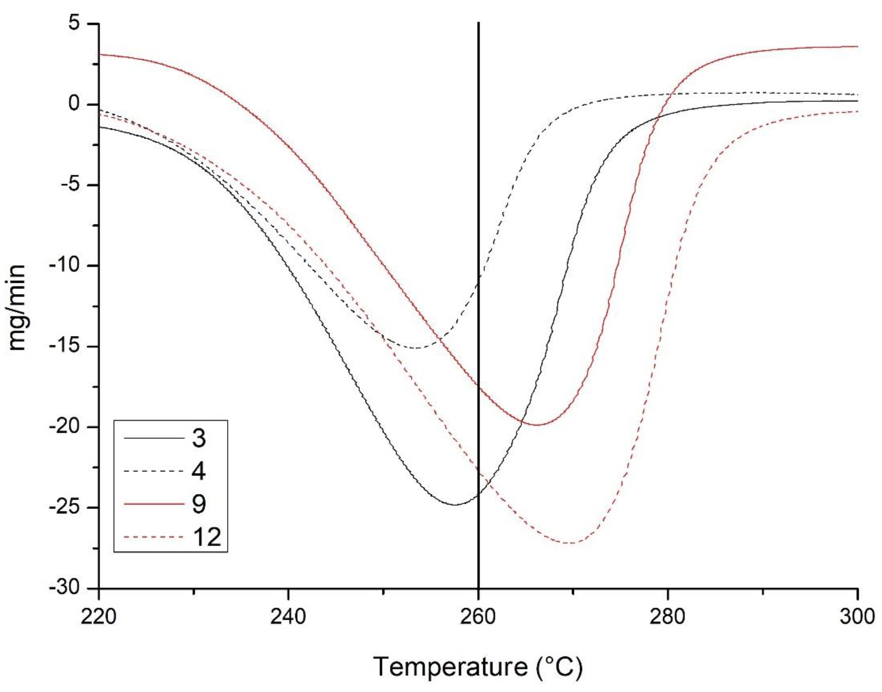
These considerations about the crystallinity of the gibbsite between the samples are also consistent with the thermal analysis results. In general, the higher the temperatures of the endothermic peak of dehydroxylation of gibbsites, the more crystalline they are [13,45]. Based on our results, the most crystalline gibbsites evaluated in this work were those from bauxites and Amazon soils, with an average of 266.52 °C (Figure 6). Samples 2, 3, 4, and 5 had the lowest dehydroxylation temperatures (an average of 255.55 ° C), and are therefore considered the least crystalline. Thus, under the conditions presented, the temperature of 260 °C can be established as that which separates gibbsites of high and low crystallinity.
There is a convergence of results, indicating a higher crystallinity in the samples from Amazon soils and bauxites, and a lower crystallinity in samples from the Southeast and Midwest Brazilian soils. The former group shows low values of the I110/002 ratio, whereas the latter shows high values of this ratio. This trend suggests that, in the soil, the more crystalline gibbsites tend to form large crystals with laminar morphology. Low crystallinity gibbsites, on the other hand, have smaller crystals and an isodimensional morphology.

5. Conclusions

The ratio between the intensity of the gibbsite XRD reflections (110) and (002) allowed us to evaluate the average orientation capacity of this mineral in the fine fraction, and it was a key factor in defining the crystallinity of this mineral. This orientation capacity was related to the morphological characteristics of the gibbsite, with lower I110/002 ratios indicating the predominance of larger and more laminar crystals, while higher ratios indicated smaller and approximately spherical-shaped crystals.
Three gibbsite crystal morphologies were distinguished: laminar, isodimensional, and prismatic. Of these, the first two shapes were the most abundant, despite not appearing to occur within the same sample.
The techniques used to assess the crystallinity of the gibbsite showed converging results, indicating a greater crystallinity in the gibbsite from the evaluated Amazon soils and bauxites, and a lower crystallinity in the samples with gibbsites from the Southeast and Midwest Brazilian soils. The dehydroxylation temperatures of gibbsites from the Amazon soils and bauxites were consistently higher than those of the remaining samples, while the poorly crystalline gibbsite from the samples of the Southeast and Midwest soils lost its hydroxyls at temperatures > 260 °C. A good correlation was observed between the values of the I110/002 ratio and the crystallinity of this mineral, with the lowest values of this ratio being observed in the samples with the most crystalline gibbsites. This suggests that the good crystallization of this mineral in the weathering environment tends to produce more laminar crystals. In addition, the results showed that the substitution of Al by Fe in natural gibbsites is quite limited and does not appear to interfere with its crystallinity, in contrast to what is generally reported for other common oxides in soil.

Author Contributions

Conceptualization, A.S.G., M.P.F.F. and J.C.K.; methodology, A.S.G., M.P.F.F. and A.A.P.; validation, A.S.G.; formal analysis, A.S.G.; investigation, A.S.G.; resources, M.P.F.F., A.A.P. and J.C.K.; data curation, A.S.G.; writing—original draft preparation, A.S.G.; writing—review and editing, M.P.F.F., A.A.P. and J.C.K.; visualization, A.S.G.; supervision, M.P.F.F. and A.A.P.; project administration, M.P.F.F. and J.C.K.; funding acquisition, M.P.F.F. All authors have read and agreed to the published version of the manuscript.

Funding

This study was financed in part by the Coordenação de Aperfeiçoamento de Pessoal de Nível Superior-Brasil (CAPES)-Finance Code 001. We also acknowledge Conselho Nacional de Desenvolvimento Científico e Tecnológico (CNPq) for financial support.

Data Availability Statement

Not applicable.

Conflicts of Interest

The authors declare no conflict of interest.

References

  1. Ferreira, M.M.; Fernandes, B.; Curi, N. Mineralogia da fração argila e estrutura de Latossolos da região sudeste do Brasil. Rev. Bras. Cienc. Solo. 1999, 23, 507–514. [Google Scholar] [CrossRef] [Green Version]
  2. Vitorino, A.C.T.; Ferreira, M.M.; Curi, N.; Lima, J.M.; Silva, M.L.N.; Motta, P.E.F. Mineralogia, química e estabilidade de agregados do tamanho de silte de solos da Região Sudeste do Brasil. Pesqui. Agropecu. Bras. 2003, 38, 133–141. [Google Scholar] [CrossRef]
  3. Leal, J.R.; Velloso, A.C.X. Adsorção de fosfato em Latossolos sob vegetação de Cerrado. Pesqui. Agropecu. Bras. 1973, 8, 81–88. [Google Scholar]
  4. Fontes, M.P.F.; Weed, S.B. Phosphate adsorption by clays from Brazilian Oxisols: Relationships with specific surface area and mineralogy. Geoderma 1996, 72, 37–51. [Google Scholar] [CrossRef]
  5. Moreira, F.L.M.; Mota, F.O.B.; Clemente, C.A.; Azevedo, B.M.; Bomfim, G.B. Adsorção de fósforo em solos do Estado do Ceará. Cienc. Agron. 2006, 37, 7–12. [Google Scholar]
  6. Goldberg, S.; Lebron, I.; Seaman, J.C.; Suarez, D.L. Soil colloidal behavior. In Handbook of Soil Sciences: Properties and Processes, 2nd ed.; Huang, P.M., Li, Y., Sumner, M.E., Eds.; CRC Press: Boca Raton, FL, USA, 2012; Chapter 15; pp. 15-1–15-39. [Google Scholar]
  7. Fontes, M.P.F.; Weed, S.B. Iron oxides in selected Brazilian oxisols: I. Mineralogy. Soil. Sci. Soc. Am. J. 1991, 55, 1143–1149. [Google Scholar] [CrossRef]
  8. Ker, J.C. Latossolos do Brasil: Uma Revisão. Geonomos 1997, 5, 17–40. [Google Scholar] [CrossRef]
  9. Macías-Vazquez, F. Formation of gibbsite in soils and saprolites of temperatehumid zones. Clay Miner. 1981, 16, 43–52. [Google Scholar] [CrossRef]
  10. Kämpf, N.; Scheinost, A.C.; Schulze, D.G. Oxide Minerals in Soils. In Handbook of Soil Sciences: Properties and Processes, 2nd ed.; Huang, P.M., Li, Y., Sumner, M.E., Eds.; CRC Press: Boca Raton, FL, USA, 2012; pp. 22-1–22-34. [Google Scholar]
  11. Caner, L.; Radtke, L.M.; Vignol-Lelarge, M.L.; Inda, A.V.; Bortoluzzi, E.C.; Mexias, A.S. Basalt and rhyo-dacite weathering and soil clay formation under subtropical climate in southern Brazil. Geoderma 2014, 235, 100–112. [Google Scholar] [CrossRef]
  12. Kloprogge, J.T.; Ruan, H.D.; Frost, R.L. Thermal decomposition of bauxite minerals: Infrared emission spectroscopy of gibbsite, boehmite and diaspore. J. Mater. Sci. 2002, 37, 1121–1129. [Google Scholar] [CrossRef] [Green Version]
  13. Melfi, A.J.; Carvalho, A.; Boulange, B.; Lucas, Y. Brazilian Bauxites; USP: São Paulo, Brazil; FAPESP: São Paulo, Brazil; ORSTOM: Paris, France, 1997; 331p. [Google Scholar]
  14. Hsu, P.H. Aluminum Hydroxides and Oxyhydroxides. In Minerals in Soil Environments, 2nd ed.; Dixon, J.B., Weed, S.B., Eds.; Soil Science Society of America: Madison, WI, USA, 1989; pp. 331–378. [Google Scholar]
  15. Huang, P.M.; Wang, M.K.; Kämpf, N.; Schulze, D.G. Aluminum hydroxides. In Soil Mineralogy with Environmental Applications; Dixon, J.B., Schulze, D.G., Eds.; Soil Science Society of America Inc.: Madison, WI, USA, 2002; pp. 261–289. [Google Scholar]
  16. Schulze, D.G. An Introduction to Soil Mineralogy. In Soil Mineralogy with Environmental Applications; Dixon, J.B., Schulze, D.G., Eds.; Soil Science Society of America Inc.: Madison, WI, USA, 2002; pp. 1–35. [Google Scholar]
  17. Oliveira Junior, J.C.; Souza, L.C.P.; Melo, V.F.; Rocha, H.O. Variabilidade espacial de atributos mineralógicos de solos da formação Guabirotuba, Curitiba (PR). Rev. Bras. Cienc. Solo. 2011, 35, 1481–1490. [Google Scholar] [CrossRef] [Green Version]
  18. Schoen, R.; Roberson, C.E. Structures of Aluminum Hydroxide and Geochemical Implications. Am. Min. 1970, 55, 43–77. [Google Scholar]
  19. Freij, S.J.; Parkinson, G.M.; Reyhani, M.M. Direct observation of the growth of gibbsite crystals by atomic force microscopy. J. Cryst. Growth. 2004, 260, 232–242. [Google Scholar] [CrossRef]
  20. Fontes, M.P.F.; Camargo, O.A.; Sposito, G. Eletroquímica das partículas coloidais e sua relação com a mineralogia de solos altamente intemperizados. Sci. Agric. 2001, 58, 627–646. [Google Scholar] [CrossRef] [Green Version]
  21. Kloprogge, J.T.; Duong, L.V.; Wood, B.J.; Frost, R.L. XPS study of the major minerals in bauxite: Gibbsite, bayerite and (pseudo-) boehmite. J. Colloid Interface Sci. 2006, 296, 572–576. [Google Scholar] [CrossRef] [Green Version]
  22. Melo, V.F.; Fontes, M.P.F.; Novais, R.F.; Singh, B.; Schaefer, C.E.G.R. Características dos óxidos de ferro e de alumínio de diferentes classes de solos. Rev. Bras. Cienc. Solo. 2001, 25, 19–32. [Google Scholar] [CrossRef]
  23. Sweegers, C.; Connick, H.C.; Meekes, H.; Enckervort, W.J.P.; Hiralal, I.D.K.; Rijkeboer, A. Morphology, evolution and other characteristics of gibbsite crystals grown from pure and impure aqueous sodium aluminate solutions. J. Cryst. Growth. 2001, 233, 567–582. [Google Scholar] [CrossRef]
  24. Braga, M.A.S.; Paquet, H.; Begonha, A. Weathering of granites in a temperate climate (NW Portugal): Granitic saprolites and arenization. Catena 2002, 49, 41–56. [Google Scholar] [CrossRef]
  25. Saalfeld, H.; Wedde, M. Refinement of the crystal structure of gibbsite, Al(OH)3. Z. Krist. 1974, 139, 120–135. [Google Scholar] [CrossRef]
  26. Ksenofontov, D.A.; Kabalov, Y.K. Structure refinement and thermal stability of gibbsite. Inorg. Mater. 2012, 48, 142–144. [Google Scholar] [CrossRef]
  27. Peskleway, C.D.; Henderson, G.S.; Wicks, F.J. Dissolution of gibbsite: Direct observations using fluid cell atomic force microscopy. Am. Min. 2003, 88, 18–26. [Google Scholar] [CrossRef]
  28. Schwertmann, U. Properties of Goethites of Varying Crystallinity. Clays Clay Miner. 1985, 33, 369–378. [Google Scholar] [CrossRef]
  29. Plançon, A.; Tchoubar, C. Determination of structural defects in phyllosilicates by X-ray powder diffraction-II. Nature and proportion of defects in natural kaolinites. Clays Clay Miner. 1977, 25, 436–450. [Google Scholar] [CrossRef]
  30. Coffin, D.E. A method for the determination of free iron in soils and clays. Can. J. Soil Sci. 1963, 43, 7–17. [Google Scholar] [CrossRef]
  31. Harris, W.; White, G.N. X-ray Diffraction Techniques for Soil Mineral Identification. In Methods of Soil Analysis Part 5: Mineralogical Methods; Ulery, A.L., Drees, L.R., Eds.; Soil Science Society of America Inc.: Madison, WI, USA, 2008; Volume 5, pp. 81–116. [Google Scholar]
  32. White, G.N. Scanning Electron Microscopy. In Methods of Soil Analysis Part 5: Mineralogical Methods; Ulery, A.L., Drees, L.R., Eds.; Soil Science Society of America Inc.: Madison, WI, USA, 2008; Volume 5, pp. 81–116. [Google Scholar]
  33. Norrish, K.; Taylor, R.M. The isomorphous replacement of iron by aluminium in soil goethites. J. Soil Sci. 1961, 12, 294–306. [Google Scholar] [CrossRef]
  34. Kämpf, N.; Schwertmann, U. The 5M NaOH concentration treatment for iron oxides in soils. Clays Clay Miner. 1982, 30, 401–408. [Google Scholar] [CrossRef]
  35. Schwertmann, U.; Carlson, L. Aluminum Influence on Iron Oxides: XVII. Unit-Cell Parameters and Aluminum Substitution of Natural Goethites. Soil Sci. Soc. Am. J. 1994, 58, 256. [Google Scholar] [CrossRef]
  36. Neumann, R.; Avelar, A.N.; Costa, G.M. Refinement of the isomorphic substitutions in goethite and hematite by the Rietveld method, and relevance to bauxite characterisation and processing. Miner. Eng. 2014, 55, 80–86. [Google Scholar] [CrossRef]
  37. Singh, B.; Gilkes, R.J. Properties of soil kaolinites from south-western Australia. J. Soil Sci. 1992, 43, 645–667. [Google Scholar] [CrossRef]
  38. Ghidin, A.A.; Melo, V.D.F.; Lima, V.C.; Lima, J.M.J.C. Toposseqüências de Latossolos originados de rochas basálticas no Paraná: I-mineralogia da fração argila. Rev. Bras. Cienc. Solo. 2006, 30, 293–306. [Google Scholar] [CrossRef] [Green Version]
  39. Pacheco, A.A.; Ker, J.C.; Schaefer, C.E.G.R.; Fontes, M.P.F.; Andrade, F.V.; Martins, E.D.S.; Oliveira, F.S.D. Mineralogy, micromorphology, and genesis of soils with varying drainage along a hillslope on granitic rocks of the Atlantic Forest Biome, Brazil. Rev. Bras. Cienc. Solo. 2018, 42, e0170291. [Google Scholar] [CrossRef] [Green Version]
  40. Schwertmann, U. Goethite and hematite formation in the presence of clay minerals and gibbsite at 25 C. Soil Sci. Soc. Am. J. 1988, 52, 288–291. [Google Scholar] [CrossRef]
  41. Camargo, L.A.; Júnior, J.M.; Pereira, G.T.; Horvat, R.A. Variabilidade Espacial de Atributos Mineralógicos De Um Latossolo Sob Diferentes Formas Do Relevo. I–Mineralogia da Fração Argila. Rev. Bras. Cienc. Solo. 2008, 32, 2279–2288. [Google Scholar] [CrossRef]
  42. Montanari, R.; Júnior, J.M.; Campos, M.C.C.; Souza, Z.M.; Camargo, L.A. Caracterização mineralógica de Latossolos em diferentes feições do relevo na região de Jaboticabal, SP. Cienc. Agron. 2010, 41, 191–199. [Google Scholar] [CrossRef]
  43. Mesquita Filho, M.V.; Torrent, J. Phosphate sorption as related to mineralogy of a hydrosequence of soils from the Cerrado region (Brazil). Geoderma 1993, 58, 107–123. [Google Scholar] [CrossRef]
  44. Fitzpatrick, R.W.; Schwertmann, U. Al-substituted goethite-An indicator of pedogenic and other weathering environments in South Africa. Geoderma 1982, 27, 335–347. [Google Scholar] [CrossRef]
  45. Frost, R.L.; Kloprogge, J.T.; Russel, S.C.; Szetu, J.L. Vibrational spectroscopy and dehydroxylation of aluminum (oxo)hydroxides: Gibbsite. Appl. Spectrosc. 1999, 53, 423–434. [Google Scholar] [CrossRef]
Figure 1. Geographic location of the soil samples in Brazil.
Minerals 12 01441 g001
Figure 2. X-ray diffraction patterns of the raw fine fraction of sample 3. The sample was prepared using the oriented suspension mount. Kt: Kaolinite, Gb: Gibbsite, An: Anatase, and Gt: Goethite.
Minerals 12 01441 g002
Figure 3. X-ray diffraction patterns of the raw fine fraction of sample 12. The sample was prepared using the oriented suspension mount. Kt: Kaolinite, Gb: Gibbsite, Bh: Boehmite, An: Anatase, and Gt: Goethite.
Minerals 12 01441 g003
Figure 4. Micrographs of the iron-removed fine fraction: (AC) show the isodiametric shape of the gibbsite crystals in sample 3, while (D) shows the general appearance of aggregates in this sample under lower magnifications. (E,F)-1 show the laminar-shaped gibbsite crystals of sample 8, while (F)-2 shows a prismatic or blocky crystal. (G,H) show an aggregate of sample 10, whose laminar crystals of gibbsite and kaolinite were arranged in a face-to-face layered set.
Minerals 12 01441 g004
Figure 5. X-ray diffraction pattern sections showing the difference in intensity between the analyzed peaks of the gibbsite: (a) X-ray diffraction pattern of non-oriented clays; (b) X-ray diffraction pattern of oriented clays; (c) X-ray diffraction pattern of oriented clays showing peak 004 magnified in sample 8B, in contrast to the same unmagnified peak in sample 3B.
Minerals 12 01441 g005
Figure 6. Thermograms sections (DTA) showing the differences in the temperature of the gibbsite dehydroxylation peak for low (3B and 4B) and high (8B and 11) crystallinity samples.
Minerals 12 01441 g006
Table 1. Identification, mineralogical composition of the fine fraction, and soil classification (World Reference Base, Soil Taxonomy, and Sistema Brasileiro de Classificação de Solos).
IdentificationMinerals in the Fine FractionWRB 1Soil TaxonomySiBCS 2
Southeast and Midwest Brazilian Soils
1Gb, Kt, An, Hm, Gt, MhGibbsic FerralsolAcrudoxLV Acriférrico
2Gb, Kt, An, Hm, Gt, MhGibbsic FerralsolAcrudoxLV Acriférrico
3Gb, Kt, An, GtGibbsic FerralsolAcrudoxLA Ácrico
4Gb, Kt, An, 2:1, Il, Hm, GtGibbsic FerralsolAcrudoxLVA Acriférrico
5Gb, Kt, An, Hm, GtGibbsic FerralsolAcrudoxLVA Ácrico
Amazon Soils
6Gb, Kt, Gt, AnHaplic FerralsolHapludoxLA Distrófico
7Gb, Kt, Gt, AnHaplic FerralsolHapludoxLA Distrófico
8Gb, Kt, Gt, AnHaplic FerralsolHapludoxLVA Distrófico
Soil over bauxite
9Gb, Kt, Gt, Hm, An---
Bauxites
10Gb, Kt, Il, Gt, Hm, Dp---
11Gb, Kt, An, Gt, Hm---
12Gb, Kt, An, Gt, Bh---
13Gb, Kt, Gt, Hm, An, Bh---
Gb: Gibbsite, Kt: Kaolinite, An: Anatase, Hm: Hematite, and Gt: Goethite. Mh: Maghemite, 2:1: 2:1 clay minerals, Il: Illite, Dp: Diaspore, and Bh: Boehmite. 1 World Reference Base. 2 Sistema Brasileiro de Classificação de Solos (Brazilian Soil Classification System).
Table 2. Gibbsite and kaolinite contents in selected iron-removed fine fraction samples, obtained by TG, and the gibbsite dehydroxylation temperatures (DT) from the DTA curves.
SampleDT-GibbsiteGibbsiteKaolinite
°Cdag kg−1
2B255.1351.7334.14
3B257.4262.0226.64
4B253.3339.4647.21
5B256.3041.1551.21
8B266.3055.4535.00
9A261.0246.0333.21
10263.6240.2936.64
11269.5273.4022.86
12265.3668.5930.57
13267.7560.6729.00
Table 3. Corrected interplanar distance (dc), Width at Half Height (WHH), Mean Crystallite Dimensions (MCD), ratio between the intensity of the reflections (110) and (002) of gibbsite (where O and R refer to the x-ray diffraction patterns of oriented and non-oriented (random) materials, respectively).
Sampledc (nm)WHH (°2θ)MCD (nm)I110/002
(002)(110)(002)(021)(313)(31-4)(002)(021)(313)(31-4)OR
Soils
1A0.48420.43770.230.560.560.5583.6122.5023.1024.680.420.46
1B0.48440.43690.240.630.650.59104.7820.9220.6824.570.430.44
2A0.48380.43730.200.590.430.4781.7819.4830.1127.930.480.50
2B0.48460.43800.230.580.480.53186.2824.6533.8630.830.490.48
3A0.48440.43770.200.590.440.47204.9922.8335.6633.680.550.41
3B0.48390.43690.220.600.400.50129.3121.6740.2330.080.560.39
4B0.48380.43710.240.680.470.57146.2319.6834.8627.370.500.54
5B0.48380.43700.190.640.360.46228.9920.2849.3934.280.480.51
6A0.48420.43670.190.470.310.36305.8031.6365.6351.970.080.46
6B0.48400.43710.180.480.340.36533.3931.1557.0854.140.070.48
7A0.48460.43730.210.480.380.38321.5133.3152.0754.860.040.51
7B0.48420.43740.170.460.340.381031.6632.7254.7948.120.020.46
8A0.48430.43740.170.450.330.35383.1932.1654.9452.630.050.46
8B0.48460.43740.190.490.330.37279.3929.3456.9149.210.040.44
Soil over bauxite
9A0,48440.43740.190.540.320.41261.5025.6460.3841.920.070.44
Bauxites
100.48420.43730.210.530.530.571618.4629.1029.7927.860.110.37
110.48430.43710.180.510.360.41502.0228.3850.4142.420.070.43
120.48460.43740.200.530.460.50204.9826.0332.7430.340.060.39
130.48460.43740.160.490.400.41530.3929.0040.5140.760.020.36
Publisher’s Note: MDPI stays neutral with regard to jurisdictional claims in published maps and institutional affiliations.