
Diversity of Pyrite-Hosted Solid Inclusions and Their Metallogenic Implications—A Case Study from the Myszków Mo–Cu–W Porphyry Deposit (the Kraków–Lubliniec Fault Zone, Poland)


 , ,
, ,
Abstract
:1. Introduction
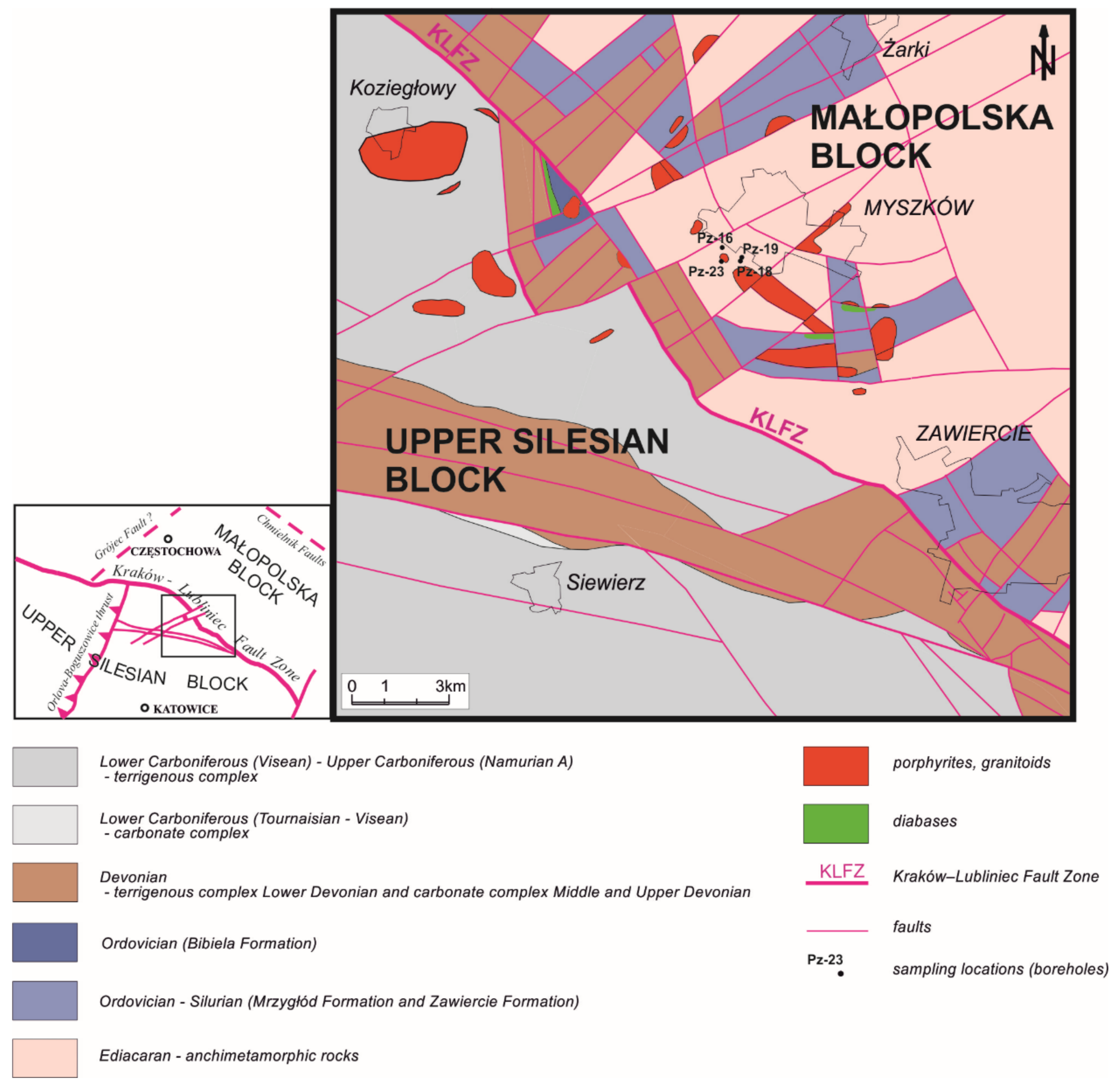
2. Geological Settings
3. Sampling and Methods
4. Results

| Mineral | Raman bands [cm−1] | References |
|---|---|---|
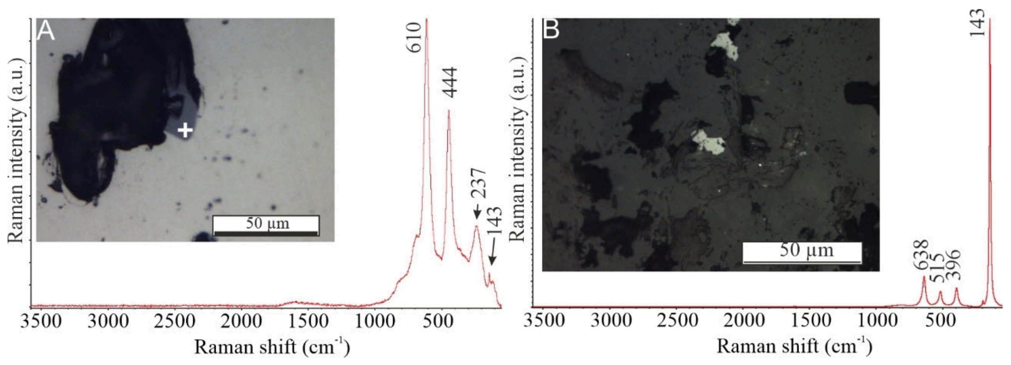
| Anatase | 143, 396, 515, 638 | [50,51] |
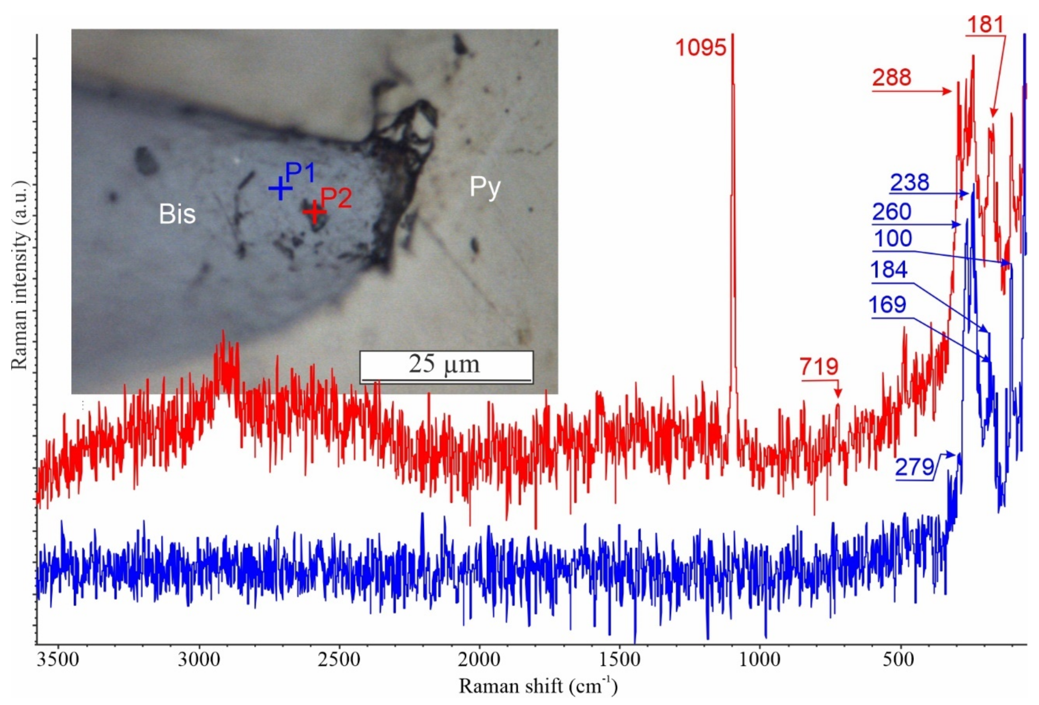
| Bastnaesite | 181, 288, 719, 1095 | [44] |
| Bismuthinite | 100, 169, 184, 238, 260, 279 | [42,43,52] |
| Hematite | s 223, vs 409, m 609, vs 1313, vs 290 | [50] |
| Rutile | 143, 237, 444, 610 | [53] |
| Xenotime | 381, 431, 522, 610, 664, 1002, 1028, 1059, 2022, 2160, 3247, 3255, 3340, 3430, 3480, 3515 | [54] |
5. Discussion
5.1. Evidence of Boiling Indicated by Pyrite’s Inclusions and Microtextures
5.2. Physicochemical Conditions of Mineralizing Fluids
6. Conclusions
- Different generations of pyrite from the Myszków Mo–Cu–W deposit exhibit diverse mineral inclusions. This points toward physicochemical changes that occurred as the ore-forming system evolved with time.
- The number and set of solid inclusions hosted in pyrite indicate precipitation from a vigorous boiling system during the early, skarn-forming stage of mineralization and the formation of pegmatitic veins. Younger episodes of fluid discharges tended to take place under gentle to non-boiling conditions.
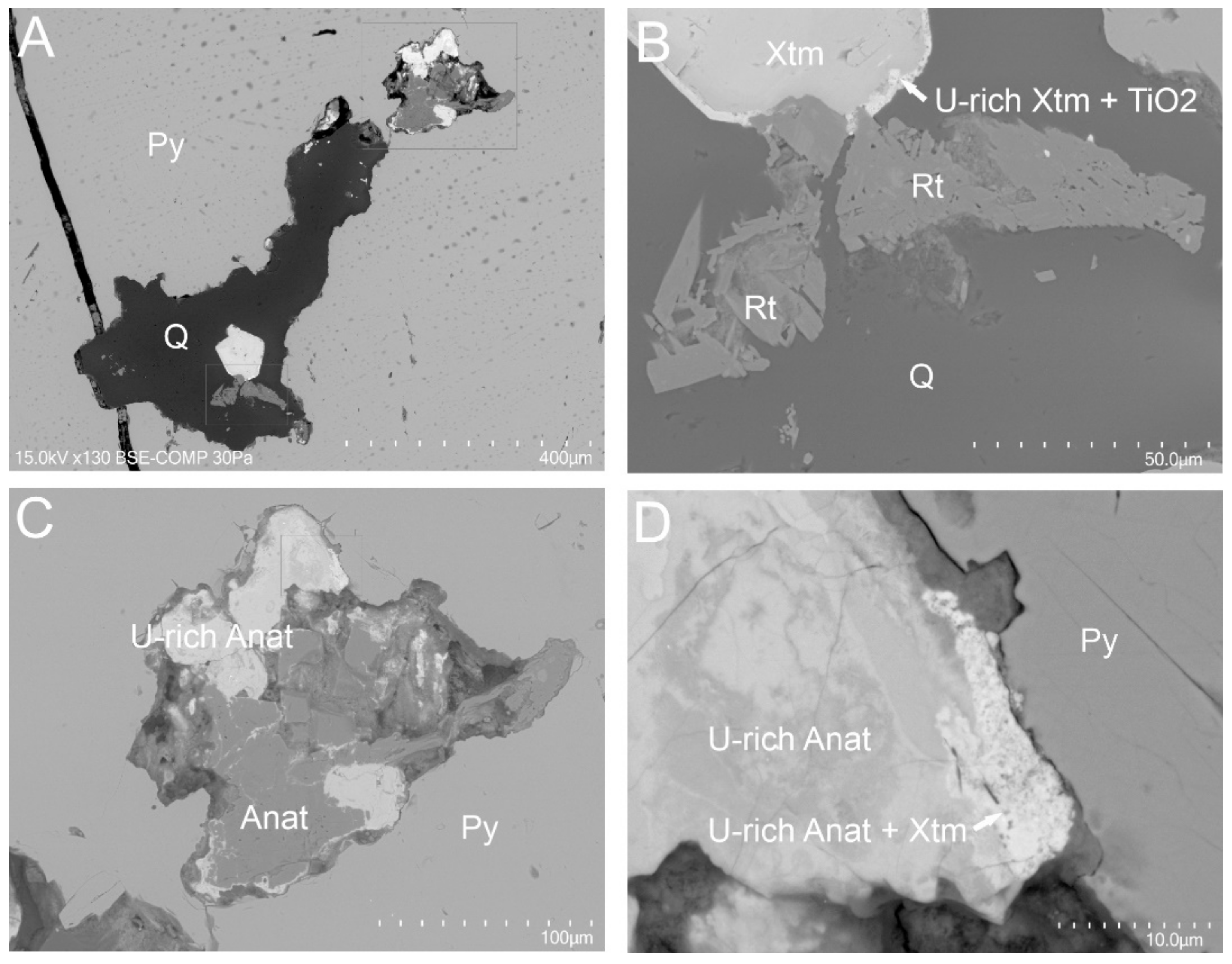
- Solid-inclusion assemblages hosted in pyrite from a stockwork system (i.e., rutile, cassiterite, REE-bearing minerals) and in associated quartz (i.e., anatase, xenotime with hematite inserts) indicate different fluid conditions (temperature, redox conditions) during the precipitation of ores and gangue phases. Moreover, the spatial differentiation of solid-inclusion assemblages in pyrites suggest some zonality of their crystallization environment. Thus, pyrite and its inclusions could be good tracers for vectoring horizons with the most vigorous mineral deposition. However, further studies are needed to support this conclusion, especially with NIR microthermometry as a key technique.
Author Contributions
Funding
Institutional Review Board Statement
Informed Consent Statement
Data Availability Statement
Acknowledgments
Conflicts of Interest
References
- Reich, M.; Deditius, A.; Chryssoulis, S.; Li, J.W.; Ma, C.Q.; Parada, M.A.; Barra, F.; Mittermayr, F. Pyrite as a record of hydrothermal fluid evolution in a porphyry copper system: A SIMS/EMPA trace element study. Geochim. Cosmochim. Acta 2013, 104, 42–62. [Google Scholar] [CrossRef]
- Huston, D.L.; Sie, S.H.; Suter, G.F.; Cooke, D.R.; Both, R.A. Trace elements in sulfide minerals from eastern Australian volcanic- hosted massive sulfide deposits: Part I. Proton microprobe analyses of pyrite, chalcopyrite, and sphalerite, and part II. Selenium levels in pyrite: Comparison with δ34S values and impli. Econ. Geol. 1995, 90, 1167–1196. [Google Scholar] [CrossRef]
- Basori, M.B.I.; Gilbert, S.; Large, R.R.; Zaw, K. Textures and trace element composition of pyrite from the Bukit Botol volcanic-hosted massive sulphide deposit, Peninsular Malaysia. J. Asian Earth Sci. 2018, 158, 173–185. [Google Scholar] [CrossRef]
- Leach, D.L.; Taylor, R.D.; Fey, D.L.; Diehl, S.F.; Saltus, R.W. A deposit model for Mississippi Valley-type lead-zinc ores. In Chapter A of Mineral Deposit Models for Resource Assessment; USGS: Reston, VA, USA, 2010; Volume 107. [Google Scholar]
- Ho, S.E.; McNaughton, N.J.; Groves, D.I. Criteria for determining initial lead isotopic compositions of pyrite in Archaean lode-gold deposits: A case study at Victory, Kambalda, Western Australia. Chem. Geol. 1994, 111, 57–84. [Google Scholar] [CrossRef]
- Yang, J.H.; Zhou, X.H. Rb-Sr, Sm-Nd, and Pb isotopes systematics of pyrite: Implications for the age and genesis of lode gold deposits. Geology 2002, 29, 711–714. [Google Scholar] [CrossRef]
- Shikazono, N. Precipitation mechanisms of barite in sulfate-sulfide deposits in back-arc basins. Geochim. Cosmochim. Acta 1994, 58, 2203–2213. [Google Scholar] [CrossRef]
- Kullerud, G.; Yoder, S. Pyrite stability relations in the Fe-S system. Econ. Geol. 1959, 54, 533–572. [Google Scholar] [CrossRef]
- Djon, M.L.N.; Barnes, S.J. Changes in sulfides and platinum-group minerals with the degree of alteration in the Roby, Twilight, and High Grade Zones of the Lac des Iles Complex, Ontario, Canada. Miner. Depos. 2012, 47, 875–896. [Google Scholar] [CrossRef]
- Lindaas, S.E.; Kulis, J.; Campbell, A.R. Near-infrared observation and microthermometry of pyrite-hosted fluid inclusions. Econ. Geol. 2002, 97, 603–618. [Google Scholar] [CrossRef]
- Large, R.R.; Maslennikov, V.V.; Robert, F.; Danyushevsky, L.V.; Chang, Z. Multistage sedimentary and metamorphic origin of pyrite and gold in the Giant Sukhoi log deposit, Lena Gold Province, Russia. Econ. Geol. 2007, 102, 1233–1267. [Google Scholar] [CrossRef]
- Large, R.R.; Danyushevsky, L.; Hollit, C.; Maslennikov, V.; Meffre, S.; Gilbert, S.; Bull, S.; Scott, R.; Emsbo, P.; Thomas, H.; et al. Gold and Trace Element Zonation in Pyrite Using a Laser Imaging Technique: Implications for the Timing of Gold in Orogenic and Carlin-style Sediment-Hosted Deposits. Econ. Geol. 2009, 104, 635–668. [Google Scholar] [CrossRef]
- Dmitrijeva, M.; Cook, N.J.; Ehrig, K.; Ciobanu, C.L.; Metcalfe, A.V.; Kamenetsky, M.; Kamenetsky, V.S.; Gilbert, S. Multivariate statistical analysis of trace elements in pyrite: Prediction, bias and artefacts in defining mineral signatures. Minerals 2020, 10, 61. [Google Scholar] [CrossRef] [Green Version]
- Deditius, A.P.; Utsunomiya, S.; Reich, M.; Kesler, S.E.; Ewing, R.C.; Hough, R.; Walshe, J. Trace metal nanoparticles in pyrite. Ore Geol. Rev. 2011, 42, 32–46. [Google Scholar] [CrossRef]
- Deditius, A.P.; Reich, M.; Kesler, S.E.; Utsunomiya, S.; Chryssoulis, S.L.; Walshe, J.; Ewing, R.C. The coupled geochemistry of Au and As in pyrite from hydrothermal ore deposits. Geochim. Cosmochim. Acta 2014, 140, 644–670. [Google Scholar] [CrossRef] [Green Version]
- Mancano, D.P.; Campbell, A.R. Microthermometry of enargite-hosted fluid inclusions from the Lepanto, Philippines, high-sulfidation Cu-Au deposit. Geochim. Cosmochim. Acta 1995, 59, 3909–3916. [Google Scholar] [CrossRef]
- Lüders, V.; Ziemann, M. Possibilities and limits of infrared light microthermometry applied to studies of pyrite-hosted fluid inclusions. Chem. Geol. 1999, 154, 169–178. [Google Scholar] [CrossRef]
- Kouzmanov, K.; Bailly, L.; Ramboz, C.; Rouer, O.; Bény, J.M. Morphology, origin and infrared microthermometry of fluid inclusions in pyrite from the Radka epithermal copper deposit, Srednogorie zone, Bulgaria. Miner. Depos. 2002, 37, 599–613. [Google Scholar] [CrossRef]
- Cook, N.J.; Ciobanu, C.L.; Mao, J. Textural control on gold distribution in As-free pyrite from the Dongping, Huangtuliang and Hougou gold deposits, North China Craton (Hebei Province, China). Chem. Geol. 2009, 264, 101–121. [Google Scholar] [CrossRef]
- Cook, N.J.; Ciobanu, C.L.; Meria, D.; Silcock, D.; Wade, B. Arsenopyrite-pyrite association in an orogenic gold ore: Tracing mineralization history from textures and trace elements. Econ. Geol. 2013, 108, 1273–1283. [Google Scholar] [CrossRef]
- Voute, F.; Hagemann, S.G.; Evans, N.J.; Villanes, C. Sulfur isotopes, trace element, and textural analyses of pyrite, arsenopyrite and base metal sulfides associated with gold mineralization in the Pataz-Parcoy district, Peru: Implication for paragenesis, fluid source, and gold deposition mechanisms. Miner. Depos. 2019, 54, 1077–1100. [Google Scholar] [CrossRef]
- Mikulski, S.Z.; Markowiak, M.; Zieliński, G.; Giro, L. New data on tellurium-bismuth mineralization from the Myszków and Mysłów regions in the contact zone of the Małopolska Block with the Upper Silesia Block. Biul. Panstw. Inst. Geol. 2015, 465, 99–122. [Google Scholar] [CrossRef]
- Buła, Z. The lower palaeozoic of upper Silesia and West Małopolska. Pr. Państwowego Inst. Geol. 2000, 171, 1–89. [Google Scholar]
- Buła, Z.; Zaba, J.; Habryn, R. Tectonic subdivison of Poland: Southern Poland (Upper Silesian Block and Małopolska Block). Prz. Geol. 2008, 56, 912–920. [Google Scholar]
- Finger, F.; Hanžl, P.; Pin, C.; von Quadt, A.; Steyrer, H.P. The Brunovistulian: Avalonian Precambrian sequence at the eastern end of the Central European Variscides? Geol. Soc. Spec. Publ. 2000, 179, 103–112. [Google Scholar] [CrossRef]
- Zelaźniewicz, A.; Buła, Z.; Fanning, M.; Seghedi, A.; Żaba, J. More evidence on Neoproterozoic terranes in Southern Poland and southeastern Romania. Geol. Q. 2009, 53, 93–124. [Google Scholar]
- Belka, Z.; Valverde-Vaquero, P.; Dörr, W.; Ahrendt, H.; Wemmer, K.; Franke, W.; Schäfer, J. Accretion of first Gondwana-derived terranes at the margin of Baltica. Geol. Soc. Spec. Publ. 2002, 201, 19–36. [Google Scholar] [CrossRef]
- Nawrocki, J.; Dunlap, J.; Pecskay, Z.; Krzemiński, L.; Zylińska, A.; Fanning, M.; Kozłowski, W.; Salwa, S.; Szczepanik, Z.; Trela, W. Late Neoproterozoic to Early Palaeozoic palaoegeography of the Holy Cross Mountains (Central Europe): An integrated approach. J. Geol. Soc. London 2007, 164, 405–423. [Google Scholar] [CrossRef]
- Habryn, R.; Krzemińska, E.; Krzemiński, L.; Markowiak, M.; Zieliński, G. Detrital zircon age data from the conglomerates in the upper silesian and małopolska blocks, and their implications for the pre-Variscan tectonic evolution (S Poland). Geol. Q. 2020, 64, 321–341. [Google Scholar] [CrossRef]
- Matte, P. Tectonics and plate tectonics model for the Variscan belt of Europe. Tectonophysics 1986, 126, 329–374. [Google Scholar] [CrossRef]
- McCann, T.; Pascal, C.; Timmerman, M.J.; Krzywiec, P.; López-Gómez, J.; Wetzel, A.; Krawczyk, C.M.; Rieke, H.; Lamarche, J. Post-Variscan (end Carboniferous-Early Permian) basin evolution in Western and Central Europe. In European Lithosphere Dynamics; Gee, D.G., Stephenson, R.A., Eds.; Geological Society: London, UK, 2006; Volume 32, pp. 355–388. ISBN 1862392129. [Google Scholar]
- Wolfgang, F. The Variscan orogen in Central Europe: Construction and collapse. In European Lithosphere Dynamics; Gee, D.C., Stephenson, R.A., Eds.; Geological Society: London, UK, 2006; pp. 333–343. [Google Scholar]
- Mazur, S.; Aleksandrowski, P.; Turniak, K.; Krzemiński, L.; Mastalerz, K.; Górecka-Nowak, A.; Kurowski, L.; Krzywiec, P.; Zelaźniewicz, A.; Fanning, M.C. Uplift and late orogenic deformation of the Central European Variscan belt as revealed by sediment provenance and structural record in the Carboniferous foreland basin of western Poland. Int. J. Earth Sci. 2010, 99, 47–64. [Google Scholar] [CrossRef]
- Słaby, E.; Breitkreuz, C.; Zaba, J.; Domańska-Siuda, J.; Gaidzik, K.; Falenty, K.; Falenty, A. Magma generation in an alternating transtensional-transpressional regime, the Kraków-Lubliniec Fault Zone, Poland. Lithos 2010, 119, 251–268. [Google Scholar] [CrossRef]
- Zelaźniewicz, A.; Oberc-Dziedzic, T.; Fanning, C.M.; Protas, A.; Muszyński, A. Late Carboniferous-early Permian events in the Trans-European Suture Zone: Tectonic and acid magmatic evidence from Poland. Tectonophysics 2016, 675, 227–243. [Google Scholar] [CrossRef]
- Ślósarz, J. Mineralogical characteristics of the mineralization. In Palaeozoic Porphyry Molybdenum-Ungsten Deposit in the Myszków area, Southern Poland; Podemski, M., Ed.; Polish Geological Institute, Special Papers: Warsaw, Poland, 2001; Volume 6, pp. 1–88. [Google Scholar]
- Mikulski, S.Z.; Williams, I.S.; Markowiak, M. Carboniferous–Permian magmatism and Mo–Cu (W) mineralization in the contact zone between the Małopolska and Upper Silesia Blocks (south Poland): An echo of the Baltica–Gondwana collision. Int. J. Earth Sci. 2019, 108, 1467–1492. [Google Scholar] [CrossRef] [Green Version]
- Ślósarz, J. Stages and zonality of ore mineralization in Paleozoic rocks of the environs of Myszków. Ann. Soc. Geol. Pol. 1985, 53, 267–288. [Google Scholar]
- Truszel, M.; Karwowski, Ł.; Lasoń, K.; Markiewicz, J.; Żaba, J. Magmatism and metamorphism of the Kraków–Lubliniec tectonic zone as a clue to the occurrence of polymetallic deposits. Biul. Inst. Geol. 2006, 418, 55–103. [Google Scholar]
- Markowiak, M. Description of ore mineralization against the background of thermal-metasomatic alterations of rocks in the Żarki–Kotowice area. Pr. Panstw. Inst. Geol. 2015, 203, 436–443. [Google Scholar]
- Buła, Z.; Byś, I.; Florek, R.H.R.; Jachowicz, M.; Kwarcinski, J.; Laskowicz, R.; Liszka, B.; Madej, K.; Maksym, A.; Markowiak, M.; et al. Atlas Geologiczno-Strukturalny Paleozoicznego Podłoża Karpat Zewnętrznych I Zapadliska Przedkarpackiego; Bula, Z.H.R., Ed.; Państwowy Instytut Geologiczny: Warsaw, Poland, 2008. [Google Scholar]
- Kharbish, S.; Libowitzky, E.; Beran, A. Raman spectra of isolated and interconnected pyramidal XS3 groups (X = Sb,Bi) in stibnite, bismuthinite, kermesite, stephanite and bournonite. Eur. J. Mineral. 2009, 21, 325–333. [Google Scholar] [CrossRef]
- Buzatu, A.; Damian, G.; Buzgar, N.; Andráš, P.; Apopei, A.I.; Maftei, A.E.; Milovská, S. Structural key features of bismuth and Sb-As sulfosalts from hydrothermal deposits—micro-Raman spectrometry. Vib. Spectrosc. 2017, 89, 49–56. [Google Scholar] [CrossRef]
- Frost, R.L.; Dickfos, M.J. Recent Advances in linear and nonlinear Raman spectroscopy I. J. Raman Spectrosc. 2007, 38, 1516–1522. [Google Scholar] [CrossRef] [Green Version]
- Moncada, D.; Mutchler, S.; Nieto, A.; Reynolds, T.J.; Rimstidt, J.D.; Bodnar, R.J. Mineral textures and fluid inclusion petrography of the epithermal Ag-Au deposits at Guanajuato, Mexico: Application to exploration. J. Geochemical Explor. 2012, 114, 20–35. [Google Scholar] [CrossRef]
- Román, N.; Reich, M.; Leisen, M.; Morata, D.; Barra, F.; Deditius, A.P. Geochemical and micro-textural fingerprints of boiling in pyrite. Geochim. Cosmochim. Acta 2019, 246, 60–85. [Google Scholar] [CrossRef]
- Williams-Jones, A.E.; Bowell, R.J.; Migdisov, A.A. Gold in solution. Elements 2009, 5, 281–287. [Google Scholar] [CrossRef]
- Hanesch, M. Raman spectroscopy of iron oxides and (oxy)hydroxides at low laser power and possible applications in environmental magnetic studies. Geophys. J. Int. 2009, 177, 941–948. [Google Scholar] [CrossRef]
- Marshall, C.P.; Marshall, A.O. Raman Hyperspectral Imaging of Microfossils: Potential Pitfalls. Astrobiology 2013, 13, 920–931. [Google Scholar] [CrossRef] [Green Version]
- Frezzotti, M.L.; Tecce, F.; Casagli, A. Raman spectroscopy for fluid inclusion analysis. J. Geochemical Explor. 2012, 112, 1–20. [Google Scholar] [CrossRef]
- Andò, S.; Garzanti, E. Raman spectroscopy in heavy-mineral studies. Geol. Soc. London Spec. Publ. 2014, 386, 395–412. [Google Scholar] [CrossRef]
- Buzatu, A.; Damian, G.; Dill, H.G.; Buzgar, N.; Apopei, A.I. Mineralogy and geochemistry of sulfosalts from Baia Sprie ore deposit (Romania)—New bismuth minerals occurrence. Ore Geol. Rev. 2015, 65, 132–147. [Google Scholar] [CrossRef]
- Swamy, V.; Muddle, B.C.; Dai, Q. Size-dependent modifications of the Raman spectrum of rutile TiO2. Appl. Phys. Lett. 2006, 89, 163118. [Google Scholar] [CrossRef]
- Lafuente, B.; Downs, R.T.; Yang, H.; Stone, N. The power of databases: The RRUFF project. In Highlights in Mineralogical Crystallography.; Armbruster, T., Danisi, R.M., Eds.; De Gruyter: Berlin, Germany, 2015; pp. 1–30. [Google Scholar]
- Kouzmanov, K.; Pokrovski, G.S.; Hedenquist, J.W.; Harris, M.; Camus, F. Hydrothermal controls on metal distribution in porphyry Cu (-Mo-Au) systems. Soc. Econ. Geol. Spec. Publ. 2012, 16, 573–618. [Google Scholar]
- Hedenquist, J.W.; Arribas, A.R.; Gonzalez-Urien, E. Exploration for Epithermal Gold Deposits. Rev. Econ. Geol. 2000, 13, 245–277. [Google Scholar]
- Cunningham, C.G. Pressure gradients and boiling as mechanisms for localization ore in porphyry system. J. Res. U.S. Geol. Surv. 1978, 6, 745–754. [Google Scholar]
- Nash, J.T. Fluid-Inclusion Petrology: Data from Porphyry Copper Deposits and Applications to Exploration; No 907; US Government Printing Office: Washington, DC, USA, 1976.
- Drummond, S.E.; Ohmoto, H. Chemical evolution and mineral deposition in boiling hydrothermal systems. Econ. Geol. 1985, 80, 126–147. [Google Scholar] [CrossRef]
- Moncada, D.; Baker, D.; Bodnar, R.J. Mineralogical, petrographic and fluid inclusion evidence for the link between boiling and epithermal Ag-Au mineralization in the La Luz area, Guanajuato Mining District, México. Ore Geol. Rev. 2017, 89, 143–170. [Google Scholar] [CrossRef]
- Haas, J.L. The effect of salinity on the maximum thermal gradient of a hydrothermal system at hydrostatic pressure. Econ. Geol. 1971, 66, 940–946. [Google Scholar] [CrossRef]
- Cruz-Pérez, M.A.; Canet, C.; Franco, S.I.; Camprubí, A.; González-Partida, E.; Rajabi, A. Boiling and depth calculations in active and fossil hydrothermal systems: A comparative approach based on fluid inclusion case studies from Mexico. Ore Geol. Rev. 2016, 72, 603–611. [Google Scholar] [CrossRef]
- Bodnar, R.J.; Reynolds, T.J.; Kuhen, C.A. Fluid inclusion systematics in epithermal systems. In Geology and Geochemistry of Epithermal Systems; Berger, B.R., Bethke, P.M., Eds.; Society of Economic Geologists, INC.: Littleton, CO, USA, 1985; pp. 73–97. [Google Scholar]
- Etoh, J.; Izawa, E.; Watanabe, K.; Taguchi, S.; Sekine, R. Bladed quartz and its relationship to gold mineralization in the Hishikari low-sulfidation epithermal gold. Econ. Geol. 2002, 97, 1841–1851. [Google Scholar] [CrossRef]
- Thompson, T.B.; Trippel, A.D.; Dwelley, P.C. Mineralized veins and breccias of the Cripple Creek district, Colorado. Econ. Geol. 1985, 80, 1669–1688. [Google Scholar] [CrossRef]
- Keith, M.; Smith, D.J.; Doyle, K.; Holwell, D.A.; Jenkin, G.R.T.; Barry, T.L.; Becker, J.; Rampe, J. Pyrite chemistry: A new window into Au-Te ore-forming processes in alkaline epithermal districts, Cripple Creek, Colorado. Geochim. Cosmochim. Acta 2020, 274, 172–191. [Google Scholar] [CrossRef]
- Kadel-Harder, I.M.; Spry, P.G.; Layton-Matthews, D.; Voinot, A.; von der Handt, A.; McCombs, A.L. Paragenetic relationships between low- and high-grade gold mineralization in the Cripple Creek Au-Te deposit, Colorado: Trace element studies of pyrite. Ore Geol. Rev. 2020, 127, 103847. [Google Scholar] [CrossRef]
- Karowski Łukasz Mineral-forming fluids of the Variscan Cu–porphyry formation from the Myszków-Mrzygłód area Cracovides (Silesian–Cracow region). Pr. Nauk. Uniw. Śląskiego 1988, 929, 3–88.
- McNaughton, N.J.; Rasmussen, B. Geochemical characterisation of xenotime formation environments using U-Th. Chem. Geol. 2018, 484, 109–119. [Google Scholar] [CrossRef]
- Ciesielczuk, J.; Janeczek, J. metasomatic pyrite from hydrothermally altered Borów granite (Strzegom-Sobótka Massif). Preliminary report. Spec. Pap. Mineral. Soc. Pol. 2004, 24, 119–122. [Google Scholar]
- Sondergeld, C.H.; Turcotte, D.L. A laboratory study of mineral deposition in a boiling environment. Econ. Geol. 1979, 74, 109–115. [Google Scholar] [CrossRef]
- Rowins, S.M. Reduced porphyry copper-gold deposits: A new variation on an old theme. Geology 2000, 28, 491–494. [Google Scholar] [CrossRef]
- Wilkinson, J.J. Fluid Inclusions in Hydrothermal Ore Deposits. Lithos 2001, 55, 229–272. [Google Scholar] [CrossRef]
- Lüders, V. Contribution of infrared microscopy to studies of fluid inclusions hosted in some opaque ore minerals: Possibilities, limitations, and perspectives. Miner. Depos. 2017, 52, 663–673. [Google Scholar] [CrossRef] [Green Version]
- Campbell, A.R.; Robinson-Cook, S. Infrared fluid inclusion m1crothermometry on coexisting wolframite and quartz. Econ. Geol. 1987, 82, 1640–1645. [Google Scholar] [CrossRef]
- Roedder, E. Occurrence and significance of magmatic inclusions and silicate liquid immiscibility. Acta Geol. Pol. 1984, 34, 139–178. [Google Scholar]
- Kampmann, T.C.; Jansson, N.F.; Stephens, M.B.; Majka, J.; Lasskogen, J. Systematics of hydrothermal alteration at the falun base metal sulfide deposit and implications for ore genesis and exploration, Bergslagen Ore District, Fennoscandian Shield, Sweden. Econ. Geol. 2017, 112, 1111–1152. [Google Scholar] [CrossRef] [Green Version]
- Rabbia, O.M.; Hernández, L.B.; French, D.H.; King, R.W.; Ayers, J.C. The El Teniente porphyry Cu-Mo deposit from a hydrothermal rutile perspective. Miner. Depos. 2009, 44, 849–866. [Google Scholar] [CrossRef]
- Pal, D.C.; Chaudhuri, T.; McFarlane, C.; Mukherjee, A.; Sarangi, A.K. Mineral chemistry and in situ dating of allanite, and geochemistry of its host rocks in the Bagjata uranium mine, Singhbhum shear zone, India-implications for the chemical evolution of REE mineralization and mobilization. Econ. Geol. 2011, 106, 1155–1171. [Google Scholar] [CrossRef]
- Hezarkhani, A.; Williams-Jones, A.E.; Gammons, C.H. Factors controlling copper solubility and chalcopyrite deposition in the Sungun porphyry copper deposit, Iran. Miner. Depos. 1999, 34, 770–783. [Google Scholar] [CrossRef]
- Pieczka, A.; Gołębiowska, B.; Parafiniuk, J. Formation conditions of sulphide mineralization in the Rędziny area (West Sudetes, Poland). Spec. Pap. Mineral. Soc. Pol. 2005, 26, 229–231. [Google Scholar]
- Xiang Ping, G.; Watanabe, M.; Ohkawa, M.; Hoshino, K.; Shibata, Y.; Desong, C. Felbertalite and related bismuth sulfosalts from the Funiushan copper skarn deposit, Nanjing, China. Can. Mineral. 2001, 39, 1641–1652. [Google Scholar] [CrossRef] [Green Version]
- McClay, K.R.; Ellis, P.G. Deformation of pyrite. Econ. Geol. 1984, 79, 400–403. [Google Scholar] [CrossRef]
- Ortelli, M.; Kouzmanov, K.; Wälle, M.; Dilles, J.H. Comparative study of fluid inclusions trapped in co-genetic ore and gangue minerals. In Proceedings of the Mineral Resources in a Sustainable World 13th SGA Biennial Meeting 2015, Nancy, France, 24–27 August 2015; pp. 505–508. [Google Scholar]


| Pyrite Generation | Stage of Mineralization (According to Ślósarz, 2001) | Form of Mineralization | Vein Mineral Assemblage (Identified by SEM–EDS and Raman Spectroscopy) | Pyrite-Hosted Solid Inclusion (Identified by SEM–EDS and Raman Spectroscopy) | Fracture-Filling Minerals in Pyrite | |||
|---|---|---|---|---|---|---|---|---|
| Gangue | Ore | Gangue | Ore | |||||
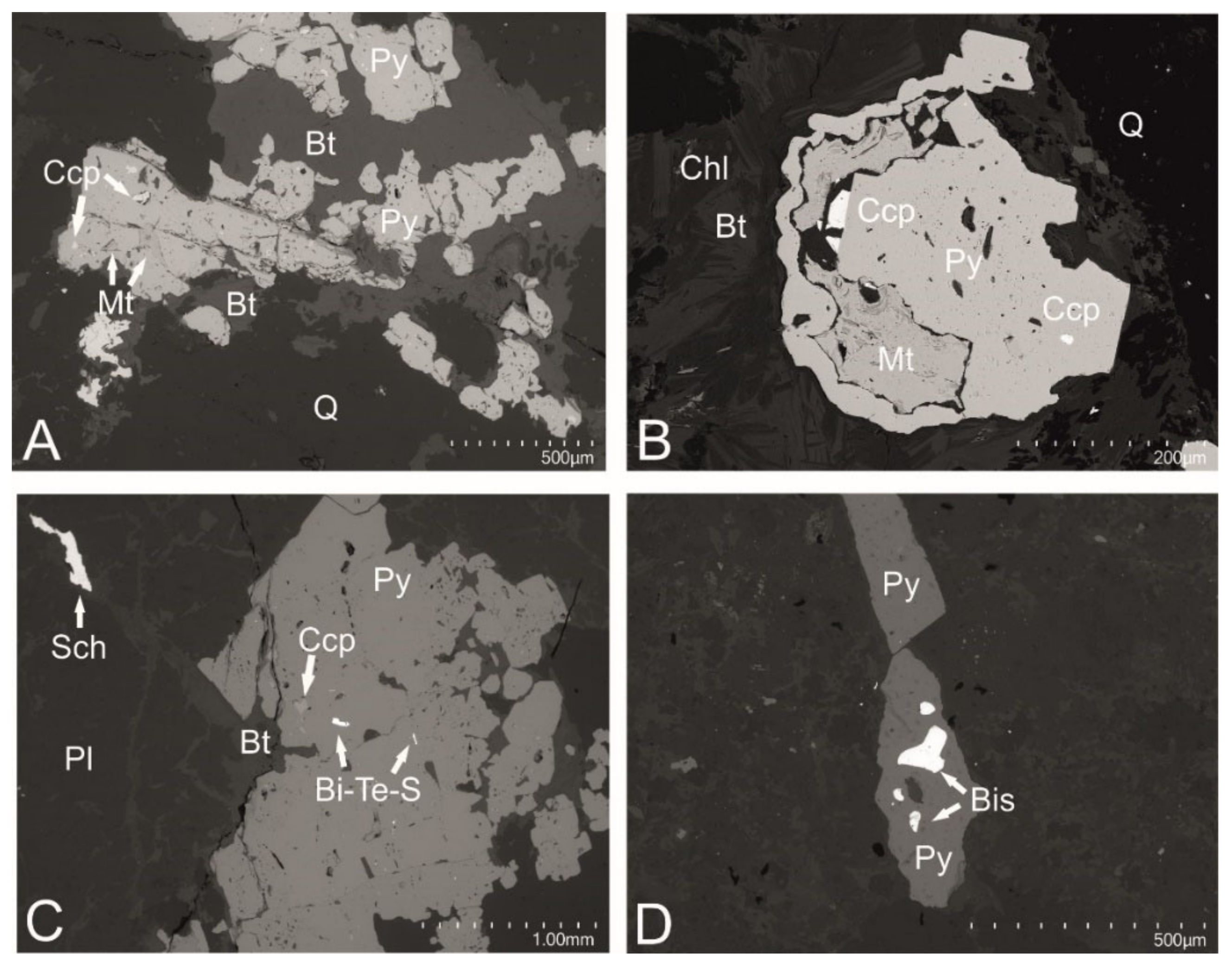
| I-type pyrite | Early, skarn-forming | - | Quartz–chlorite–biotite veins with magnetite, molybdenite, and scheelite | plagioclase, biotite, chlorite, epidote, quartz, calcite, apatite | magnetite, chalcopyrite, molybdenite, ilmenite, scheelite | quartz, chlorite, plagioclase, biotite | magnetite, chalcopyrite, molybdenite, ilmenite, Bi-tellurosulfides and tellurides, bismuthinite | not observed |
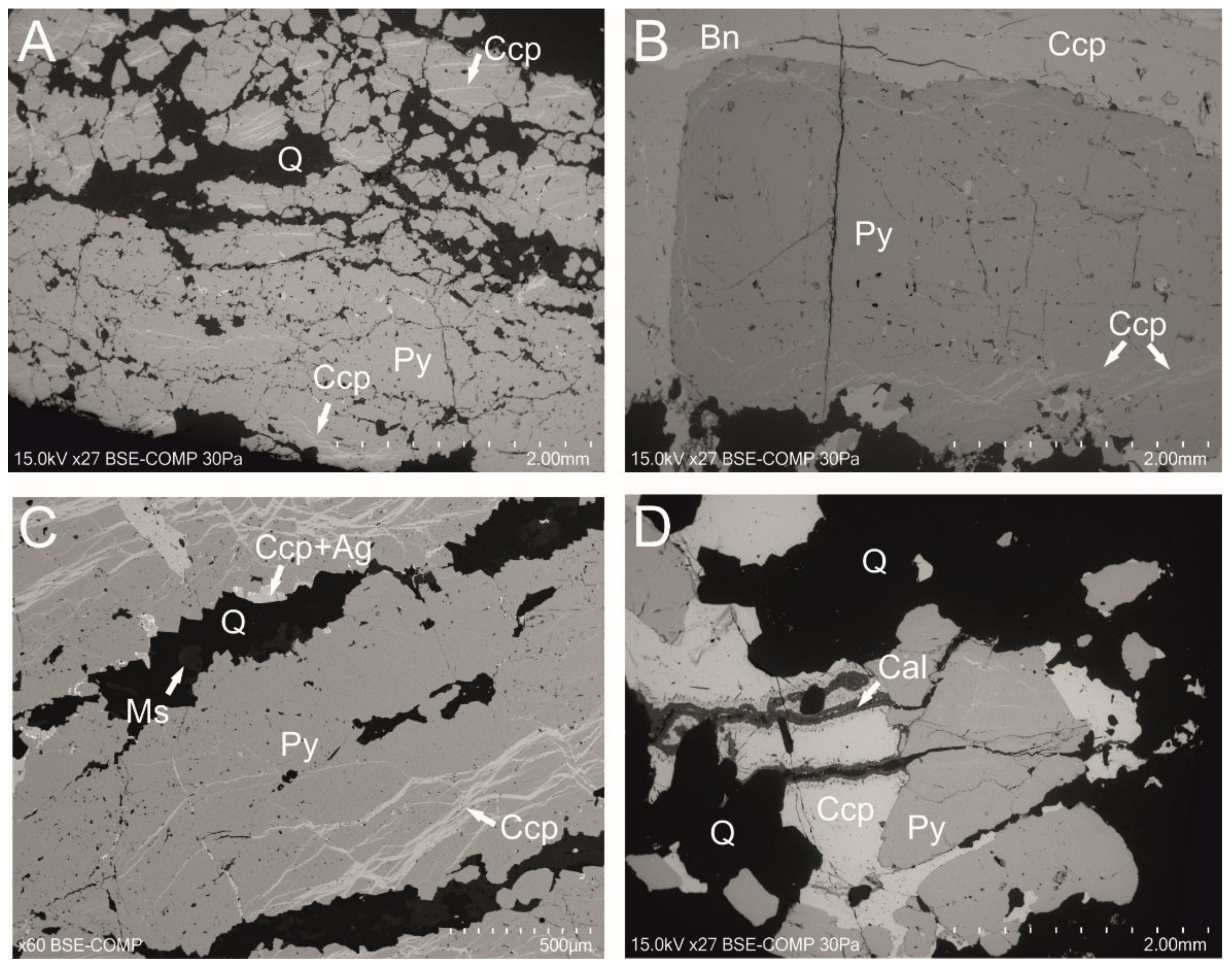
| II-type pyrite | Main, hydrothermal | Feldspathic, pegmatitic quartz veins | Pegmatitic veins | quartz, K-feldspars, calcite, galena | chalcopyrite, molybdenite, scheelite, bornite (veinlets in chalcopyrite) | quartz | galena, chalcopyrite, native Ag | chalcopyrite |
| III-type pyrite | Main, hydrothermal | Quartz veins, with molybdenite and scheelite | Stockwork veins | quartz, calcite, K-feldspars | molybdenite, chalcopyrite, bornite, scheelite, REE-bearing minerals | quartz, K-feldspars, K-mica, illite, apatite | chalcopyrite, galena, cassiterite, REE-bearing minerals (monazite, cerianite), molybdenite, Bi-tellurides, Sn+Cu, rutile, titanite | not observed |
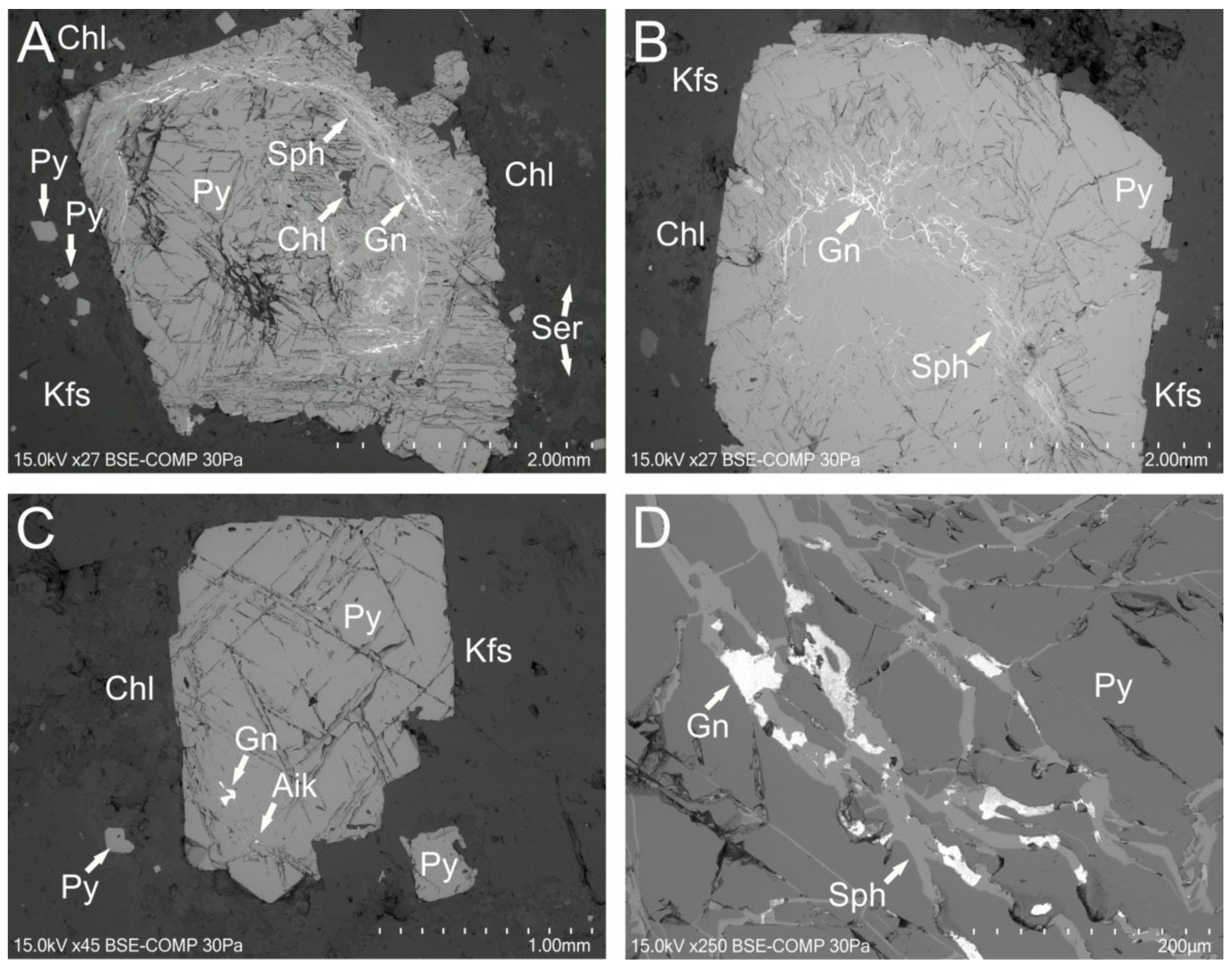
| IV-type pyrite | Main, hydrothermal | Polymetallic quartz | Impregnations | - | - | chlorite | galena, sphalerite, minerals from aikinite-group | galena, sphalerite |
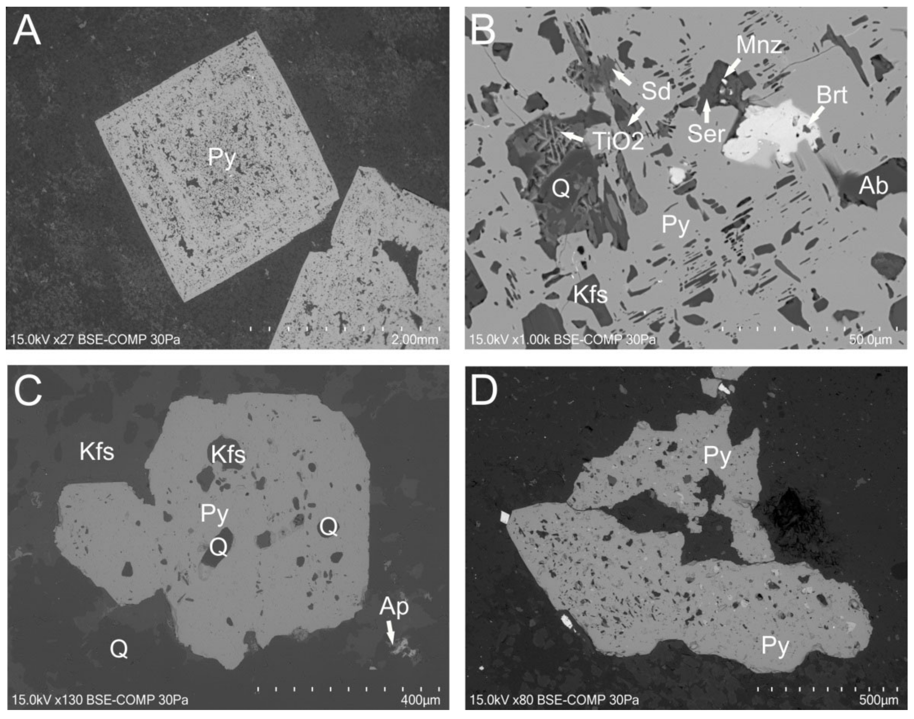
| V-type pyrite | Metasomatic pyrite from different mineralization stages | Near-vein mineralization | - | - | quartz, K-feldspars, calcite, sericite, chlorite, albite, siderite, apatite, gypsum, clay minerals | galena, sphalerite, chalcopyrite, molybdenite, REE-bearing minerals (monazite), Ti-oxides, Bi-tellurides | chalcopyrite | |
Publisher’s Note: MDPI stays neutral with regard to jurisdictional claims in published maps and institutional affiliations. |
© 2021 by the authors. Licensee MDPI, Basel, Switzerland. This article is an open access article distributed under the terms and conditions of the Creative Commons Attribution (CC BY) license (https://creativecommons.org/licenses/by/4.0/).
Share and Cite
Naglik, B.; Dumańska-Słowik, M.; Toboła, T.; Derkowski, P.; Habryn, R.; Markowiak, M. Diversity of Pyrite-Hosted Solid Inclusions and Their Metallogenic Implications—A Case Study from the Myszków Mo–Cu–W Porphyry Deposit (the Kraków–Lubliniec Fault Zone, Poland). Minerals 2021, 11, 1426. https://doi.org/10.3390/min11121426
Naglik B, Dumańska-Słowik M, Toboła T, Derkowski P, Habryn R, Markowiak M. Diversity of Pyrite-Hosted Solid Inclusions and Their Metallogenic Implications—A Case Study from the Myszków Mo–Cu–W Porphyry Deposit (the Kraków–Lubliniec Fault Zone, Poland). Minerals. 2021; 11(12):1426. https://doi.org/10.3390/min11121426
Chicago/Turabian StyleNaglik, Beata, Magdalena Dumańska-Słowik, Tomasz Toboła, Paweł Derkowski, Ryszard Habryn, and Marek Markowiak. 2021. "Diversity of Pyrite-Hosted Solid Inclusions and Their Metallogenic Implications—A Case Study from the Myszków Mo–Cu–W Porphyry Deposit (the Kraków–Lubliniec Fault Zone, Poland)" Minerals 11, no. 12: 1426. https://doi.org/10.3390/min11121426
APA StyleNaglik, B., Dumańska-Słowik, M., Toboła, T., Derkowski, P., Habryn, R., & Markowiak, M. (2021). Diversity of Pyrite-Hosted Solid Inclusions and Their Metallogenic Implications—A Case Study from the Myszków Mo–Cu–W Porphyry Deposit (the Kraków–Lubliniec Fault Zone, Poland). Minerals, 11(12), 1426. https://doi.org/10.3390/min11121426