Article

Preliminary Data on Geochemical Characteristics of Major and Trace Elements in Typical Biominerals: From the Perspective of Human Kidney Stones

1
Department of Urology, Peking University Third Hospital, Beijing 100191, China
2
Institute of Earth Sciences, China University of Geosciences (Beijing), Beijing 100083, China
3
Institute of Geographic Sciences and Natural Resources Research, Chinese Academy of Sciences, Beijing 100101, China
4
Nu Instruments, Wrexham LL13 9XS, UK
*
Author to whom correspondence should be addressed.
Minerals 2021, 11(12), 1396; https://doi.org/10.3390/min11121396
Submission received: 10 November 2021 / Revised: 6 December 2021 / Accepted: 8 December 2021 / Published: 10 December 2021
(This article belongs to the Special Issue Biominerals and Bio-Inspired Materials)

Abstract

:
The chemical composition of biominerals is essential for understanding biomineral formation and is regarded as an attractive subject in bio-mineralogical research on human kidney stones (urinary calculi). In order to obtain more geochemically interpreted data on biogenic minerals, mineralogical compositions and major and trace element concentrations of sixty-six kidney stone samples derived from kidney stone removal surgeries were measured. Infrared spectroscopy results showed that calcium oxalate monohydrate (COM) and calcium oxalate dihydrate (COD) were the two main mineral components of kidney stones. Geochemical results indicated that major and trace element concentrations were present in the following order: Ca > Mg > Na > K > Zn > Fe > Pb > Ba > Cu > Ti > Mo > Cd > Cr. With the exception of Ca, Mg was the second-most abundant element. Zn exhibited higher concentrations relative to other trace elements, which suggests a potential substitution of calcium by metal ions with a similar charge and radius rather than by metals in kidney stone formation. Pb appeared in significantly higher concentrations than in previous studies, which indicates Pb enrichment in the environment. In order to discern multi-element relationships within kidney stones, principal component analysis was applied. Three principal components (PCs, eigenvalues >1) were extracted to explain 64.4% of the total variance. The first component exhibited positively correlated Na-Zn-Cr-Mo-Cd-Pb, while the second component exhibited more positively weighted Mg-K-Ba-Ti. Fe-Cu demonstrated a positive correlation in the third component. This study suggests that Ca exhibits a preference for uptake by oxalates during human urinary stone crystallization, while other alkali metals and alkaline earth metals precipitate with phosphate.

1. Introduction

As one of the most typical biominerals, the biomineralogy of human kidney stones (urinary calculi) has rapidly developed in recent years [1,2,3]. Kidney stones, regarded as one of the most challenging medical issues, have become a global scourge and can even result in the deterioration of renal function and permanent kidney damage [4,5,6,7]. However, some of the causes of kidney stone formation remain unknown [4,6,8,9,10]. Kidney stones can be classified according to mineral composition, which includes four most common types: calcium phosphate, calcium oxalate, uric acid, and struvite kidney stones [2,5]. Oxalates of calcium and calcium phosphate are regarded as the two most common types of kidney stones, and a combination of both types is also included. As an essential component of kidney stones, calcium can undergo a potential combination reaction with oxalate and phosphate and can further precipitate white alkali and alkaline elements with calcium. Incidences of different kidney stone types are related to various factors including geographic location, climate, ethnicity, and social and environmental characteristics [4,10,11]. In addition, incidences of urinary tract infections and obstruction of the chronic urinary tract also contribute to variances [6,8,12].
Kidney stones are a common public health issue. The causes of kidney stone formation are well understood based on previous findings from studies over the past decades [13,14]. Research has shown that C, O, N, P, and Ca are major components of kidney stones, while other elements, such as Sr, Cu, Fe, Zn, Pb, Cr, and Mn, are found in trace amounts [15,16,17]. In the public’s view of the environmental factors, water hardness is a strong candidate; however, this has not yet been proven. Most research to date has focused on the role of calcium oxalate crystalluria in kidney stone formation [10,12].
Since kidney stone composition directly impacts secondary prevention, medical treatment, and prognostication, its characterization should be carried out in a broad sense. Therefore, it is important to identify the chemical composition of kidney stones and to explore the relationship between different components. In this respect, geochemical and mineralogical characterizations can reveal personal dietary intake, ambient environmental conditions and patient residence [18,19], and predict potential metabolic abnormalities [20]. In order to obtain more geochemically interpreted data of biogenic minerals, this study investigated mineralogical, major, and trace element compositions of human kidney stones obtained from patients in Beijing and assessed the potential effects of elements on nucleation, or any other crystallization process, of the main components of kidney stone formation.

2. Materials and Methods

In this study, urologists collected kidney stone samples during removal surgeries from 66 patients who were admitted to the Peking University Third Hospital. Ethical approval was provided by the hospital’s ethical review board before the study. Written consent with relevant information was obtained from the patients before surgery. Patient details, such as gender (19 females and 47 males) and age (20–83 with an average value of 49), are presented in Supplementary Materials Table S1. Deionized water was used to rinse all kidney stone samples, and samples were transferred to dry bottles after air-drying on sterile gauze. In the laboratory, deionized water was used to re-rinse all samples. In order to remove residuals and remaining blood clots, samples were sonicated in a 15-min water bath. Samples were then sliced with a sterile saw and partly powdered by agate mortar and pestle after the air-drying process.
Kidney stone samples were first analyzed by using infrared spectroscopy (LIIR-20, Lambda scientific, Tianjin, China, more details can be found in the Supplementary Materials) to obtain mineralogical compositions. All kidney stone samples from each patient (defined as an individual sample) were completely digested by HF-HNO3-HClO4 following digestion procedures published in the literature [21,22,23]. Briefly, the sample powder was weighed and poured into a PFA beaker, then added to the acid mixture. Next, the beakers were heated and digested on a hotplate. All solutions were finally dried and re-dissolved (constant volume) before measurement. Concentrations of major and trace elements were measured by an Inductively Coupled Plasma Optical Emission Spectrometer (ICP-OES, Optima 5300DV, Perkin Elmer, Waltham, MA, USA) and Inductively Coupled Plasma Mass Spectrometry (ICP-MS, Elan DRC-e, Perkin Elmer, Waltham, MA, USA) at the Institute of Geographic Sciences and Natural Resources Research at the Chinese Academy of Sciences [24,25]. Quality control was guaranteed by using method blanks, duplicates, and standard reference materials (Alfa 046318 and Alfa 036371). Concentrations of K, Na, Ca, and Mg were determined by using ICP-OES, and other trace elements (e.g., Cu and Zn) were determined by using ICP-MS. It is noteworthy that toxic elements (Pb) were also measured by using ICP-MS in this study. Procedural and reagent blanks were measured along with samples treated by identical procedures. Quality control (QC) standards were evaluated in each calibration curve during and after each sample set. The precision of the analysis was greater than ±5%. Infrared spectroscopy was also applied in this study. Mineralogical compositions of most typical kidney stones were characterized based on a comparison of infrared spectra of sample and mineral standards (reference database, Supplementary Materials Figure S1).
Several indicators were used to evaluate the effects of metal concentrations on human health. Pearson correlation analysis was used to explore relationships among different elements, and principal component analysis (PCA) was used to identify possible sources by using a commercially available statistics software package (SPSS 22.0, IBM, Armonk, NY, USA).

3. Results and Discussion

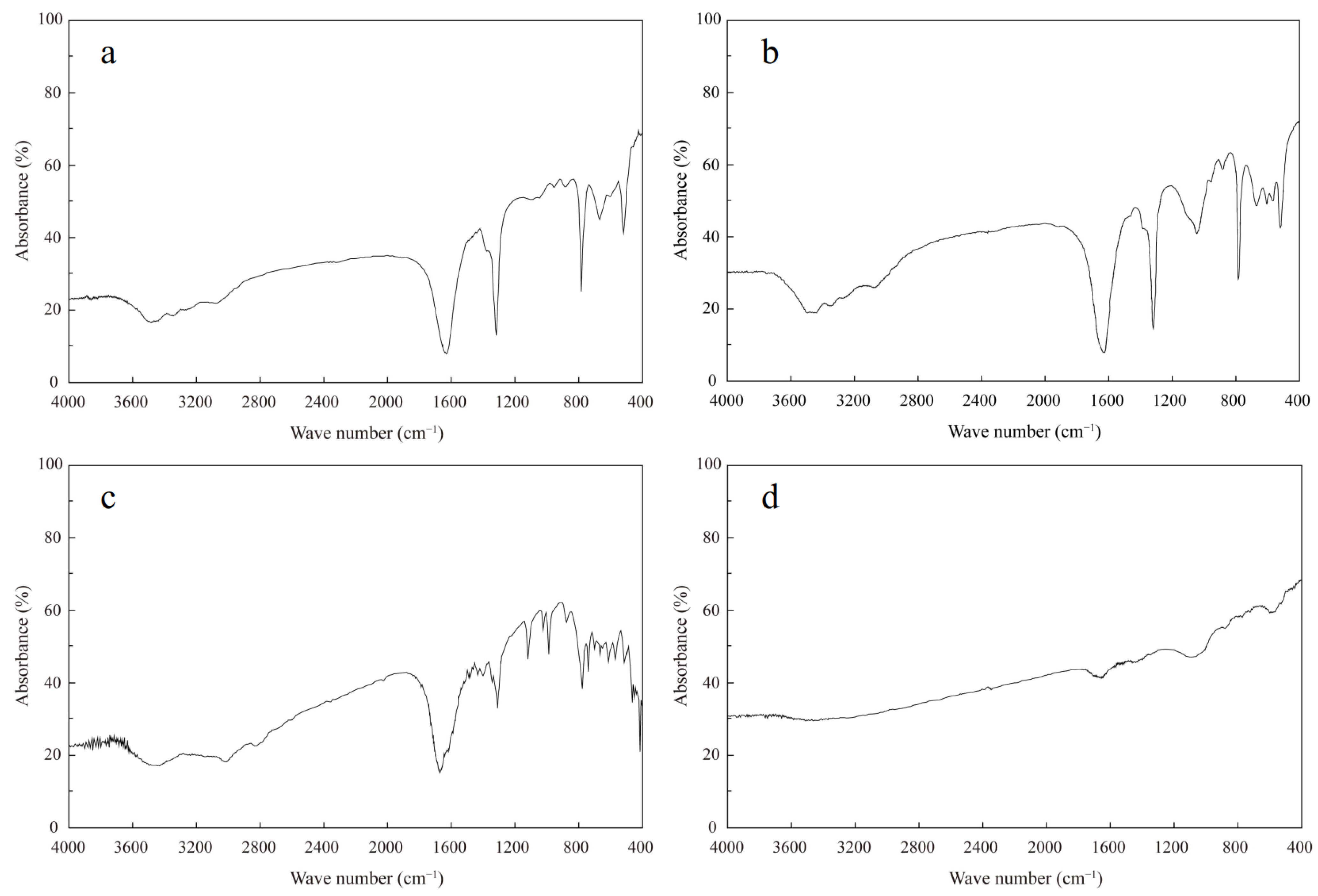
3.1. Mineralogical Compositions of Kidney Stones

Infrared spectroscopy is a simple and convenient method for analyzing the mineralogical composition of kidney stones [26,27,28]. Infrared spectra obtained by using an infrared spectrometer can identify the following crystalline compounds in collected kidney stones: calcium oxalate monohydrate (COM, CaC2O4·H2O), calcium oxalate dihydrate (COD, CaC2O4·2H2O), carbonate apatite (CA, Ca10(PO4)6CO3·H2O), uric acid (UA, C5H4N4O3), and ammonium acid urate (AAU, C5H7N5O3). For example, symmetric and asymmetric absorption bands, which appeared at 3480, 3423, and 3055 cm−1 possibly due to asymmetric O-H stretching, characterize the calcium oxalate monohydrate kidney stone [29]. C=O stretching contributes to a sharp absorption band at 1616 cm−1, whereas C-O stretching contributes to the band at ~1315 cm−1 [30]. Bands at ~780 and ~660 cm−1 are produced by C-H bending together with out-of-plane O-H bending [31]. Representative infrared spectra from this study are presented in Figure 1. Calcium oxalate monohydrate commonly mixes with calcium oxalate dihydrate kidney stones. As shown in Table 1, COM and COM + COD kidney stones were the dominant (74.2%, n = 49) types, carbonate apatite-type kidney stones were rare (3%, n = 2), and uric acid (n = 1) and ammonium acid urate (n = 1) kidney stones were less common than compared to oxalate types (COM and COD) and may only account for 3% of the kidney stones in this study. COD + Mix kidney stones (mainly COD with a small amount of observable mixture of carbonate apatite, ammonium acid urate, and uric acid) were also found in this study, with a frequency of 19.7% (n = 13, Table 1). Most of the elements of COD + Mix kidney stones exhibited lower concentrations when compared to COM and COM + COD kidney stones, which reflects potential differences in the formation processes of these kidney stones.

3.2. Chemical Compositions of Kidney Stones

Major and trace element compositions of kidney stones provide basic information on their bio-mineralization process. Concentrations of some alkali, alkaline earth, and transition elements together with phosphate, which is considered to be more environmentally significant, were determined. Chemical compositions of 66 kidney stone samples are summarized in Supplementary Materials Table S1. It should be noted that average concentrations of major and trace elements were ordered as follows: Ca > Mg > Na > K > Zn > Fe > Pb > Ba > Cu > Ti > Mo > Cd > Cr. Magnesium is an essential element in biological processes and acts as one of the most important low molecular inhibitors of stone formation in urine [13,32]. In this study, Mg appears to be the second-most abundant element with an average concentration of 2688.3 μg/g (range from 8.1 to 55,480.8 μg/g). Na concentrations ranged from 29.9 to 16,666.7 μg/g with an average concentration of 2436.0 μg/g. K concentrations varied from 21.9 to 25,475.6 μg/g with an average concentration of 880.0 μg/g. Fe concentrations varied from 0 to 271.0 μg/g with an average concentration of 23.3 μg/g. Iron in urine is commonly derived from human urinary channel cells and can increase in terms of percentage in the urinary system if it is allowed to be incorporated in calcium phosphate stones and oxalates [33]. Zn concentrations varied from 1.4 to 2678.0 μg/g with an average concentration of 345.7 μg/g. Mixed kidney stones made from a combination of oxalates and apatite showed higher Zn concentrations. Cu concentrations varied from 0 to 5.8 μg/g with an average concentration of 0.8 μg/g. Ba concentrations varied from 0.04 to15.4 μg/g with an average concentration of 2.5 μg/g. Concentrations of Pb, a toxic element, varied from 0 to 25.8 μg/g with an average concentration of 5.3 μg/g, which was significantly higher than concentrations observed in previous studies (0.8~2.6 μg/g) [2]. Concentrations of Ti, Cr, Mo, and Cd were relatively low with average concentrations of 0.67, 0.19, 0.44, and 0.30 μg/g, respectively. Compared with previous investigations from the Novosibirsk region in Russia (Ti = 3~3459 μg/g, Mo = 0.2~225.3 μg/g, Cd = 0.5~33.6 μg/g, Zn = 12.8~908.8 μg/g, Fe = 18.8~138.3 μg/g, and Cu = 5.2~34.1 μg/g) [34], concentrations of Ti, Mo, Cd, and Zn were higher in our study, while concentrations of Fe and Cu were relatively lower. In contrast, an appreciable amount of Sr was found in most urinary stones from the Novosibirsk region with concentrations of up to ~415 μg/g [34]. These results may reflect differences in the formation process and provenance of kidney stones in patients from different regions. Therefore, Sr concentrations in kidney stones should also be further studied in the Beijing area.
Kidney stones were classified according to the minerals identified by infrared spectroscopy. Composition statistics of major and trace elements are presented in Table 1. These results indicate an important role for these elements in bio-mineralization processes, such as combining with inorganic or organic parts of kidney stones. As shown in Table 1, significant differences were observed in element concentrations although the samples came from the same geological area (Beijing). Almost all major and trace elements, with the exception of Cu, were present in greater concentrations in calcium oxalate monohydrate (COM) than in other types of kidney stones. Generally, calcium oxalate dihydrate (COD) formation was associated with rapid CaOx crystallization due to hypercalciuria, while COM formation is a significantly longer process with a relatively slow formation rate that relies on several factors [13]. Therefore, more potential trace elements can be captured during COM formation, which results in higher trace element concentrations in COM. The arithmetic mean value of element concentrations was higher than the median value, which indicates that frequency distribution is asymmetric and skewed by high concentration values. This suggests different chemical compositions of kidney stones, which can be attributed to the environment or the composition of food and water ingested by the patient or accidentally ingested through the food chain [35,36]. It is worth noting that Zn concentrations in calcium oxalate were higher than other trace element concentrations.

3.3. Statistical Analysis and Source Identification

The relationship between major and trace elements in the pathogenesis of kidney stones is still under debate. However, previous studies have shown that the element composition of kidney stones can provide basic information about their sources [2,4].

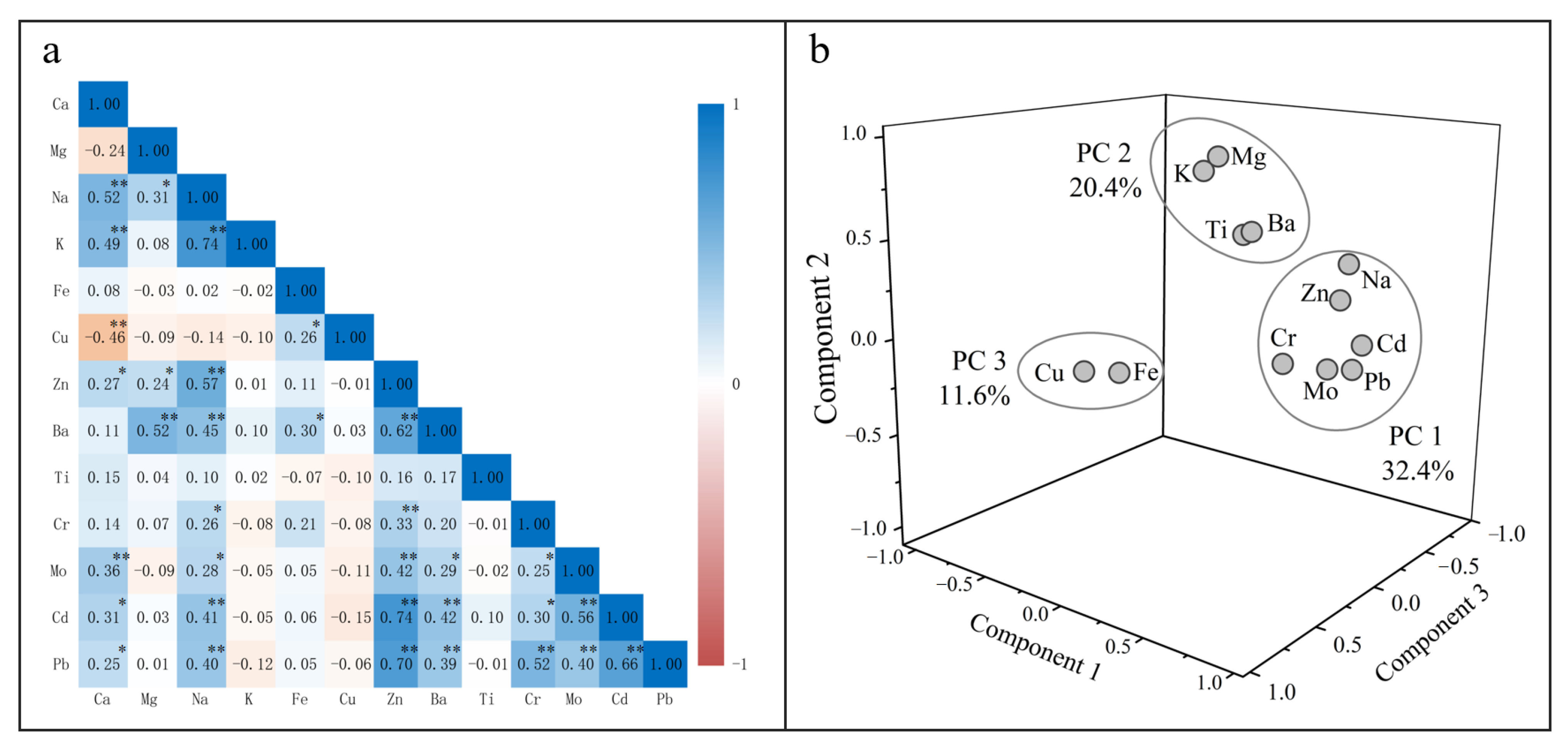
3.3.1. Correlation Analysis

In order to find possible connections and sources among elements in the kidney stone samples, a Pearson correlation coefficient was calculated for the analyzed elements (Figure 2a). Positive correlations were observed between Ca and Na (R = 0.52), Ca and K (R = 0.49), and Ca and Mo (R = 0.36), which suggests a potential thermodynamically favorable substitution pathway or a preference for uptake by the oxalate crystal structure of these elements (Na, K, and Mo) in the human body. Mg displays a strong positive correlation with trace element Ba (R = 0.52) and displays a weak negative correlation with Fe, Cu, and Mo. Previous studies have shown that Cu affects the crystallization rate and external morphology of growing crystals [37], while Fe and Cu affect calcium oxalate growth at very low concentrations [38]. A similar relationship was observed in different types of kidney stones (individual groups of kidney stones) where positive correlations between Ca and Na (R = 0.84), Ca and K (R = 0.92), and Mg and Ba (R = 0.60) were observed within COM-type kidney stones. Ba concentrations in calcium oxalate monohydrate (2.8 μg/g, Table 1) were higher than in calcium oxalate dihydrate and the mixture, which indicates a preference for Ba uptake by the oxalate structure during kidney stone crystallization processes. Among biochemical toxic elements, Pb concentrations were higher than other elements, whereas Cd concentrations were lower. Therefore, these trace elements in some kidney stone materials may be related to individual behavioral factors, such as food and water, rather than their geological background. This theory is also supported by the positive correlations between most trace elements (including Zn, Ba, Cd, and Pb) and Na (a major element in dietary intake) shown in Figure 2a. As a biomineral, kidney stones crystallize in the human body and are essential for discriminating dietary patterns, even though studies have not yet been widely reported. Further studies on the connection between the chemical composition of kidney stones and dietary intake are required. To date, as a typical chronic kidney disease, the occurrence of kidney stones is not only considered to be related to geographical distribution, geological environment, and occupational factors [39,40,41] but is also widely suspected to be controlled by long-term exposure to various nephrotoxic elements in drinking water, specifically groundwater sources with high alkalinity and hardness [42,43,44,45]. Water hardness is mainly controlled by Ca and Mg, and it is affected by various dissolved metallic ions [36]. Previous studies in regions with high incidences of chronic kidney disease have shown that the occurrence of kidney disease (including kidney stones) is positively correlated with hard water consumption and that more than 90% of patients have used extremely hard water as their main water source for at least five years [46,47]. Ingestion of hard water, together with other chemical components (such as Cd and As), produces various compounds that are harmful to human health and can cause further kidney stone formation [43,44]. In northern China, many people rely exclusively on groundwater as their main source of drinking water, which may be a factor that potentially affects kidney stone occurrences.

3.3.2. Principal Component Analysis

PCA exhibited varimax rotation, eigenvalues, and communality derivation of the chemical composition of human kidney stones. Prior to PCA analysis, each element index was normalized by removing the mean value and was divided by its standard deviation. The normalization process was applied to reduce discrepancies in different chemical index ranges. PCA results are displayed in Table 2 and Figure 2b. Three main principal components (PCs) with high eigenvalues, which accounted for 64.4% of the total variance, were extracted (all extracted eigenvalues were >1). The first principal component (PC1) accounted for 32.4% of the total variance. Table 2 and Figure 2b show the highest Na, Zn, Cr, Mo, Cd, and Pb positive loads and Cu negative load in PC1. The second principal component (PC2) accounted for ~20.4% of the total variance, with the highest positive load relative to Mg, K, Ba, and Ti. The negative load of Cu, Cr, Mo, and Pb was also present in PC2. The third principal component (PC3) accounted for 11.6% of the total variance, including the highest positive load of Fe and Cu. In summary, the three PCs can be explained by the potentially favorable substitution pathway or by a preference for the uptake process in the human body, which can reveal co-precipitation or substitution to a certain extent during kidney stone formation.

4. Conclusions

Chemical analyses showed that Ca is the major component of kidney stones. Infrared spectroscopy analysis revealed that kidney stones in the Beijing area are mineralogically whewellite (calcium oxalate), but they also contain phosphates, which indicates that kidney stones are mainly found in inorganic phases, such as Ca-oxalate and Ca-phosphates, with only a tiny portion exhibited in organic phases (e.g., uric acid stone). In addition, we have observed that whewellite-containing calcium kidney stones retain more trace metals than weddellite-containing calcium kidney stones, which is mainly a result of the duration of kidney stone formation.
During the formation of human urinary tract stones, Ca precipitates as oxalate in the urethra, while other alkali metals and alkaline earth metals are co-precipitated or replaced by Ca. This result is supported by a significant positive correlation between those elements. The formation process of kidney stones may be similar to the crystallization process of precipitated particles, such as pisoids and oncoids, which are triggered by material precipitation around one or more nuclei. Nevertheless, further research on drinking water chemistry and eating habits of kidney stone patients is also required in order to discriminate potential pathogenic factors of kidney stone biomineralization. Well-preserved component stratification that was observed in kidney stones implies that differences in kidney stone composition may be related to individual distinctions among diet, water intake, medication, or other supplementary intakes.

Supplementary Materials

The following data are available online at https://www.mdpi.com/article/10.3390/min11121396/s1, Table S1: major and trace constituents of all kidney stones and details of corresponding patients (gender and age), Figure S1: comparison of infrared spectra of kidney stones and mineral standards, Text S1: parameters of infrared spectroscopy.

Author Contributions

Conceptualization, Y.T. and G.H.; methodology, Y.T., G.H., J.Z., Q.Z., L.X., K.L., C.X. and L.M.; software, G.H. and J.Z.; formal analysis, G.H. and Q.Z.; investigation, G.H., L.X., K.L., C.X. and L.M.; resources, G.H.; data curation, G.H. and Q.Z.; writing—original draft, Y.T., G.H., J.Z. and Y.Z.; writing—review and editing, Y.T., G.H. and J.Z.; visualization, J.Z.; supervision, G.H.; funding acquisition, G.H. All authors have read and agreed to the published version of the manuscript.

Funding

This study was funded by the National Natural Science Foundation of China (No. 41325010).

Institutional Review Board Statement

The study was conducted according to the guidelines of the Declaration of Helsinki, and approved by the Ethics Committee of Peking University Third Hospital (protocol code (2021)MSREC 475-1, 8 November 2021).

Informed Consent Statement

Patient consent was waived due to the Informed consent exemption is included in the Ethics Review Approval Notice ((2021)MSREC 475-1).

Data Availability Statement

The data presented in this study are available upon request from the corresponding author.

Acknowledgments

The authors gratefully acknowledge Jinke Liu and Yikai Li from the China University of Geosciences for their laboratory work and Shitong Zhang for English editing.

Conflicts of Interest

The authors declare no conflict of interest.

References

  1. Gebauer, D.; Jansson, K.; Oliveberg, M.; Hedin, N. Indications that Amorphous Calcium Carbonates Occur in Pathological Mineralisation—A Urinary Stone from a Guinea Pig. Minerals 2018, 8, 84. [Google Scholar] [CrossRef] [Green Version]
  2. Chandrajith, R.; Weerasingha, A.; Premaratne, K.M.; Gamage, D.; Abeygunasekera, A.M.; Joachimski, M.M.; Senaratne, A. Mineralogical, compositional and isotope characterization of human kidney stones (urolithiasis) in a Sri Lankan population. Environ. Geochem. Health 2019, 41, 1881–1894. [Google Scholar] [CrossRef] [PubMed]
  3. Izatulina, A.R.; Gurzhiy, V.V.; Krzhizhanovskaya, M.G.; Chukanov, N.V.; Panikorovskii, T.L. Thermal Behavior and Phase Transition of Uric Acid and Its Dihydrate Form, the Common Biominerals Uricite and Tinnunculite. Minerals 2019, 9, 373. [Google Scholar] [CrossRef] [Green Version]
  4. Chandrajith, R.; Wijewardana, G.; Dissanayake, C.B.; Abeygunasekara, A. Biomineralogy of human urinary calculi (kidney stones) from some geographic regions of Sri Lanka. Environ. Geochem. Health 2006, 28, 393–399. [Google Scholar] [CrossRef]
  5. Wrobel, A.; Rokita, E.; Taton, G.; Thor, P. Chemical composition and morphology of renal stones. Folia Med. Crac. 2013, 53, 5–15. [Google Scholar]
  6. Heilberg, I.P.; Schor, N. Renal stone disease: Causes, evaluation and medical treatment. Arq. Bras. Endocrinol. Metabol. 2006, 50, 823–831. [Google Scholar] [CrossRef] [PubMed] [Green Version]
  7. Krouse, H.R.; Levinson, A.A.; Piggott, D.; Ueda, A. Further stable isotope investigations of human urinary stones: Comparison with other body components. Appl. Geochem. 1987, 2, 205–211. [Google Scholar] [CrossRef]
  8. Reynolds, T.M. ACP Best Practice No 181: Chemical pathology clinical investigation and management of nephrolithiasis. J. Clin. Pathol. 2005, 58, 134–140. [Google Scholar] [CrossRef] [PubMed]
  9. Parks, J.H.; Worcester, E.M.; Coe, F.L.; Evan, A.P.; Lingeman, J.E. Clinical implications of abundant calcium phosphatein routinely analyzed kidney stones. Kidney Int. 2004, 66, 777–785. [Google Scholar] [CrossRef] [PubMed] [Green Version]
  10. Blaschko, S.D.; Miller, J.; Chi, T.; Flechner, L.; Fakra, S.; Kahn, A.; Kapahi, P.; Stoller, M.L. Microcomposition of Human Urinary Calculi Using Advanced Imaging Techniques. J. Urol. 2013, 189, 726–734. [Google Scholar] [CrossRef] [PubMed]
  11. Han, G.; Tang, Y.; Liu, M.; Van Zwieten, L.; Yang, X.; Yu, C.; Wang, H.; Song, Z. Carbon-nitrogen isotope coupling of soil organic matter in a karst region under land use change, Southwest China. Agric. Ecosyst. Environ. 2020, 301, 107027. [Google Scholar] [CrossRef]
  12. Abeywickarama, B.; Ralapanawa, U.; Chandrajith, R. Geoenvironmental factors related to high incidence of human urinary calculi (kidney stones) in Central Highlands of Sri Lanka. Environ. Geochem. Health 2016, 38, 1203–1214. [Google Scholar] [CrossRef] [PubMed]
  13. Kustov, A.V.; Strelnikov, A.I. Quantitative Mineralogical Composition of Calculi and Urine Abnormalities for Calcium Oxalate Stone Formers: A Single-Center Results. Urol. J. 2018, 15, 87–91. [Google Scholar] [CrossRef]
  14. Cloutier, J.; Villa, L.; Traxer, O.; Daudon, M. Kidney stone analysis: “Give me your stone, I will tell you who you are!”. World J. Urol. 2015, 33, 157–169. [Google Scholar] [CrossRef] [PubMed] [Green Version]
  15. Kustov, A.V.; Berezin, B.D.; Strel’nikov, A.I.; Shevyrin, A.A.; Trostin, V.N. Interaction of a complexing agent with urolith as the basis for efficient little-invasive therapy of phosphaturia. Dokl. Phys. Chem. 2009, 428, 175–177. [Google Scholar] [CrossRef]
  16. Kustov, A.V.; Berezin, B.D.; Trostin, V.N. The Complexon−Renal Stone Interaction: Solubility and Electronic Microscopy Studies. J. Phys. Chem. B 2009, 113, 9547–9550. [Google Scholar] [CrossRef]
  17. Kustov, A.V.; Shevyrin, A.A.; Strel’nikov, A.I.; Smirnov, P.R.; Trostin, V.N. Chemolysis of calcium oxalate stones: Study in vitro and possible clinical application. Urol. Res. 2012, 40, 205–209. [Google Scholar] [CrossRef] [PubMed]
  18. Fraser, I.; Meier-Augenstein, W.; Kalin, R.M. The role of stable isotopes in human identification: A longitudinal study into the variability of isotopic signals in human hair and nails. Rapid Commun. Mass Spectrom. 2006, 20, 1109–1116. [Google Scholar] [CrossRef]
  19. Chen, L.; Wang, J.; Beiyuan, J.; Guo, X.; Wu, H.; Fang, L. Environmental and health risk assessment of potentially toxic trace elements in soils near uranium (U) mines: A global meta-analysis. Sci. Total Environ. 2021, 151556, in press. [Google Scholar] [CrossRef]
  20. Hareendra, P.P.G.K.; Hunais, M.M.; Suvendiran, S.; Palihakkara, S.D.; Abeygunasekera, A.M. Chemical composition of kidney stones obtained from a cohort of Sri Lankan patients. Sri Lanka J. Surg. 2015, 33, 14–19. [Google Scholar] [CrossRef]
  21. Li, X.; Han, G. One-step chromatographic purification of K, Ca, and Sr from geological samples for high precision stable and radiogenic isotope analysis by MC-ICP-MS. J. Anal. At. Spectrom. 2021, 36, 676–684. [Google Scholar] [CrossRef]
  22. Zeng, J.; Han, G.; Yang, K. Assessment and sources of heavy metals in suspended particulate matter in a tropical catchment, northeast Thailand. J. Clean. Prod. 2020, 265, 121898. [Google Scholar] [CrossRef]
  23. Li, X.; Han, G.; Liu, M.; Liu, J.; Zhang, Q.; Qu, R. Potassium and its isotope behaviour during chemical weathering in a tropical catchment affected by evaporite dissolution. Geochim. Cosmochim. Acta 2022, 316, 105–121. [Google Scholar] [CrossRef]
  24. Zeng, J.; Han, G. Preliminary copper isotope study on particulate matter in Zhujiang River, southwest China: Application for source identification. Ecotoxicol. Environ. Saf. 2020, 198, 110663. [Google Scholar] [CrossRef] [PubMed]
  25. Zeng, J.; Han, G. Tracing zinc sources with Zn isotope of fluvial suspended particulate matter in Zhujiang River, southwest China. Ecol. Indic. 2020, 118, 106723. [Google Scholar] [CrossRef]
  26. Chatterjee, P.; Chakraborty, A.; Mukherjee, A.K. Phase composition and morphological characterization of human kidney stones using IR spectroscopy, scanning electron microscopy and X-ray Rietveld analysis. Spectrochim. Acta Part A Mol. Biomol. Spectrosc. 2018, 200, 33–42. [Google Scholar] [CrossRef] [PubMed]
  27. Bhatt, P.A.; Paul, P. Analysis of urinary stone constituents using powder X-ray diffraction and FT-IR. J. Chem. Sci. 2008, 120, 267–273. [Google Scholar] [CrossRef]
  28. Afaj, A.H.; Sultan, M.A. Mineralogical Composition of the Urinary Stones from Different Provinces in Iraq. Sci. World J. 2005, 5, 792841. [Google Scholar] [CrossRef] [PubMed] [Green Version]
  29. Matsuzaki, S.; Matsushita, K.; Tanikawa, K.; Masuda, A.; Matsunaga, J. Sequential analysis of recurrent calcium calculi by infrared spectroscopy. Int. J. Urol. 1995, 2, 235–237. [Google Scholar] [CrossRef] [PubMed]
  30. Sekkoum, K.; Cheriti, A.; Taleb, S.; Belboukhari, N. FTIR spectroscopic study of human urinary stones from El Bayadh district (Algeria). Arab. J. Chem. 2016, 9, 330–334. [Google Scholar] [CrossRef] [Green Version]
  31. Levinson, A.A.; Mino, M.P.Y.; Stams, U.K.; Hariharan, A. The mineralogy of human urinary stones from Calgary, Quito and Honolulu. Am. Mineral. 1985, 70, 630–635. [Google Scholar]
  32. Ries, J.B. Review: Geological and experimental evidence for secular variation in seawater Mg/Ca (calcite-aragonite seas) and its effects on marine biological calcification. Biogeosciences 2010, 7, 2795–2849. [Google Scholar] [CrossRef] [Green Version]
  33. Wandt, M.A.E.; Underhill, L.G. Covariance Biplot Analysis of Trace Element Concentrations in Urinary Stones. J. Urol. 1989, 141, 1275. [Google Scholar] [CrossRef]
  34. Pal’chik, N.A.; Moroz, T.N.; Maksimova, N.V.; Dar’in, A.V. Mineral and microelement compositions of urinary stones. Russ. J. Inorg. Chem. 2006, 51, 1098–1105. [Google Scholar] [CrossRef]
  35. Abboud, I.A. Mineralogy and chemistry of urinary stones: Patients from North Jordan. Environ. Geochem. Health 2008, 30, 445–463. [Google Scholar] [CrossRef]
  36. Xu, S.; Li, S.-L.; Yue, F.; Udeshani, C.; Chandrajith, R. Natural and Anthropogenic Controls of Groundwater Quality in Sri Lanka: Implications for Chronic Kidney Disease of Unknown Etiology (CKDu). Water 2021, 13, 2724. [Google Scholar] [CrossRef]
  37. Muñoz, J.A.; Valiente, M. Effects of trace metals on the inhibition of calcium oxalate crystallization. Urol. Res. 2005, 33, 267–272. [Google Scholar] [CrossRef]
  38. Bazin, D.; Chevallier, P.; Matzen, G.; Jungers, P.; Daudon, M. Heavy elements in urinary stones. Urol. Res. 2007, 35, 179–184. [Google Scholar] [CrossRef]
  39. Jayatilake, N.; Mendis, S.; Maheepala, P.; Mehta, F.R. Chronic kidney disease of uncertain aetiology: Prevalence and causative factors in a developing country. BMC Nephrol. 2013, 14, 180. [Google Scholar] [CrossRef] [Green Version]
  40. Vlahos, P.; Schensul, S.L.; Nanayakkara, N.; Chandrajith, R.; Haider, L.; Anand, S.; Silva, K.T.; Schensul, J.J. Kidney progression project (KiPP): Protocol for a longitudinal cohort study of progression in chronic kidney disease of unknown etiology in Sri Lanka. Glob. Public Health 2019, 14, 214–226. [Google Scholar] [CrossRef]
  41. Rango, T.; Jeuland, M.; Manthrithilake, H.; McCornick, P. Nephrotoxic contaminants in drinking water and urine, and chronic kidney disease in rural Sri Lanka. Sci. Total Environ. 2015, 518–519, 574–585. [Google Scholar] [CrossRef] [PubMed] [Green Version]
  42. Chandrajith, R.; Nanayakkara, S.; Itai, K.; Aturaliya, T.N.C.; Dissanayake, C.B.; Abeysekera, T.; Harada, K.; Watanabe, T.; Koizumi, A. Chronic kidney diseases of uncertain etiology (CKDue) in Sri Lanka: Geographic distribution and environmental implications. Environ. Geochem. Health 2011, 33, 267–278. [Google Scholar] [CrossRef] [PubMed]
  43. Balasooriya, S.; Munasinghe, H.; Herath, A.T.; Diyabalanage, S.; Ileperuma, O.A.; Manthrithilake, H.; Daniel, C.; Amann, K.; Zwiener, C.; Barth, J.A.C.; et al. Possible links between groundwater geochemistry and chronic kidney disease of unknown etiology (CKDu): An investigation from the Ginnoruwa region in Sri Lanka. Expo. Health 2020, 12, 823–834. [Google Scholar] [CrossRef]
  44. Wickramarathna, S.; Balasooriya, S.; Diyabalanage, S.; Chandrajith, R. Tracing environmental aetiological factors of chronic kidney diseases in the dry zone of Sri Lanka—A hydrogeochemical and isotope approach. J. Trace Elem. Med. Biol. 2017, 44, 298–306. [Google Scholar] [CrossRef] [PubMed]
  45. Nikagolla, C.; Meredith, K.T.; Dawes, L.A.; Banati, R.B.; Millar, G.J. Using water quality and isotope studies to inform research in chronic kidney disease of unknown aetiology endemic areas in Sri Lanka. Sci. Total Environ. 2020, 745, 140896. [Google Scholar] [CrossRef]
  46. Wasana, H.M.S.; Aluthpatabendi, D.; Kularatne, W.M.T.D.; Wijekoon, P.; Weerasooriya, R.; Bandara, J. Drinking water quality and chronic kidney disease of unknown etiology (CKDu): Synergic effects of fluoride, cadmium and hardness of water. Environ. Geochem. Health 2016, 38, 157–168. [Google Scholar] [CrossRef] [PubMed]
  47. Jayasumana, C.; Gunatilake, S.; Senanayake, P. Glyphosate, Hard Water and Nephrotoxic Metals: Are They the Culprits behind the Epidemic of Chronic Kidney Disease of Unknown Etiology in Sri Lanka? Int. J. Environ. Res. Public Health 2014, 11, 2125–2147. [Google Scholar] [CrossRef] [PubMed] [Green Version]
Figure 1. Infrared spectra of typical kidney stones that contain the following: (a) calcium oxalate monohydrate + calcium oxalate dihydrate; (b) calcium oxalate dihydrate + calcium oxalate monohydrate + carbonate apatite (a few); (c) calcium oxalate dihydrate + uric acid + calcium oxalate monohydrate (a few); (d) ammonium acid urate.
Minerals 11 01396 g001
Figure 2. Correlation analysis (a) and principal component analysis (b) of the chemical components of kidney stones. ** p < 0.01; * p < 0.05.
Minerals 11 01396 g002
Table 1. Statistical results of major and trace constituents in different kidney stone types.
Kidney Stone TypeParametersCaMgNaKFeCuZnBaTiCrMoCdPb
%μg/gμg/gμg/gμg/gμg/gμg/gμg/gng/gng/gng/gng/gμg/g
COM (n = 26)Min20.598.229.988.80.00.019.90.20.00.068.122.00.0
Max54.413,499.916,666.725,475.6271.05.52678.013.520,694.4215.11391.11524.925.8
Mean28.01880.43168.11563.228.00.7506.22.81192.118.1461.5384.86.6
Median27.31147.62462.8400.314.80.3345.01.5306.20.0371.8273.16.2
COM + COD (n = 23)Min24.2135.1578.164.04.40.112.50.10.00.0112.558.40.0
Max28.34469.24765.7830.2191.41.9873.115.3845.14247.41264.5780.124.0
Mean26.4831.21817.7373.323.30.5269.92.3221.8359.1502.0307.66.1
Median26.1355.21405.7362.313.90.479.70.587.30.0494.1176.82.7
COD + Mix
(n = 13)
Min0.38.1108.821.95.20.11.40.10.00.085.511.40.0
Max28.02753.53251.6768.679.32.3508.12.21625.91043.4515.8477.84.8
Mean18.9743.81436.1254.417.01.0149.30.8347.391.9315.8164.71.6
Median23.7228.81158.4221.310.80.624.00.555.00.0297.698.01.0
AAU (n = 1) 9.055,480.84417.02576.31.80.51466.115.41969.60.093.296.91.63
CA-1 (n = 1) 30.73568.56685.11697.615.30.13578.08.121576.90.01043.3566.312.59
CA-2 (n = 1) 14.840,219.16010.6915.630.80.13461.24.911283.82883.1376.080.30.64
UA (n = 1) 0.3481.2813.2351.73.35.752.30.040.00.0143.59.50.00
Note: COM = calcium oxalate monohydrate, COD = calcium oxalate dihydrate, Mix = mixture, AAU = ammonium acid urate, CA = carbonate apatite, UA = uric acid.
Table 2. Varimax rotation component matrix of the chemical composition of kidney stones.
VariablePC1PC2PC3
Eigenvalues3.892.441.39
Variance (%)32.420.411.6
Mg0.020.840.06
Na0.800.47−0.04
K0.010.89−0.07
Fe0.130.010.79
Cu−0.15−0.060.72
Zn0.840.320.07
Ba0.500.630.31
Ti0.000.46−0.30
Cr0.57−0.030.14
Mo0.67−0.09−0.10
Cd0.850.06−0.11
Pb0.87−0.030.00
Publisher’s Note: MDPI stays neutral with regard to jurisdictional claims in published maps and institutional affiliations.

