Odikhinchaite, Na9Sr3[(H2O)2Na]Ca6Mn3Zr3NbSi (Si24O72)O(OH)3(CO3)·H2O, a New Eudialyte-Group Mineral from the Odikhincha Intrusion, Taimyr Peninsula, Russia


 ,
,  ,
,
Abstract
1. Introduction
2. Geological Setting
3. Materials and Methods
4. Results
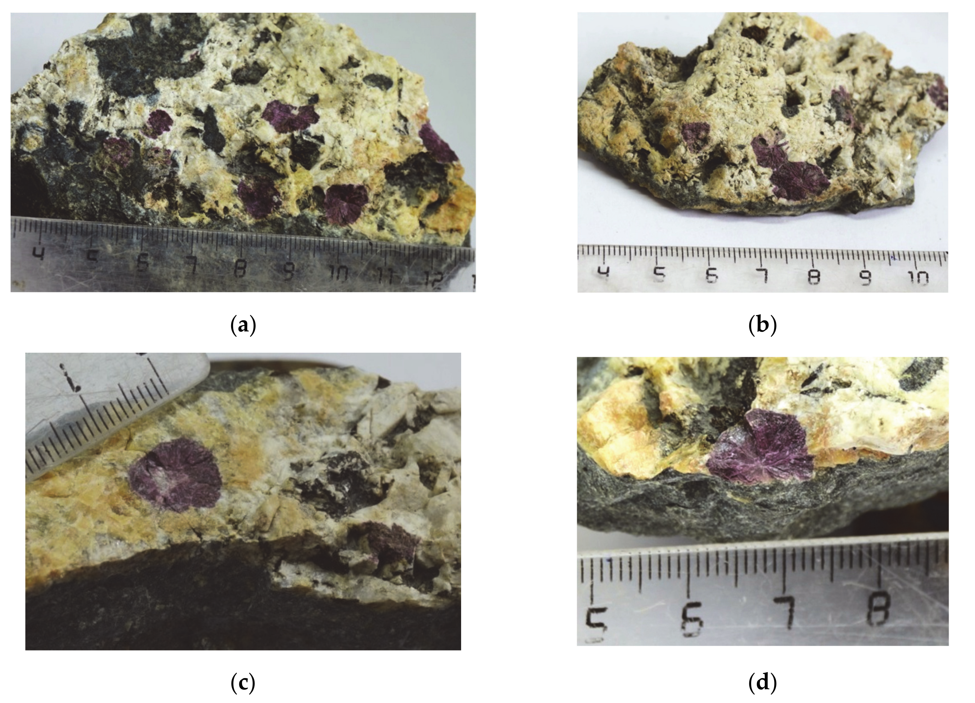
4.1. Physical Properties
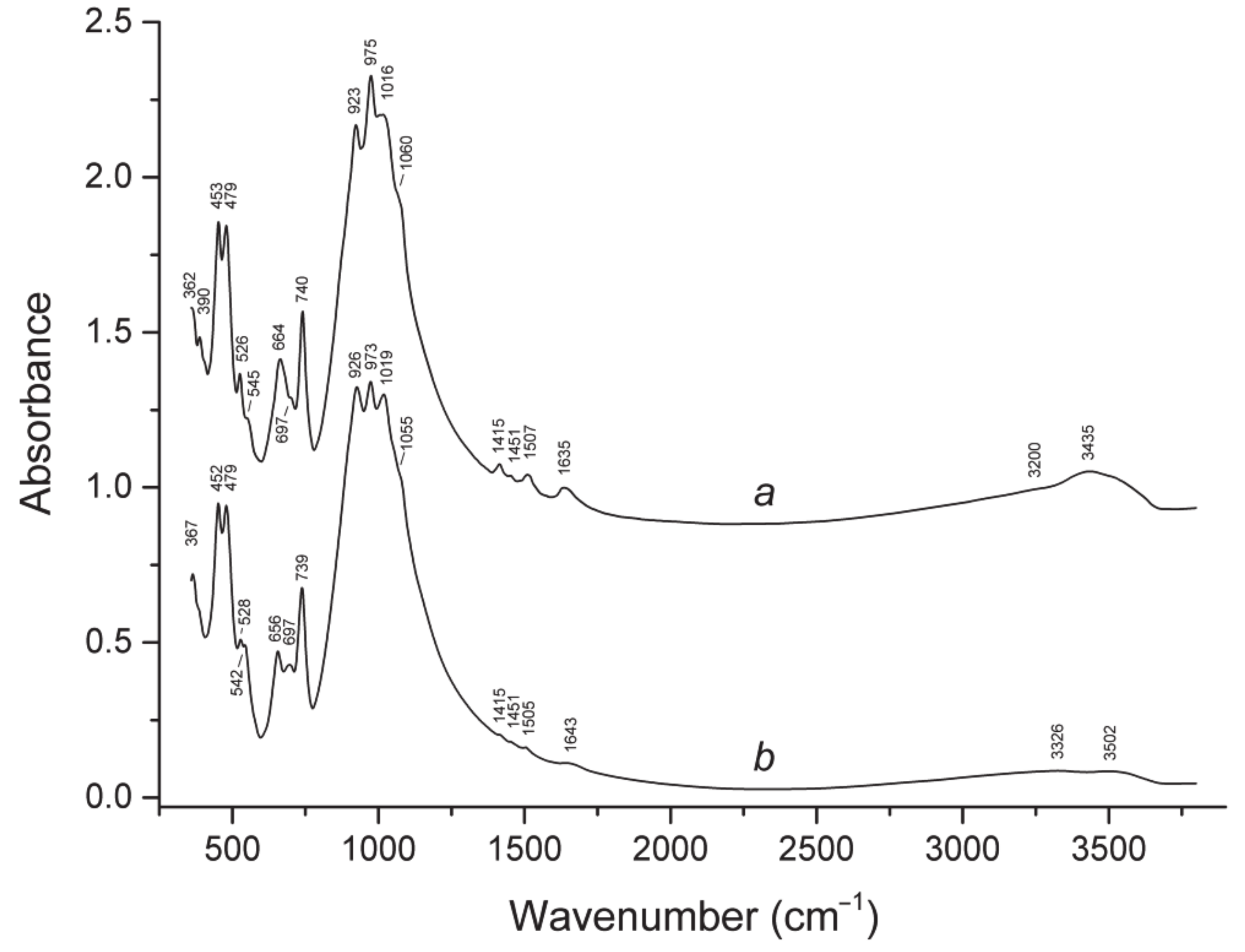
4.2. Infrared Spectroscopy
4.3. Optical Properties
4.4. Chemical Composition
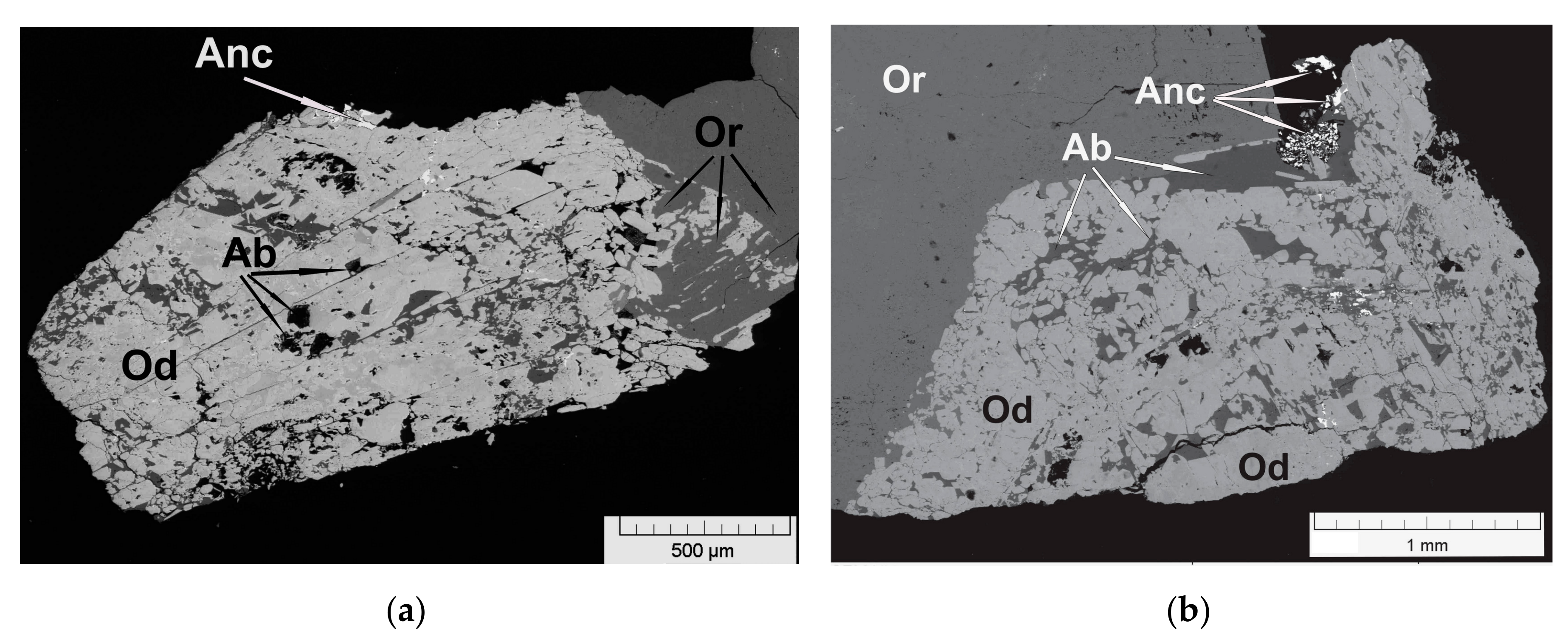
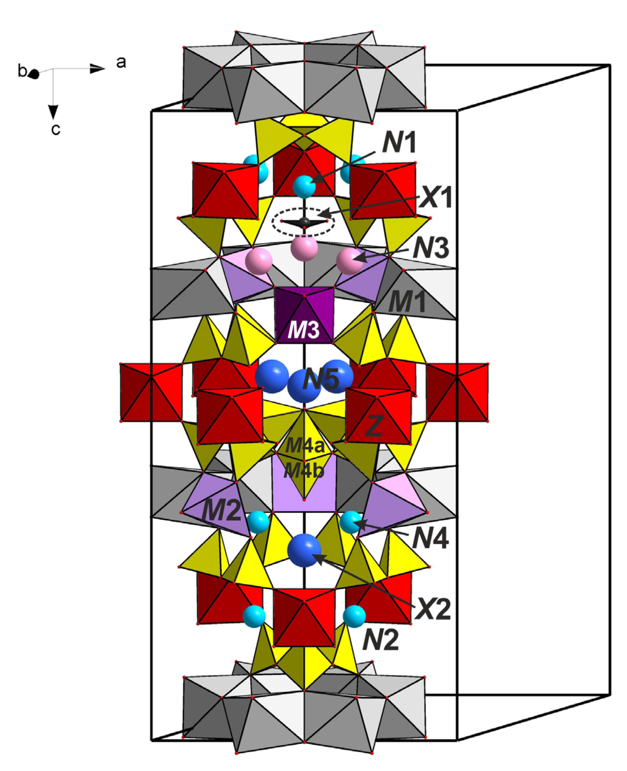
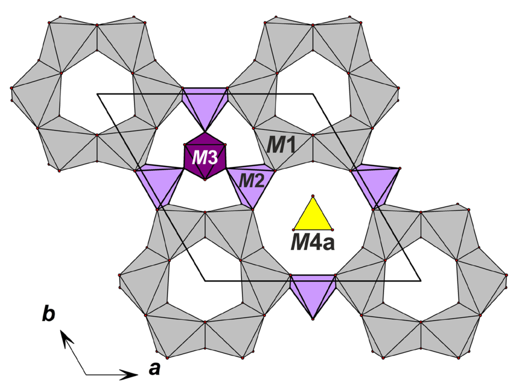
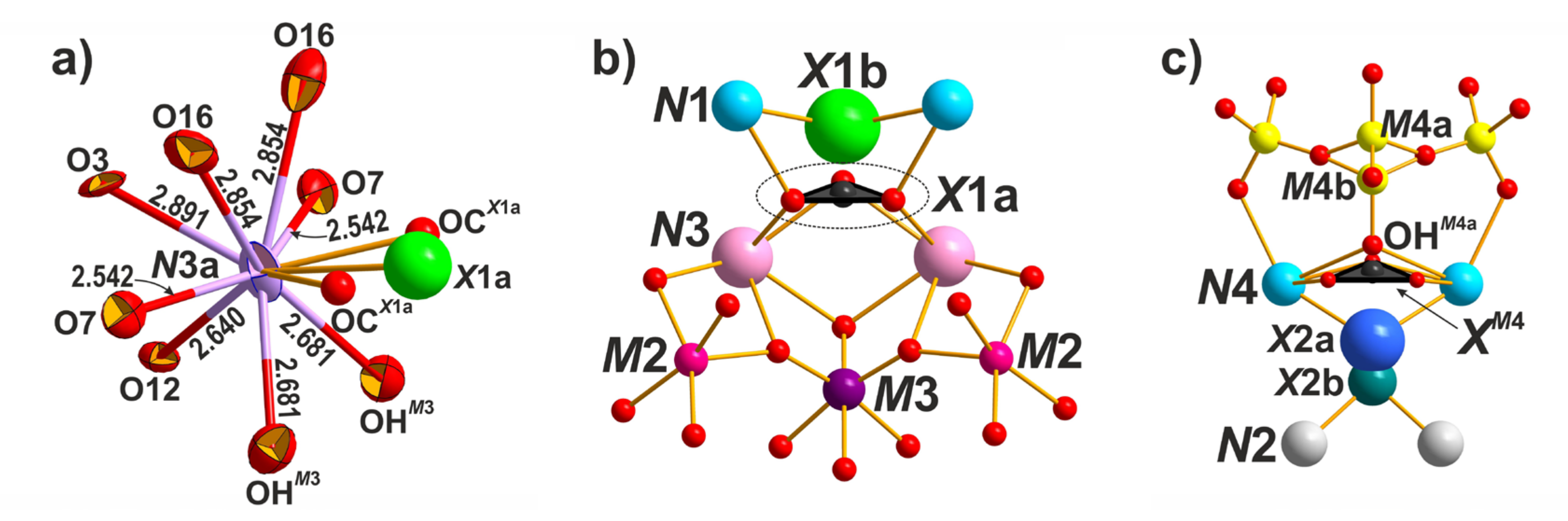
4.5. X-Ray Diffraction Data and Crystal Structure
5. Discussion
Author Contributions
Funding
Acknowledgments
Conflicts of Interest
References
- Sjöqvist, A.S.L. The tale of greenlandite: Commemorating the two-hundredth anniversary of eudialyte (1819–2019). Minerals 2019, 9, 497–510. [Google Scholar] [CrossRef]
- Rastsvetaeva, R.K.; Chukanov, N.V.; Aksenov, S.M. Minerals of Eudialyte Group: Crystal Chemistry, Properties, Genesis; University of Nizhniy Novgorod: Nizhniy Novgorod, Russia, 2012; p. 230. ISBN 9785913262073. (In Russian) [Google Scholar]
- Johnsen, O.; Grice, J.D.; Gault, R.A. The crystal chemistry of the eudialyte group. Can. Mineral. 1999, 37, 865–891. [Google Scholar]
- Johnsen, O.; Ferraris, G.; Gault, R.A.; Grice, J.D.; Kampf, A.R.; Pekov, I.V. The nomenclature of eudialyte group minerals. Can. Mineral. 2003, 41, 785–794. [Google Scholar] [CrossRef]
- Rastsvetaeva, R.K.; Khomyakov, A.P. Crystal chemistry of modular eudialytes. Crystallogr. Rep. 2003, 48, S78–S90. [Google Scholar]
- Rastsvetaeva, R.K.; Chukanov, N.V. Classification of eudialyte-group minerals. Geol. Ore Depos. 2012, 54, 487–497. [Google Scholar] [CrossRef]
- Rastsvetaeva, R.K.; Chukanov, N.V.; Pekov, I.V.; Schäfer, C.; Van, K.V. New data on the isomorphism in eudialyte-group minerals. 1. Crystal chemistry of eudialyte-group members with Na incorporated into the framework as a marker of hyperagpaitic conditions. Minerals 2020, 10, 587. [Google Scholar] [CrossRef]
- Rastsvetaeva, R.K.; Chukanov, N.V. New data on the isomorphism in eudialyte-group minerals. 2. Crystal-chemical mechanisms of blocky isomorphism at the key sites (a review). Minerals 2020, 10, 720. [Google Scholar] [CrossRef]
- Egorov, L.S. Iiolite-Carbonatite Pluton (For Example Maimeche-Kotuy Province Polar Siberia); Nedra: Moscow, Russia, 1991; p. 260. (In Russian) [Google Scholar]
- Rastsvetaeva, R.K.; Chukanov, N.V.; Zaitsev, В.А.; Aksenov, S.M.; Viktorova, K.A. Crystal structure of Cl-deficiente analog of tasekite from the Odihincha massif. Crystallogr. Rep. 2018, 63, 392–400. [Google Scholar] [CrossRef]
- Britvin, S.N.; Dolivo-Dobrovolsky, D.V.; Krzhizhanovskaya, M.G. Software for processing the X-ray powder diffraction data obtained from the curved image plate detector of Rigaku RAXIS Rapid II diffractometer. Zap. Ross. Mineral. Obs. (Proc. Russ. Mineral. Soc.) 2017, 146, 104–107. (In Russian) [Google Scholar]
- Oxford Diffraction. CrysAlisPro; Oxford Diffraction Ltd.: Abingdon, UK, 2009. [Google Scholar]
- Petřiček, V.; Dušek, M.; Palatinus, L. Structure Determination Software Programs; Institute of Physics: Praha, Czech Republic, 2006. [Google Scholar]
- Prince, E. (Ed.) International Tables for Crystallography, Volume C: Mathematical, Physical and Chemical Tables, 3rd ed.; Kluwer Academic Publishers: Dordrecht, The Netherlands, 2004. [Google Scholar]
- Brandenburg, K.; Putz, H. DIAMOND Version 3; Crystal Impact GbR: Bonn, Germany, 2005. [Google Scholar]
- Brown, I.D.; Altermatt, D. Bond-valence parameters obtained from a systematic analysis of the Inorganic Crystal Structure Database. Acta Crystallogr. Sect. B 1985, 41, 244–247. [Google Scholar] [CrossRef]
- Brese, N.E.; O’Keeffe, M. Bond-valence parameters for solids. Acta Crystallogr. Sect. B 1991, 47, 192–197. [Google Scholar] [CrossRef]
- Petersen, O.V.; Johnsen, O.; Gault, R.A.; Niederayr, G.; Grice, A.D. Taseqite, a new member of the eudialyte group from the Ilímaussaq alkaline complex, South Greenland. Neues Jahrb. Mineral. Mon. 2004, 2, 83–96. [Google Scholar] [CrossRef]
- Khomyakov, A.P.; Dusmatov, V.D.; Ferraris, G.; Gula, A.; Ivaldi, G.; Nechelyustov, G.N. Zirsilite-(Ce), (Na,⎕)12(Ce,Na)3Ca6Mn3Zr3Nb(Si25O73)(OH)3(CO3)·H2O, and carbokentbrooksite, (Na,⎕)12(Na,Ce)3Ca6Mn3Zr3Nb(Si25O73)(OH)3(CO3)·H2O, two new eudialyte-group minerals from the Dara-i-Pioz alkaline massif, Tajikistan. Zap. Vserossiyskogo Mineral. Obs. (Proc. Russ. Mineral. Soc.) 2003, 132, 40–51. (In Russian) [Google Scholar]
- Johnsen, O.; Gault, R.A.; Grice, J.D.; Ercit, T.S. Khomyakovite and manganokhomyakovite, two new members of the eudialyte group, from Mont Saint-Hilaire, Quebec. Can. Mineral. 1999, 37, 893–899. [Google Scholar]


| Crystal Data | |
| Simplified formula | Na9Sr3[(H2O)2Na]Ca6Mn3Zr3NbSi(Si24O72)O(OH)3(CO3)·H2O |
| Formula weight, Mr (g) | 3270.52 |
| Crystal system, space group | Trigonal, R3m (#143) |
| Temperature (K) | 293 |
| a, c (Å) | 14.2837(2), 30.0697(3) |
| V (Å3) | 5313.04(12) |
| Z | 3 |
| Radiation type; λ | MoKα; 0.71073 |
| Absorption coefficient, μ (mm−1) | 3.91 |
| Crystal size (mm) | 0.3 × 0.1 × 0.05 |
| Data Collection | |
| Diffractometer | Xcalibur, Sapphire3 (CCD detector) |
| Absorption correction | Multi scan |
| Tmin, Tmax | 0.773; 1 |
| No. of measured, independent and observed [I > 2σ(I)] reflections | 27,254, 2345, 2312 |
| Rint | 0.053 |
| (sin θ/λ)max (Å−1) | 0.759 |
| Refinement | |
| Refinement on | Full-matrix least squares on F |
| R[I > 2σ(I)], wR(I), S | 0.031, 0.043, 1.63 |
| Weight scheme | 1/(σ2|F| + 0.0004F2) |
| No. of refinement parameters (Npar) | 265 |
| Nref/Npar | 8.73 |
| Δρmax, Δρmin (e Å–3) | −1.25/1.10 |
| Constituent | Mean Content, wt% | Range | Standard Deviation | Probe Standard |
|---|---|---|---|---|
| Na2О | 9.26 | 8.97–9.68 | 0.29 | Albite |
| K2О | 0.59 | 0.41–0.76 | 0.24 | Orthoclase |
| CaО | 12.77 | 12.42–13.58 | 0.15 | Wollastonite |
| MnО | 5.49 | 5.21–6.49 | 0.18 | Mn |
| FeО | 0.75 | 0–1.26 | 0.13 | Fe |
| MgО | 0.24 | 0.08–0.35 | 0.14 | MgO |
| SrО | 5.81 | 4.55–6.63 | 0.09 | SrF2 |
| La2О3 | 0.38 | 0.11–0.63 | 0.12 | LaPO4 |
| Ce2О3 | 0.39 | 0–1.41 | 0.15 | CePO4 |
| Nd2O3 | 0.15 | 0–0.42 | 0.21 | NdPO4 |
| Al2O3 | 0.07 | 0–0.17 | 0.11 | Albite |
| SiО2 | 44.80 | 44.12–45.74 | 0.74 | SiO2 |
| ZrО2 | 11.13 | 10.82–11.72 | 0.19 | Zr |
| TiО2 | 0.07 | 0–0.41 | 0.14 | Ti |
| Nb2О5 | 4.17 | 3.21–4.74 | 0.21 | Nb |
| Cl | 0.69 | 0.61–0.83 | 0.02 | NaCl |
| CО2 | 0.90 | - | - | - |
| Н2О | 2.22 | - | - | - |
| –O = Cl | –0.16 | - | - | - |
| Total | 99.72 | - | - | - |
| Iobs | dobs | Icalc * | dcalc ** | h k l | Iobs | dobs | Icalc * | dcalc ** | h k l |
|---|---|---|---|---|---|---|---|---|---|
| 64 | 11.42 | 100 | 11.439 | 101 | 17 | 2.138 | 4 4 8 8 | 2.144 2.141 2.138 2.131 | 057 0.3.12 3.1.11 514 |
| 9 | 10.01 | 12 | 10.023 | 003 | 2 | 2.085 | 3 | 2.084 | 155 |
| 36 | 9.54 | 51 | 9.552 | 012 | 7 | 2.118 | 6 | 2.116 | 0.1.14 |
| 24 | 7.13 | 15 | 7.142 | 110 | 3 | 2.098 | 4 | 2.100 | 149 |
| 38 | 6.42 | 23 | 6.424 | 104 | 9 | 2.062 | 6 | 2.064 2.062 | 3.2.10 600 |
| 24 | 6.05 | 22 | 6.058 | 021 | 8 | 2.055 | 5 2 1 | 2.053 2.051 2.048 | 247 2.2.12 0.4.11 |
| 39 | 5.71 | 26 | 5.720 | 202 | 4 | 2.027 | 5 | 2.029 | 431 |
| 3 | 5.42 | 1 | 5.409 | 015 | 6 | 2.019 | 5 3 | 2.019 2.015 | 603 342 |
| 6 | 5.01 | 6 | 5.012 | 006 | 19 | 1.984 | 17 3 | 1.985 1.981 | 428 520 |
| 3 | 4.774 | 2 | 4.776 | 024 | 3 | 1.952 | 4 | 1.952 | 1.2.14 |
| 3 | 4.611 | 3 | 4.620 | 211 | 9 | 1.938 | 2 9 | 1.943 1.939 | 253 339 |
| 41 | 4.309 | 22 | 4.312 | 205 | 6 | 1.918 | 5 1 | 1.918 1.913 | 1.3.13 158 |
| 19 | 4.100 | 13 | 4.102 | 116 | 13 | 1.907 | 1 12 | 1.910 1.907 | 0.5.10 606 |
| 25 | 3.967 | 22 | 3.970 | 214 | 3 | 1.881 | 2 | 1.883 | 161 |
| 19 | 3.810 | 11 | 3.813 | 303 | 3 | 1.872 | 2 | 1.872 | 612 |
| 7 | 3.689 | 7 | 3.691 | 125 | 4 | 1.856 | 2 1 | 1.858 1.852 | 1.0.16 4.0.13 |
| 28 | 3.567 | 7 19 | 3.596 3.571 | 018 220 | 20 | 1.839 | 5 9 5 6 | 1.846 1.842 1.838 1.836 | 2.4.10 526 437 1.4.12 |
| 30 | 3.529 | 19 | 3.528 | 027 | 8 | 1.798 | 3 4 2 2 | 1.803 1.800 1.798 1.793 | 0.3.15 615 0.2.16 3.2.13 |
| 53 | 3.405 | 46 | 3.409 | 131 | 27 | 1.785 | 2 4 22 4 | 1.788 1.787 1.785 1.777 | 348 5.1.10 440 4.2.11 |
| 15 | 3.344 | 9 1 | 3.345 3.341 | 312 009 | 13 | 1.764 | 13 | 1.764 | 701 |
| 45 | 3.208 | 37 23 | 3.212 3.184 | 208 036 | 6 | 1.749 | 1 1 4 | 1.755 1.755 1.748 | 072 609 2.2.15 |
| 44 | 3.167 | 36 | 3.163 | 217 | 7 | 1.722 | 1 3 6 | 1.727 1.726 1.720 | 167 3.3.12 704 |
| 3 | 3.121 | 6 | 3.121 | 134 | 8 | 1.713 | 5 | 1.713 | 621 |
| 31 | 3.025 | 12 16 | 3.029 3.026 | 042 119 | 7 | 1.704 | 5 1 1 | 1.704 1.704 1.701 | 262 259 2.0.17 |
| 100 | 2.978 | 91 | 2.980 | 315 | 12 | 1.682 | 1 4 9 | 1.686 1.685 1.682 | 618 4.3.10 446 |
| 21 | 2.908 | 18 | 2.908 | 226 | 2 | 1.670 | 1 | 1.671 | 0.0.18 |
| 86 | 2.858 | 74 | 2.860 | 404 | 10 | 1.647 | 5 3 4 | 1.650 1.648 1.644 | 265 1.3.16 2.4.13 |
| 5 | 2.787 | 7 | 2.789 | 232 | 5 | 1.627 | 2 4 1 | 1.631 1.627 1.622 | 3.4.11 1.1.18 5.0.14 |
| 3 | 2.747 | 4 | 2.750 | 045 | 14 | 1.606 | 2 13 | 1.609 1.606 | 4.1.15 4.0.16 |
| 19 | 2.699 | 11 7 8 | 2.704 2.699 2.681 | 0.2.10 410 137 | 10 | 1.596 | 3 6 4 1 | 1.599 1.598 1.593 1.592 | 538 1.6.10 627 0.6.12 |
| 12 | 2.654 | 11 | 2.655 | 324 | 4 | 1.581 | 5 3 1 | 1.582 1.575 1.575 | 541 452 449 |
| 30 | 2.595 | 3 32 | 2.607 2.596 | 143 309 | 7 | 1.567 | 6 | 1.567 | 3.2.16 |
| 13 | 2.530 | 8 7 | 2.534 2.529 | 318 2.1.10 | 9 | 1.552 | 4 2 5 1 3 | 1.558 1.557 1.554 1.553 1.550 | 630 176 2.5.12 6.1.11 544 |
| 7 | 2.508 | 5 3 | 2.510 2.506 | 407 0.0.12 | 8 | 1.546 | 1 6 1 | 1.548 1.544 1.540 | 0.3.18 081 363 |
| 8 | 2.464 | 7 | 2.466 | 051 | 3 | 1.527 | 3 1 | 1.527 1.524 | 4.3.13 3.5.10 |
| 5 | 2.439 | 1 3 | 2.441 2.440 | 502 229 | 1 | 1.513 | 1 1 | 1.514 1.513 | 084 2.2.18 |
| 19 | 2.378 | 5 9 9 | 2.388 2.380 2.377 | 048 330 416 | 3 | 1.497 | 2 1 | 1.497 1.493 | 0.5.16 0.1.20 |
| 13 | 2.329 | 13 | 2.331 | 241 | 11 | 1.484 | 2 8 2 4 | 1.486 1.484 1.482 1.477 | 547 0.7.11 274 3.4.14 |
| 6 | 2.264 | 3 2 | 2.265 2.261 | 238 1.3.10 | 5 | 1.465 | 2 3 2 | 1.466 1.465 1.460 | 725 2.4.16 458 |
| 3 | 2.213 | 4 | 2.216 | 511 | 2 | 1.454 | 1 1 | 1.454 1.453 | 4.4.12 2.6.11 |
| 7 | 2.196 | 7 | 2.198 | 152 | 5 | 1.437 | 3 1 1 | 1.437 1.437 1.435 | 0.6.15 1.3.19 5.1.16 |
| 6 | 2.178 | 5 | 2.179 | 425 | 9 | 1.430 | 1 2 1 5 1 | 1.432 1.431 1.430 1.428 1.426 | 0.0.21 1.2.20 808 550 277 |
| 16 | 2.154 | 10 2 | 2.156 2.150 | 4.0.10 336 | 5 | 1.417 | 1 5 2 | 1.420 1.417 1.413 | 4.1.18 461 642 |
| Site | x | y | z | Uiso*/Ueq | Mult | Occupancy | BVS |
|---|---|---|---|---|---|---|---|
| Z | 0.16245(2) | 0.32489(5) | 0.13811(4) | 0.0066(2) | 9 | Zr | 4.01 |
| M1 | −0.00293(8) | 0.2588(1) | 0.30584(5) | 0.0089(3) | 18 | Ca | 2.05 |
| Si1 | 0.07001(7) | 0.1400(1) | 0.22528(7) | 0.0077(5) | 9 | Si | 3.98 |
| Si2 | −0.07891(14) | 0.46054(7) | 0.05013(7) | 0.0074(5) | 9 | Si | 3.92 |
| Si3 | −0.05666(10) | 0.2693(1) | 0.06888(6) | 0.0071(4) | 18 | Si | 3.97 |
| Si4 | 0.26280(7) | 0.5256(1) | 0.05556(7) | 0.0072(5) | 9 | Si | 4.11 |
| Si5 | 0.41983(15) | 0.20992(8) | 0.23062(7) | 0.0098(5) | 9 | Si | 4.18 |
| Si6 | 0.39110(10) | 0.3957(1) | 0.20748(5) | 0.0078(4) | 18 | Si | 4.11 |
| O1 | −0.1813(2) | 0.1813(2) | 0.0858(2) | 0.0106(14) | 9 | O | - |
| O2 | 0.0245(3) | 0.2497(3) | 0.0990(1) | 0.0151(13) | 18 | O | - |
| O3 | 0.1174(2) | 0.2349(4) | 0.2615(2) | 0.0127(15) | 9 | O | - |
| O4 | 0.2335(2) | 0.4671(5) | 0.1031(2) | 0.0199(18) | 9 | O | - |
| O5 | 0.3578(4) | 0.1789(2) | 0.2768(2) | 0.0137(16) | 9 | O | - |
| O6 | −0.2081(4) | 0.3960(2) | 0.0555(2) | 0.0148(15) | 9 | O | - |
| O7 | 0.3757(3) | 0.4102(3) | 0.2595(1) | 0.0139(13) | 18 | O | - |
| O8 | 0.2099(4) | 0.6050(2) | 0.0542(2) | 0.0123(14) | 8 | O | - |
| O9 | 0.3876(4) | 0.2816(3) | 0.1985(1) | 0.0206(16) | 18 | O | - |
| O10 | 0.0905(2) | 0.1809(4) | 0.1746(2) | 0.0159(16) | 9 | O | - |
| O11 | −0.0472(3) | 0.2548(3) | 0.0167(1) | 0.0153(13) | 18 | O | - |
| O12 | −0.0340(4) | 0.4830(2) | 0.0003(2) | 0.0094(14) | 9 | O | - |
| O13 | −0.0338(3) | 0.3921(3) | 0.0792(1) | 0.0112(12) | 18 | O | - |
| O14 | 0.5067(2) | 0.4933(2) | 0.1892(2) | 0.0130(15) | 9 | O | - |
| O15 | −0.0616(2) | 0.0616(2) | 0.2322(2) | 0.0137(15) | 9 | O | - |
| O16 | 0.2981(3) | 0.3975(3) | 0.1793(2) | 0.0194(14) | 18 | O | - |
| O17 | 0.2234(2) | 0.4468(5) | 0.0149(2) | 0.0179(18) | 9 | O | - |
| O18 | 0.5462(5) | 0.2731(2) | 0.2387(2) | 0.036(2) | 9 | O | - |
| M2 | 0.4832(1) | 0.5168(1) | 0.3077(5) | 0.0122(3) | 9 | Mn0.83Fe0.17 | 2.10 |
| M3 | 1/3 | 2/3 | 0.34260(5) | 0.0114(2) | 3 | Nb | 5.06 |
| OHM3 | 0.3981(2) | 0.6019(2) | 0.3092(2) | 0.0162(15) | 9 | (OH)0.607O0.393 | - |
| M4a | 0 | 0 | 0.8834(1) | 0.0093(8) | 3 | Si0.82 | - |
| OHM4a | 0 | 0 | 0.8303(4) | 0.016(3) | 3 | (OH)0.82 | - |
| M4b | 0 | 0 | 0.9192(7) | 0.026(6) | 3 | Si0.18 | - |
| OHM4b | 0 | 0 | 0.975(2) | 0.0612(6) * | 3 | (OH)0.18 | - |
| N1 | 0.4366(2) | 0.5634(5) | 0.1300(1) | 0.0299(9) | 9 | Na0.79Ca0.17(H2O)0.04 | - |
| N2a | −0.1093(3) | 0.1093(3) | 0.1525(2) | 0.0415(17) | 9 | Na0.86 | - |
| N2b | −0.0782(7) | 0.0782(7) | 0.1343(6) | 0.033(4) | 9 | Ca0.14 | - |
| N3a | 0.23412(1) | 0.46830(7) | 0.25630(4) | 0.0167(2) | 9 | Sr0.667K0.15Ln0.067 | - |
| N3b | 0.2357(14) | 0.471(3) | 0.2267(12) | 0.031(7) * | 9 | Na0.116 | - |
| N4 | 0.1018(4) | 0.2036(8) | 0.0182(2) | 0.084(5) | 9 | Na | - |
| N5 | −0.2630(2) | 0.2630(2) | 0.1180(2) | 0.037(2) | 9 | (H2O)0.6Na0.4 | - |
| X1a | 1/3 | 2/3 | 0.2027(3) | 0.0237(17) | 3 | C0.53Cl0.35 | - |
| OCX1a | 0.3827(3) | 0.6173(3) | 0.2125(8) | 0.091(8) * | 9 | O0.53 | - |
| X1b | 1/3 | 2/3 | 0.1541(13) | 0.039(7) * | 3 | Cl0.12 | - |
| X2a | 0 | 0 | 0.0556(12) | 0.056(10) | 3 | (H2O)0.6 | - |
| X2b | 0 | 0 | 0.0898(18) | 0.064(12) * | 3 | (OH,F)0.4 | - |
| CXM4 | 0 | 0 | 0.997(8) | 0.057(12) * | C0.15 | - | |
| OCXM4 | −0.0509(2) | 0.0509(2) | 0.999(3) | 0.08(2) * | O0.15 | - |
| Bond | Distance | Bond | Distance | ||
|---|---|---|---|---|---|
| Z | O4 | 2.050(6) | N1 | O14 | 2.484(6) |
| O2 × 2 | 2.074(5) | O8 | 2.500(7) | ||
| O16 × 2 | 2.086(4) | O4 | 2.639(7) | ||
| O10 | 2.093(5) | O4 | 2.641(4) | ||
| - | 2.077 | O16 × 2 | 2.652(4) | ||
| M1 | O17 | 2.287(5) | X1b | 2.655(11) | |
| O3 | 2.332(5) | OCX1a | 2.816(22) | ||
| O5 | 2.348(5) | O13 × 2 | 2.823(5) | ||
| O11 | 2.378(5) | - | 2.651 | ||
| O7 | 2.397(4) | N2a | O2 × 2 | 2.536(6) | |
| O12 | 2.464(5) | O10 | 2.590(7) | ||
| - | 2.368 | O10 | 2.591(5) | ||
| Si1 | O3 | 1.601(5) | O15 | 2.671(8) | |
| O10 | 1.605(6) | O1 | 2.683(7) | ||
| O15 × 2 | 1.651(4) | O9 × 2 | 2.802(7) | ||
| - | 1.627 | - | 2.651 | ||
| Si2 | O12 | 1.598(5) | N1b | 0.945(16) | |
| O6 | 1.606(5) | N2b | X2b | 2.352(34) | |
| O13 | 1.663(5) | O2 × 2 | 2.385(11) | ||
| O13 | 1.663(4) | O10 | 2.427(12) | ||
| - | 1.633 | O10 | 2.428(12) | ||
| Si3 | O11 | 1.599(4) | O1 | 2.938(15) | |
| O2 | 1.603(5) | O15 | 2.973(19) | ||
| O13 | 1.645(5) | X2a | 3.057(32) | ||
| O1 | 1.665(3) | - | 2.618 | ||
| - | 1.628 | N3a | OCX1a × 2 | 2.500(13) | |
| Si4 | O17 | 1.565(6) | O7 × 2 | 2.542(4) | |
| O4 | 1.602(6) | O12 | 2.639(5) | ||
| O8 | 1.647(5) | OHM3 × 2 | 2.681(5) | ||
| O8 | 1.648(6) | O16 × 2 | 2.854(5) | ||
| - | 1.616 | O3 | 2.891(5) | ||
| Si5 | O18 | 1.582(7) | X1a | 2.936(5) | |
| O5 | 1.587(6) | - | 2.693 | ||
| O9 | 1.633(6) | N3b | 0.891(36) | ||
| O9 | 1.633(5) | N3b | OCX1a | 2.138(25) | |
| - | 1.609 | OCX1a | 2.141(44) | ||
| Si6 | O16 | 1.588(5) | O16 | 2.206(34) | |
| O7 | 1.608(4) | O16 | 2.210(27) | ||
| O9 | 1.627(5) | X1a | 2.526(37) | ||
| O14 | 1.635(3) | O7 | 2.732(37) | ||
| - | 1.615 | O7 | 2.737(24) | ||
| - | - | - | 2.384 | ||
| M2 | OHM3 | 2.105(4) | N4 | OCXM4 × 2 | 2.256(24) |
| O7 × 2 | 2.106(4) | O11 × 2 | 2.574(11) | ||
| O11 × 2 | 2.113(4) | O5 | 2.597(9) | ||
| - | 2.109 | X2a | 2.758(17) | ||
| M3 | OHM3 × 3 | 1.891(4) | OHM4b | 2.835(9) | |
| O6 × 3 | 2.081(6) | O2 × 2 | 2.878(9) | ||
| - | 1.986 | O17 | 3.010(12) | ||
| M4a | OHM4a | 1.598(13) | - | 2.662 | |
| O18 × 3 | 1.630(4) | N5 | OHM4a | 2.217(9) | |
| - | 1.622 | O1 | 2.241(6) | ||
| M4b | 1.076(22) | O6 × 2 | 2.503(6) | ||
| M4b | OH18 × 3 | 1.547(8) | N5 × 2 | 3.014(5) | |
| OM4b | 1.675(21) | O13 × 2 | 3.072(5) | ||
| - | 1.579 | - | 2.705 | ||
| X1a | OCX1a × 3 | 1.256(7) | XM4 | OCXM4 × 3 | 1.260(5) |
| X1b | 1.461(40) | OHM4b | 0.676(12) | ||
| Mineral | Odikhinchaite | Taseqite | Kentbrook- Site | Carbokent- Brooksite | Zirsilite-(Ce) |
| Formula | Na9Sr3[(H2O)2Na] Ca6Mn3Zr3NbSi (Si24O72)O(OH)3 (CO3)·H2O | Na12Sr3Ca6Fe2+3 Zr3NbSi(Si24O72) (O,OH,H2O)4Cl2 | Na15Ca6Mn3 Zr3NbSi (Si24O72)O2 F2·2H2O | (Na,⎕,REE)15Ca6 Mn3Zr3NbSi (Si24O72)O(OH)3 (CO3)·H2O | (Na,⎕)12(Ce,Na)3 Ca6Mn3Zr3Nb Si(Si24O72)O (OH)3(CO3)·H2O |
| N3 site M2 site | Sr Mn2+ | Sr Fe2+ | Na Mn2+ | Na Mn2+ | Ce3+ Mn2+ |
| a, Å c, Å V, Å3 | 14.2179 30.349 5313.1 | 14.2828 30.0222 5303.9 | 14.1686 30.0847 5260 | 14.239 30.039 5274 | 14.248 30.076 5288 |
| Optical data | Uniaxial (–) ω = 1.638 ε = 1.630 | Uniaxial (–) ω = 1.6494 ε = 1.6378 | Uniaxial (–) ω = 1.628 ε = 1.623 | Uniaxial (–) ω = 1.645 ε = 1.635 | Uniaxial (–) ω = 1.648 ε = 1.637 |
| Density, g·cm–3 | 2.97 (measured) 3.04 (calculated) | 3.24 (measured) 3.20 (calculated) | 3.10 (measured) 3.08 (calculated) | 3.14 (measured) 3.10 (calculated) | 3.15 (measured) 3.10 (calculated) |
Publisher’s Note: MDPI stays neutral with regard to jurisdictional claims in published maps and institutional affiliations. |
© 2020 by the authors. Licensee MDPI, Basel, Switzerland. This article is an open access article distributed under the terms and conditions of the Creative Commons Attribution (CC BY) license (http://creativecommons.org/licenses/by/4.0/).
Share and Cite
Gritsenko, Y.D.; Chukanov, N.V.; Aksenov, S.M.; Pekov, I.V.; Varlamov, D.A.; Pautov, L.A.; Vozchikova, S.A.; Ksenofontov, D.A.; Britvin, S.N. Odikhinchaite, Na9Sr3[(H2O)2Na]Ca6Mn3Zr3NbSi (Si24O72)O(OH)3(CO3)·H2O, a New Eudialyte-Group Mineral from the Odikhincha Intrusion, Taimyr Peninsula, Russia. Minerals 2020, 10, 1062. https://doi.org/10.3390/min10121062
Gritsenko YD, Chukanov NV, Aksenov SM, Pekov IV, Varlamov DA, Pautov LA, Vozchikova SA, Ksenofontov DA, Britvin SN. Odikhinchaite, Na9Sr3[(H2O)2Na]Ca6Mn3Zr3NbSi (Si24O72)O(OH)3(CO3)·H2O, a New Eudialyte-Group Mineral from the Odikhincha Intrusion, Taimyr Peninsula, Russia. Minerals. 2020; 10(12):1062. https://doi.org/10.3390/min10121062
Chicago/Turabian StyleGritsenko, Yuliya D., Nikita V. Chukanov, Sergey M. Aksenov, Igor V. Pekov, Dmitry A. Varlamov, Leonid A. Pautov, Svetlana A. Vozchikova, Dmitry A. Ksenofontov, and Sergey N. Britvin. 2020. "Odikhinchaite, Na9Sr3[(H2O)2Na]Ca6Mn3Zr3NbSi (Si24O72)O(OH)3(CO3)·H2O, a New Eudialyte-Group Mineral from the Odikhincha Intrusion, Taimyr Peninsula, Russia" Minerals 10, no. 12: 1062. https://doi.org/10.3390/min10121062
APA StyleGritsenko, Y. D., Chukanov, N. V., Aksenov, S. M., Pekov, I. V., Varlamov, D. A., Pautov, L. A., Vozchikova, S. A., Ksenofontov, D. A., & Britvin, S. N. (2020). Odikhinchaite, Na9Sr3[(H2O)2Na]Ca6Mn3Zr3NbSi (Si24O72)O(OH)3(CO3)·H2O, a New Eudialyte-Group Mineral from the Odikhincha Intrusion, Taimyr Peninsula, Russia. Minerals, 10(12), 1062. https://doi.org/10.3390/min10121062