Crystal Structure Refinements of Four Monazite Samples from Different Localities


Abstract
1. Introduction
2. Experimental Methods
2.1. Sample Description
2.2. Electron-Probe Microanalysis (EPMA)
2.3. Single-Crystal X-ray Diffraction (SCXRD)
2.4. Structure Refinements of SCXRD Data
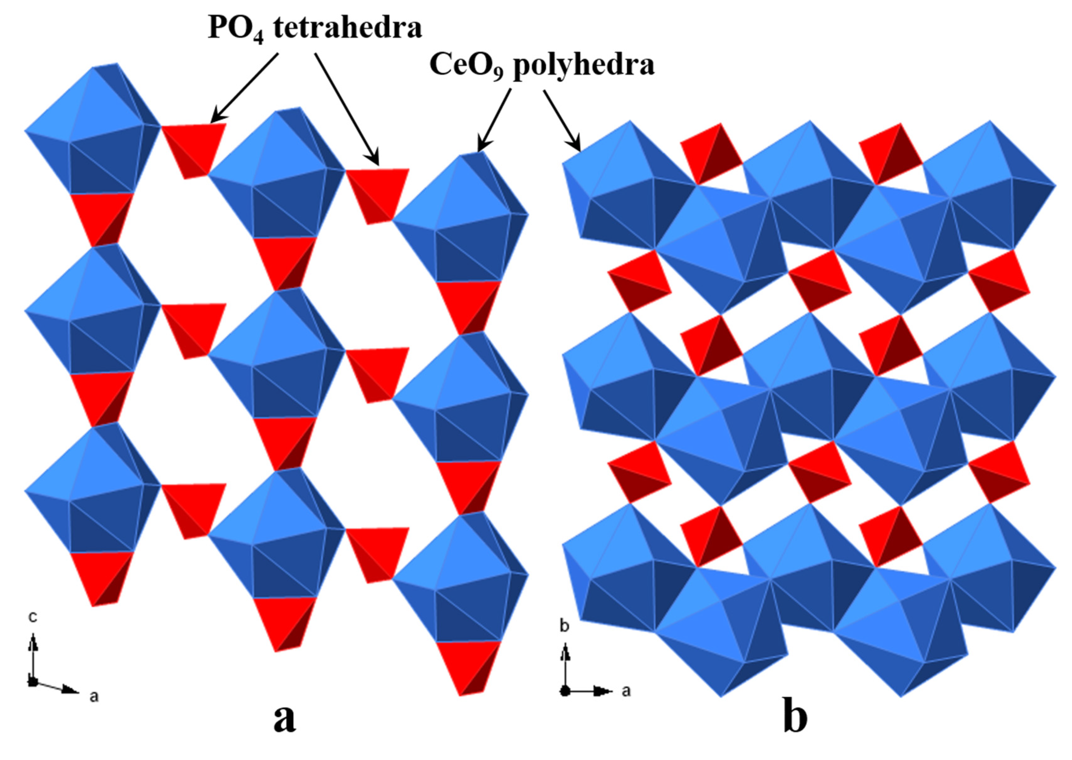
3. Results
3.1. Chemical Composition of Monazite-Ce and Sm-Rich Monazite
- 1:
- (Ce0.40La0.20Nd0.17Ca0.08Th0.06Pr0.04Sm0.02Gd0.01Y0.01)Ʃ0.99(P0.96S0.03Si0.02)Ʃ1.01O4,
- 2:
- (Ce0.33Nd0.22La0.11Y0.08Th0.08Sm0.06Pr0.05Gd0.03Dy0.01Ca0.01)Ʃ0.98(P0.92Si0.08)Ʃ1.00O4,
- 3:
- (Ce0.37La0.17Nd0.16Th0.09Y0.07Pr0.04Ca0.04Sm0.03Gd0.02Dy0.01)Ʃ1.00(P0.95Si0.06)Ʃ1.01O4, and
- 4:
- (Sm0.19Ce0.19Th0.17Ca0.13Nd0.10Gd0.08La0.06Pr0.03Y0.02Pb0.01)Ʃ0.98(P0.96Si0.05)Ʃ1.01O4.
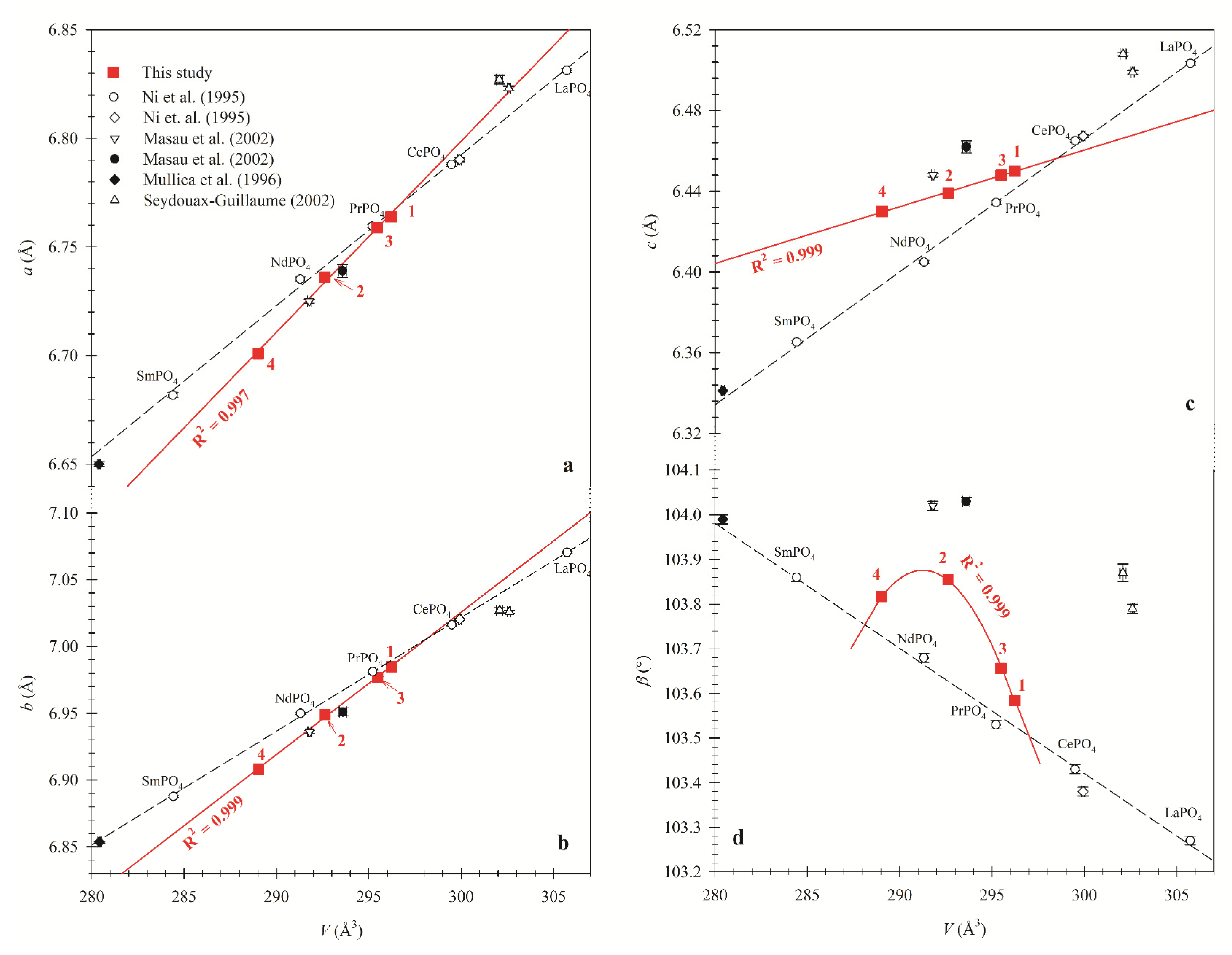
3.2. Variations of Unit-Cell Parameters
3.3. Site Occupancy Factor (sof) and Chemical Composition
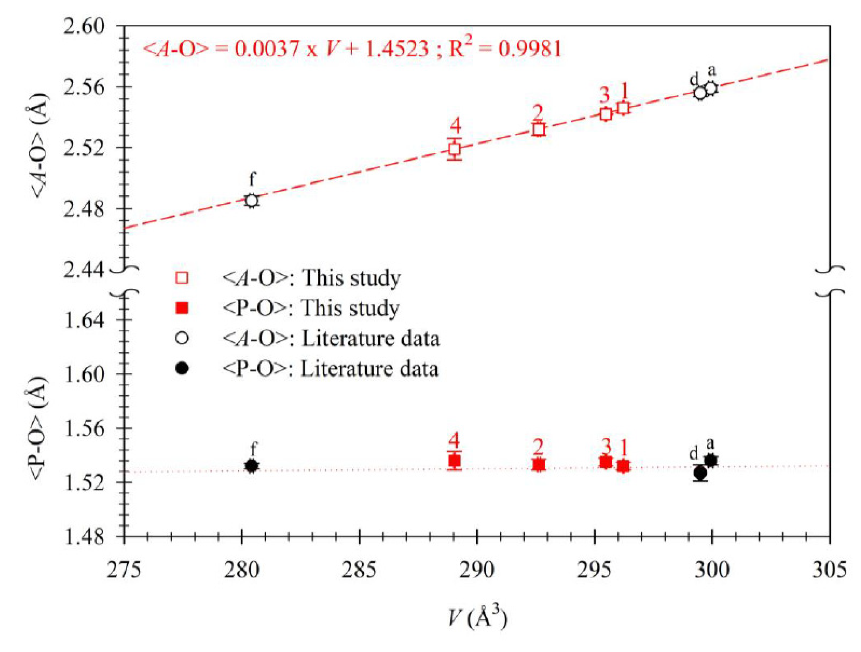
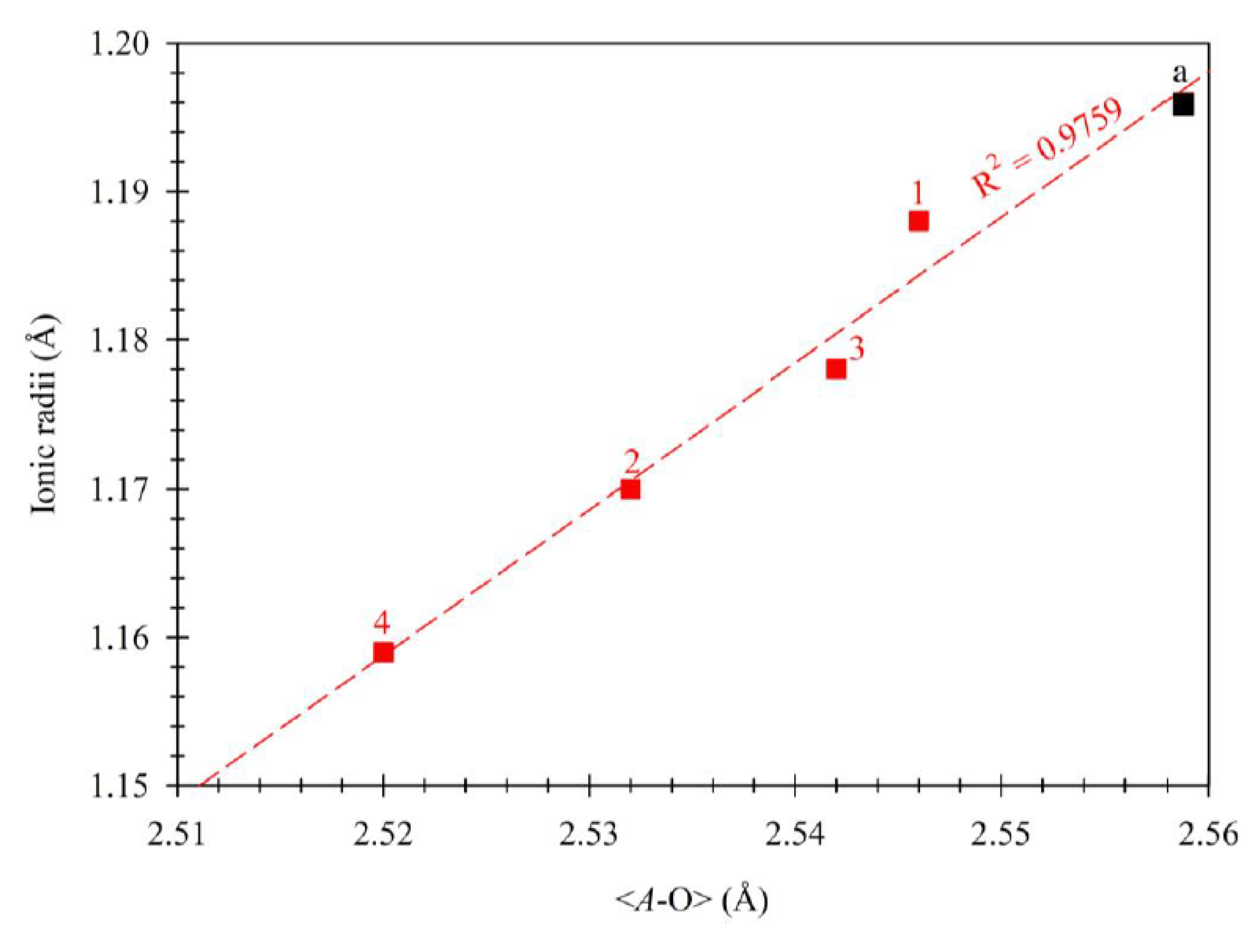
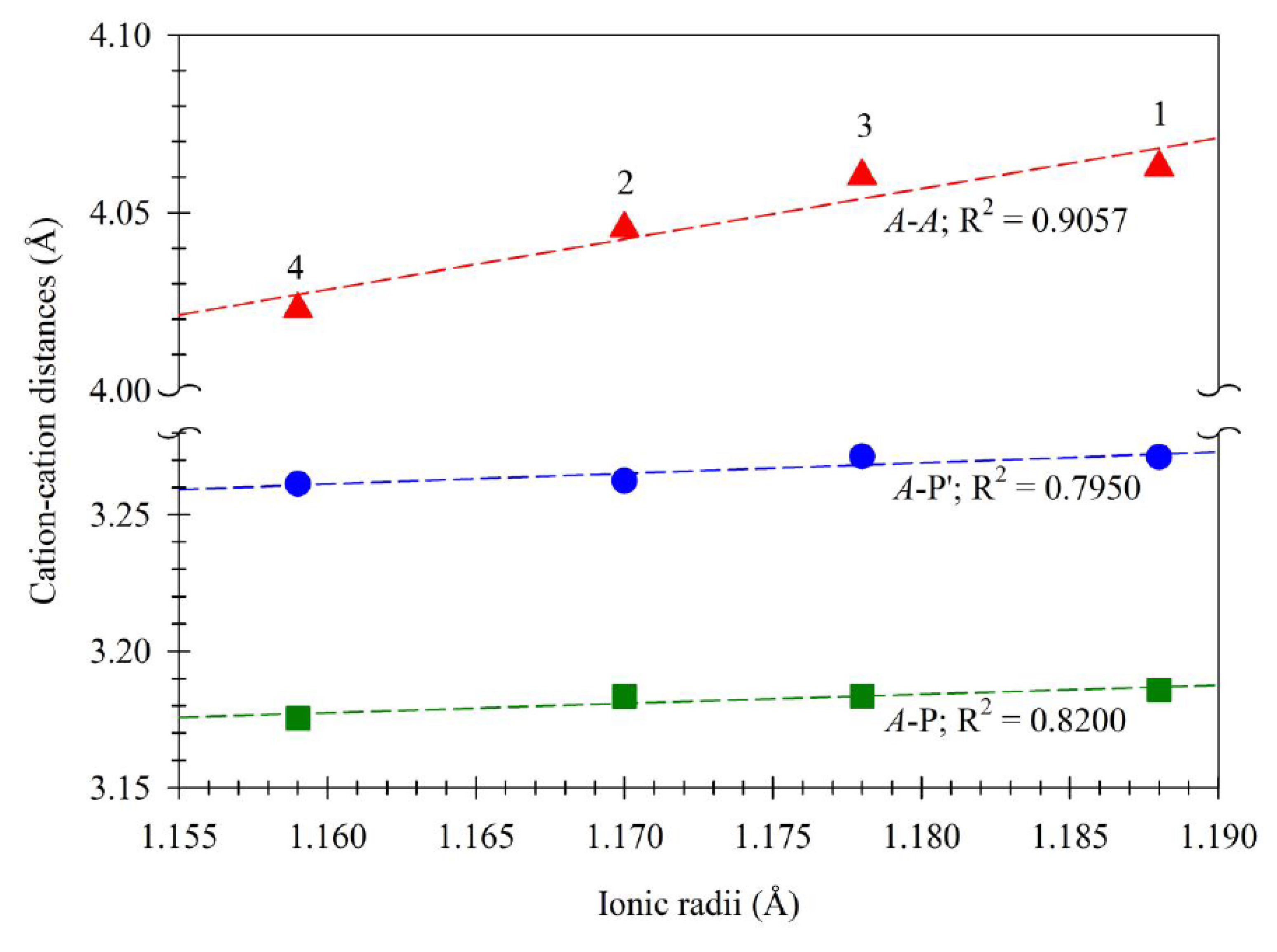
3.4. Bond Distances and Chemical Compositions
4. Conclusions
Author Contributions
Funding
Acknowledgments
Conflicts of Interest
References
- Boatner, L.A.; Sales, B.C. Monazite. In Radioactive Waste Forms for the Future; Lutze, W., Ewing, R.C., Eds.; Elsevier: Amsterdam, The Netherlands, 1988; pp. 495–564. [Google Scholar]
- Clavier, N.; Podor, R.; Dacheux, N. Crystal chemistry of the monazite structure. J. Eur. Ceram. Soc. 2011, 31, 941–976. [Google Scholar] [CrossRef]
- Gramaccioli, C.M.; Segalstad, T.V. A uranium- and thorium-rich monazite from a south-alpine pegmatite at Piona, Italy. Am. Mineral. 1978, 63, 757–761. [Google Scholar]
- Van-Emden, B.; Graham, J.; Lincoln, F.J. The incorporation of actinides in monazite and xenotime from placer deposits in Western Australia. Can. Mineral. 1997, 35, 95–104. [Google Scholar]
- Mooney, R.C.L. Crystal structures of a series of rare earth phosphates. J. Chem. Phys. 1948, 16, 1003. [Google Scholar] [CrossRef]
- Ueda, T. Re-examination of the crystal structure of monazite. J. Jpn. Assoc. Mineral. Petrol. Econ. Geol. 1967, 58, 170–179. [Google Scholar] [CrossRef][Green Version]
- Ghouse, K.M. Refinement of the crystal structure of heat-treated monazite crystal. Indian J. Pure Appl. Phys. 1968, 2, 265–268. [Google Scholar]
- Ni, Y.; Hughes, J.M.; Mariano, A.N. Crystal chemistry of the monazite and xenotime structures. Am. Mineral. 1995, 80, 21–26. [Google Scholar] [CrossRef]
- Mullica, D.F.; Grossie, D.A.; Boatner, L.A. Coordination geometry and structural determinations of SmPO4, EuPO4 and GdPO4. Inorg. Chim. Acta 1985, 109, 105–110. [Google Scholar] [CrossRef]
- Masau, M.; Cerny, P.; Cooper, M.A.; Chapman, R.; Grice, J.D. Monazite-Sm, a new member of the monazite group from the Annie claim #3 granite pegmatite, southeastern Manitoba. Can. Mineral. 2002, 40, 1649–1655. [Google Scholar]
- Seydoux-Guillaume, A.M.; Wirth, R.; Nasdala, L.; Gottschalk, M.; Montel, J.M.; Heinrich, W. An XRD, TEM and Raman study of experimentally annealed natural monazite. Phys. Chem. Miner. 2002, 29, 240–253. [Google Scholar] [CrossRef]
- Boatner, L.A. Synthesis, structure and properties of monazite, pretulite and xenotime. In Phosphates. Reviews in Mineralogy and Geochemistry; Kohn, M.L., Rakovan, J., Hughes, J.M., Eds.; Mineralogical Society of America: Chantilly, VA, USA, 2002; Volume 48, pp. 87–120. [Google Scholar]
- Dacheux, N.; Clavier, N.; Podor, R. Monazite as a promising long-term radioactive waste matrix: Benefits of high-structural flexibility and chemical durability. Am. Mineral. 2013, 98, 833–847. [Google Scholar] [CrossRef]
- Crichton, W.A.; Parise, J.B.; Antao, S.M.; Grzechnik, A. Evidence for monazite-, barite- and AgMnO4 (distorted barite)-type structures of CaSO4 at high pressure and temperature. Am. Mineral. 2005, 90, 22–27. [Google Scholar] [CrossRef]
- Beall, G.W.; Boatner, L.A.; Mullica, D.F.; Milligan, W.O. The structure of cerium ortho-phosphate, a synthetic analog of monazite. J. Inorg. Nucl. Chem. 1981, 43, 101–105. [Google Scholar] [CrossRef]
- Zaman, M.; Schubert, M.; Antao, S. Elevated radionuclide concentrations in heavy mineral-rich beach sands in the Cox’s Bazar region, Bangladesh and related possible radiological effects. Isot. Environ. Health Stud. 2012, 48, 512–525. [Google Scholar] [CrossRef] [PubMed]
- Zaman, M.M.; Antao, S.M. Crystal chemistry and structural variations for zircon samples from various localities. Minerals 2020, 10, 947. [Google Scholar] [CrossRef]
- Pyle, J.M.; Spear, F.S.; Wark, D.A. Electron microprobe analysis of REE in apatite, monazite, and xenotime: Protocols and pitfalls. In Phosphates. Reviews in Mineralogy and Geochemistry; Kohn, M.J., Rakovan, J., Hughes, J.M., Eds.; Mineralogical Society of America: Washington, DC, USA, 2002; Volume 48, pp. 337–362. [Google Scholar]
- Otwinowski, Z.; Minor, W. Processing of X-ray diffraction data collected in oscillation mode. In Methods in Enzymology: Macromolecular crystallography A276; Carter, C.W., Jr., Sweet, R.M., Eds.; Academic Press: New York, NY, USA, 1997; pp. 307–326. [Google Scholar]
- Antao, S.M.; Dhaliwal, I. Growth Oscillatory Zoning in Erythrite, Ideally Co3(AsO4)2·8H2O: Structural Variations in Vivianite-Group Minerals. Minerals 2017, 7, 136. [Google Scholar] [CrossRef]
- Antao, S.M.; Hassan, I.; Crichton, W.A.; Parise, J.B. Effects of high pressure and temperature on cation ordering in magnesioferrite, MgFe2O4, using in situ synchrotron X-ray powder diffraction up to 1430 K and 6 GPa. Am. Mineral. 2005, 90, 1500–1505. [Google Scholar] [CrossRef]
- Antao, S.M.; Hassan, I.; Mulder, W.H.; Lee, P.L. The R-3c→R-3m transition in nitratine, NaNO3, and implications for calcite, CaCO3. Phys. Chem. Miner. 2008, 35, 545–557. [Google Scholar] [CrossRef]
- Ehm, L.; Michel, F.M.; Antao, S.M.; Martin, C.D.; Lee, P.L.; Shastri, S.D.; Chupas, P.J.; Parise, J.B. Structural changes in nanocrystalline mackinawaite (FeS) at high pressure. J. Appl. Crystallogr. 2009, 42, 15–21. [Google Scholar] [CrossRef]
- Hassan, I.; Antao, S.M.; Hersi, A.A. Single-crystal XRD, TEM, and thermal studies of the satellite reflections in nepheline. Can. Mineral. 2003, 41, 759–783. [Google Scholar] [CrossRef]
- Hassan, I.; Antao, S.M.; Parise, J.B. Haüyne: Phase transition and high-temperature structures obtained from synchrotron radiation and Rietveld refinements. Mineral. Mag. 2004, 68, 499–513. [Google Scholar] [CrossRef]
- Parise, J.B.; Antao, S.M.; Michel, F.M.; Martin, C.D.; Chupas, P.J.; Shastri, S.; Lee, P.L. Quantitative high-pressure pair distribution function analysis. J. Synchrotron Radiat. 2005, 12, 554–559. [Google Scholar] [CrossRef] [PubMed]
- Sheldrick, G.M. A short history of SHELX. Acta Crystallogr. 1998, A64, 112–122. [Google Scholar]
- Farrugia, L.J. WinGX and ORTEP for Windows: An update. J. Appl. Crystallogr. 2012, 45, 849–854. [Google Scholar] [CrossRef]
- Brown, I.D. The Chemical Bond in Inorganic Chemistry: The Bond Valence Model; Oxford University Press: New York, NY, USA, 2002. [Google Scholar]
- Brown, I.D.; Altermatt, D. Bond-valence parameters obtained from a systematic analysis of the inorganic crystal structure database. Acta Crystallogr. 1985, B41, 244–247. [Google Scholar] [CrossRef]
- Shannon, R.D. Revised effective ionic radii and systematic studies of interatomic distances in halides and chalcogenides. Acta Crystallogr. 1976, A32, 75l–767. [Google Scholar] [CrossRef]
- Antao, S.M. Three cubic phases intergrown in a birefringent andradite-grossular garnet and their implications. Phys. Chem. Miner. 2013, 40, 705–716. [Google Scholar] [CrossRef]
- Antao, S.M. The mystery of birefringent garnet: Is the symmetry lower than cubic? Powder Diffr. 2013, 28, 281–288. [Google Scholar] [CrossRef]
- Antao, S.M. Crystal chemistry of birefringent hydrogrossular. Phys. Chem. Miner. 2015, 42, 455–474. [Google Scholar] [CrossRef]
- Antao, S.M.; Klincker, A.M. Origin of birefringence in andradite from Arizona, Madagascar, and Iran. Phys. Chem. Miner. 2013, 40, 575–586. [Google Scholar] [CrossRef]
- Antao, S.M.; Mohib, S.; Zaman, M.; Marr, R.A. Ti-rich andradites: Chemistry, structure, multi-phases, optical anisotropy, and oscillatory zoning. Can. Mineral. 2015, 53, 133–158. [Google Scholar] [CrossRef]
- Mullica, D.F.; Sappenfield, E.L.; Boatner, L.A. Monazite- and zircon-type structures of seven mixed (Ln/Ln) PO4 compounds. Inorg. Chinica Acta 1996, 244, 247–252. [Google Scholar] [CrossRef]
- Muller, O.; Roy, R. Crystal Chemistry of Non-Metallic Materials: Volume 4; Springer: New York, NY, USA, 1975. [Google Scholar]
- Huang, C. Rare Earth Coordination Chemistry—Fundamentals and Applications; John Wiley & Sons Pte Ltd.: Singapore, 2010. [Google Scholar]


| Sample | Localities | Descriptions and Occurrences |
|---|---|---|
| 1 | Kolatoli beach, Cox’s Bazar, Bangladesh | Detrital monazite grains separated from a bulk beach sand sample. Grains are spherical in shape and greenish yellow. |
| 2 | Iveland, Norway | Massive monazite occurs in a quartz pegmatitic rock. |
| 3 | Shaplapur paleobeach, Cox’s Bazar, Bangladesh | Same as sample 1. |
| 4 | Gunnison County, Colorado, USA | Massive dark brown Sm-rich monazite occurs with cleavelandite, feldspar, and lepidolite from the brown Derby-1 pegmatite. |
| Sample | 1 | 2 | 3 | 4 | 1 | 2 | 3 | 4 | |
|---|---|---|---|---|---|---|---|---|---|
| Oxide wt.% | apfu * | ||||||||
| La2O3 | 13.98 | 7.65 | 11.36 | 3.80 | La | 0.200 | 0.113 | 0.166 | 0.057 |
| Ce2O3 | 28.42 | 22.42 | 25.24 | 12.42 | Ce | 0.404 | 0.330 | 0.367 | 0.185 |
| Pr2O3 | 2.80 | 3.25 | 2.58 | 1.92 | Pr | 0.040 | 0.048 | 0.037 | 0.029 |
| Nd2O3 | 12.05 | 15.31 | 11.24 | 7.12 | Nd | 0.167 | 0.220 | 0.159 | 0.104 |
| Sm2O3 | 1.81 | 4.01 | 1.97 | 13.73 | Sm | 0.024 | 0.056 | 0.027 | 0.193 |
| Eu2O3 | 0.11 | bdl | bdl | bdl | Eu | 0.001 | 0.000 | 0.000 | 0.000 |
| Gd2O3 | 1.23 | 2.33 | 1.83 | 5.77 | Gd | 0.016 | 0.031 | 0.024 | 0.078 |
| Tb2O3 | bdl | 0.11 | bdl | bdl | Tb | 0.000 | 0.001 | 0.000 | 0.000 |
| Dy2O3 | 0.25 | 0.79 | 0.96 | 0.27 | Dy | 0.003 | 0.010 | 0.012 | 0.003 |
| Y2O3 | 0.51 | 3.92 | 3.31 | 0.73 | Y | 0.011 | 0.084 | 0.070 | 0.016 |
| CaO | 1.80 | 0.27 | 1.01 | 2.89 | Ca | 0.075 | 0.012 | 0.043 | 0.126 |
| FeO | bdl | bdl | bdl | bdl | Fe | 0.000 | 0.000 | 0.000 | 0.000 |
| P2O5 | 29.11 | 27.14 | 28.18 | 27.94 | P | 0.957 | 0.923 | 0.946 | 0.964 |
| SiO2 | 0.42 | 2.06 | 1.38 | 1.22 | Si | 0.016 | 0.083 | 0.055 | 0.050 |
| SO3 | 0.94 | 0.09 | bdl | 0.09 | S | 0.027 | 0.003 | 0.000 | 0.003 |
| ThO2 | 6.55 | 8.71 | 10.10 | 18.22 | Th | 0.058 | 0.080 | 0.091 | 0.169 |
| UO2 | 0.22 | 0.36 | 0.25 | 0.42 | U | 0.002 | 0.003 | 0.002 | 0.004 |
| PbO | 0.01 | 0.28 | 0.13 | 1.06 | Pb | 0.000 | 0.003 | 0.001 | 0.012 |
| Total | 100.18 | 98.70 | 99.56 | 97.61 | Total | 2.001 | 1.998 | 2.001 | 1.993 |
| ∑LREE | 0.811 | 0.710 | 0.729 | 0.375 | |||||
| ∑MREE | 0.045 | 0.098 | 0.063 | 0.274 | |||||
| ∑A | 1.001 | 0.990 | 1.000 | 0.976 | |||||
| ∑P | 1.000 | 1.009 | 1.001 | 1.017 | |||||
| Miscellaneous | 1 | 2 | 3 | 4 |
|---|---|---|---|---|
| a (Å) | 6.7640(5) | 6.7360(8) | 6.7590(4) | 6.7010(4) |
| b (Å) | 6.9850(4) | 6.9490(7) | 6.9770(4) | 6.9080(4) |
| c (Å) | 6.4500(3) | 6.4390(8) | 6.4480(3) | 6.4300(4) |
| β (°) | 103.584(2) | 103.855(6) | 103.656(3) | 103.817(3) |
| V (Å3) | 296.22(3) | 292.63(6) | 295.48(3) | 289.04(3) |
| Crystal dimension (mm3) | 0.06 × 0.06 × 0.05 | 0.08 × 0.06 × 0.06 | 0.10 × 0.10 × 0.08 | 0.05 × 0.05 × 0.04 |
| Densitycalc (g/cm3) | 5.272 | 5.336 | 5.285 | 5.638 |
| Absorption coefficient (mm−1) | 15.718 | 15.911 | 15.758 | 20.676 |
| 2θ range | 7.86–55.28° | 7.86–55.22° | 7.86–55° | 7.92–55° |
| Index ranges | −8 < = h < = 8 | −8 < = h < = 8 | −8 = < h = < 8 | −8 = < h = < 8 |
| −9 < = k < = 8 | −8 < = k < = 8 | −9 = < k = < 9 | −8 = < k = < 8 | |
| −8 < = l < = 8 | −8 < = l < = 8 | −8 = < l = < 8 | −8 = < l = < 8 | |
| Total reflections | 2307 | 2278 | 2578 | 2214 |
| Unique reflections | 692 | 676 | 680 | 659 |
| Completeness to θ = 27.7 (%) | 100 | 100 | 100 | 98.9 |
| Rint | 0.0282 | 0.0415 | 0.0327 | 0.0506 |
| Goodness of fit on F2 | 1.206 | 1.231 | 1.314 | 0.789 |
| R1 [I > 2σ(I)] | 0.0139 | 0.0237 | 0.0180 | 0.0365 |
| wR2 | 0.0350 | 0.0644 | 0.0471 | 0.1594 |
| Extinction coefficient | 0.0162(8) | 0.005(1) | 0.013(1) | 0.000(3) |
| Largest difference peak/hole (e/Å3) | 0.509/−0.529 | 0.750/−1.213 | 0.602/−0.953 | 2.430/−1.324 |
| Mosaicity (°) | 0.751(3) | 0.981(9) | 0.803(3) | 1.74(3) |
| Atom | x | y | z | Ueq | U11 | U22 | U33 | U23 | U12 | U13 | |
|---|---|---|---|---|---|---|---|---|---|---|---|
| Ce * | 1 § | 0.28155(3) | 0.15901(3) | 0.10011(3) | 0.0111(1) | 0.0115(2) | 0.0113(2) | 0.0099(1) | 0.00123(6) | 0.00018(7) | 0.00156(8) |
| Ce | 2 | 0.28047(5) | 0.15821(4) | 0.09972(5) | 0.0183(2) | 0.0193(2) | 0.0167(3) | 0.0169(2) | 0.0020(1) | 0.0000(1) | 0.0003(1) |
| Ce | 3 | 0.28129(3) | 0.15862(3) | 0.09982(3) | 0.0107(1) | 0.0103(2) | 0.0093(2) | 0.0116(2) | 0.00156(7) | 0.00004(7) | 0.0006(1) |
| Sm | 4 | 0.28004(8) | 0.15793(8) | 0.10002(7) | 0.0124(5) | 0.0119(6) | 0.0114(6) | 0.0133(6) | 0.0027(2) | 0.0001(3) | 0.0020(3) |
| P | 1 | 0.3039(1) | 0.1629(1) | 0.6122(1) | 0.0105(3) | 0.0108(5) | 0.0115(5) | 0.0090(4) | 0.0002(3) | 0.0008(3) | 0.0022(3) |
| 2 | 0.3028(2) | 0.1620(2) | 0.6115(2) | 0.0178(5) | 0.0209(8) | 0.0171(9) | 0.0141(8) | −0.0002(5) | 0.0004(5) | 0.0018(5) | |
| 3 | 0.3036(2) | 0.1625(1) | 0.6121(2) | 0.0107(4) | 0.0106(6) | 0.0104(6) | 0.0106(5) | 0.0001(2) | 0.0011(3) | 0.0017(4) | |
| 4 | 0.3020(4) | 0.1625(3) | 0.6122(4) | 0.010(1) | 0.009(1) | 0.013(2) | 0.008(2) | 0.0007(5) | 0.0009(6) | 0.000(1) | |
| O1 | 1 | 0.2488(4) | 0.0064(4) | 0.4425(4) | 0.0155(6) | 0.019(1) | 0.016(1) | 0.012(1) | −0.0021(9) | −0.000(1) | 0.0038(9) |
| 2 | 0.2474(6) | 0.0070(6) | 0.4389(7) | 0.022(1) | 0.026(2) | 0.019(2) | 0.021(2) | −0.004(2) | −0.002(2) | 0.005(2) | |
| 3 | 0.2487(5) | 0.0059(4) | 0.4413(5) | 0.0167(7) | 0.019(2) | 0.016(2) | 0.014(1) | −0.002(1) | 0.000(1) | 0.003(1) | |
| 4 | 0.249(1) | 0.002(1) | 0.439(2) | 0.017(2) | 0.014(3) | 0.022(4) | 0.013(3) | 0.004(3) | 0.004(3) | 0.000(3) | |
| O2 | 1 | 0.3816(4) | 0.3318(3) | 0.4993(4) | 0.0161(6) | 0.015(1) | 0.015(1) | 0.020(1) | 0.0030(9) | −0.0016(9) | 0.007(1) |
| 2 | 0.3817(7) | 0.3327(6) | 0.4990(7) | 0.024(1) | 0.025(2) | 0.021(2) | 0.027(2) | 0.004(2) | −0.002(2) | 0.007(2) | |
| 3 | 0.3816(5) | 0.3323(4) | 0.4997(5) | 0.0173(7) | 0.015(2) | 0.016(2) | 0.022(2) | 0.004(1) | −0.001(1) | 0.005(1) | |
| 4 | 0.381(1) | 0.3317(1) | 0.501(1) | 0.018(2) | 0.020(4) | 0.009(4) | 0.029(4) | −0.001(2) | −0.008(2) | 0.012(4) | |
| O3 | 1 | 0.4743(4) | 0.1061(4) | 0.8044(4) | 0.0170(6) | 0.015(1) | 0.019(1) | 0.015(1) | −0.000(1) | 0.003(1) | −0.002(1) |
| 2 | 0.4744(7) | 0.1053(7) | 0.8064(7) | 0.029(1) | 0.028(2) | 0.031(2) | 0.022(2) | −0.002(2) | 0.007(2) | −0.004(2) | |
| 3 | 0.4739(5) | 0.1050(5) | 0.8049(5) | 0.0185(7) | 0.016(2) | 0.020(2) | 0.017(1) | −0.000(1) | 0.005(1) | −0.003(1) | |
| 4 | 0.475(2) | 0.106(1) | 0.807(2) | 0.019(2) | 0.009(3) | 0.019(4) | 0.023(3) | 0.002(3) | 0.001(3) | −0.008(3) | |
| O4 | 1 | 0.1268(4) | 0.2134(4) | 0.7104(4) | 0.0153(6) | 0.015(1) | 0.019(1) | 0.013(1) | 0.001(1) | 0.002(1) | 0.0049(9) |
| 2 | 0.1262(6) | 0.2117(7) | 0.7100(7) | 0.024(1) | 0.022(2) | 0.032(2) | 0.018(2) | 0.002(2) | −0.003(2) | 0.003(2) | |
| 3 | 0.1267(4) | 0.2133(5) | 0.7112(5) | 0.0157(7) | 0.012(1) | 0.020(2) | 0.015(1) | 0.002(1) | 0.001(1) | 0.002(1) | |
| 4 | 0.124(1) | 0.217(1) | 0.710(1) | 0.017(2) | 0.028(4) | 0.013(4) | 0.013(3) | 0.002(2) | 0.004(3) | 0.013(3) | |
| Bond/Angle | 1 | 2 | 3 | [8] | 4 | |||||
|---|---|---|---|---|---|---|---|---|---|---|
| BV § | BV | BV | BV | BV | ||||||
| A-O1’ | 2.445(3) | 0.452 | 2.439(4) | 0.459 | 2.440(3) | 0.458 | 2.528(2) | 0.361 | 2.395(8) | 0.436 |
| -O1’’ | 2.509(3) | 0.380 | 2.481(4) | 0.410 | 2.503(3) | 0.386 | 2.461(2) | 0.433 | 2.484(7) | 0.343 |
| -O2’ | 2.554(2) | 0.336 | 2.526(4) | 0.363 | 2.544(3) | 0.346 | 2.776(3) | 0.185 | 2.515(7) | 0.315 |
| -O2’’ | 2.630(3) | 0.274 | 2.609(5) | 0.290 | 2.626(3) | 0.277 | 2.644(2) | 0.264 | 2.600(8) | 0.251 |
| -O2’’’ | 2.779(3) | 0.183 | 2.776(5) | 0.185 | 2.784(3) | 0.181 | 2.573(2) | 0.320 | 2.775(7) | 0.156 |
| -O3’ | 2.461(3) | 0.433 | 2.443(5) | 0.454 | 2.454(3) | 0.441 | 2.585(3) | 0.309 | 2.430(7) | 0.397 |
| -O3’’ | 2.577(3) | 0.316 | 2.567(5) | 0.325 | 2.573(3) | 0.320 | 2.481(2) | 0.410 | 2.565(7) | 0.275 |
| -O4’ | 2.444(3) | 0.453 | 2.440(4) | 0.458 | 2.444(3) | 0.453 | 2.526(2) | 0.363 | 2.403(7) | 0.427 |
| -O4’’ | 2.514(2) | 0.375 | 2.503(4) | 0.386 | 2.506(3) | 0.383 | 2.455(2) | 0.440 | 2.507(6) | 0.322 |
| <A-O> [9] | 2.546(3) | 3.202 † | 2.532(4) | 3.330 † | 2.542(3) | 3.244 † | 2.559(2) | 3.084 † | 2.519(7) | 2.923 † |
| P-O1 | 1.530(3) | 1.221 | 1.528(4) | 1.228 | 1.534(3) | 1.208 | 1.534(3) | 1.208 | 1.550(8) | 1.157 |
| -O2 | 1.542(3) | 1.182 | 1.548(4) | 1.163 | 1.545(3) | 1.173 | 1.545(3) | 1.173 | 1.529(7) | 1.225 |
| -O3 | 1.533(3) | 1.212 | 1.540(4) | 1.189 | 1.535(3) | 1.205 | 1.534(3) | 1.208 | 1.539(6) | 1.192 |
| -O4 | 1.522(3) | 1.248 | 1.517(5) | 1.265 | 1.524(3) | 1.241 | 1.531(3) | 1.218 | 1.526(7) | 1.235 |
| <P-O> [4] | 1.532(3) | 4.863 † | 1.533(4) | 4.845 † | 1.535(3) | 4.828 † | 1.536(3) | 4.808 † | 1.536(7) | 4.809 † |
| O1-P-O2 | 105.1(2) | 104.6(3) | 105.1(2) | 113.7 | 104.8(4) | |||||
| O1-P-O3 | 113.8(2) | 114.6(3) | 113.8(2) | 103.9 | 113.7(7) | |||||
| O1-P-O4 | 112.4(2) | 112.7(3) | 112.7(2) | 113.7 | 113.8(4) | |||||
| O2-P-O3 | 107.8(2) | 107.6(3) | 107.9(2) | 112.4 | 107.3(5) | |||||
| O2-P-O4 | 114.1(2) | 114.4(3) | 114.0(2) | 105.2 | 113.5(4) | |||||
| O3-P-O4 | 103.8(1) | 103.2(3) | 103.5(2) | 108.1 | 103.9(4) | |||||
| <O-P-O> [6] | 109.5(2) | 109.5(3) | 109.5(2) | 109.5 | 109.5(5) |
Publisher’s Note: MDPI stays neutral with regard to jurisdictional claims in published maps and institutional affiliations. |
© 2020 by the authors. Licensee MDPI, Basel, Switzerland. This article is an open access article distributed under the terms and conditions of the Creative Commons Attribution (CC BY) license (http://creativecommons.org/licenses/by/4.0/).
Share and Cite
Zaman, M.M.; Antao, S.M. Crystal Structure Refinements of Four Monazite Samples from Different Localities. Minerals 2020, 10, 1028. https://doi.org/10.3390/min10111028
Zaman MM, Antao SM. Crystal Structure Refinements of Four Monazite Samples from Different Localities. Minerals. 2020; 10(11):1028. https://doi.org/10.3390/min10111028
Chicago/Turabian StyleZaman, M. Mashrur, and Sytle M. Antao. 2020. "Crystal Structure Refinements of Four Monazite Samples from Different Localities" Minerals 10, no. 11: 1028. https://doi.org/10.3390/min10111028
APA StyleZaman, M. M., & Antao, S. M. (2020). Crystal Structure Refinements of Four Monazite Samples from Different Localities. Minerals, 10(11), 1028. https://doi.org/10.3390/min10111028