Article

Developmental Origins of Limb Developmental Instability in Human Fetuses: Many Abnormalities Make the Difference

1
Evolutionary Ecology Group, Campus Drie Eiken, University of Antwerp, Universiteitsplein 1, 2610 Wilrijk, Belgium
2
Naturalis Biodiversity Center, Endless Forms, Vondellaan 55, 2332 AA Leiden, The Netherlands
3
Department of Pathology, VU University Medical Center, De Boelelaan 1117, 1081 HV Amsterdam, The Netherlands
*
Authors to whom correspondence should be addressed.
Symmetry 2017, 9(4), 51; https://doi.org/10.3390/sym9040051
Submission received: 16 February 2017 / Revised: 20 March 2017 / Accepted: 23 March 2017 / Published: 28 March 2017
(This article belongs to the Special Issue Symmetry in Human Evolutionary Biology)

Abstract

:
Fluctuating asymmetry (FA) is the small random deviation from perfect symmetry in bilateral traits and is often used to assess developmental instability (DI) experienced by organisms. In this study, with a unique dataset of 1389 deceased human fetuses, we investigated the relationship between abnormal development and human limb FA in different ways, using a more fundamental approach than usually done. We studied whether there is an underlying developmental basis of DI, as measured by FA, by investigating, first, whether limb FA can be attributed to developmental abnormalities associated with specific organ systems, germ layers or patterning processes, and second, whether limb FA increases with increasing number of developmental abnormalities either gradually, or in a threshold-like fashion. Limb FA was found to increase in fetuses with cardiovascular and nervous system abnormalities. Fetuses with ectoderm-derived abnormalities were also found to have significantly higher limb FA, but no other germ layers were found to be associated. We found no significant correlation between specific developmental processes, such as neural crest development, segmentation, midline and left-right patterning and limb FA. Although only some congenital abnormalities were correlated with limb FA, our results do suggest that limb FA increases when an increasing number of organ systems, germ layers or developmental pathways are disrupted. Therefore, we conclude that limb FA is mainly a good indicator for DI in the case of particularly severe perturbations of development and that FA does not reflect the subtler deviations from developmental stability.

1. Introduction

For decades, there has been an interest in the biological significance of developmental instability (DI) and its associations with fitness and stress. DI causes small non-directional deviations from perfect symmetry in bilaterally paired traits, termed fluctuating asymmetry (FA) [1,2,3]. FA is considered to reflect the ability of an organism to cope with genetic and environmental stress during its development and is based on the a priori expectation that symmetry is the outcome of the bilaterally paired structures [4]. It has been recognized that organisms have the capacity to buffer disturbances of various origin that may influence development. However, increased levels of perturbations cause a general breakdown of this buffering capacity, leading to increased DI and thus measurable FA. Indeed, several studies have demonstrated associations between FA and indices of stress, health and fitness (reviewed in [5,6,7,8,9]). The patterns emerging from different studies, however, show much variation in effect sizes and underlying causes of this diversity remain poorly understood. It is difficult to pinpoint the moderators of the between–study variation as very little is known about the developmental origins of DI. For example, it is unknown whether DI is affected uniformly with increased perturbation of any developmental pathway or if disturbances of specific pathways increase DI more strongly than others. In addition, it is unclear whether DI increases gradually as more pathways are affected or if the increase occurs in a threshold-like fashion [10]. In this study, we make use of a unique combination of data on deceased human fetuses, to investigate the relationship between abnormal development and limb FA. Associations between FA and developmental abnormalities were analysed at three different levels by classifying abnormalities on the basis of (i) the affected organ systems; (ii) the germ layer of origin of the affected organ systems; and (iii) the developmental processes that were perturbed to cause the observed abnormalities. Furthermore, we quantified the number of affected organ systems, affected germ layers and perturbed developmental processes with the aim to determine whether FA increases in a threshold-like fashion with the number of abnormalities (as seen in [10]) or rather in a gradual fashion with increasing number of abnormalities.

2. Materials and Methods

2.1. Fetuses and Asymmetry Measurements

Since 1980, all deceased infants and fetuses have been routinely radiographed both ventrally and laterally, when presented for autopsy at the VU University Medical Centre in Amsterdam, the Netherlands (23 mA, 70–90 kV, 4–12 s, Agfa (Mortsel, Belgium) Gevaert D7DW Structurix films). This research was carried out on the anterior-posterior projections of 1389 deceased fetuses and infants obtained between 1990 and 2009. In total, we measured 616 male and 487 female fetuses and infants and four fetuses of unknown sex (13.6–92.1 weeks, mean: 27.8 weeks ± 10.0 weeks). We excluded some fetuses when radiographs had insufficient resolution or limb bones were not properly positioned. The radiographs were digitized using a Canon 30D digital SLR camera (Canon, Machelen, Belgium) in a fixed-distance set-up with a glass plate and a flash underneath. We measured the length of the left and right digit 2, digit 4, femur, fibula, radius, ulna and tibia from the midpoint of the proximal end of the bone to the midpoint of the distal end of the bone in Image J version 1.42q (National Institutes of Health, Bethesda, MA, USA). Digits were measured from the proximal end of the proximal phalanx to the distal end of the distal phalanx. We excluded all fetuses that had abnormally developed limbs from analyses, because otherwise the possibly higher measured FA could have directly been an artefact of the abnormalities. Standard autopsy reports were made by pathologists and filed in a national pathological archive (PALGA; www.palga.nl). We categorized the different congenital abnormalities into distinct groups referring to the different germ layers involved in development: endoderm malformations including bronchopulmonary malformations (BP) and digestive system malformations (DS), mesoderm malformations including muscular system malformations (MUS), skeletal malformations (SK), urogenital malformations (UG) and cardiovascular malformations (CV) and ectoderm malformations including nervous system defects (NS) and craniofacial malformations (CF). Moreover, we categorized congenital abnormalities resulting from different disturbed developmental events into separate groups: left-right patterning defects (LR), neural crest development defects (NC), midline pattering defects (MID) and segmentation defects (SGMD). We also quantified the number of affected organ systems, affected number of germ layers and number of affected processes in order to evaluate whether asymmetry increases with the number of malformations or disturbed processes. When autopsy was not consented or when the individuals were not in good condition due to maceration we scored them as missing values. We scored, as far as possible, malformations only in one category by distinguishing between primary and secondary causes, with only primary causes counted in the analyses [11].

2.2. Fluctuating Asymmetry Measurement, Measurement Error and Directional Asymmetry

True measurements of fluctuating asymmetry are being influenced by measurement error (ME) and directional asymmetry (DA, the consistent difference between left and right in which the same side is always larger than the other) and need to be accounted for. Three different investigators (Jessica Bots, Clara M. A. ten Broek and Frietson Galis) performed all measurements without prior knowledge of the autopsy reports. To compare the accuracy of the measurements thirty-one fetuses were re-measured independently by each investigator. Correlation tests showed that results were highly repeatable (all p < 0.001 and all r < 0.30). In addition, the procedure of positioning the fetus and taking the radiograph was repeated for 147 individuals. On the basis of mixed regression models we determined ME and DA following [12] (see [13] for details). For each trait, the levels of ME were smaller than the levels of asymmetry and we corrected for DA when necessary (see [13]). Signed asymmetries were obtained as right minus left trait values. The distributions of signed asymmetries were all leptokurtic, as often observed in FA studies (kurtosis ranging between 6.15 and 31.41). The absolute value of the difference between left and right trait sides (unsigned asymmetry) was used as proxy of DI. Because age and thus size of fetuses differed, we corrected unsigned asymmetry for individual-specific trait size. We divided levels of unsigned FA by the trait size (mean of both sides) and multiplied by 100, so that the calculated FA reflected differences in percentages of asymmetry relative to trait size. Thereafter we standardized size-corrected unsigned FA values and we calculated average asymmetry based on the available traits. The resulting average size-corrected standardized unsigned FA values were used in the statistical analyses and will be further referred to as (limb) FA.

2.3. The Number of Malformed Organ Systems, the Germ Layer of Origin and Associations, Patterning Defects with Fluctuating Asymmetry

We used limb FA as dependent variable in a linear model and we included age as continuous covariate in the model as they were found to correlate negatively with FA [13,14]. We controlled for possible effects of deficient amniotic fluid volume levels on FA by adding amniotic fluid volume as categorical variable to the model (fetuses with amniotic fluid deficiencies treated as 1, with normal levels as 0) [13]. Age was log-transformed to assure linearity and to correctly use the analysis of covariance (ANCOVA) models [15]. We tested for differences in FA between (1) individuals with malformations arising from the three different germ layers and the number of affected germ layers (2) individuals with different disturbed morphological patterning processes and the number of disturbed patterning processes and (3) individuals with different malformed organ systems and different numbers of affected organ systems. Post-hoc pairwise comparisons were carried out using contrasts with Benjamini-Hochberg adjustment [16]. In addition, the model with the number of abnormalities as factor was compared to a model assuming a linear increase of FA with numbers as continuous variable using Akaike Information Criterion (AIC) and significance levels. In this way, evidence can be found whether increases in FA are rather gradual or threshold-like. All analyses were conducted in R software (version 3.02) using, the glht function of the multcomp package for the post-hoc pairwise comparisons and the lm function for the ANCOVA models [17,18,19]. P-values smaller than 0.05 were considered significant and descriptive statistics are reported as mean ± SE.

3. Results

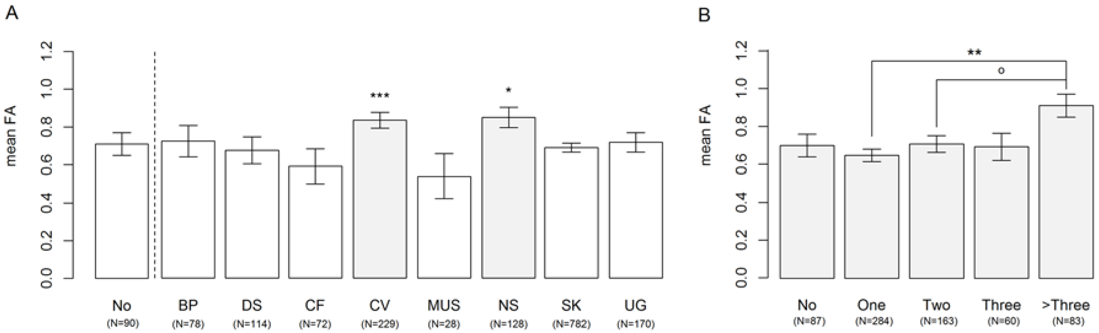
3.1. Affected Organ Systems

We compared influence of the different affected organ systems on FA and compared it with fetuses without having congenital malformations. We found no significant difference in limb FA for fetuses having bronchopulmonary abnormalities (F1,661 = 0.29, p = 0.59), digestive system abnormalities (F1,652 = 0.07, p = 0.80), craniofacial malformations (F1,664 = 0.98, p = 0.32), skeletal abnormalities (F1,670 = 1.64, p = 0.20), muscular disorders (F1,748 = 1.92, p = 0.17), urogenital malformations (F1,738 = 1.4, p = 0.24). Fetuses with cardiovascular and nervous system malformations had significantly higher FA than fetuses without malformations (F1,760 = 11.98, p < 0.001 and F1,760 = 5.47, p < 0.05, respectively).
We quantified the number of malformed organ systems and compared their associations with limb FA. Sample sizes were very low for fetuses with four, five, six, seven, eight or nine malformed organ systems (n = 34, n = 18, n = 8, n = 7, n = 1, n = 1, respectively). To increase statistical power in further analyses we merged those individuals into one group, because the correlation between four to nine malformed organ systems and limb FA were not significantly different from one another (0.99 < p < 1.00). When there were missing values for one or more organ systems, the number of affected organ systems for that individual was scored as missing. However, when there were missing values and the number of malformed organ systems was four or more, the fetuses were categorized into the group with fetuses with more than three malformed organ systems. We found significantly different correlations with limb FA for fetuses with either no, one, two, three, or four or more malformed organ systems (F4,662 = 3.64, p < 0.01). Post-hoc comparisons showed that fetuses with four or more malformed organ systems had significantly higher FA than fetuses with one malformed organ systems (t = 3.79, p < 0.01) and tended to have significantly higher limb FA than fetuses with two malformed organ systems (t = 2.49, p = 0.09). All other groups were not significantly different from one another (0.034 < t < 2.13, 0.21 < p < 1.00). The model with number of organ systems as factor showed a lower AIC value than a model assuming a gradual/linear increase in FA (−441.8 vs. −435.9) (Figure 1).

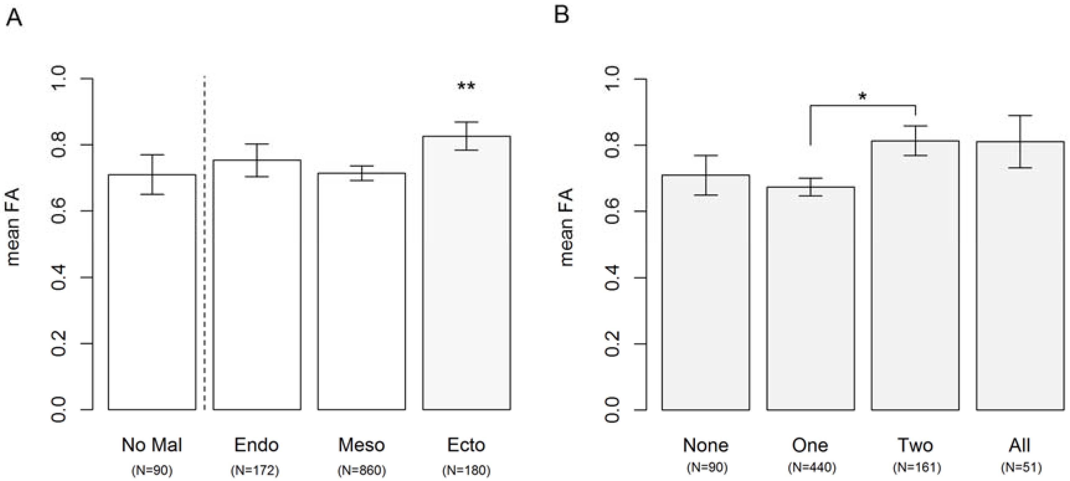
3.2. Congenital Abnormalities Arising from Different Germ Layers during Development

We compared the relationship of congenital malformations arising from the three different germ layers with limb FA. Fetuses with abnormalities in the ectoderm (n = 180) had significantly higher limb FA than fetuses without congenital abnormalities (F1,809 = 7.07, p < 0.01, see also Figure 1A). Fetuses with abnormalities in the endoderm (n = 172) and mesoderm (n = 860) did not have significantly higher limb FA than fetuses without malformations (F1,777 = 0.82, p = 0.36 and F1,726 = 0.32, p = 0.57 respectively, see also Figure 1A). Subsequently, we quantified the number of germ layers affected: no, one, two or all three germ layers with malformations. When there were missing values for one or more germ layers, the number of affected germ layers for that individual was scored as missing. We found significantly different relations with limb FA for the number of germ layers affected (F3,726 = 2.73, p < 0.05). Post-hoc comparisons showed that fetuses with abnormalities in two germ layers (n = 161) had significantly higher FA than fetuses with abnormalities arising from one germ layer (n = 440) (t = 2.64, p < 0.05, Figure 1B). All other post-hoc comparisons were not significantly different (none—one: t = 0.79, p = 0.85, none—two: t = 1.15, p = 0.64, none—all three: t = 0.75, p = 0.87, one—all: t = 1.15, p = 0.42, two—all three: t = 0.13, p = 1.00, see also Figure 1B). The model with number of germ layers as factor showed a lower AIC value than a model assuming a gradual/linear increase in FA (−481.2 vs. −480.9) (Figure 2).

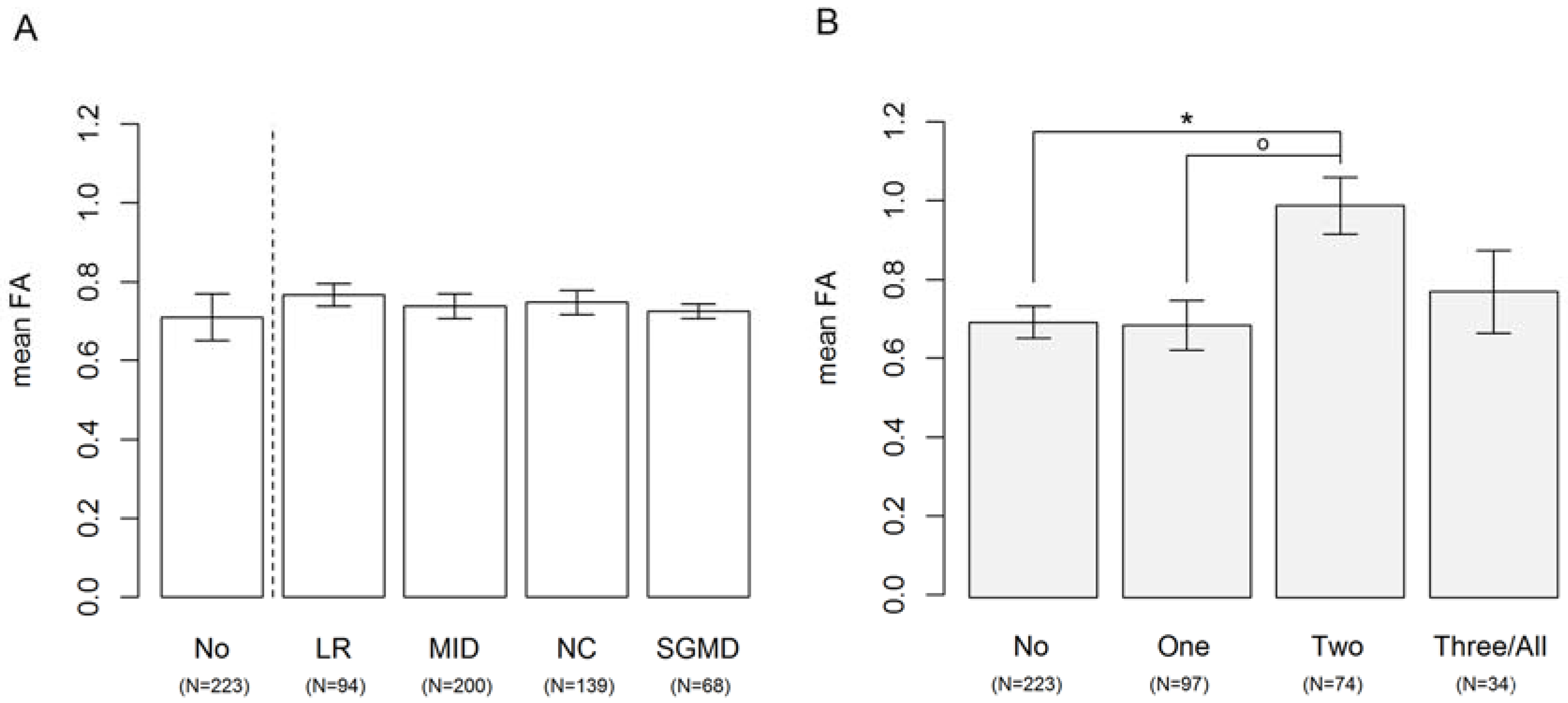
3.3. Disturbances in Development of Left-Right Patterning, Midline Development, Neural Crest Development and Segementation

We compared fetuses with malformations resulting from different developmental processes with fetuses without having these patterning defects. We found no significant relationship between limb and fetuses with segmentation defects (F1,416 = 0.002, p = 0.97), neural crest development defects (F1,446 = 0.29, p = 0.58), left-right patterning defects (F1,501 = 1.21, p = 0.27). Fetuses with midline patterning defects tended to show higher limb FA, however not reaching statistical significance (F1,542 = 3.17, p = 0.08). We quantified the number of disturbed patterning processes per individual (no, one, two, three, or four) and compared the correlation with limb FA. Since there were only eight individuals with malformations resulting from all patterning processes we added those individuals to the group of fetuses that had three disturbed patterning processes (n = 26) to increase power in the analysis. When there were missing values for one or more patterning processes, the number of affected processes for that individual was scored as missing. We found significantly different correlations between limb FA and different numbers of patterning defects (F3,417 = 3.00, p < 0.05). Post-hoc comparisons showed that fetuses with two patterning defects (n = 74) had significantly higher FA than fetuses without patterning defects (n = 224, t = 2.86, p < 0.05) and marginally higher FA than fetuses with one patterning defect (n = 97, t = 2.52, p =0.055). All other groups were not significantly different from one another (0.57 < p < 1.00). The model with number of patterning processes as factor showed a lower AIC value than a model assuming a gradual/linear increase in FA (−226.5 vs. −225.8) (Figure 3 next page).

4. Discussion

Little is known about the factors affecting the strength of association between fluctuating asymmetry and measures of stress, health and quality. An aspect that has received hardly any attention in the literature is that developmental instability may increase only when specific developmental pathways are disturbed, or that the increase in DI emerges only if disturbances of a particular magnitude take place. We investigated whether increased FA of fetal limbs is associated with specific organ systems, germ layers and developmental processes. Furthermore, we investigated whether limb FA increased with increasing number of congenital abnormalities, either gradually, or in a threshold-like fashion.
Fetuses with cardiovascular and nervous system malformations showed significantly higher limb FA than fetuses without congenital abnormalities. We found significantly increased limb FA in fetuses with malformations where the ectoderm was involved. This correlation was not unexpected because the ectodermal abnormalities most often involved the nervous system: by contrast we did not find higher limb FA for fetuses with problems emerging from the mesoderm, which include cardiovascular system abnormalities. No other specific organ system or germ layer was associated with higher FA, nor any of the developmental processes. Increases in limb FA, thus, may be more likely to arise when nervous system or cardiovascular system development is disturbed. The correlation with four included developmental processes (segmentation, midline patterning, neural crest development and left-right patterning) appeared limited. Earlier, we showed that there was no significant association with defects arising form disturbances of the head-to-tail patterning process and limb FA either [20].
We did observe significant increases in limb FA with increasing number of involved germ layers, developmental processes and organ systems. For each of the three categories, there was evidence that the increase was not linear, but rather increased with a single jump at a high threshold. Apparently, when a relatively high number of developmental processes are disturbed, a sudden rise in limb FA emerges. A similar conclusion was reached by Bots et al. [10] where FA was compared for fetuses with different chromosomal abnormalities. Human fetuses with aneuploidies with very short life expectancies (trisomy 13 or 18, monosomy X and triploidy) have higher limb FA than fetuses with trisomy 21, a normal karyotype or no congenital abnormalities at all. It was suggested that limb development is better buffered against perturbations resulting from trisomy 21 rather than perturbations resulting from trisomy 13, 18, monosomy X and triploidy, which are more general genetic aberrations involving many more genes and causing both more severe congenital abnormalities and shorter life expectancies [10,21]. Moreover, we previously published a study in which we failed to find a consistent relationship between severe developmental disorders and limb FA [14]. Higher limb FA was only found in 4 out of 17 individually investigated disorders, but fetuses with major (chromosomal number changes or not) congenital abnormalities showed significantly higher limb FA than fetuses with no or minor abnormalities [14]. Here we did not distinguish between major and minor clinical abnormalities, but rather focused on the origins of developmental errors to gain more insight in the relevant developmental pathways. We conclude that limb FA mainly increased when multiple developmental pathways were disrupted, but that no particular patterning process, germ layer or organ system is involved, except for the cardiovascular and nervous system. So generally speaking, despite studying a large population of deceased human fetuses, fetal limb FA is only moderately increased when development is disturbed dramatically. The disturbance of development or phenodeviance, as witnessed by the many morphological abnormalities, also reflects DI, but is not expressed as limb FA, possibly because intrauterine development creates a highly buffered environment for the developing extremities. Our study involves only deceased human fetuses and young infants. This has advantages, in the sense that we were able to study severe developmental abnormalities that are rare in the surviving human population, because they are often lethal. In addition, fetuses are not (or hardly) affected by mechanical loading of the limbs which in turn can affect asymmetry and blur the link with developmental instability [22]. Our overall conclusion that only severe abnormalities affect developmental instability is in line with a recent large-scale cohort study conducted in living children. Indeed, [23] concluded that subtle facial fluctuating asymmetry in children is not correlated with ill-health and that our preferences for (facial) symmetry during mate selection is likely to have evolved as a result of gross asymmetries signaling severe developmental abnormalities.

Acknowledgments

We thank Jaap van Veldhuisen and Ron Otsen of the photography division of the Institute of Pathology of the VU University Medical Centre (VUMC) in Amsterdam, the Netherlands.

Author Contributions

Clara M. A. ten Broek, Frietson Galis, Jessica Bots and Stefan van Dongen conceived and designed the experiments; Clara M. A. ten Broek performed the experiments; Marianna Bugiani acquired autopsy reports and radiographs, Clara M. A. ten Broek and Jessica Bots measured the radiographs, and Clara M. A. ten Broek, and Frietson Galis analyzed the data of the autopsy reports; Clara M. A. ten Broek, Frietson Galis, Jessica Bots, Marianna Bugiani and Stefan van Dongen wrote the paper. All authors revised the manuscript critically and all authors approved the final version of the manuscript.

Conflicts of Interest

The authors declare no conflict of interest.

Ethics Statement

Patient data and radiographs were used according the guidelines of the Medical Ethics Committee of the VU University Medical Center and patient anonymity was strictly maintained. Parental written informed consent was obtained for patients and data were handled in a coded and completely anonymous fashion, according to Dutch national ethical guidelines (Code for Proper Secondary Use of Human Data, Dutch Federation of Medical Scientific Societies; http://www.federa.org/codes-conduct).

References

  1. Klingenberg, C.P. A developmental perspective on developmental instability: Theory, models, and mechanisms. In Developmental Instability: Causes and Consequences; Polak, M., Ed.; Oxford University Press: Oxford, UK, 2003; pp. 14–34. [Google Scholar]
  2. Van Dongen, S. Fluctuating asymmetry and developmental instability in evolutionary biology: Past, present and future. J. Evol. Biol. 2006, 19, 1727–1743. [Google Scholar] [CrossRef] [PubMed]
  3. Van Valen, L. A study of fluctuating asymmetry. Evolution 1962, 16, 125–142. [Google Scholar] [CrossRef]
  4. Palmer, A.R.; Strobeck, C. Fluctuating asymmetry: Measurement, analysis, patterns. Annu. Rev. Ecol. Syst. 1986, 17, 391–421. [Google Scholar] [CrossRef]
  5. Møller, A.P. Developmental stability and fitness: A review. Am. Nat. 1997, 149, 916–932. [Google Scholar] [CrossRef] [PubMed]
  6. Møller, A.P. Developmental stability is related to fitness. Am. Nat. 1999, 153, 556–560. [Google Scholar] [CrossRef]
  7. Møller, A.P. A review of developmental instability, parasitism and disease: Infection, genetics and evolution. Infect. Genet. Evol. 2006, 6, 133–140. [Google Scholar] [CrossRef] [PubMed]
  8. Thornhill, R.; Møller, A.P. Developmental stability, disease and medicine. Biol. Rev. 1997, 72, 497–548. [Google Scholar] [CrossRef] [PubMed]
  9. Van Dongen, S.; Gangestad, S.W. Human fluctuating asymmetry in relation to health and quality: A meta-analysis. Evol. Hum. Behav. 2011, 32, 380–398. [Google Scholar] [CrossRef]
  10. Bots, J.; ten Broek, C.M.A.; Belien, J.A.M.; Bugiani, M.; Galis, F.; Van Dongen, S. Higher limb asymmetry in deceased human fetuses and infants with aneuploidy. Sci. Rep. 2014, 4, 3703. [Google Scholar] [CrossRef] [PubMed]
  11. Ten Broek, C.M.A.; Bakker, A.J.; Varela-Lasheras, I.; Bugiani, M.; Van Dongen, S.; Galis, F. Evo-devo of the human vertebral column: On homeotic transformations, pathologies and prenatal selection. Evol. Boil. 2012, 39, 456–471. [Google Scholar] [CrossRef] [PubMed]
  12. Van Dongen, S. The statistical analysis of fluctuating asymmetry: Reml estimation of a mixed regression model. J. Evol. Biol. 1999, 12, 94–102. [Google Scholar] [CrossRef]
  13. Ten Broek, C.M.A.; Bots, J.; Varela-Lasheras, I.; Bugiani, M.; Galis, F.; Van Dongen, S. Amniotic fluid deficiency and congenital abnormalities both influence fluctuating asymmetry in developing limbs of human deceased fetuses. PLoS ONE 2013, 8, e81824. [Google Scholar] [CrossRef] [PubMed]
  14. Van Dongen, S.; Wijnaendts, L.C.D.; ten Broek, C.M.A.; Galis, F. Fluctuating asymmetry does not consistently reflect severe developmental disorders in human fetuses. Evolution 2009, 63, 1832–1844. [Google Scholar] [CrossRef] [PubMed]
  15. Van Dongen, S.; ten Broek, C.M.A; Bots, J.; Galis, F. Changes of fluctuating asymmetry in human fetuses with age. Symmetry 2017, 9, 44. [Google Scholar] [CrossRef]
  16. Williams, V.S.; Jones, L.V.; Tukey, J.W. Controlling error in multiple comparisons, with examples from state-to-state differences in educational achievement. J. Educ. Behav. Stat. 1999, 24, 42–69. [Google Scholar] [CrossRef]
  17. Bates, D.; Maechler, M.; Bolker, B.; Walker, S. Lme4: Linear mixed-Effects Models Using Eigen and s4, R package version 1.0-5. 2013.
  18. Hothorn, T.; Bretz, F.; Westfall, P. Simultaneous inference in general parametric models. Biom. J. 2008, 50, 346–363. [Google Scholar] [CrossRef] [PubMed]
  19. R Core Team. R: A Language and Environment for Statistical Computing; R Core Team: Vienna, Austria, 2013. [Google Scholar]
  20. ten Broek, C.M.A.; Bots, J.; Bugiani, M.; Galis, F.; Van Dongen, S. No relationship between vertebral column shifts and limb fluctuating asymmetry in human foetuses. PeerJ. submitted for publication.
  21. Strachan, T.; Read, A. Human Molecular Genetics; Taylor and Francis Inc.: Boca Raton, FL, USA, 2003; Volume 3, p. 675. [Google Scholar]
  22. Özener, B. Fluctuating and directional asymmetry in young human males: Effect of heavy working condition and socioeconomic status. Am. J. Phys. Anthropol. 2010, 143, 112–120. [Google Scholar] [CrossRef] [PubMed]
  23. Pound, N.; Lawson, D.W.; Toma, A.M.; Richmond, S.; Zhurov, A.I.; Penton-Voak, I.S. Facial fluctuating asymmetry is not associated with childhood ill-health in a large british cohort study. Proc. R. Soc. Lond. B Biol. Sci. 2014, 281, 20141639. [Google Scholar]
Figure 1. (A) Mean limb fluctuating asymmetry (FA) (non-standardized) in fetuses with abnormalities in the different organ systems. The level of FA in fetuses with no abnormalities in the organ systems was depicted for comparison (left of the dashed line). Fetuses with cardiovascular and nervous system malformations had significantly higher limb FA than fetuses without congenital abnormalities in the organ systems (F1,760 = 12.84, p < 0.001 and F1,760 = 5.78, p < 0.05, respectively). All other organ system defects were not significantly correlated by limb FA compared to fetuses with no abnormalities; (B) FA increased in fetuses with more than three malformed organ systems. Fetuses with congenital abnormalities in more than three organ systems had significantly increased fluctuating asymmetry compared to fetuses with, one malformed organ system (p < 0.01). Fetuses with more than three malformed organ systems tended to have significantly higher limb FA than fetuses with two malformed organ systems (p = 0.09). All other pairwise comparisons between the numbers of malformed organ systems showed no significant difference (0.21 < p < 1.00). o = p-value < 0.1, ** = p-value < 0.01. BP: bronchopulmonary malformations; DS: digestive system malformations; CF: craniofacial malformations; CV: cardiovascular malformations; MUS: mesoderm malformations including muscular system malformations; NS ectoderm malformations including nervous system defects; SK: skeletal malformations; UG: urogenital malformations.
Symmetry 09 00051 g001
Figure 2. (A) Mean fluctuating asymmetry (not standardized) increased in fetuses with ectoderm abnormalities. The level of FA of fetuses without malformations was depicted for comparison. Fetuses suffering congenital abnormalities in the ectoderm (Ecto) had significantly higher FA than fetuses with no congenital abnormalities (F1,809 = 8.03, p < 0.01). Fetuses with abnormalities in the endoderm (Endo) or mesoderm (Meso) did not have higher FA than fetuses without congenital abnormalities (F1,777 = 2.05, p = 0.15 and F1,726 = 0.17, p = 0.68, respectively); (B) Relationship with the number of malformed germ layers and FA are not clear/No significant correlation between the number of malformed germ layers and FA. Fetuses with two malformed organ systems had significantly higher FA than fetuses with one malformed germ layer (p < 0.05). All other pairwise comparisons between the numbers of malformed organ systems did not differ significantly so (0.42 < p < 1.00). * = p-value < 0.05, ** = p-value < 0.01.
Symmetry 09 00051 g002
Figure 3. (A) Limb fluctuating asymmetry did not increase in fetuses with patterning defects. The level of limb FA of fetuses without malformations was depicted for comparison. Fetuses with left-right patterning, midline patterning, or neural crest development defects had no significantly higher FA than fetuses without patterning defects (F1,468 = 0.87, p = 0.35; F1,499 = 1.90, p = 0.17 and F1,416 = 0.22, p = 0.64, respectively), fetuses with segmentation defects had marginally significantly higher FA than fetuses without patterning defects; (B) Limb FA increased in fetuses with two, but not in more patterning defects. Fetuses with two disturbed patterning processes had significantly higher FA than fetuses without disturbed patterning process (p < 0.05) and marginally significantly higher FA than fetuses with one disturbed patterning process (p = 0.55). All other pairwise comparisons between the numbers of disturbed patterning processes did not show significant differences (0.57< p < 0.99). o = p-value < 0.1, * = p-value < 0.05. LR: left-right patterning defects; MID: midline pattering defects.; NC: neural crest development defects; SGMD: segmentation defects.
Symmetry 09 00051 g003

