Article

Synthesis, Structural Characterization, EPR Analysis and Antimicrobial Activity of a Copper(II) Thiocyanate Complex Based on 3,7-Di(3-pyridyl)-1,5-dioxa-3,7-diazacyclooctane

1
School of Chemistry and Life Sciences, Suzhou University of Science and Technology, Suzhou 215009, China
2
School of Material Science and Engineering, Suzhou University of Science and Technology, Suzhou 215009, China
*
Author to whom correspondence should be addressed.
Symmetry 2025, 17(5), 791; https://doi.org/10.3390/sym17050791
Submission received: 10 April 2025 / Revised: 15 May 2025 / Accepted: 15 May 2025 / Published: 20 May 2025
(This article belongs to the Section Chemistry: Symmetry/Asymmetry)

Abstract

:
The reaction of bipyridine 3,7-di(3-pyridyl)-1,5-dioxa-3,7-diazacyclooctane (L) with copper thiocyanate produces a discrete metallamacrocycle [Cu(L)(SCN)2(DMF)]2 (1). In complex 1, two cis-coordinated ligands combine with two copper ions to form an unabridged 24-membered macrocycle. Each copper ion is five-coordinated with two nitrogens from separate ligands, two nitrogens from thiocyanates and one oxygen from the dimethylformamide (DMF) solvent. Complex 1 has been characterized using single-crystal X-ray diffraction, optical and thermal analyses and antimicrobial activity measurements. The solid electron paramagnetic resonance (EPR) analysis of complex 1 yielded a characteristic structural g factor value of 2.147. In addition, the thermal analysis established that the complex is thermally stable at up to 176 °C. The antimicrobial activity measurements demonstrated that both the ligand and complex 1 exhibit an inhibitory effect on two strains, where the complex exhibits a significantly greater inhibition relative to that of the free ligand (p < 0.05).

1. Introduction

Coordination complexes of copper thiocyanate with N-heterocyclic ligands have undergone rapid development. The thiocyanate anion serves as a versatile ambidentate ligand, bridging the metal ion with nitrogen and sulfur donors [1,2,3,4]. Copper thiocyanate complexes have demonstrated diverse biochemical activities, including antioxidant, antitumor and antimicrobial effects. Copper is an essential human micronutrient, playing a critical role as a cofactor in key enzymes involved in oxidative metabolism, including superoxide dismutase (SOD), tyrosinase, cytochrome c oxidase, ascorbic acid oxidase and ceruloplasmin. Due to its redox-active character, copper participates in numerous electron transfer reactions. In particular, copper (II) complexes exhibit significant SOD-like activity, making them promising candidates for mitigating oxidative stress [5]. Transition metal (Ru, Ir, Cu, Ni, Zn and Co) complexes have also demonstrated significant anticancer potential. Copper, which is essential for cellular growth and development, is integral to enzymes involved in energy metabolism, respiration and DNA synthesis. The versatile coordination geometries of Cu(II) (square planar, trigonal bipyramidal and distorted octahedral) and Cu(I) (tetrahedral, trigonal planar and linear) enable the design of a range of copper-based chemotherapeutic agents [6]. A novel mixed-valence Cu(II)/Cu(I) coordination polymer, [CuI3CuIIL2(SCN)5]n, was synthesized using 2,5-bis(pyridine-2-yl)-1,3,4-thiadiazole (L) as a ligand and thiocyanate as a co-ligand. This polymer exhibited significant antibacterial activity against the phytopathogen Agrobacterium tumefaciens [7,8].
The bipyridine ligand, 3,7-di(3-pyridyl)-1,5-dioxa-3,7-diazacyclooctane (L), exhibits a semi-rigid character, with rich coordination features that include monodentates, cis-bridges and trans-bridges when assembling with metal ions. The ligand can be adjusted freely by rotating it along the C-N bond or widening/narrowing the two pyridine rings [9,10,11]. In this study, we have synthesized a discrete dinuclear complex ([Cu(L)(SCN)2(DMF)]2) by reacting CuSCN/KSCN or CuSO4/2KSCN with L in DMF solution, as shown in Scheme 1. The complex was characterized using single-crystal X-ray diffraction. The infrared (IR) and UV-Vis spectra were also recorded and are discussed. The solid EPR spectrum in the X-band frequency was determined at room temperature, with an associated g-value of 2.147. In addition, antibacterial tests are reported based on two typical microorganisms (E. coli and B. subtilis).

2. Materials and Methods

2.1. Materials

All of the reagents were obtained from commercial sources and used without further purification. L·H2O was synthesized through the reaction of 3-aminopyridine (Sinopharm Chemical Reagent Co., Ltd., Shanghai, China) and 37% formaldehyde (Sinopharm Chemical Reagent Co., Ltd.) solution to give a yield of 60%. The anhydrous ligand was recrystallized in hot methanol. Fourier-transform infrared (FT-IR) spectra were recorded on a Perkin Elmer Spectrum BXII spectrometer. The UV–visible spectra for the diluted samples (10−3 M and 10−4 M in DMF) were recorded using a UV-2450 spectrophotometer (SHIMADZU, Kyoto City, Japan). The X-band EPR spectrum was recorded on a Bruker (Bill Rica, MA, USA) EMX PLUS spectrometer; the center field was 3480 G with a sweep width of 50 G and a modulation frequency of 100 kHz. Thermal gravimetry analyses (TGAs) were conducted in a nitrogen atmosphere using a Rigaku (Tokyo, Japan) TG-DTA8122 thermogravimetric analyzer at a rate of 10 °C/min from 30 to 800 K. The antibacterial tests involved E. coli (CICC 24188) and B. subtilis (CICC 10721). The microbial isolates were maintained on an agar slant at 4 °C and stored in tubes with 20% glycerol at −80 °C.

2.2. Methods

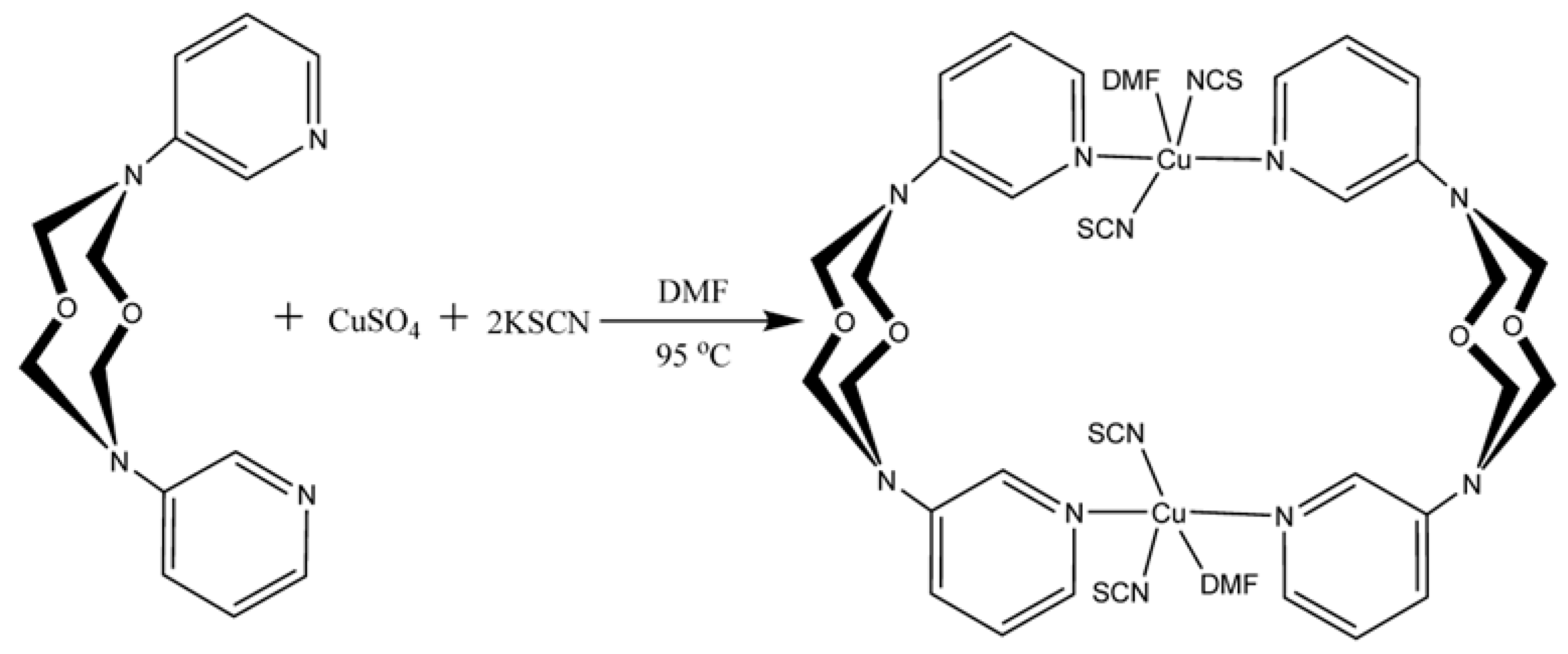
2.2.1. Synthesis of the [Cu(L)(SCN)2(DMF)]2 Complex

CuSCN (0.5 mmol, 60 mg), KSCN (0.6 mmol, 60 mg) and 3,7-di(3-pyridyl)-1,5-dioxa-3,7-diazacyclooctane (L) (0.5 mmol, 145 mg) were dissolved in 12 mL of DMF, stirred and refluxed at 90 °C for 30 min. The resultant solution was cooled to room temperature, generating 223 mg green rhombus crystals (yield = 85%). The FT-IR (in KBr) spectra exhibited the following peaks (cm−1): 2956(w), 2936(w), 2077(s), 1656(s), 1601(w), 1577(w), 1498(m), 1443(w), 1364(m), 1306(m), 1260(w), 1229(s), 1197(w), 1138(s), 1101(w), 1067(m), 1037(vs), 1017(s), 994(m), 944(s), 900(w), 878(w), 800(s), 692(vs), 671(s), 622(w) and 510(w). The following UV-Vis peaks were found: 267, 292 and 373 nm. In the alternative synthesis of complex 1, 3,7-di(3-pyridyl)-1,5-dioxa-3,7-diazacyclooctane (290 mg, 1 mmol), CuSO4·5H2O (249 mg, 1 mmol) and KSCN (195 mg, 2 mmol) were placed into a 25 mL flask, 18 mL of DMF was added and the mixture was stirred for 30 min at 90 °C. The resultant green powder was filtered and dried, giving a yield of complex 1 of 83%.

2.2.2. Single-Crystal X-Ray Crystallographic Study

Single crystals of L and complex 1 were obtained directly from the above syntheses. The measurements were made using a Bruker APEXII DUO X-ray diffractometer with graphite-monochromated Mo- radiation (λ = 0.71073 Å) in ω-2θ scan mode. The single crystal was mounted onto a glass fiber at 293(2) K. The cell parameters were refined using the Bruker SMART software (https://www.pubcompare.ai/product/qijiCZIBPBHhf-iFLWme/, accessed on 13 April 2025) with data reduction using Bruker SHELXTL. The reflection data were corrected for Lorentz and polarization effects. The structure of 1 was solved through direct methods and refined using full-matrix least squares calculations on F2 using the SHELX-97 package [12]. All non-hydrogen atoms were refined anisotropically. All H atoms were placed in geometrically idealized positions (C−H = 0.98 Å for methylene groups and 0.94 Å for pyridyl groups) and constrained to ride on the parent atoms with Uiso(H) = 1.2 Ueq(C). The crystallographic data and pertinent information are presented in Table 1; selected bond lengths and angles are given in Table 2.

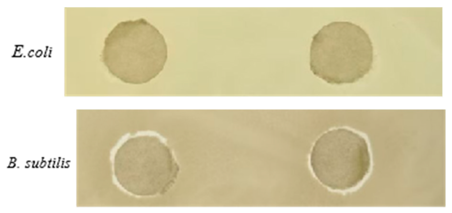
2.2.3. Antibacterial Tests

The antibacterial tests were conducted at the Laboratory of Suzhou University of Science and Technology (China). The tests involved two standard microorganisms, E. coli (CICC 24188) and B. subtilis (CICC 10721), which were stored in our laboratory. The selected microorganisms represent common microbial environmental contaminants and are representative Gram-negative and -positive bacteria. The microbial isolates were kept in tubes containing 20% glycerol at −80 °C. The strains were sub-cultured on fresh broth agar plates in incubators 24 h before the antimicrobial test and then transferred into the broth medium for cultivation until the logarithmic growth phase. A sample (100 μL) of the test microorganism suspension (OD650 = 0.5) was used to seed Petri dishes containing solidified broth agar with the bacterial strains. The tested compounds (20 mg/mL) were diluted in deionized formamide (DFM). In each case, 10 μL of the test product solution was used to soak 5.5 mm sterile filter paper discs, resulting in a 200 μg load per disc. The solvent was evaporated from the discs under an air flow for 2 h, which were then impregnated with the test products on the surface of the inoculated Petri dishes and lightly pressed to aid adhesion. The plates were pre-incubated for 30 min to facilitate the diffusion of the test product and then incubated aerobically at 37 °C for 48 h. Three replicate measurements were taken for each sample, and the mean value of the growth inhibition zone was determined.

3. Results and Discussion

3.1. The Structural Analysis

The L·H2O was a yellow rhombus crystal, whereas ligand L crystallized in methanol exhibited a white cubic structure. As presented in Figure 1A, the latter shows two parallel pyridine rings, unlike the anti-parallel conformation reported previously for L·H2O. In the case of the free ligand L, the centroid-to-centroid distance between two pyridine rings is 5.112 Å, and the interplanar angle is 52.98°, whereas these values for L·H2O are 4.939 Å and 46.35°, respectively. Compound L was stabilized by multiple hydrogen bonds. These included intra-molecular hydrogen bonds of C-H⋯O (C1-H1⋯O1, C8-H8⋯O1) and inter-molecular hydrogen bonds C-H⋯O (C5-H5⋯O1a) of and C-H⋯N (C7-H7a⋯N4,b C13-H13a⋯N4,b C13-H13b⋯N1c). These hydrogen bonds were in the range of 2.44–2.58 Å. No classic hydrogen bonds were found. The minimum distance between two centroids in the eight-membered ring in L is 4.624 Å. Complex 1 can also be prepared using CuSCN as the starting material, where copper is present as Cu+, attributed to its partial oxidation by ambient O2. The X-ray diffraction analysis established that complex 1 crystallizes as the space group P-1. The asymmetric unit comprises half a [Cu(L)(SCN)2(DMF)]2 molecule, as shown in Figure 1B. The Cu(II) center is five-coordinated with one oxygen from DMF occupying the axial position and four nitrogen atoms (two from SCN- anions and two from separate pyridine ligands) located at the square position, forming a [CuON4] distorted square–pyramid (τ = 0.098) [13]. The shortest Cu···Cu distance is 3.858 Å, which is greater than that reported for [Cu2Cl4(L)2] (3.625 Å) and [Cu(L)(µ-OAc)(OAc)]2 (3.513 Å) [14]. It can be seen that the bridging capability of the pseudohalide is inferior to that of the chloride and acetate anions. The shortest Cu···S distance is 4.595 Å, where the S atoms of SCN- do not bind with copper ions. The centroid-to-centroid distance between two pyridine rings is 3.805 Å, and the interplanar angle is 21.29°. The average Cu-N bond length is 2.030 Å, close to that in [Cu2Cl4(L)2] (2.023 Å) [14]. The bond length of Cu–O3 is 2.83(5) Å. Supplementing crystallographic data can be found in Supplementary Materials.

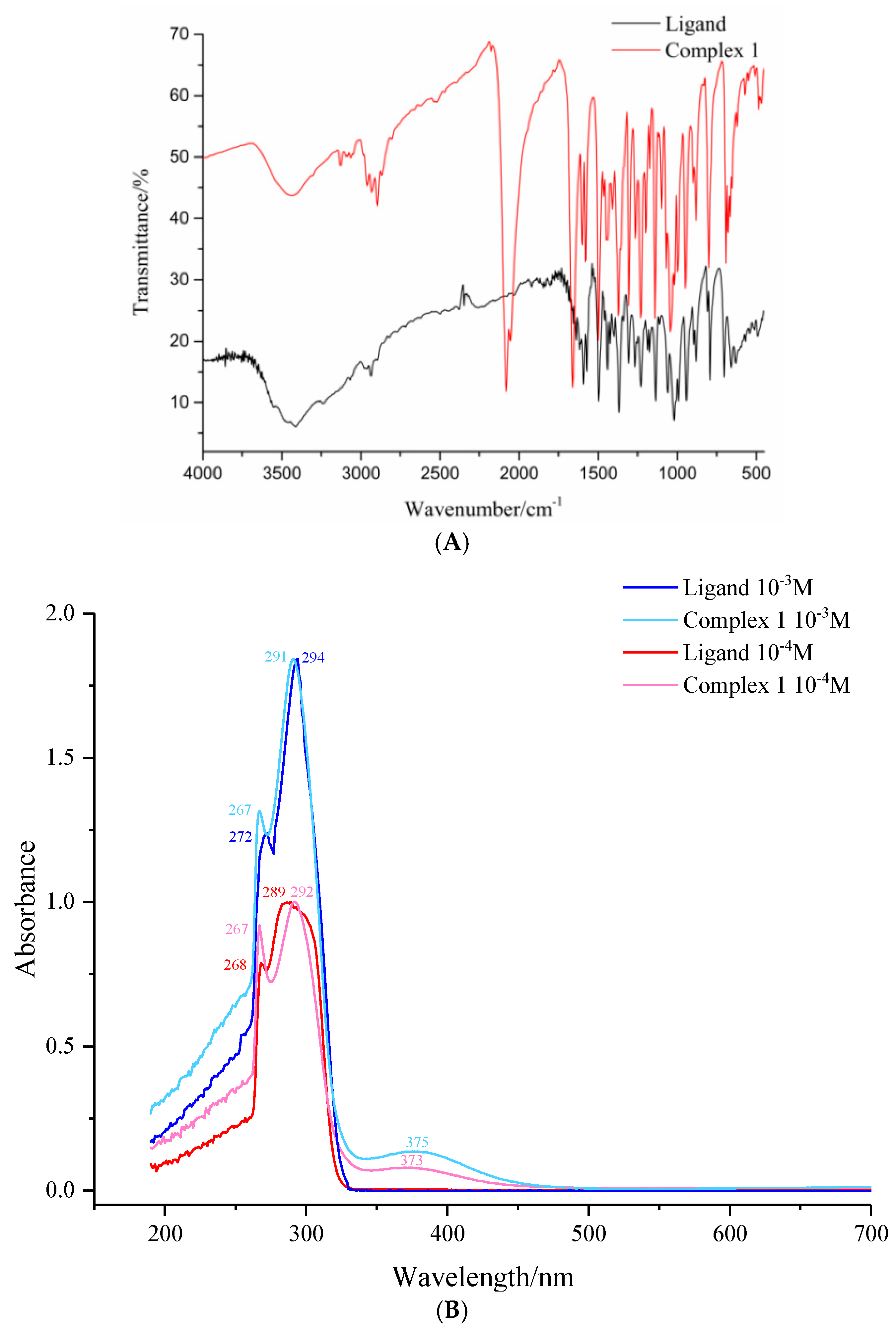
3.2. The IR and UV-Vis Spectra of Complex 1

The IR spectra of 1 and L were measured under the same conditions and are presented in Figure 2A. The bands at 2936 cm−1 (1) and 2936 cm−1 (L) are attributed to methylene symmetric/asymmetric stretching vibrations. The strong absorption at 2077 cm−1 represents the characteristic absorption peak for SCN. This is consistent with the spectra reported for [Cu(SCN)2(Bpybc)] (Bpybc = 1,1′-bis(4-carboxybenzyl)-4,4′-bipyridine) (2086 cm−1) [8] and [Cu(py)2(SCN)2] (2084 cm−1) [15]. The peak at 1660 cm−1 is ascribed to C=O stretching vibrations in DMF [16]. The bands at 1604, 1580 and 1498 cm−1 are assigned to the stretching vibrations associated with pyridine groups. The strong peak at 1138 cm−1 is due to C-O-C bond stretching vibrations.
The UV–visible spectra of different concentrations of the ligand and complex 1 in DMF were measured at room temperature. The associated peak shapes and normalized peak heights are equivalent, as shown in Figure 2B. The maximum absorption bands are at 250–350 nm in the low=energy region. In 10−4 M of complex 1, the peaks at 267 and 292 nm correspond to π-π* and n-π* transitions in the ligand, respectively. The small absorption peak at 373 nm is attributed to O-Cu charge transfer transition absorption in the DMF [8].

3.3. DSC-TGA Profiles for Complex 1

In order to evaluate the thermal stability of complex 1, a thermogravimetric analysis (TGA) was conducted for the crystalline samples, and the resulting profiles are shown in Figure 3A. The TGA curve of 1 shows that if the mass loss is due to the removal of the DMF solvent and the decomposition of the four formaldehyde molecules associated with the two ligands in the backbone (144–182 °C, exp.), the complex is stable up to 176 °C. The thermal stability of the ligand (L·H2O) was also assessed, as illustrated in Figure 3B. The endothermic peak observed at 203 °C is ascribed to the decomposition of methyleneaminopyridine associated with the ligand (182~700 °C, exp. 38.43%, calc. 40.80%). The thermal responses are in accordance with those reported for Cu2Cl4(L)2 and Cu2(CH3COO)4(L)2, with a thermal peak at approximately 200 °C [14], generating Cu(SCN)2 as residue (700 °C, exp. 34.83%, calc. 34.21%).

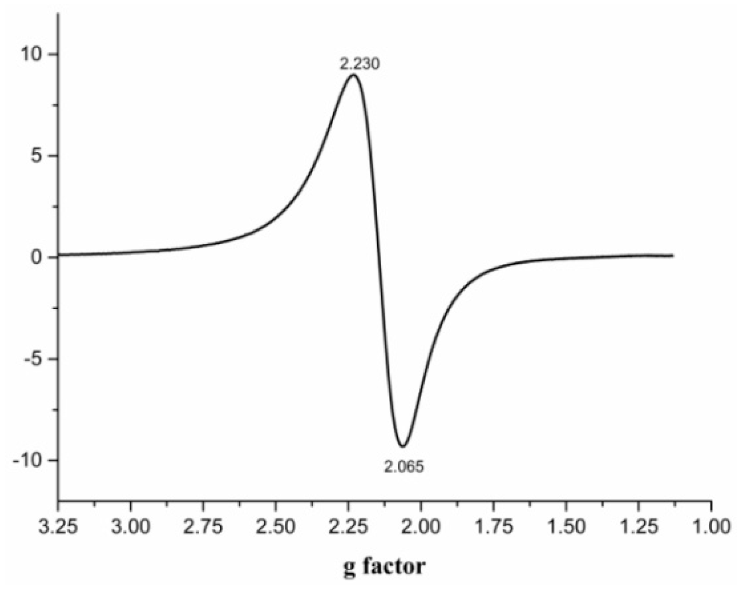
3.4. The EPR Spectra of Complex 1

The powder X-band EPR spectrum for complex 1 was recorded for powder crystals at 100 K. As shown in Figure 4, the sample exhibits a typical axial spectrum with well-resolved Cu hyperfine splitting in the parallel region [17,18]. No band was observed in the spectra corresponding to Ms = +2 transitions, suggesting that there is no Cu…Cu interaction. The values of gII and gI are 2.230 and 2.065, respectively, which are consistent with the values reported for monomeric [CuL4NO3]NO3·2H2O (L4 = N-methyl-benzimidazol-2-ylmethyl)amine) (2.24, 2.01) [19] and [CuL3Cl] (HL3 = 2-hydroxy-4-methyl-2′-(2-picolyl-thio)azobenzene) (2.23, 2.05). These findings suggest that there is no magnetic interaction between the two copper ions.

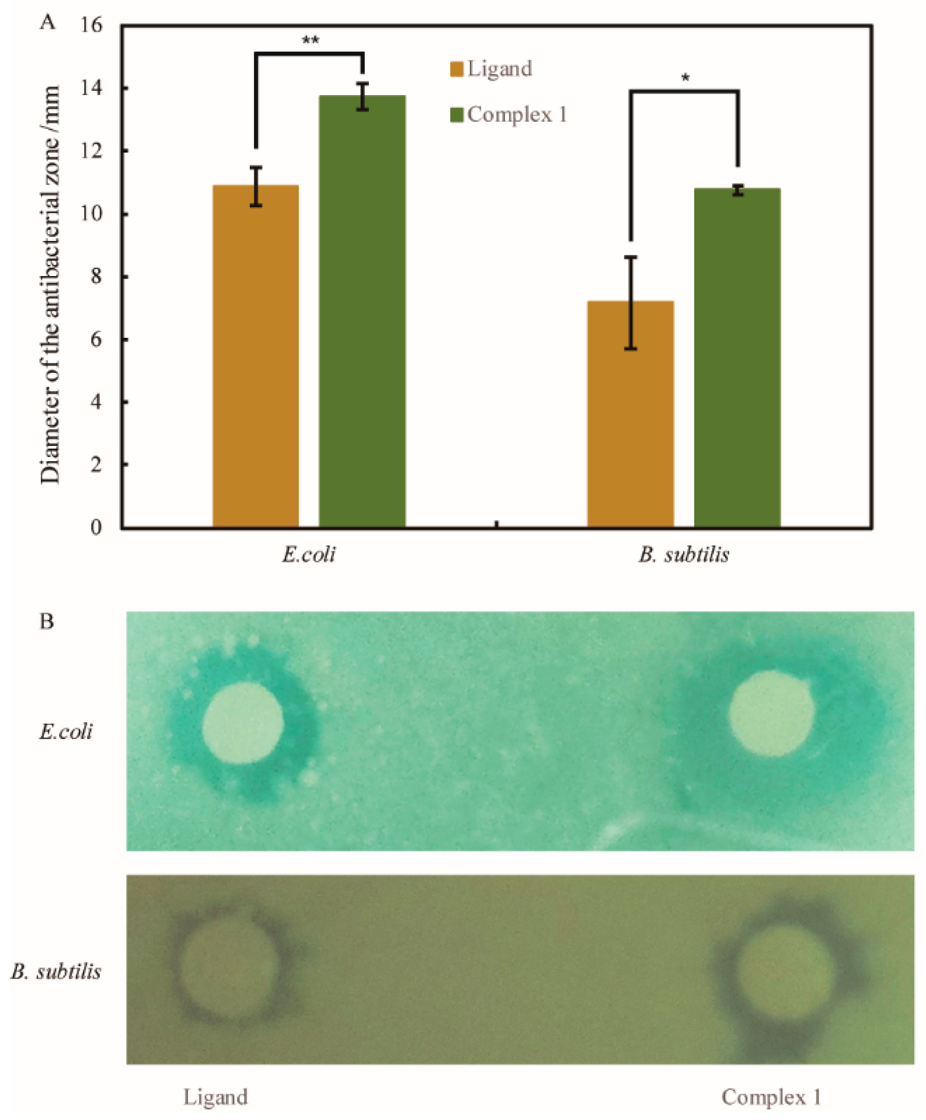
3.5. The Antimicrobial Activities of L and Complex 1

The disc diffusion method was employed for an in vitro evaluation of antibacterial activities against E. coli and B. subtilis. The inhibition diameter was recorded 48 h after incubation at 37 °C, using sterile filter paper discs impregnated with 200 µg of the polymeric complex dissolved in deionized formamide (DFM). The inhibitory effect of the ligand and complex 1 with respect to the two strains is illustrated in Figure 5.
The zones of inhibition (mm) of the ligand and complex 1 for both microorganisms are presented in Figure 5A. In the case of the Gram-negative bacteria, the zones of inhibition were 10.87 ± 0.60 mm (L) and 13.73 ± 0.40 mm (1). The zone was smaller for the Gram-positive bacteria, with average diameters for the antibacterial circles of 7.17 ± 1.47 mm and 10.77 ± 0.15 mm in the cases of L and 1, respectively. At the same time, the antibacterial experiments with DMF on E. coli and B. subtilis are presented in Figure 6, which showed that DFM had no antibacterial effect on E. coli; however, it had a certain inhibitory effect on B. subtilis, and the diameter of the inhibition circle was 5.62 ± 0.13 mm (Table 3).
The images associated with the antibacterial tests are shown in Figure 5B. The zone of inhibition (mm) of the ligand with respect to E. coli was significantly greater than that recorded for B. subtilis (p < 0.05). There is also a significant difference in the inhibitory effect of the metal complex on the growth of the two forms of bacteria (p < 0.01). The antimicrobial screening data established that the metal complex delivers an appreciably greater inhibitory effect than that of the free ligand (p < 0.05).

4. Conclusions

A copper pseudohalide complex, [Cu(L)(SCN)2(DMF)]2, has been synthesized using Cu2+/+, SCN and 3,7-di(3-pyridyl)-1,5-dioxa-3,7-diazacyclooctane and characterized using X-ray diffraction and optical and thermal analyses. The powder X-band EPR spectra suggest that there is no magnetic interaction between the two copper ions. The thermogravimetric analysis revealed that complex 1 is thermally stable up to 176 °C. Moreover, the antimicrobial screening of complex 1 established its superior inhibitory effect when compared with that of the free ligand (p < 0.05).
In this study, copper(II) thiocyanate was innovatively proposed to be coordinated with 3,7-di(3-pyridyl)-1,5-dioxa-3,7-diazacyclooctane, and a series of structural analyses, characterizations and antimicrobial experiments was carried out. However, the redox properties of complex 1 were not systematically investigated and analyzed. It is expected that future studies will find more ligands that can be synthesized using 3,7-di(3-pyridyl)-1,5-dioxa-3,7-diazacyclooctane. Meanwhile, it is expected that its antimicrobial action will be studied further and eventually put into commercial application.

Supplementary Materials

CCDC 2344183 and CCDC 2344184 contain the supplementary crystallographic data for this paper. These data can be obtained free of charge from the Cambridge Crystallographic Data Centre via www.ccdc.cam.ac.uk/data_request/cif, accessed on 13 April 2025.

Author Contributions

L.L.: Supervision; methodology; software; writing—original draft; visualization. W.Q. and Z.W.: Writing—review and editing. J.X., H.W. and S.D.: Investigation and validation. S.L. and P.D.: Writing—original draft. All authors have read and agreed to the published version of the manuscript.

Funding

This research was funded by the National Natural Science Foundation of China (This project is led by Professor Li), grant number 21676173, the Postgraduate Research & Practice Innovation Program of Jiangsu Province, grant number SJCX24_1905, and the Postgraduate Research & Practice Innovation Program of Jiangsu Province, grant number SJCX25_1853.

Data Availability Statement

The original contributions presented in the study are included in the article and Supplementary Materials, further inquiries can be directed to the corresponding author.

Conflicts of Interest

The authors declare that they have no known competing financial interests or personal relationships that could have appeared to influence the work reported in this paper.

References

  1. Wechwithayakhlung, C.; Packwood, D.M.; Harding, D.J.; Pattanasattayavong, P. Structures, bonding, and electronic properties of metal thiocyanates. J. Phys. Chem. Solids 2021, 154, 110085. [Google Scholar] [CrossRef]
  2. Li, L.-L.; Yuan, R.-X.; Liu, L.-L.; Ren, Z.-G.; Zheng, A.-X.; Cheng, H.-J.; Li, H.-X.; Lang, J.-P. Formation of [CuSCN]n-Based Topological Structures via a Family of Flexible Benzimidazolyl-Based Linkers with Different Spacer Lengths. Cryst. Growth Des. 2010, 10, 1929–1938. [Google Scholar] [CrossRef]
  3. Pramanik, S.; Mandal, P.C.; Das, K.; Pathak, S.; Mukhopadhyay, S. Synthetic, Structural and Supramolecular Features of a New Dithiocyanato-κ-N, κ-S-Copper(II) Complex: Insights Through Computational Studies. J. Chem. Crystallogr. 2023, 53, 417–430. [Google Scholar] [CrossRef]
  4. Qin, Y.; Jianfang, Q. Synthesis, Structure and Properties of 3D Copper(I) Pseudohalide Compound Constructed by Conformationally Flexible N-Heterocylic Ligand. J. Synth. Cryst. 2022, 51, 1059–1068. [Google Scholar]
  5. Han, X.; Shen, K.; Huang, G.; Li, C.; Mao, S.; Shi, X.; Wu, H. Synthesis, structure, electrochemical properties and superoxide radical scavenging activities of two thiocyanate copper(II) complexes with different pyridyl-benzoxazole ligands. J. Mol. Struct. 2018, 1169, 18–24. [Google Scholar] [CrossRef]
  6. El-bendary, M.M.; Akhdhar, A.; Ali, E.M.M.; Kalantan, A.A.; Davaasuren, B.; Jaremko, M.; Babgi, B.A. Synthesis, structure characterization and antitumor activities of copper and cobalt thiocyanate complexes with 3-acetlpyridine ligand. Polyhedron 2023, 242, 116511. [Google Scholar] [CrossRef]
  7. Laachir, A.; Rhoufal, F.; Guesmi, S.; Ketatni, E.M.; Saadi, M.; Ammari, L.E.; Mentré, O.; Bentiss, F. Novel Cu coordination polymer self-assembled from 2,5-bis(pyridin-4-yl)-1,3,4-oxadiazole and thiocyanate ions: Synthesis, structural characterization, Hirshfeld surface analysis, thermal and magnetic studies. J. Mol. Struct. 2022, 1269, 133790. [Google Scholar] [CrossRef]
  8. Laachir, A.; Zine, H.; Guesmi, S.; Mostafa Ketatni, E.; Saadi, M.; El Ammari, L.; Mentré, O.; Bentiss, F. Unusual mixed-valence CuII/CuI coordination polymer based on 2,5-bis(pyridine-2-yl)-1,3,4-thiadiazole and thiocyanate: Synthesis, structural characterization and antimicrobial in vitro activity assessment. Polyhedron 2021, 209, 115494. [Google Scholar] [CrossRef]
  9. Abalintsina, S.A.; Conde, M.A.; Eni, D.B.; Agwara, M.O. Synthesis, characterization and antimicrobial properties of Cobalt(III), Nickel(III), Copper(II) and Zinc(II) complexes with pyridine and thiocyanate. Sci. Afr. 2022, 18, e01402. [Google Scholar] [CrossRef]
  10. Gashu, M.; Aragaw, B.A.; Tefera, M.; Abebe, A. Poly(bis(2,2′-bipyridine) hydroxy Copper(II) iodide modified glassy carbon electrode for electrochemical determination of chloroquine in pharmaceuticals and biological samples. Sens. Bio-Sens. Res. 2023, 42, 100598. [Google Scholar] [CrossRef]
  11. Tsague Chimaine, F.; Mbom, Y.D.; Amah, C.B.Y.; Bekindaka, E.D.; Agwara, M.O. Synthesis, crystal structure, photoluminescent and antimicrobial properties of a thiocyanato-bridged copper(II) coordination polymer. Cogent Chem. 2016, 2, 1253905. [Google Scholar] [CrossRef]
  12. Li, L.; Dong, S.; He, W.; Wang, H.; Ding, P.; Liu, S.; Qian, W. Synthesis, crystal structures, electrochemistry and thermal stabilities of two copper complexes built by 3,7-di(3-pyridyl)-1,5-dioxa-3,7-diazacyclooctane. Polyhedron 2023, 231, 116268. [Google Scholar] [CrossRef]
  13. Sheldrick, G.M. A short history of SHELX. Acta Crystallogr. Sect. A Found. Crystallogr. 2008, 64 Pt 1, 112–122. [Google Scholar] [CrossRef] [PubMed]
  14. Yang, H.; Sun, X.-M.; Ren, X.-M. Syntheses, crystal structures and magnetic properties of two halogen bridged dinuclear copper(II) complexes [(4,4′-diethylester-2,2′-biquinoline)2Cu2(μ-X)2X2] (X−=Cl−, Br−). Polyhedron 2014, 83, 24–29. [Google Scholar] [CrossRef]
  15. Li, L. Syntheses, crystal structures and thermal stabilities of two copper complexes based on 3,7-di(3-pyridyl)-1,5-dioxa-3,7-diazacyclooctane. J. Struct. Chem. 2022, 63, 1929–1937. [Google Scholar] [CrossRef]
  16. Qiu, L.X.; Wan, F.; Zhu, B.B.; Sun, Y.Q.; You, Y.; Chen, Y.P. Synthesis, Structure and Spectroscopy Study of a 1D Copper Coordination Polymer Based on a Carboxybenzyl Viologen Ligand and SCN-Anion. Guang Pu Xue Yu Guang Pu Fen Xi = Guang Pu 2015, 35, 1340–1344. [Google Scholar] [PubMed]
  17. Li, H.H.; Chen, Z.R.; Li, J.Q.; Huang, C.C.; Hu, X.L. Crystal and electronic structure of novel organic-inorganic hybrid coordination polymer {[C12H28N2][(Pb 3I8) (DMF)2]•2DMF}n. Acta Chim. Sin.-Chin. Ed. 2005, 63, 697–702. [Google Scholar]
  18. Dhara, P.K.; Pramanik, S.; Lu, T.-H.; Drew, M.G.B.; Chattopadhyay, P. Copper(II) complexes of new tetradentate NSNO pyridylthioazophenol ligands: Synthesis, spectral characterization and crystal structure. Polyhedron 2004, 23, 2457–2464. [Google Scholar] [CrossRef]
  19. Karlin, K.D.; Hayes, J.C.; Juen, S.; Hutchinson, J.P.; Zubieta, J. Tetragonal vs. trigonal coordination in copper(II) complexes with tripod ligands: Structures and properties of [Cu(C21H24N4)Cl]PF6 and [Cu(C18H18N4)Cl]PF6. Inorg. Chem. 1982, 21, 4106–4108. [Google Scholar] [CrossRef]
Scheme 1. Preparation of complex 1.
Symmetry 17 00791 sch001
Figure 1. The molecular structures of L (A) and complex 1 (B) (30% probability ellipsoids; H atoms were omitted from 1 for clarify).
Symmetry 17 00791 g001
Figure 2. Infrared (A) and UV-Vis (B) spectra of L and complex 1.
Symmetry 17 00791 g002
Figure 3. DSC-TGA curves for complex 1 under a N2 atmosphere (A) and the TG curve for L·H2O under the atmosphere (B).
Symmetry 17 00791 g003
Figure 4. The EPR spectrum of complex 1 (100 K).
Symmetry 17 00791 g004
Figure 5. Antibacterial experiments with L and complex 1. Data are the means and confidence intervals from three replicates; asterisks indicate significant differences according to Tukey’s HSD test (A). Images of the results of the antibacterial experiment are shown in (B).
Symmetry 17 00791 g005
Figure 6. Antibacterial experiments with DMF.
Symmetry 17 00791 g006
Table 1. Crystal parameters of L and 1.
ComplexL1
Empirical formulaC14H16N4O2C38H46Cu2N14O6S4
Formula weight272.311050.21
Crystal systemMonoclinicTriclinic
Space groupP21/nP−1
a (Å)8.8945(13)10.4637(13)
b (Å)11.3334(16)11.0234(13)
c (Å)13.575(2)11.7951(15)
α (°)9062.755(2)
β (°)102.522(3)75.035(2)
γ (°)9076.914(2)
V3)1335.9(3)1 159.2(2)
Z21
F(000)576540
μ (mm−1)0.0941.158
GoF on F21.0711.080
Final R indices [I > 2σ(I)]: R1, wR2R1 = 0.042 9, wR2 = 0.136 6R1 = 0.056 1, wR2 = 0.167 6
R indices (all data): R1, wR2R1 = 0.056 6, wR2 = 0.158 6R1 = 0.078 5, wR2 = 0.205 1
Table 2. Selected bonds (Å) and angles (°) in 1.
BondLength/ÅBondAngle (°)
Cu(1)—O(3)2.383(5)O(3)—Cu(1) -N(1)92.6(2)
Cu(1)—N(1)2.030(4)O(3)—Cu(1) -N(2)89.4(2)
Cu(1)—N(2)2.037(4)O(3)—Cu(1) -N(5)88.2(2)
Cu(1)—N(5)1.992(4)O(3)—Cu(1) -N(6)99.0(2)
Cu(1)—N(6)1.971(4)N(1)—Cu(1) -N(5)89.4(1)
S(1)—C(15)1.611(5)N(1)—Cu(1) -N(6)87.71(1)
N(5)—C(15)1.162(6)N(5)—Cu(1) -N(2)91.51(1)
N(6)—C(16)1.146(6)N(5)—Cu(1) -N(6)172.41(2)
N(6)—Cu(1) -N(2)91.12(1)
Symmetry codes: -x,-y,-z.
Table 3. Antibacterial circle data in antibacterial experiments with DMF.
MicroorganismAntibacterial Circle/mm
E. coli0
B. subtilis5.62 ± 0.13
Disclaimer/Publisher’s Note: The statements, opinions and data contained in all publications are solely those of the individual author(s) and contributor(s) and not of MDPI and/or the editor(s). MDPI and/or the editor(s) disclaim responsibility for any injury to people or property resulting from any ideas, methods, instructions or products referred to in the content.

