Article

Molecular Dynamics Simulation from Symmetry Breaking Changing to Asymmetrical Phospholipid Membranes Due to Variable Capacitors during Resonance with Helical Proteins

by
Dung My Thi Dang
1,2,
Majid Monajjemi
1,3,*,
Fatemeh Mollaamin
4 and
Chien Mau Dang
1,2,*
1
Institute for Nanotechnology (INT) Community 6, Vietnam National University, Linh Trung Ward, Thu Duc District, Ho Chi Minh City 700000, Vietnam
2
Vietnam National University Ho Chi Minh City, Community 6, Linh Trung Ward, Thu Duc District, Ho Chi Minh City 700000, Vietnam
3
Department of Chemical Engineering, Central Tehran Branch, Islamic Azad University, Tehran 1477893855, Iran
4
Department of Biology, Faculty of Science, Kastamonu University, Kastamonu 37100, Turkey
*
Authors to whom correspondence should be addressed.
Symmetry 2023, 15(6), 1259; https://doi.org/10.3390/sym15061259
Submission received: 8 May 2023 / Revised: 5 June 2023 / Accepted: 12 June 2023 / Published: 14 June 2023
(This article belongs to the Section Chemistry: Symmetry/Asymmetry)

Abstract

:
Biological symmetry breaking is a mechanism in biosystems that is necessary for human survival, and depends on chemical physics concepts at both microscopic and macroscopic scales. In this work, we present a few mechanisms of the signaling phenomenon that have been studied in various tissues of human origin. We exhibit that anatomical asymmetry in the structure of a membrane can produce a flow of extracellular fluid. Furthermore, we exhibit that membrane asymmetry is a misbalance in the composition of the aqueous phases and interaction forces with the protein trans-membrane. Various biological membranes such as DPPC, DMPC, DLPC, and so on, have considerable electrostatic voltages that extend across the phosphor lipids bilayer. For studying these phenomena, we modeled DPPC, DMPC, and DLPC lipid bilayers with a net charge misbalance across the phospholipids. Because asymmetric membranes create the shifted voltages among the various aqueous tissues, this effect makes the charge misbalances cause a voltage of 1.3 V across the DPPC bilayer and 0.8 V across the DMPC bilayer. This subject exhibits the importance of membrane structures on electrostatic potential gradients. Finally, we exhibited that a quantum effect was created in small parts of the cell’s thickness due to the symmetry breaking of asymmetrical phospholipid bilayers.

1. Introduction

Symmetry in nature, from a pair of hands, a pair of feet, or a pair of eyes in human, up to the bilateral stripes of the tiger, is beautiful and awe inspiring, and indicates natural balances in a wide range of biological phenomenon. However, biological symmetry breaking due to asymmetry in macromolecules is a process by which the dissimilarity between biological macromolecules generates a more structured position [1,2,3,4].
As listed in Table 1, symmetry breaking in biological systems has been discussed considerably in recent decades. Mostly, relationships between inducing phenomena and biophysical results have been studied. It was indicated that the symmetry breaking of actomyosin and microtubule structures and the PAR genes exhibiting Wnt and Notch signaling caused a huge number of ancillary agents, and that this can produce extremely different species-specific results on the macroscopic biological system level [3,4,5].
Symmetry breaking is related to diversification on all scales, from molecular assemblies up to sub-membrane structures, including tissue architecture [6,7]. Symmetry breaking at the sub-membrane level causes different cell growth shapes such as membrane division, membrane fusion, and axon–dendrites specification. In biological systems, the origins of macromolecules’ asymmetry usually are related to the asymmetries of smaller parts.
As instances actin filaments, microtubules [8] and bilayer phospholipids are intrinsically polar due to their being assembled from different subunits. This polarity [3,4] plays important roles in their symmetry breaking mechanism, such as providing extra force generation and guiding the subunit assemblies towards producing the macromolecules and also creating the organelles in the cell. Obviously, cytoskeletal macromolecules similar to actin and alpha and beta tubulins also play important roles in polarization and asymmetrical cell division. Cell polarities result from membrane asymmetry along a special axis, and, in turn, determine asymmetry at the tissue or organismal levels [3]. Phospholipids separate proteins and liposomes from their exterior environs. They can automatically backlog into the bilayers, which is the fundamental structure of a membrane. They act as a suitable barrier to the entry/exit of molecules outside/inside the cells and perform channelization of various cell mechanisms. Due to their special scale, twisting, and fluid phase characteristics, it is hard to study the structures of this structure directly, exclusively in vivo. In the past decades, it has been distinguished that most phospholipid cell membranes have an asymmetric distribution in tissues. The flip-flop of these compounds when they exchange their structures indicates how cells construct and maintain asymmetry to prepare their biological and physical effects. The asymmetry mechanism is created based on several factors, such as different types of phospholipids, helical proteins, and chemically different and aqueous compartments. In other words, deleting lipids from one plasma membrane makes a strain in the cell membrane and consequently causes a curvature. For instance, small vesicles that have a large curvature are included in several different kinds of phospholipids in each plasma membrane. Inside the membrane cells, regions of stable curvature, which are similar to microtubules, exosomes appear during a vesicle fission/fusion process that manufacture asymmetry [9,10,11]. Van Meer [12] and Lorent [13] exhibited that mammalian plasma membranes strongly control the asymmetry of trans-bilayers’ distribution of lipids using phosphatidyl-choline and serine in the outer and inner plasma membrane. Although, up to now, the position of cholesterol, with its huge flip-flop, remains little discussed, the disruption of the phospholipids’ asymmetry with phosphatidyl-serine occurs during apoptosis [12]. This mechanism is due to the lipid rafts where an asymmetry can be caused and to a heterogeneous lipid distribution within the several-layer plasma membrane. Remarkable attempts have been undertaken to study the interaction between the lipid phase segregation in plasma membranes and the asymmetric distribution of phospholipids [14,15]. A major unknown issue is how arranged domains enriched by cholesterol in plasma membranes can induce domains in the inner plasma membrane. Particularly, the question that remains is how phospholipids can be transported across membranes via related proteins (trans-membrane proteins). Moreover, cells strongly control the osmotic gradients along the cells with trans-membrane protein channels via electrostatic potential in nerve cells using minimum energy [16,17]. Although wide, fundamental studies in the biological sciences have been accomplished using various methods, such as combinatorial chemistry, protein synthesis for self-organization, prebiotic biology, self-biological assembling, and the metabolism of organs, only a few studies were reported for asymmetric membranes [16,17,18]. In evolved cell membranes, enantiopure layers were grown in live tissue, with super-molecular systems that automatically kept persistent positions rather than achieving thermo-dynamic stability [19]. Symmetric and asymmetric cell membranes exhibited considerably different activities in lipid rafts and in membrane permeability. Recent research indicated that the symmetric segment of the cell membrane is located in prebiotic proto-cells [20]. In other words, the permeability property in the asymmetric phospholipids (as instanced in the vesicles’ bilayer) and the permeability of the phospholipids was lower than the more symmetric lipids [21]. This type of permeability exclusivity could be a mechanism for selecting suitable components for building up primitive organic ingredients, such as amino acids for phospholipids, nucleic acids, and their derivatives. This raises the question, why are most membranes made of symmetric phospholipids? The “advantage” of symmetric cell membranes rather than asymmetric ones is probably due to trans-cell-membrane proteins. In fact, the fluidity and flip-flop of cell membranes with lipid rafts can indicate tuned-up enzymatic activities that are considerably different in symmetric and in asymmetric cell membranes. Phospholipid structures can moderate enzyme activity, even within symmetrical membranes [22]. Via a hypothesis regarding earlier life membrane structures [23,24], it has been supposed that in the initial life, there was a presence of a cell membrane iso-prenoid hydrocarbon core, and fatty acids’ metabolism was retarded. In other words, the most diversity appeared in the glycerol phosphate backbone [24,25,26]. It has been proven that the pro-chiral di-hydroxyl acetone phosphate (DHAP) is the main root for the biosynthesis of phospholipids in living organs [27,28,29]. Advanced cells are worked similarly to bio machines, with the reaction processes accomplished through enzymes. However, prebiotic mechanisms in the last common ancestor (LCA) proto-cells [30,31,32,33], perhaps were not the same as in the actual ones. In addition, system proto-biology indicates that phospholipids had a basic role in the emergence of living organs in the initial life.

1.1. Prebiotic Approaches for the Asymmetry of Phospholipids Structures

Obviously, all chiral molecules can appear in both enantiomer types, including with asymmetry between the two stereoisomers demonstrated. It is notable that during the formation of prebiotic peptides (1) and di-hydroxyl-acetone (2), glycerol (3) associates with phosphate derivatives (Figure 1).
The redox reactions (Equations (1)–(6)) are a key for various biological reactions in the reduction process that are conducted via FADH2 or NAD (P) H, and which can recycle the cofactors for the next round of oxidation [34].
R1-C = (O)-R2 + 2H+ + 2e = R1-CH(OH)-R2
NAD(P)H + H+ =NAD(P)+ + 2e + 2H+
R1-C = (O)-R2 +NAD(P)H + H+ = R1-CH(OH)-R2 + NAD(P)+
Concerning the oxidation of 3, where R 1 and R 2 are C H 2 O H , respectively:
R1-CH(OH)-R2 = R1-C = (O)-R2 +2H+ + 2e
NAD(P)+ + 2e + 2H+ = NAD(P)H + H+
R1-CH(OH)-R2 +NAD(P)+ =R1-C=(O)-R2 +NAD(P)H + H+
It is notable that early membranes were more likely formed from derivatives of alkanols [35,36,37], fatty acids [38], mono-alkyl phosphates [39], and isoprenoids [40,41] (Figure 2).

1.2. Achiral and Racemic Amphiphiles

Various acceptable prebiotic syntheses were considered and all the supposed pathways accomplished in enzyme-free conditions [42,43,44,45,46,47] or racemic mixtures of glycerol phosphates [48,49,50,51]. The asymmetry between the R: S ratio of mono phosphates and cyclic glycerol-phosphates [52,53] from di-acyl glycerol 12 or glycerol 3, respectively, were not discussed (Figure 3).

2. Materials and Methods

2.1. Lipid Membrane Simulations

We modeled the phospholipid bilayers with several numbers of asymmetry using the Martini tool [54,55], containing 500 lipids in each cell membrane of liver tissue, through running asymmetrical forms of DPCC, POPC, DMPC, and PAPE mixed lipids with a 30/40/30 ratio in the top leaflet and 20/60/20 in the lower leaflet of the cell membrane of the liver tissue. Simulations were done with 40 mol% cholesterol content with lipid number asymmetry (Table 2).

2.2. Computational Details

CHARMM, the bimolecular simulation program, was used for this work as a useful molecular simulation and modeling program. CHARMM36 lipids [56] with TIP3P water [57] were simulated with GROMACS [58].
The structures were taken from the 150 ns frame of the Martini model. The backbone was simulated by CHARMM36 using the Backward structure model [59,60]. Outer time steps of between 1 and 5.0 fs were used for non-bonded forces beyond the cutoff. The function of the molecular dynamics method was restricted by a final time step between 1–5 fs, that was needed to define the best degrees of freedom in the simulation, namely, the vibration of bonds and angles involving hydrogen atoms. Algorithms for most lipids in the CHARMM36 force field were in a suitable agreement with those calculated in both simulated and experimental phospholipid bilayers. This method has an ability to make longer time and length scales accessible in all-atom simulations of membrane–protein complexes.
Simulations were then started using a 1–5 fs time step. Lennard-Jones interactions were cut off after 1.3 nm [61,62] and a temperature of 300 K. Part of the systems containing di-palmitoylphosphatidylcholine (DPPC)n and (DMPC)n were simulated with a mixture of quantum mechanics and molecular mechanics (QM/MM) methods. Each system contained 60 phospholipids [62,63,64]. At 300 K, the clusters had finite vapor pressures that were generally unstable to evaporation. Each of 60 DPPC and DMPC molecules was optimized with their lateral parts for a proper simulation. In this investigation, various force fields were used through CHARMM (OPLS force field) software. In addition, the Hyper Chem professional 7.01 program was also used for the simulated calculations. All optimization of DPPC, DOPC, DMPC, and PAPE monomers was done by Gaussian 09. The main focus in this study was to obtain the results from DFT methods, such as m062x, m06-L, and m06 for the phosphor lipids. The ab-initio and DFT methods were used for the nonbonded interactions between two parts of the upper lateral phospholipids side (P+) and the lower lateral phospholipids side (P−) (Figure 4). Based on our previous works, we simulated our systems in the viewpoint of computational biochemistry using biochemistry software [65,66,67,68,69,70,71,72,73].

3. Results and Discussion

3.1. Asymmetry with Fewer Lipids

For finding the lipid number asymmetry for complex asymmetrical bilayers, we systematically simulated the asymmetric PPC/POPC/DMPC mixture containing 40 mol% of cholesterol (Figure 5). In this mixture, the upper cell membrane of liver tissue had significantly more lipids with saturated tails (DPPC) and fewer with a high level of unsaturation (DMPC) compared to the lower cell membrane of liver tissue. To distinguish what the number asymmetry should be, we systematically reduced the number of lipids in the lower cell membrane of liver tissue, simulating 5–6 repeats at each asymmetry value ranging from 0 to 35 random lipids removed from the lower cell membrane of liver tissue. For the cholesterol, both cell membranes of the liver tissue began with an equal amount that then flip-flopped between the cell membrane of liver tissue. Figure 5A exhibits the cholesterol among the bilayer phospholipids. There was a net drive towards cholesterol from the lower cell membrane of liver tissue into the upper one (Figure 5B). Figure 5C shows the average area per lipid (APL) for systems with (right) and without (left) cholesterol as the number asymmetry increases. As can be seen in Figure 5B–D, cholesterol has a high ability among the various lipids to flip from one leaflet to another one. Bennett and coworkers [74,75,76,77] investigated the effect of cholesterol mobility on asymmetry based on the number of cholesterol molecules in each leaflet during their simulations of the symmetry situation towards asymmetry. They considered cholesterols as belonging to each leaflet, established by MD analysis with a leaflet finder using the non-flip-flopping lipids’ PO4 units. Marrink and coworkers exhibited a Martini coarse-grained (CG) model [78] that has been used for analyzing the data and situation of CG membrane asymmetry. As can be seen in Figure 5C,D symmetric simulation provided values of 13.8 and 12.5 for without and with cholesterol, respectively, and the asymmetry simulation provided values of 16.5 and 15.8 for without and with cholesterol, respectively (vertical lines).
In Table 3, the APLs for each of phospholipids in both the upper and lower cell membrane of liver tissue are listed. [79,80,81,82].

3.2. Membrane Asymmetry

We calculated the effect of membrane asymmetry on the phospholipid structure of atomistic lipid membranes through the bilayers model with CHARMM36, with lipids after 150 ns for each one. Reducing the electrostatic potential and the number of lipids on one side of DPPC caused an increase in potential of ∼80 mV, whereas having DMPC or DOPC on the other cell membrane of liver tissue caused a potential of ∼−60 mV. We estimated the coefficient parameters for DPPC in the mixed structure membranes. We also calculated the effect of asymmetric membranes with different head groups, with DPPC in one cell membrane of liver tissue and DOPC in the other. The DPPC in the DOPC (upper)/DPPC (lower) bilayer had a high coefficient, with a similar structure to the DPPC_5 upper. This result shows how one cell membrane of liver tissue can compensate for the other, with the DPPC becoming more numerous to accommodate the DMPC in the opposite cell membrane of liver tissue. With 40 mol% cholesterol, there are interesting differences, with the DPPC lipids in the DMPC (upper)/DPPC (lower) mixed membranes having a higher coefficient compared with the pure DPPC bilayer. Asymmetric membranes make it possible to shift among the various aqueous tissues. Reducing the number of lipids on one side (DPPC_10) made an increase in potential of around110 mV, whereas having DMPC or DOPC on the other cell membrane of liver tissue made a potential of 80 mV. We also exhibited the electrostatic potential across a DMPC bilayer based on the Martini model that did not reproduce the positive potential at the membrane center.

3.3. Electrostatic of Symmetry Breaking in Bilayers

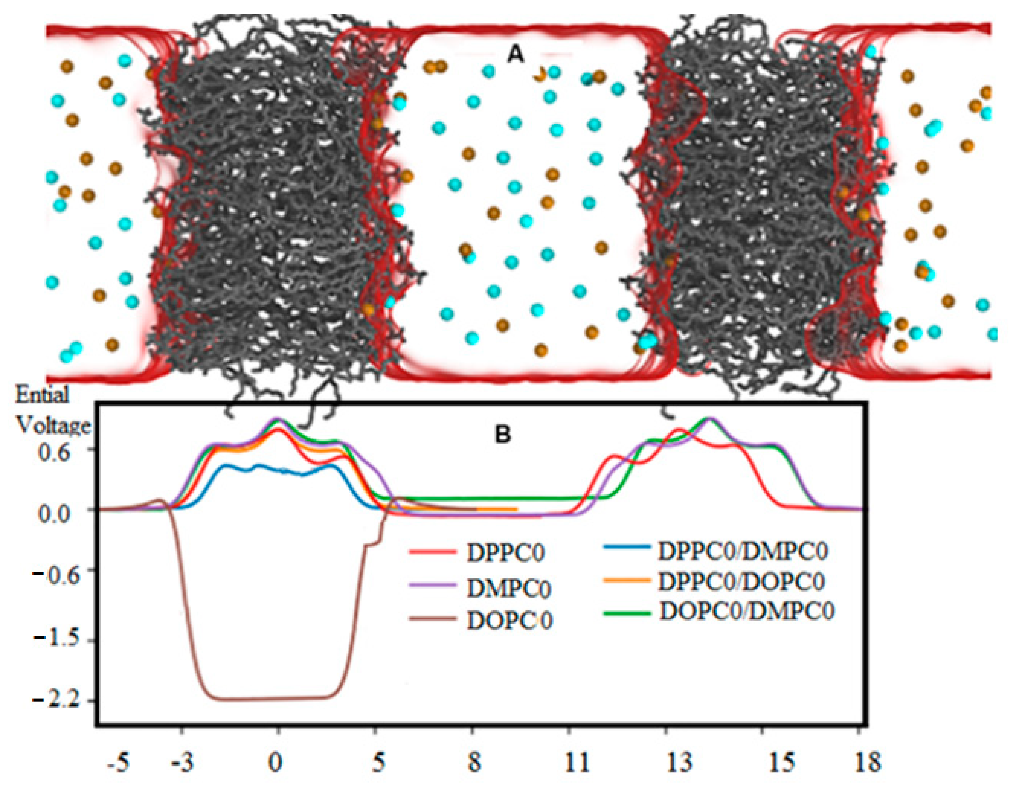
Symmetry breaking causes substantial electrostatic voltage differences on either side of the lipid bilayer membrane, leading to significant structural modifications. This phenomenon is valid across numerous biological processes. This mechanism can occur in either leaflet, and is also referred to as a trans-leaflet exchange (flip-flop). It is how cells create the biological and physical effects of membrane asymmetry, invoked to produce electrostatic charges between two sides of a phospholipid membrane. Consequently, based on these electrostatic forces (that are different in variants of phospholipids such as DPPC, DLPC, DMPC, and so on), different voltages appear in each part of the cell membrane that influence the transferring proteins. There are several models that are able to change a membrane towards the asymmetry situation, including different numbers of lipids, different types of lipids, asymmetric proteins, and, finally, symmetry breaking, mostly near the lipid raft and cholesterol zone. In addition, in membranes with different numbers of lipids, and also in cell membrane regions of high curvature, such as microtubules, symmetry breaking appears. Figure 6 and Figure 7 exhibit the electrostatic potential across lipid membranes during symmetry breaking. The power of the potential voltage is related to the bilayer composition and also to unsaturated bilayers. For symmetric bilayers, the voltage is equal to zero between both sides of the cell membrane. The DMPC bilayer has a lower voltage compared to the DPPC bilayer. Asymmetric membranes and their interactions with helical protein trans-membranes cause the potential to shift considerably. Although DPPC_10 makes an increase in potential of ∼90 mV on one side, having DMPC or DOPC on the other cell membrane of liver tissue causes a potential of ∼−65 mV. Another item to note about membrane asymmetry is the misbalances in the composition of the aqueous phases and interaction forces with the protein trans-membrane. Various biological membranes have an electrostatic voltage across the bilayer. To investigate these phenomena, we simulated lipid bilayers with net charge misbalances across their phospholipids. Figure 6 exhibits the electrostatic potential across pure DPPC, DMPC, and DOPC lipid bilayers, compared with a mixture of DPPC/DMPC, DPPC/DOPC, and DMPC/DOPC.
It is realistic to state that lipid distribution changes the membrane’s electrostatic properties, which are important behaviors in numerous biological processes. It is interesting that these membranes’ large electrostatic voltage gradients can promote structural fluctuations.

3.4. Membrane Capacitor Model

In the symmetrical bilayers’ membranes, the medium value of charges in both down and up leaflet were equal, but due to symmetry breaking and an asymmetry in the structures, this equality disappeared and, consequently, based on the following equations, the potential difference arose from this parity. The membrane variable capacitance, C m e m ( t ) , defines the amount of charge (q) which is stored on bilayers. Generally, in the nano scale of the cell membrane, quantum electrostatic force must be taken into consideration. This model indicates that the geometric capacitance C m e m g e o is related to the applied voltage, V m e m based on Equation (7):
C m e m g e o = σ V m e m ( t ) = ε r ε 0 d m e m ( t )
where σ is the surface of charge density, ε 0 is ∼8.85 × 10⁻¹² F.m−1, and d m e m ( t ) is the thickness of the dielectric between two sides of the membrane. Energy stored in the membrane capacitor can be written like this:
E i = Q 2 2 C
Based on the quantum tunneling effect, the electrons move through the insulating layer from the negative side to the positive side; consequently, (Q + ∆qe) resides on the top plate and (−Q −∆qe) resides on the bottom of the two membranes’ sides, and therefore, the stored energies are equal ( E f = ( Q + q ) 2 2 C ) inside the membrane capacitor. Since the charge is quantized, it is polarized on the membrane capacitor and, consequently, the energy cannot be saved before a single electron tunnels through the alkyl layers; therefore, the energy can be calculated as Equation (9):
E s = E f E i = q ( Q + q 2 ) C
Additionally, the large voltage is located between q 2 C < V < + q 2 C , and the tunneling current will only flow if the voltage is sufficiently large, i.e., | V | > | q e 2 C | . This effect is known as the Coulomb blockade [83]. Since, for a macroscopic capacitor, the amount of C ~ 1013 F is related to voltages larger than V > 1.5   μ V , the tunneling effect can occur in voltages as V < 1.5   μ . For nano-scale capacitors with capacitance in the range of “C ~ 1017 F”, the amount of 1.5   m V > V < 0.73   V is required to have a tunneling effect. By supposing
R T u n = V I
as tunneling resistance, it theoretically allows the electrons to cross the insulating junction as discrete occurrences, where “I” is the resulting current due to the tunneling effect. Although the tunneling resistance is a virtual resistance or an imaginary item, it allows the electrons to cross the insulating junction during
t = R T u n C Q
where C Q indicates the quantum capacitance and “t” is a characteristic time [84]. Hence, the hybrid capacitance of any nano-capacitor architecture is:
C m e m = ( 1 c m e m q u a ( o n e h a n d ) + 1 c m e m g e o + 1 c m e m q u a ( o p p o s i t e h a n d ) ) 1
where c m e m q u a (top side) and c m e m q u a (down side) due to the changes in the charge on the capacitor, which is given by:
d Q d t = d d t [ C m e m × V m e m = C m e m d V m e m d t + V m e m d C m e m d t
where:
d C m e m d t = f ( d C m e m g e o d t , d C m e m q u a d t )
For large dielectric thicknesses, the classical capacitance rule of the “ C g 1 d ” is adaptable. This adaptability is not valid for short distances, which is attributed to the quantum effect. The dielectric permittivity as a function of dielectric size, through C Q F × 10 20 = q ( Q + q 2 ) E S   a n d   C n e t F × 10 20 = C g C Q C Q + 2 C g and quantum effect by R T u n ( K ) E S P 2 , using the QM/MM calculations, is identified (Table 4 and Table 5). The quantum effect of different thicknesses has also been listed in Table 4.
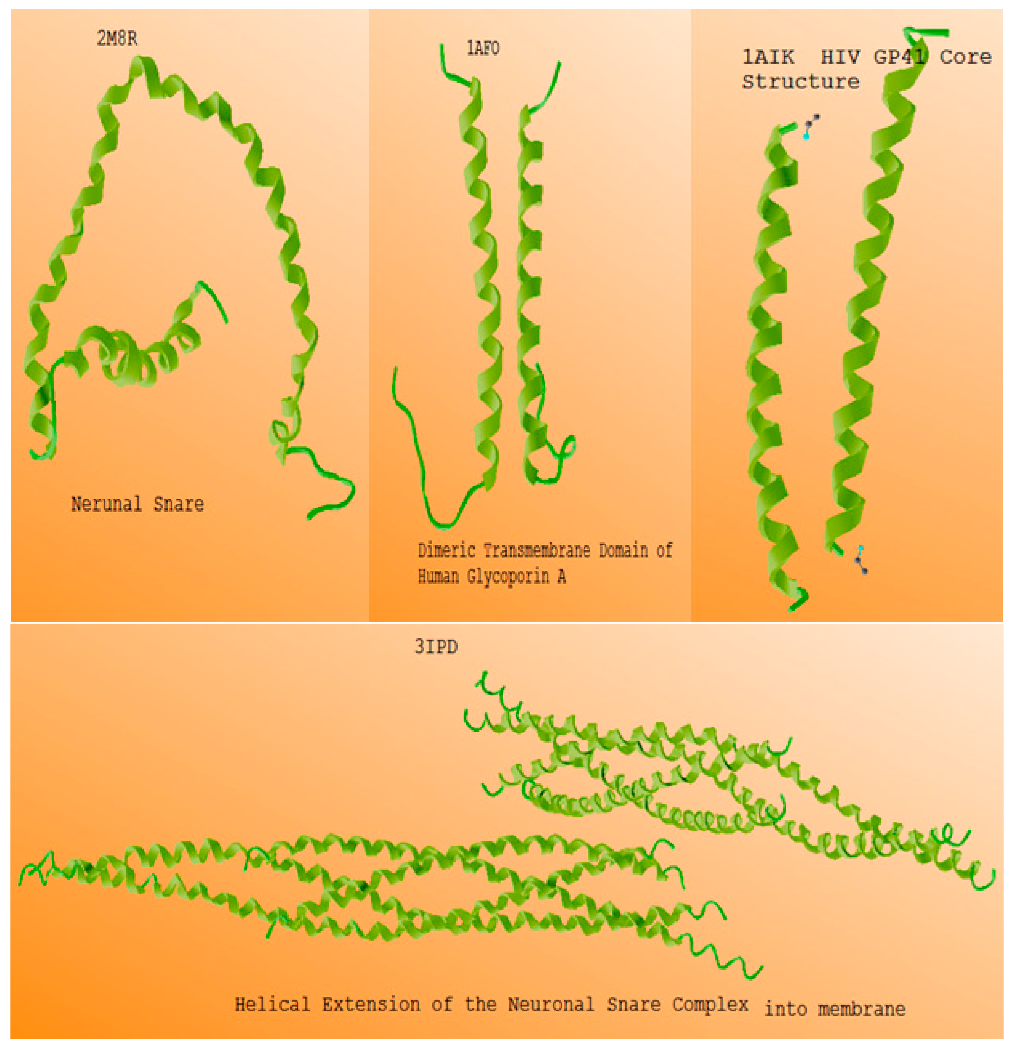
Table 6 and Figure 8 indicate the calculated helical proteins’ self induction through resonance with the capacitance of the DPPC phospholipids due to symmetry breaking.

4. Conclusions

Due to symmetry breaking, various biological membranes such as DPPC, DMPC, DLPC, and so on, have considerable electrostatic voltages across the phosphor lipids bilayer. It is realistic to state that lipid distribution changes the membrane’s electrostatic properties, which are important behaviors in numerous biological processes. It is notable that the large electrostatic potentials have a medium effect on the membranes’ overall structure [84,85,86,87]. In physics, in an electrical circuit including a capacitor with a capacitance of “C” and electrical coils such as an electrical bobbin with self-induction coefficient “L”, there are two virtual resistances; the first is X c = 1 C ω and the second is X l = L ω and ω = 2 π ϑ , and ϑ is the resonance frequency. During the parity of these two resistances, the phenomenon of resonance will appear, and its frequency can be calculated as: X l = X c , then L ω = 1 C ω and consequently L C ω 2 = 1 and ϑ = 1 2 π L C . Radio and television and many other similar instruments work based on the mentioned phenomenon. In biological systems, the phospholipid membrane works in such a way, as a variable capacitor. Additionally, alpha helical proteins are similar to electrical coils, such as electrical bobbins with self-induction coefficient “L”, but in the form of biological self induction. The root of many internal wave pulses such as HER2 or HER3 in cancer cells and also the external propagation of waves such as telepathy and so on are resonated between variable phospholipid membranes as capacitors and alpha helical proteins.
In magnetic external fields through membrane proteins, charges produce the forces that potentially affect the asymmetrical situation of bilayer membranes; consequently, by this external pressure, the variable electrical fields create a variable capacitance. This phenomenon permits the system to make a resonance force with helical coils such as alpha helical proteins during the self-induction effect. Due to the large inductance compared with conductance in coils, the alpha helical proteins of the bilayer membrane resonate with a capacitive susceptibility as phospholipid capacitors in the biological system. Due to proteins being coiled, the magnetic field created a strong current across neighboring coil rings, and this current changed bit by bit, and consequently induced an internal magnetic field, and then forces that opposed the changes in the current. This effect did not appear in symmetrical membranes and it was only possible for it to occur in variable capacitors arising from symmetry breaking. When the current flowed quickly, the magnetic field also collapsed quickly, and was capable of generating high induced forces. During the interaction of the cell membrane with helical proteins as electrical inductors, we were able to produce suitable electrical voltages due to the asymmetry phospholipids and the symmetry breaking phenomenon.

Author Contributions

Conceptualization, M.M and C.M.D.; methodology, D.M.T.D., F.M., M.M. and C.M.D.; software, D.M.T.D., F.M., M.M. and C.M.D.; validation, D.M.T.D., F.M., M.M. and C.M.D.; formal analysis, D.M.T.D., F.M., M.M. and C.M.D.; investigation, D.M.T.D., F.M., M.M. and C.M.D.; resources, D.M.T.D. and F.M.; data curation, D.M.T.D., F.M., M.M. and C.M.D.; writing—original draft preparation, M.M. and C.M.D.; writing—review and editing, D.M.T.D. and F.M.; visualization, D.M.T.D., F.M., M.M. and C.M.D.; supervision, M.M. and C.M.D.; project administration, M.M. and C.M.D. All authors have read and agreed to the published version of the manuscript.

Funding

This research is funded by Vietnam National University Ho Chi Minh City (VNU-HCM) under grant number TX2023-32-01.

Data Availability Statement

Not appreciable.

Acknowledgments

The authors thank the Vietnam National University Ho Chi Minh City (VNU-HCM) under grant number TX2023-32-01 for their support.

Conflicts of Interest

The authors declare no conflict of interest.

References

  1. Li, R.; Bowerman, B. Symmetry breaking in biology. Cold Spring Harb. Perspect. Biol. 2010, 2, a003475. [Google Scholar] [CrossRef]
  2. Wodarz, A. Establishing cell polarity in development. Nat. Cell Biol. 2002, 4, E39–E44. [Google Scholar] [CrossRef]
  3. Li, R.; Gundersen, G.G. Beyond polymer polarity: How the cytoskeleton builds a polarized cell. Nat. Rev. Mol. Cell Biol. 2008, 9, 860–873. [Google Scholar] [CrossRef]
  4. Munro, E.; Nance, J.; Priess, J.R. Cortical flows powered by asymmetrical contraction transport PAR proteins to establish and maintain anterior-posterior polarity in the early C. elegans embryo. Dev. Cell 2004, 7, 413–424. [Google Scholar] [CrossRef] [PubMed] [Green Version]
  5. Borovina, A.; Superina, S.; Voskas, D.; Ciruna, B. Vangl2 directs the posterior tilting and asymmetric localization of motile primary cilia. Nat. Cell Biol. 2010, 12, 407–412. [Google Scholar] [CrossRef]
  6. Van der Gucht, J.; Sykes, C. Physical model of cellular symmetry breaking. Cold Spring Harb. Perspect. Biol. 2009, 1, a001909. [Google Scholar] [CrossRef]
  7. Motegi, F.; Seydoux, G. The PAR network: Redundancy and robustness in a symmetry-breaking system. Philos. Trans. R. Soc. Lond. B Biol. Sci. 2013, 368, 20130010. [Google Scholar] [CrossRef] [PubMed] [Green Version]
  8. Padinhateeri, R.; Kolomeisky, A.B.; Lacoste, D. Random hydrolysis controls the dynamic instability of microtubules. Biophys. J. 2012, 102, 1274–1283. [Google Scholar] [CrossRef] [Green Version]
  9. Callan-Jones, A.; Sorre, B.; Bassereau, P. Curvature-driven lipid sorting in biomembranes. Cold Spring Harb. Perspect. Biol. 2011, 3, a004648. [Google Scholar] [CrossRef] [PubMed] [Green Version]
  10. Skotland, T.; Sandvig, K.; Llorente, A. Lipids in exosomes: Current knowledge and the way forward. Prog. Lipid Res. 2017, 66, 30–41. [Google Scholar] [CrossRef] [PubMed]
  11. Gruenberg, J. Life in the lumen: The multivesicular endosome. Traffic 2020, 21, 76–93. [Google Scholar] [CrossRef] [Green Version]
  12. Van Meer, G.; Voelker, D.R.; Feigenson, G.W. Membrane lipids: Where they are and how they behave. Nat. Rev. Mol. Cell Biol. 2008, 9, 112–124. [Google Scholar] [CrossRef]
  13. Lorent, J.H.; Levental, K.R.; Ganesan, L.; Rivera-Longsworth, G.; Sezgin, E.; Doktorova, M.; Levental, I. Plasma membranes are asymmetric in lipid unsaturation, packing and protein shape. Nat. Chem. Biol. 2020, 16, 710. [Google Scholar] [CrossRef]
  14. Collins, M.D.; Keller, S.L. Tuning lipid mixtures to induce or suppress domain formation across leaflets of unsupported asymmetric bilayers. Proc. Natl. Acad. Sci. USA 2008, 105, 124–128. [Google Scholar] [CrossRef] [Green Version]
  15. Perlmutter, J.D.; Sachs, J.N. Interleaflet interaction and asymmetry in phase separated lipid bilayers: Molecular dynamics simulations. J. Am. Chem. Soc. 2011, 133, 6563–6577. [Google Scholar] [CrossRef]
  16. Becker, S.; Feldmann, J.; Wiedemann, S.; Okamura, H.; Schneider, C.; Iwan, K.; Crisp, A.; Rossa, M.; Amatov, T.; Carell, T. Unified prebiotically plausible synthesis of pyrimidine and purine RNA ribonucleotides. Science 2019, 366, 76–82. [Google Scholar] [CrossRef] [Green Version]
  17. Eghiaian, F.; Rico, F.; Colom, A.; Casuso, I.; Scheuring, S. High-speed atomic force microscopy: Imaging and force spectroscopy. FEBS Lett. 2014, 588, 3631–3638. [Google Scholar] [CrossRef] [PubMed]
  18. Böhm, C.; Möhwald, H.; Leiserowitz, L.; Als-Nielsen, J.; Kjaer, K. Influence of chirality on the structure of phospholipid monolayers. Biophys. J. 1993, 64, 553–559. [Google Scholar] [CrossRef] [PubMed] [Green Version]
  19. Eschenmoser, A.; Loewenthal, E. Chemistry of potentially prebiological natural products. Chem. Soc. Rev. 1992, 21, 1. [Google Scholar] [CrossRef]
  20. Subramanian, H.; Gatenby, R.A. Evolutionary advantage of directional symmetry breaking in self-replicating polymers. J. Theor. Biol. 2018, 446, 128–136. [Google Scholar] [CrossRef]
  21. Altamura, E.; Comte, A.; D’Onofrio, A.; Roussillon, C.; Fayolle, D.; Buchet, R.; Mavelli, F.; Stano, P.; Fiore, M.; Strazewski, P. Racemic Phospholipids for Origin of Life Studies. Symmetry 2020, 12, 1108. [Google Scholar] [CrossRef]
  22. Schreiber, A.; Huber, M.C.; Schiller, S.M. Prebiotic Protocell Model Based on Dynamic Protein Membranes Accommodating Anabolic Reactions. Langmuir 2019, 35, 9593–9610. [Google Scholar] [CrossRef] [PubMed]
  23. Koga, Y.; Kyuragi, T.; Nishihara, M.; Sone, N. Did Archaeal and Bacterial Cells Arise Independently from Noncellular Precursors? A Hypothesis Stating that the Advent of Membrane Phospholipid with Enantiomeric Glycerophosphate Backbones Caused the Separation of the Two Lines of Descent. J. Mol. Evol. 1998, 46, 54–63. [Google Scholar] [CrossRef] [PubMed]
  24. Yokobori, S.; Nakajima, Y.; Akanuma, S.; Yamagishi, A. Birth of Archaeal Cells: Molecular Phylogenetic Analyses of G1P Dehydrogenase, G3P Dehydrogenases, and Glycerol Kinase Suggest Derived Features of Archaeal Membranes Having G1P Polar Lipids. Archaea 2016, 2016, 1802675. [Google Scholar] [CrossRef] [PubMed] [Green Version]
  25. Koga, S.; Williams, D.S.; Perriman, A.W.; Mann, S. Peptide-nucleotide microdroplets as a step towards a membrane-free protocell model. Nat. Chem. 2011, 3, 720–724. [Google Scholar] [CrossRef]
  26. Tahan, A.; Mollaamin, F.; Monajjemi, M. Thermochemistry and NBO analysis of peptide bond: Investigation of basis sets and binding energy. Russ. J. Phys. Chem. A 2009, 83, 587–597. [Google Scholar] [CrossRef]
  27. Khalili Hadad, B.; Mollaamin, F.; Monajjemi, M. Biophysical chemistry of macrocycles for drug delivery: A theoretical study. Russ. Chem. Bull. 2011, 60, 238–241. [Google Scholar] [CrossRef]
  28. Zadeh, M.A.A.; Lari, H.; Kharghanian, L.; Balali, E.; Khadivi, R.; Yahyaei, H.; Mollaamin, F.; Monajjemi, M. Density functional theory study and anti-cancer properties of shyshaq plant: In view point of nano biotechnology. J. Comput. Theor. Nanosci. 2015, 12, 4358–4367. [Google Scholar] [CrossRef]
  29. Peretó, J.; López-García, P.; Moreira, D. Ancestral lipid biosynthesis and early membrane evolution. Trends Biochem. Sci. 2004, 29, 469–477. [Google Scholar] [CrossRef]
  30. Lopez, A.; Fiore, M. Investigating prebiotic protocells for a comprehensive understanding of the origins of life: A prebiotic systems chemistry perspective. Life 2019, 9, 49. [Google Scholar] [CrossRef] [Green Version]
  31. Stano, P.; Altamura, E.; Mavelli, F. Novel directions in molecular systems design: The case of light-transducing synthetic cells. Commun. Integr. Biol. 2017, 10, e1365993. [Google Scholar] [CrossRef] [PubMed] [Green Version]
  32. Altamura, E.; Milano, F.; Tangorra, R.R.; Trotta, M.; Omar, O.H.; Stano, P.; Mavelli, F. Highly oriented photosynthetic reaction centers generate a proton gradient in synthetic protocells. Proc. Natl. Acad. Sci. USA 2017, 114, 3837–3842. [Google Scholar] [CrossRef] [PubMed] [Green Version]
  33. Altamura, E.; Albanese, P.; Marotta, R.; Milano, F.; Fiore, M.; Trotta, M.; Stano, P.; Mavelli, F. Light-driven ATP production promotes mRNA biosynthesis inside hybrid multi-compartment artificial protocells. bioRxiv 2020, 1–24. [Google Scholar] [CrossRef]
  34. Pasek, M.A. Thermodynamics of Prebiotic Phosphorylation. Chem. Rev. 2019, 120, 4690–4706. [Google Scholar] [CrossRef]
  35. Walde, P. Surfactant Assemblies and Their Various Possible Roles for the Origin(s) of Life. Orig. Life Evol. Biosph. 2006, 36, 109–150. [Google Scholar] [CrossRef]
  36. Douliez, J.-P.; Gaillard, C. Self-assembly of fatty acids: From foams to protocell vesicles. New J. Chem. 2014, 38, 5142–5148. [Google Scholar] [CrossRef]
  37. Fiore, M.; Strazewski, P. Prebiotic lipidic amphiphiles and condensing agents on the early earth. Life 2016, 6, 17. [Google Scholar] [CrossRef]
  38. Hargreaves, W.R.; Deamer, D.W. Liposomes from Ionic, Single-Chain Amphiphiles. Biochemistry 1978, 17, 3759–3768. [Google Scholar] [CrossRef]
  39. Fayolle, D.; Altamura, E.; D’Onofrio, A.; Madanamothoo, W.; Fenet, B.; Mavelli, F.; Buchet, R.; Stano, P.; Fiore, M.; Strazewski, P. Crude phosphorylation mixtures containing racemic lipid amphiphiles self-assemble to give stable primitive compartments. Sci. Rep. 2017, 7, 18106. [Google Scholar] [CrossRef] [Green Version]
  40. Ourisson, G.; Nakatani, Y. The terpenoid theory of the origin of cellular life: The evolution of terpenoids to cholesterol. Chem. Biol. 1994, 1, 11–23. [Google Scholar] [CrossRef]
  41. Jordan, S.F.; Rammu, H.; Zheludev, I.N.; Hartley, A.M.; Maréchal, A.; Lane, N. Promotion of protocell self-assembly from mixed amphiphiles at the origin of life. Nat. Ecol. Evol. 2019, 3, 1705–1714. [Google Scholar] [CrossRef]
  42. Hargreaves, W.R.; Mulvihill, S.J.; Deamer, D.W. Synthesis of phospholipids and membranes in prebiotic conditions. Nature 1977, 266, 78–80. [Google Scholar] [CrossRef] [PubMed]
  43. Epps, D.E.; Sherwood, E.; Eichberg, J.; Oró, J. Cyanamide mediated syntheses under plausible primitive earth conditions. J. Mol. Evol. 1978, 11, 279–292. [Google Scholar] [CrossRef]
  44. Rao, M.; Eichberg, J.; Oró, J. Synthesis of phosphatidylcholine under possible primitive Earth conditions. J. Mol. Evol. 1982, 18, 196–202. [Google Scholar] [CrossRef]
  45. Rao, M.; Eichberg, J.; Oró, J. Synthesis of phosphatidylethanolamine under possible primitive earth conditions. J. Mol. Evol. 1987, 25, 1–6. [Google Scholar] [CrossRef] [PubMed]
  46. Rushdi, A.I.; Simoneit, B.R.T.T. Abiotic condensation synthesis of glyceride lipids and wax esters under simulated hydrothermal conditions. Orig. Life Evol. Biosph. 2006, 36, 93–108. [Google Scholar] [CrossRef]
  47. Maheen, G.; Tian, G.; Wang, Y.; He, C.; Shi, Z.; Yuan, H.; Feng, S. Resolving the enigma of prebiotic C-O-P bond formation: Prebiotic hydrothermal synthesis of important biological phosphate esters. Heteroat. Chem. 2010, 21, 161–167. [Google Scholar] [CrossRef]
  48. Bonfio, C.; Caumes, C.; Duffy, C.D.; Patel, B.H.; Percivalle, C.; Tsanakopoulou, M.; Sutherland, J.D. Length-Selective Synthesis of Acylglycerol-Phosphates through Energy-Dissipative Cycling. J. Am. Chem. Soc. 2019, 141, 3934–3939. [Google Scholar] [CrossRef] [Green Version]
  49. Pasek, M.A.; Gull, M.; Herschy, B. Phosphorylation on the early earth. Chem. Geol. 2017, 475, 149–170. [Google Scholar] [CrossRef]
  50. Xu, J.; Green, N.J.; Gibard, C.; Krishnamurthy, R.; Sutherland, J.D. Prebiotic phosphorylation of 2-thiouridineprovides either nucleotides or DNA building blocks via photoreduction. Nat. Chem. 2019, 11, 457–462. [Google Scholar] [CrossRef] [PubMed]
  51. Pross, A. Toward a general theory of evolution: Extending Darwinian theory to inanimate matter. J. Syst. Chem. 2011, 2, 1. [Google Scholar] [CrossRef] [Green Version]
  52. Toparlak, D.; Karki, M.; Ortuno, V.E.; Krishnamurthy, R.; Mansy, S.S. Cyclophospholipids Increase Protocellular Stability to Metal Ions. Small 2020, 16, 1903381. [Google Scholar] [CrossRef] [Green Version]
  53. Gibard, C.; Bhowmik, S.; Karki, M.; Kim, E.-K.K.; Krishnamurthy, R. Phosphorylation, oligomerization and self-assembly in water under potential prebiotic conditions. Nat. Chem. 2018, 10, 212–217. [Google Scholar] [CrossRef] [PubMed]
  54. Wassenaar, T.A.; Ingólfsson, H.I.; Böckmann, R.A.; Tieleman, D.P.; Marrink, S.J. Computational lipidomics with insane: A versatile tool for generating custom membranes for molecular simulations. J. Chem. Theory Comput. 2015, 11, 2144–2155. [Google Scholar] [CrossRef]
  55. Wassenaar, T.A.; Pluhackova, K.; Böckmann, R.A.; Marrink, S.J.; Tieleman, D. Going backward: A flexible geometric approach to reverse transformation from coarse grained to atomistic models. J. Chem. Theory Comput. 2014, 10, 676–690. [Google Scholar] [CrossRef] [PubMed] [Green Version]
  56. KKlauda, J.B.; Venable, R.M.; Freites, J.A.; O’Connor, J.W.; Tobias, D.J.; Mondragon-Ramirez, C.; Vorobyov, I.; MacKerell, A.D., Jr.; Pastor, R.W. Update of the CHARMM all-atom additive force field for lipids: Validation on six lipid types. J. Phys. Chem. B 2010, 114, 7830–7843. [Google Scholar] [CrossRef] [PubMed] [Green Version]
  57. Jorgensen, W.L.; Chandrasekhar, J.; Madura, J.D.; Impey, R.W.; Klein, M.L. Comparison of simple potential functions for simulating liquid water. J. Chem. Phys. 1983, 79, 926–935. [Google Scholar] [CrossRef]
  58. Abraham, M.J.; Murtola, T.; Schulz, R.; Páll, S.; Smith, J.C.; Hess, B.; Lindahl, E. GROMACS: High performance molecular simulations through multilevel parallelism from laptops to supercomputers. SoftwareX 2015, 1–2, 19–25. [Google Scholar] [CrossRef] [Green Version]
  59. Monajjemi, M.; Farahani, N.; Mollaamin, F. Thermodynamic study of solvent effects on nanostructures: Phosphatidylserine and phosphatidylinositol membranes. Phys. Chem. Liq. 2012, 50, 161–172. [Google Scholar] [CrossRef]
  60. Mollaamin, F.; Ilkhani, A.R.; Sakhaei, N.; Bonsakhteh, B.; Faridchehr, A.; Tohidi, S.; Monajjemi, M. Thermodynamic and solvent effect on dynamic structures of nano bilayer-cell membrane: Hydrogen bonding study. J. Comput. Theor. Nanosci. 2015, 12, 3148–3154. [Google Scholar] [CrossRef]
  61. Darden, T.; York, D.; Pedersen, L. Particle mesh Ewald: An N·log (N) method for Ewald sums in large systems. J. Chem. Phys. 1983, 98, 10089–10092. [Google Scholar] [CrossRef] [Green Version]
  62. Tieleman, D.P.; Marrink, S.J.; Berendsen, H.J. A computer perspective of membranes: Molecular dynamics studies of lipid bilayer systems. Biochim. Biophys. Acta 1997, 1331, 235–270. [Google Scholar] [CrossRef]
  63. Feller, S.E.; Pastor, R.W.; Rojnickarin, A.; Bogusz, S.; Brooks, B.R. Effect of electrostatic force truncation on interfacial and transport properties of water. J. Phys. Chem. 1996, 100, 17011. [Google Scholar] [CrossRef]
  64. Frisch, M.J.; Trucks, G.W.; Schlegel, H.B.; Scuseria, G.E.; Robb, M.A.; Cheeseman, J.R.; Zakrzewski, V.G.; Montgomery, J.A., Jr.; Stratmann, R.E.; Burant, J.C.; et al. Gaussian 98; Gaussian, Inc.: Pittsburgh, PA, USA, 1998. [Google Scholar]
  65. Monajjemi, M. Liquid-phase exfoliation (LPE) of graphite towards graphene: An ab initio study. J. Mol. Liq. 2017, 230, 461–472. [Google Scholar] [CrossRef]
  66. Jalilian, H.; Monajjemi, M. Capacitor simulation including of X-doped graphene (X = Li, Be, B) as two electrodes and (h-BN)m (m = 1–4) as the insulator. Jpn. J. Appl. Phys. 2015, 54, 085101. [Google Scholar] [CrossRef]
  67. Monajjemi, M. Non-covalent attraction of B2N(−, 0) and repulsion of B2N(+) in the BnNnring: A quantum rotatory due to an external field. Theor. Chem. Acc. 2015, 134, 77. [Google Scholar] [CrossRef]
  68. Monajjemi, M.; Lee, V.S.; Khaleghian, M.; Honarparvar, B.; Mollaamin, F. Theoretical Description of Electromagnetic Nonbonded Interactions of Radical, Cationic, and Anionic NH2BHNBHNH2 Inside of the B18N18 Nanoring. J. Phys. Chem. C 2010, 114, 15315–15330. [Google Scholar] [CrossRef]
  69. Monajjemi, M.; Boggs, J.E. A New Generation of BnNn Rings as a Supplement to Boron Nitride Tubes and Cages. J. Phys. Chem. A 2013, 117, 1670–1684. [Google Scholar] [CrossRef] [PubMed]
  70. Monajjemi, M. Non bonded interaction between BnNn (stator) and BN(−, 0, +)B (rotor) systems: A quantum rotation in IR region. Chem. Phys. 2013, 425, 29–45. [Google Scholar] [CrossRef]
  71. Monajjemi, M. Metal-doped graphene layers composed with boron nitride–graphene as an insulator: A nano-capacitor. J. Mol. Model. 2014, 20, 2507. [Google Scholar] [CrossRef] [PubMed]
  72. Mollaamin, F.; Monajjemi, M.; Salemi, S.; Baei, M.T. A Dielectric Effect on Normal Mode Analysis and Symmetry of BNNT Nanotube. Full. Nanotub. Carbon Nanostructures 2011, 19, 182–196. [Google Scholar] [CrossRef]
  73. Mahdavian, L.; Monajjemi, M. Alcohol sensors based on SWNT as chemical sensors: Monte Carlo and Langevin dynamics simulation. Microelectron. J. 2010, 41, 142–149. [Google Scholar] [CrossRef]
  74. Bennett, W.F.D.; MacCallum, J.L.; Hinner, M.J.; Marrink, S.J.; Tieleman, D.P. Molecular view of cholesterol flip-flop and chemical potential in different membrane environments. J. Am. Chem. Soc. 2009, 131, 12714–12720. [Google Scholar] [CrossRef] [Green Version]
  75. Bennett, W.F.D.; MacCallum, J.L.; Tieleman, D.P. Thermodynamic analysis of the effect of cholesterol on dipalmitoylphosphatidylcholine lipid membranes. J. Am. Chem. Soc. 2009, 131, 1972–1978. [Google Scholar] [CrossRef] [PubMed]
  76. Bennett, W.F.D.; Sapay, N.; Tieleman, D.P. Atomistic simulations of pore formation and closure in lipid bilayers. Biophys. J. 2014, 106, 210–219. [Google Scholar] [CrossRef] [Green Version]
  77. Bennett, W.F.D.; Tieleman, D.P. Molecular simulation of rapid translocation of cholesterol, diacylglycerol, and ceramide in model raft and nonraft membranes. J. Lipid Res. 2012, 53, 421–429. [Google Scholar] [CrossRef] [Green Version]
  78. Marrink, S.J.; Risselada, H.J.; Yefimov, S.; Tieleman, D.P.; De Vries, A.H. The MARTINI force field: Coarse grained model for biomolecular simulations. J. Phys. Chem. B 2007, 111, 7812–7824. [Google Scholar] [CrossRef] [Green Version]
  79. Klein, D.L.; Roth, R.; Lim, A.K.L.; Alivisatos, A.P.; McEuen, P.L. A single-electron transistor made from a cadmium selenide nanocrystal. Nature 1997, 389, 699. [Google Scholar] [CrossRef] [Green Version]
  80. Liu, Z.; Song, L.; Zhao, S.Z.; Huang, J.Q.; Ma, L.L.; Zhang, J.N.; Lou, J.; Ajayan, P.M. Direct Growth of Graphene/Hexagonal Boron Nitride Stacked Layers. Nano Lett. 2011, 11, 2032–2037. [Google Scholar] [CrossRef]
  81. Liu, Z.; Zhan, Y.J.; Shi, G.; Moldovan, S.; Gharbi, M.; Song, L.; Ma, L.L.; Gao, W.; Huang, J.Q.; Vajtai, R.; et al. Anomalous high capacitance in a coaxial single nanowire capacitor. Nat. Commun. 2012, 3, 879. [Google Scholar] [CrossRef] [Green Version]
  82. Macucci, M.; Hess, K.; Iafrate, G.J. Electronic energy spectrum and the concept of capacitance in quantum dots. Phys. Rev. B 1993, 48, 17354–17363. [Google Scholar] [CrossRef] [PubMed]
  83. Wang, B.; Zhao, X.; Wang, J.; Guo, H. Nonlinear quantum capacitance. Appl. Phys. Lett. 1999, 74, 2887–2889. [Google Scholar] [CrossRef] [Green Version]
  84. Monajjemi, M. Cell membrane causes the lipid bilayers to behave as variable capacitors: A resonance with self-induction of helical proteins. Biophys. Chem. 2015, 207, 114–127. [Google Scholar] [CrossRef]
  85. Monajjemi, M.; Mollaamin, F. Intermolecular simulation of nanobiological structures in point of potential energy and second virial coefficient. J. Comput. Theor. Nanosci. 2012, 9, 2208–2214. [Google Scholar] [CrossRef]
  86. Monajjemi, M.; Baheri, H.; Mollaamin, F. A percolation model for carbon nanotube-polymer composites using the Mandelbrot-Given curve. J. Struct. Chem. 2011, 52, 54–59. [Google Scholar] [CrossRef]
  87. Monajjemi, M.M.; Bagheri, S.; Moosavi, M.S.; Moradiyeh, N.; Zakeri, M.; Attarikhasraghi, N.; Saghayimarouf, N.; Niyatzadeh, G.; Shekarkhand, M.; Khalilimofrad, M.S.; et al. Symmetry Breaking of B2N(−, 0, +): An Aspect of the Electric Potential and Atomic Charges. Molecules 2015, 20, 21636–21657. [Google Scholar] [CrossRef] [Green Version]
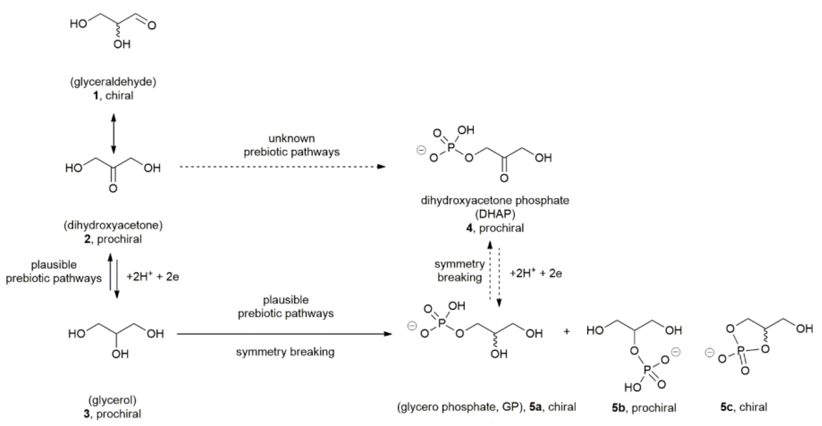
Figure 1. Plausibly prebiotic pathways for glycerophosphates.
Symmetry 15 01259 g001
Figure 2. Various lipid derivatives including the polar head group.
Symmetry 15 01259 g002
Figure 3. Relationship between prebiotic phospholipid esters and glycerol phosphates; (A) prebiotic pathways explored during pioneering research (1977–1982); (B) phosphorylation of glycerol; (C) recent results obtained in phosphorylation; (D) concomitant acylation of glycerol in the fatty acids; The asterisk (*) indicates the stereo genic carbon C2 of any phospholipid and phospholipid precursors; Pi indicates for any phosphorous salt or plausibly phosphate-containing mineral able to promote the phosphorylation of primary or secondary alcohols [34]; ca, stands for any condensing agents [36]; Δ, stands for temperatures between 65 and 130 °C; DAP indicates for diamidophosphate [50,53].
Symmetry 15 01259 g003
Figure 4. Upper and downer DPPC in membrane; (A) one molecule of DPPC, blue is nitrogen; red is oxygen, gray is carbon and pink is phosphorus; (B) exhibited the situation of four DPPC in both down and up leaflet of membrane.
Symmetry 15 01259 g004
Figure 5. Tuning a critical point of symmetry; lipids converting to asymmetry: lipids, for DPPC/POPC/DMPC/30:40:30 upper and 20:60:20 lower mixture, with and without 40% cholesterol. (A) Conformational diagram. (B) Cholesterol fraction in the upper and lower leaflet of the simulation. (C) Upper and lower leaflet APL for promoting amounts for symmetry/asymmetry situation in two sides of phospholipid membrane without cholesterol. (D), Upper and lower leaflet APL to promote amounts for the symmetry/asymmetry situation in two sides of phospholipid membrane with cholesterol.
Symmetry 15 01259 g005
Figure 6. Scheme of simulation for 60 number of phospholipids (blue is nitrogen; red is oxygen, gray is carbon and pink is phosphorus) as a membrane capacitor including band diagram under applied voltage V (between C2 with C1 or C3 parts) and voltage U (between C1 and C3 parts); C1 and C3 are the thicknesses of upper and lower phosphate groups and C2 is the thickness of the hydrocarbon chain of phospholipids that was used as dielectric for the model of the membrane capacitor.
Symmetry 15 01259 g006
Figure 7. Electrostatic potential for the lipid membranes; (A) the DPPC lipids, (B) bilayers with no charge misbalances.
Symmetry 15 01259 g007
Figure 8. Helical membrane proteins.
Symmetry 15 01259 g008
Table 1. Several instance of the symmetry breaking processes in biological systems.
ItemPolarizing CuePhysical EffectEffect Molecular
S. cerevisiaesCdc42 activationTransportation of plasmaF-actin
D. meleanogasterTollPlanar polarizationNon-muscle tissues
C. elegansSperms entryA/P polarization
Node of RanvierCanonical and non-canonical of WntParkinsonNodal cilia structure
Danio rerioFluctuating adhesionLarge actin disassemblyCortical-actomyosin
Table 2. The phospholipid bilayers modeled with several numbers of asymmetry using the Martini tool.
PhospholipidsModel 1
40 mol% Cholesterol
Model 2
40 mol% Cholesterol
Model 2
40 mol% Cholesterol
Mol Ratio
(Lower)
Mol Ratio
(Upper)
Mol Ratio
(Lower)
Mol Ratio
(Upper)
Mol Ratio
(Lower)
Mol Ratio
(Upper)
DPPC2030 2030
DOPC60402030
DMPC203060406040
PAPE 20302030
Table 3. Average area per lipid (APL) for each of the lipids in either the upper or lower cell membrane of liver tissue.
Phospholipids Mol% DPCC (Lower) DPCC (Upper) DMPC (Lower) DMPC (Upper)
DPPC0 0 0.655 0.655
DPCC5 0 0.620 0.665
DPCC10 0 0.715 0.763
DMPC0 0 0.534 0.564
DMPC5 0 0.555 0.532
DMPC10 0 0.543 0.454
DMPC/DPPC 0 0.333 0.438
DMPC/POPC 0 0.435 0.432
DPPC/POPC 0 0.435 0.549
Table 4. Physical and electrical properties of membrane capacitors.
DPPC& Number of Atoms V =
( V P + 1 t o N V P 1 t o N )
Q =
( Q P + 1 N Q P 1 N )
Expectation
of Dielectric Thickness
C g F × 10 20 Dielectric Constant
(N = 50)-----
(N = 100)5.341.4332.432.68.35
(N = 200)4.121.7636.354.555.44
(N = 400)8.441.3433.433.113.65
(N = 500)12.431.5532.445.224.33
(N = 600)17.331.4336.51.987.42
(N = 3000)14.751.0838.431.343.54
Table 5. Dielectrics and capacitance of various numbers of DPPC; C Q is the quantum capacitance, C g is the geometry capacitance, and C n e t is the net capacitance.
DPPC& Number of Atoms C Q F × 10 19
= q ( Q + q 2 ) E S
C n e t F × 10 19 =
C g C Q C Q + 2 C g
ESPMullikenESPMulliken
(N = 500)1.161.361.451.32
(N = 600)1.381.980.910.96
(N = 2000)1.761.231.091.25
(N = 30001.641.770.980.99
Table 6. The self induction of DPPC modeled capacitors (N = 300) with some helical proteins.
Helical ProteinsNumber of CoilsMillimeter WavesSelf Induction Per coil
1AFO1665 GHZ43.55
1AIK2755GHZ27.6
2M8R3270 GHZ11.8
Disclaimer/Publisher’s Note: The statements, opinions and data contained in all publications are solely those of the individual author(s) and contributor(s) and not of MDPI and/or the editor(s). MDPI and/or the editor(s) disclaim responsibility for any injury to people or property resulting from any ideas, methods, instructions or products referred to in the content.