
Subjective and Objective Evaluation of the Symmetry of Maxillary Incisors among Residents of Southwest Poland


Abstract
:
1. Introduction
1.1. Macroaesthetics
1.2. Miniesthetics
2. Materials and Methods
- Analysis of variance with post hoc tests to find significant differences between groups;
- Student’s t-test to assess the significance of differences between the measurements in two groups;
- ROC curve analysis to determine threshold values for continuous variables
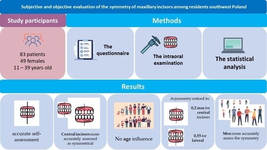
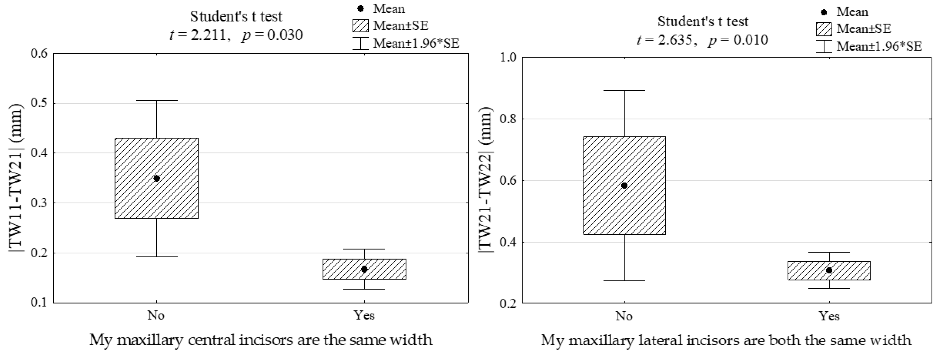
- Subjective assessment of the width of the upper incisors by patients coincides with the actual width of the incisors.
- Patients define the symmetry of maxillary central incisors more accurately than maxillary lateral incisors.
- Patients are able to notice only a large disproportion in the width of the upper incisors (e.g., over 1 mm).
- Younger people more accurately assess the symmetry of the upper incisors.
- Women more accurately assess the symmetry of the upper incisors.
3. Results
Verification of Hypotheses
- Subjective assessment of the width of the upper incisors by patients coincides with the actual width of the incisors.
- 2.
- Patients define the symmetry of maxillary central incisors more accurately than that of maxillary lateral incisors.
- 3.
- Patients are able to notice only a large disproportion in the width of the upper incisors (e.g., over 1 mm).
- 4.
- Younger people more accurately assess the symmetry of the upper incisors.
- 5.
- Women more accurately assess the symmetry of their upper incisors.
4. Discussion
5. Conclusions
Author Contributions
Funding
Institutional Review Board Statement
Informed Consent Statement
Data Availability Statement
Conflicts of Interest
References
- Lukez, A.; Pavlic, A.; Trinajstic Zrinski, M.; Spalj, S. The Unique Contribution of Elements of Smile Aesthetics to Psychosocial Well-Being. J. Oral Rehabil. 2015, 42, 275–281. [Google Scholar] [CrossRef] [PubMed]
- Prado, L.H.; Previato, K.; Delgado, R.Z.R.; Nelson Filho, P.; Bezerra Segato, R.A.; Nakane Matsumoto, M.A.; Neuppmann Feres, M.F. Adolescents’ Perception of Malocclusion, Their Motivations, and Expectations Concerning the Orthodontic Treatment. Is It All about Attractiveness? A Qualitative Study. Am. J. Orthod. Dentofac. Orthop. 2022, 161, e345–e352. [Google Scholar] [CrossRef] [PubMed]
- Geoghegan, F.; Birjandi, A.A.; Xavier, G.M.; Dibiase, A.T. Motivation, Expectations and Understanding of Patients and Their Parents Seeking Orthodontic Treatment in Specialist Practice. J. Orthod. 2019, 46, 46–50. [Google Scholar] [CrossRef] [PubMed]
- Jefferson, Y. Facial Beauty—Establishing a Universal Standard. Int. J. Orthod. Milwaukee. 2004, 15, 9–22. [Google Scholar] [PubMed]
- Ricketts, R.M. Divine Proportion in Facial Esthetics. Clin. Plast. Surg. 1982, 9, 401–422. [Google Scholar] [CrossRef]
- Lombardi, R.E. The Principles of Visual Perception and Their Clinical Application to Denture Esthetics. J. Prosthet. Dent. 1973, 29, 358–382. [Google Scholar] [CrossRef]
- Levin, E.I. Dental Esthetics and the Golden Proportion. J. Prosthet. Dent. 1978, 40, 244–252. [Google Scholar] [CrossRef]
- Snow, S.R. Esthetic Smile Analysis of Maxillary Anterior Tooth Width: The Golden Percentage. J. Esthet. Dent. 1999, 11, 177–184. [Google Scholar] [CrossRef]
- Rayyan, M.R. Effect of the Interproximal Contact Level on the Perception of Smile Esthetics. Dent. Med. Probl. 2019, 56, 251–255. [Google Scholar] [CrossRef] [Green Version]
- Magne, P.; Salem, P.; Magne, M. Influence of Symmetry and Balance on Visual Perception of a White Female Smile. J. Prosthet. Dent. 2018, 120, 573–582. [Google Scholar] [CrossRef]
- Londono, J.; Ghasemi, S.; Lawand, G.; Dashti, M. Evaluation of the Golden Proportion in the Natural Dentition: A Systematic Review and Meta-Analysis. J. Prosthet. Dent. 2021, in press. [Google Scholar] [CrossRef] [PubMed]
- Ahmed, N.; Halim, M.S.; Khalid, S.; Ghani, Z.A.; Jamayet, N. Bin Evaluation of Golden Percentage in Natural Maxillary Anterior Teeth Width: A Systematic Review. J. Prosthet. Dent. 2021, in press. [Google Scholar] [CrossRef]
- Sarul, M.; Antoszewska-Smith, J.; Park, H.S. Self-Perception of Smile Attractiveness as a Reliable Predictor of Increased Patient Compliance with an Orthodontist. Adv. Clin. Exp. Med. 2019, 28, 1633–1638. [Google Scholar] [CrossRef] [PubMed]
- Srivastava, D.; Singh, H.; Mishra, S.; Sharma, P.; Kapoor, P.; Chandra, L. Facial Asymmetry Revisited: Part I—Diagnosis and Treatment Planning. J. Oral Biol. Craniofac. Res. 2018, 8, 7–14. [Google Scholar] [CrossRef] [PubMed] [Green Version]
- Thiesen, G.; Gribel, B.F.; Freitas, M.P.M. Facial Asymmetry: A Current Review. Dent. Press J. Orthod. 2015, 20, 110–125. [Google Scholar] [CrossRef]
- Zaidel, D.W.; Hessamian, M. Asymmetry and Symmetry in the Beauty of Human Faces. Symmetry 2010, 2, 136–149. [Google Scholar] [CrossRef] [Green Version]
- Zaidel, D.W.; Deblieck, C. Attractiveness of Natural Faces Compared to Computer Constructed Perfectly Symmetrical Faces. Int. J. Neurosci. 2007, 117, 423–431. [Google Scholar] [CrossRef]
- Bertamini, M.; Rampone, G.; Makin, A.D.J.; Jessop, A. Symmetry Preference in Shapes, Faces, Flowers and Landscapes. PeerJ 2019, 7, e7078. [Google Scholar] [CrossRef]
- Zheng, R.; Ren, D.; Xie, C.; Pan, J.; Zhou, G. Normality Mediates the Effect of Symmetry on Facial Attractiveness. Acta Psychol. 2021, 217, 103311. [Google Scholar] [CrossRef]
- Kobus, K.; Kobus-Zaleśna, K. The Treatment of Facial Asymmetry: Review. Adv. Clin. Exp. Med. 2017, 26, 1301–1311. [Google Scholar] [CrossRef] [Green Version]
- Scheuerle, A.E.; Firth, R.M. Asymmetric Faces: Symbolic, Spiritual, and Representative. Am. J. Med. Genet. C Semin. Med. Genet. 2021, 187, 278–282. [Google Scholar] [CrossRef] [PubMed]
- Severt, T.R.; Proffit, W.R. The Prevalence of Facial Asymmetry in the Dentofacial Deformities Population at the University of North Carolina. Int. J. Adult Orthodon. Orthognath. Surg. 1997, 12, 171–176. [Google Scholar] [CrossRef]
- Bernabe, E.; Flores-Mir, C. Influence of Anterior Occlusal Characteristics on Self-Perceived Dental Appearance in Young Adults. Angle Orthod. 2007, 77, 831–836. [Google Scholar] [CrossRef] [PubMed] [Green Version]
- Parrini, S.; Rossini, G.; Castroflorio, T.; Fortini, A.; Deregibus, A.; Debernardi, C. Laypeople’s Perceptions of Frontal Smile Esthetics: A Systematic Review. Am. J. Orthod. Dentofac. Orthop. 2016, 150, 740–750. [Google Scholar] [CrossRef] [PubMed]
- Brough, E.; Donaldson, A.N.; Naini, F.B. Canine Substitution for Missing Maxillary Lateral Incisors: The Influence of Canine Morphology, Size, and Shade on Perceptions of Smile Attractiveness. Am. J. Orthod. Dentofac. Orthop. 2010, 138, 705.e1–705.e9. [Google Scholar] [CrossRef]
- Bjelopavlovic, M.; Weyhrauch, M.; Erbe, C.; Burkard, F.; Petrowski, K.; Lehmann, K.M. Influencing Factors on Aesthetics: Highly Controlled Study Based on Eye Movement and the Forensic Aspects in Computer-Based Assessment of Visual Appeal in Upper Front Teeth. Appl. Sci. 2021, 11, 6797. [Google Scholar] [CrossRef]
- Wang, Y.; Song, Y.; Zhong, Q.; Xu, C. Evaluation of Influence Factors on the Width, Length, and Width to Length Ratio of the Maxillary Central Incisor: A Systematic Review and Meta-Analysis. J. Esthet. Restor. Dent. 2021, 33, 351–363. [Google Scholar] [CrossRef] [PubMed]
- Ormianer, Z.; Solodukhin, A.L.; Lauritano, D.; Segal, P.; Lavi, D.; Carinci, F.; Block, J. Bilateral Symmetry of Anterior Maxillary Incisors: Evaluation of a Community-Based Population. J. Biol. Regul. Homeost. Agents 2017, 31, 37–43. [Google Scholar]
- Gallão, S.; Ortolani, C.L.F.; Santos-Pinto, A.; Santos-Pinto, L.A.M.; Faltin, K., Jr. Análise Fotográfica Da Simetria e Da Proporção Estética Dos Dentes Anteriores—Photographic Analysis of Symmetry and Aesthetic Proportion of Anterior Teeth. Rev. Inst. Ciênc. 2010, 27, 400–404. [Google Scholar]
- Paolone, G.; Scolavino, S.; Gherlone, E.; Spagnuolo, G. Direct Esthetic Composite Restorations in Anterior Teeth: Managing Symmetry Strategies. Symmetry 2021, 13, 797. [Google Scholar] [CrossRef]
- Spinell, T.; Tarnow, D. Restoring Lost Gingival Pigmentation in the Esthetic Zone: A Case Report. J. Am. Dent. Assoc. 2015, 146, 402–405. [Google Scholar] [CrossRef] [PubMed]
- Afrashtehfar, K.I.; Assery, M.K. Five Considerations in Cosmetic and Esthetic Dentistry. J. N. J. Dent. Assoc. 2014, 85, 14–15. [Google Scholar] [PubMed]
- Frese, C.; Staehle, H.J.; Wolff, D. The Assessment of Dentofacial Esthetics in Restorative Dentistry: A Review of the Literature. J. Am. Dent. Assoc. 2012, 143, 461–466. [Google Scholar] [CrossRef] [PubMed]
- Calamia, J.R.; Trushkowsky, R.D.; Wolff, M.S. Esthetic and Cosmetic Dentistry for Modern Dental Practice: Update 2011. Preface Dent. Clin. N. Am. 2011, 55, xiii–xiv. [Google Scholar] [CrossRef] [PubMed]
- Aldeeri, A.A.; Alhababi, K.A.; Algahtani, F.A.; Tounsi, A.A.; Albadr, K.I. Perception of Altered Smile Esthetics by Orthodontists, Dentists, and Laypeople in Riyadh, Saudi Arabia. Clin. Cosmet. Investig. Dent. 2020, 12, 563–570. [Google Scholar] [CrossRef]
- Pinzan-Vercelino, C.R.M.; Costa, A.C.S.; Ferreira, M.C.; Bramante, F.S.; Fialho, M.P.N.; de Gurgel, J.A. Comparison of Gingival Display in Smile Attractiveness among Restorative Dentists, Orthodontists, Prosthodontists, Periodontists, and Laypeople. J. Prosthet. Dent. 2020, 123, 314–321. [Google Scholar] [CrossRef]
- Ntovas, P.; Diamantopoulou, S.; Gogolas, N.; Sarri, V.; Papandreou, A.; Sakellaridi, E.; Petrakos, G.; Papazoglou, E. Influence of Lightness Difference of Single Anterior Tooth to Smile Attractiveness. J. Esthet. Restor. Dent. 2021, 33, 856–864. [Google Scholar] [CrossRef]
- Malicka, B.; Skośkiewicz-Malinowska, K.; Kaczmarek, U. The Impact of Socioeconomic Status, General Health and Oral Health on Health-Related Quality of Life, Oral Health-Related Quality of Life and Mental Health among Polish Older Adults. BMC Geriatr. 2022, 22, 2. [Google Scholar] [CrossRef]
- Alzabibi, B.A.; Burhan, A.S.; Hajeer, M.Y.; Nawaya, F.R. Short-Term Effects of the Orthodontic Removable Traction Appliance in the Treatment of Skeletal Class III Malocclusion: A Randomized Controlled Trial. Dent. Med. Probl. 2021, 58, 163–172. [Google Scholar] [CrossRef]
- Chiche, G.J.; Pinault, A. Esthetics of Anterior Fixed Prosthodontics, 1st ed.; Elsevier: Amsterdam, The Netherlands, 1994. [Google Scholar]
- Goldstein, R.E. Change Your Smile, 3rd ed.; Quintessence: Chicago, IL, USA, 1997. [Google Scholar]
- MacHado, A.W.; Moon, W.; Gandini, L.G. Influence of Maxillary Incisor Edge Asymmetries on the Perception of Smile Esthetics among Orthodontists and Laypersons. Am. J. Orthod. Dentofac. Orthop. 2013, 143, 658–664. [Google Scholar] [CrossRef] [Green Version]
- Wolfart, S.; Brunzel, S.; Freitag, S.; Kern, M. Assessment of Dental Appearance Following Changes in Incisor Angulation. Int. J. Prosthodont. 2004, 17, 150–154. [Google Scholar] [PubMed]
- Thomas, J.L.; Hayes, C.; Zawaideh, S. The Effect of Axial Midline Angulation on Dental Esthetics. Angle Orthod. 2003, 73, 359–364. [Google Scholar] [PubMed]
- Ma, W.; Preston, B.; Asai, Y.; Guan, H.; Guan, G. Perceptions of Dental Professionals and Laypeople to Altered Maxillary Incisor Crowding. Am. J. Orthod. Dentofac. Orthop. 2014, 146, 579–586. [Google Scholar] [CrossRef] [PubMed]
- Martinez Florez, D.; Rinchuse, D.; Zullo, T. Influence of Maxillary Lateral Incisor Width Ratio on Perception of Smile Esthetics among Orthodontists and Laypersons. J. Esthet. Restor. Dent. 2021, 33, 510–515. [Google Scholar] [CrossRef]
- Cracel-Nogueira, F.; Pinho, T. Assessment of the Perception of Smile Esthetics by Laypersons, Dental Students and Dental Practitioners. Int. Orthod. 2013, 11, 432–444. [Google Scholar] [CrossRef]
- Alomari, S.A.; Abu Alhaija, E.S.; AlWahadni, A.M.; Al-Tawachi, A.K. Smile Microesthetics as Perceived by Dental Professionals and Laypersons. Angle Orthod. 2022, 92, 101–109. [Google Scholar] [CrossRef]
- Liliana Musskopf, M.; Mariano da Rocha, J.; Kuchenbecker Rösing, C. Perception of Smile Esthetics Varies Between Patients and Dental Professionals When Recession Defects Are Present. Braz. Dent. J. 2013, 24, 385–390. [Google Scholar] [CrossRef]
- Ousehal, L.; Aghoutan, H.; Chemlali, S.; Anssari, I.F.; Talic, N. Perception of Altered Smile Esthetics among Moroccan Professionals and Lay People. Saudi Dent. J. 2016, 28, 174–182. [Google Scholar] [CrossRef] [Green Version]
- Pinho, S.; Ciriaco, C.; Faber, J.; Lenza, M.A. Impact of Dental Asymmetries on the Perception of Smile Esthetics. Am. J. Orthod. Dentofac. Orthop. 2007, 132, 748–753. [Google Scholar] [CrossRef]
- Sobral, M.C.; Crusoé-Rebello, I.M.; Machado, A.W. Does the Presence of Maxillary Central Incisor Edge Asymmetry Influence the Perception of Dentofacial Esthetics in Video Analysis? Angle Orthod. 2019, 89, 775–780. [Google Scholar] [CrossRef] [Green Version]
- Sriphadungporn, C.; Chamnannidiadha, N. Perception of Smile Esthetics by Laypeople of Different Ages. Prog. Orthod. 2017, 18, 8. [Google Scholar] [CrossRef] [PubMed] [Green Version]
- Lacerda-Santos, R.; Pereira, T.B.; Pithon, M.M. PercepÇÃo EstÉtica Do Corredor Bucal Em Diferentes Tipos Faciais Por Leigos de Diferentes Idades. Biosci. J. 2015, 31, 1283–1290. [Google Scholar] [CrossRef] [Green Version]
- Chou, J.C.; Nelson, A.; Katwal, D.; Elathamna, E.N.; Durski, M.T. Effect of Smile Index and Incisal Edge Position on Perception of Attractiveness in Different Age Groups. J. Oral Rehabil. 2016, 43, 855–862. [Google Scholar] [CrossRef] [PubMed]
- Gerritsen, A.E.; Sarita, P.; Witter, D.J.; Kreulen, C.M.; Mulder, J.; Creugers, N.H. Esthetic Perception of Missing Teeth among a Group of Tanzanian Adults. Int. J. Prosthodont. 2008, 2, 169–173. [Google Scholar]
- Lira dos Santos, E.J.; Dantas, A.M.X.; Vilela, R.M.; de Lima, K.J.R.S.; Beltrão, R.T.S. The Influence of Varying Maxillary Central Incisor Vertical Dimension on Perceived Smile Aesthetics. J. Orthod. 2019, 46, 137–142. [Google Scholar] [CrossRef]
- Pithon, M.M.; Bastos, G.W.; Miranda, N.S.; Sampaio, T.; Ribeiro, T.P.; Andrade Gomes Do Nascimento, L.E.; Da Silva Coqueiro, R. Esthetic Perception of Black Spaces between Maxillary Central Incisors by Different Age Groups. Am. J. Orthod. Dentofac. Orthop. 2013, 143, 371–375. [Google Scholar] [CrossRef]
- Carlsson, G.E.; Wagner, I.V.; Odman, P.; Ekstrand, K.; MacEntee, M.; Marinello, C.; Nanami, T.; Ow, R.K.; Sato, H.; Speer, C.; et al. An International Comparative Multicenter Study of Assessment of Dental Appearance Using Computer-Aided Image Manipulation. Int. J. Prosthodont. 1998, 11, 246–254. [Google Scholar]
- Bolas-Colvee, B.; Tarazona, B.; Paredes-Gallardo, V.; Luxan, S.A. De Relationship between Perception of Smile Esthetics and Orthodontic Treatment in Spanish Patients. PLoS ONE 2018, 13, e0201102. [Google Scholar] [CrossRef] [Green Version]
- Althagafi, N. Esthetic Smile Perception Among Dental Students at Different Educational Levels. Clin. Cosmet. Investig. Dent. 2021, 13, 163–172. [Google Scholar] [CrossRef]
- Wolfart, S.; Quaas, A.C.; Freitag, S.; Kropp, P.; Gerber, W.D.; Kern, M. Subjective and Objective Perception of Upper Incisors. J. Oral Rehabil. 2006, 33, 489–495. [Google Scholar] [CrossRef]
- Pease, A.; Pease, B. Why Men Don’t Listen & Women Can’t Read Maps: How to Spot the Differences in the Way Men & Women Think; Orion: London, UK, 2016. [Google Scholar]

| Variable | Statistics |
|---|---|
| Sex: | |
| F, n (%) | 49 (59.0) |
| M, n (%) | 34 (41.0) |
| Age (years), M ± SD: | 20.5 ± 5.3 |
| Age from 11 to 18 years, n (%) | 30 (34.9) |
| Age from 19 to 23 years, n (%) | 27 (31.4) |
| Age from 24 to 39 years, n (%) | 26 (30.2) |
| Place of residence: | |
| Village | 38 (45.8) |
| A city with up to 50,000 inhabitants, n (%) | 28 (33.7) |
| A city with 50,000 to 100,000 inhabitants, n (%) | 10 (12.1) |
| A city with a population of 100,000 and more, n (%) | 7 (8.4) |
| Education: | |
| Student, n (%) | 35 (42.2) |
| Basic, n (%) | 7 (8) |
| Professional, n (%) | 4 (4.8) |
| Secondary, n (%) | 18 (21.47) |
| Higher, n (%) | 19 (22.9) |
| The Questions of the Original Questionnaire | N | (%) |
|---|---|---|
| 1. The upper arch of my teeth is symmetrical | ||
| Strongly disagree | 9 | 10.8 |
| Disagree | 14 | 16.9 |
| Neither agree nor disagree | 16 | 19.3 |
| Agree | 33 | 39.8 |
| Strongly agree | 11 | 13.2 |
| 2. My maxillary central incisors are the same width | ||
| Strongly disagree | 2 | 2.4 |
| Disagree | 3 | 3.6 |
| Neither agree nor disagree | 7 | 8.4 |
| Agree | 59 | 71.1 |
| Strongly agree | 12 | 14.5 |
| 3. My maxillary lateral incisors are both the same width | ||
| Strongly disagree | 2 | 2.4 |
| Disagree | 6 | 7.2 |
| Neither agree nor disagree | 18 | 21.7 |
| Agree | 47 | 56.6 |
| Strongly agree | 10 | 12.1 |
| Objective Assessment of the Asymmetry of the Width of the Tooth | My Maxillary Incisors Are the Same Width | p | Sens. | Spec. | OR (95% CI) | |||
|---|---|---|---|---|---|---|---|---|
| “Definitely Not” or “Rather Not” | “Definitely Yes”, “Rather Yes” or “Hard to Say” | |||||||
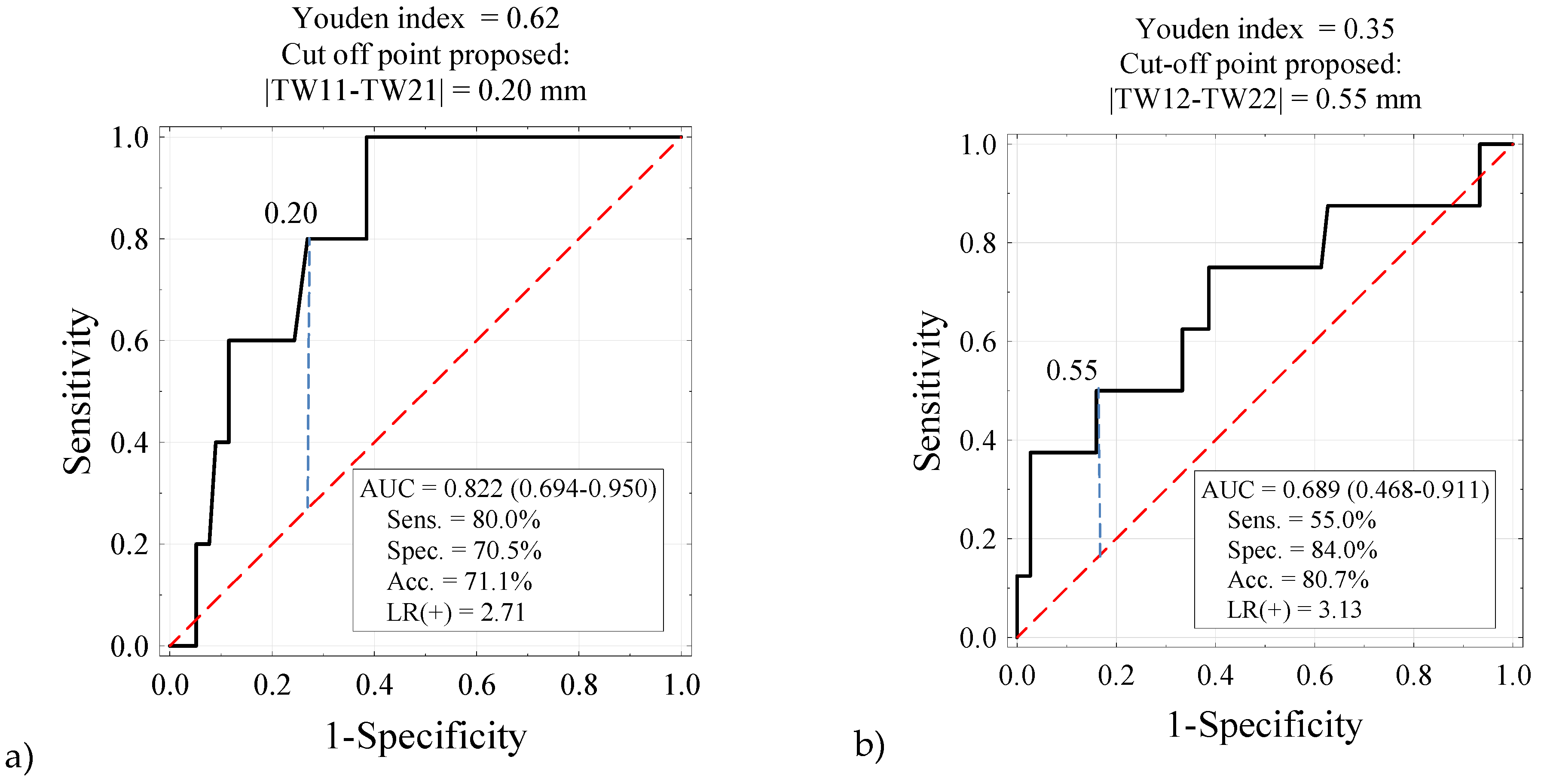
| ΔW1 ≥ 0.20 mm | 4 | 80.0% | 22 | 28.2% | 0.032 | 80.0% | 71.3% | 10.2 |
| ΔW1 < 0.20 mm | 1 | 20.0% | 56 | 71.8% | (1.08–96.2) | |||
| ΔW2 ≥ 0.20 mm | 6 | 75.0% | 45 | 60.0% | 0.477 | 80.0% | 71.3% | 2.00 |
| ΔW2 < 0.20 mm | 2 | 25.0% | 30 | 40.0% | (0.38–10.6) | |||
| ΔW2 ≥ 0.55 mm | 6 | 75.0% | 29 | 38.7% | 0.038 | 80.0% | 71.3% | 5.42 |
| ΔW2 < 0.55 mm | 2 | 25.0% | 46 | 61.3% | (1.19–24.7) | |||
| Age (Years) | Incisor Symmetry Assessment | p | OR (95% CI) | |||
|---|---|---|---|---|---|---|
| Accurate | Inaccurate | |||||
| From 11 to 18 | 14 | 31.1 | 16 | 42.1 | 0.358 | 0.46 (0.16–1.37) |
| From 19 to 23 | 14 | 31.1 | 13 | 34.2 | 0.57 (0.19–1.72) | |
| From 24 to 39 | 17 | 37.8 | 9 | 23.7 | 1.00 (ref.) | |
| Gender | Incisor Symmetry Assessment | p | OR (95% CI) | |||
|---|---|---|---|---|---|---|
| Accurate | Inaccurate | |||||
| Men | 23 | 51.1 | 11 | 28.9 | 0.047 | 2.57 (1.03–6.39) |
| Women | 22 | 48.9 | 27 | 71.1 | 1.00 (ref.) | |
Publisher’s Note: MDPI stays neutral with regard to jurisdictional claims in published maps and institutional affiliations. |
© 2022 by the authors. Licensee MDPI, Basel, Switzerland. This article is an open access article distributed under the terms and conditions of the Creative Commons Attribution (CC BY) license (https://creativecommons.org/licenses/by/4.0/).
Share and Cite
Chrapla, P.; Paradowska-Stolarz, A.; Skoskiewicz-Malinowska, K. Subjective and Objective Evaluation of the Symmetry of Maxillary Incisors among Residents of Southwest Poland. Symmetry 2022, 14, 1257. https://doi.org/10.3390/sym14061257
Chrapla P, Paradowska-Stolarz A, Skoskiewicz-Malinowska K. Subjective and Objective Evaluation of the Symmetry of Maxillary Incisors among Residents of Southwest Poland. Symmetry. 2022; 14(6):1257. https://doi.org/10.3390/sym14061257
Chicago/Turabian StyleChrapla, Paulina, Anna Paradowska-Stolarz, and Katarzyna Skoskiewicz-Malinowska. 2022. "Subjective and Objective Evaluation of the Symmetry of Maxillary Incisors among Residents of Southwest Poland" Symmetry 14, no. 6: 1257. https://doi.org/10.3390/sym14061257
APA StyleChrapla, P., Paradowska-Stolarz, A., & Skoskiewicz-Malinowska, K. (2022). Subjective and Objective Evaluation of the Symmetry of Maxillary Incisors among Residents of Southwest Poland. Symmetry, 14(6), 1257. https://doi.org/10.3390/sym14061257