Abstract
The purpose of this study is to determine if there is asymmetry in the brain activity between both hemispheres while watching cuts in movies. We presented videos with cuts to 36 participants, registered electrical brain activity through electroencephalography (EEG) and analyzed asymmetry in frontal, somatomotor, temporal, parietal and occipital areas. EEG power and alpha (8–13 Hz) asymmetry were analyzed based on 4032 epochs (112 epochs from videos × 36 participants) in each hemisphere. On average, we found negative asymmetry, indicating a greater alpha power in the left hemisphere and a greater activity in the right hemisphere in frontal, temporal and occipital areas. The opposite was found in somatomotor and temporal areas. However, with a high inter-subjects variability, these asymmetries did not seem to be significant. Our results suggest that cuts in audiovisuals do not provoke any specific asymmetrical brain activity in the alpha band in viewers. We conclude that brain asymmetry when decoding audiovisual content may be more related with narrative content than with formal style.
1. Introduction
1.1. Neural Processing of Visual Content
Visual perception is one of the most studied topics in neuroscience [,,]. Learning how the brain processes visual content has been of interest for decades. Visual processing is considered in a hierarchical network [] with different stages []. However, after several experimental approaches, it is not convincingly clear what happens in the brain after a visual stimulus is presented. It is believed that the processing of visual content is a very rapid processing with a large number of stages involved, presumably based on feed-forward mechanisms []. Currently, there are, at least, two plausible frameworks of how perception occurs: the outside-in and the inside-out []. While in the former the stimulus reaches the eyes with a response of the brain that causes neurons to fire, in the latter we understand the external world by taking actions to learn about an object []. On the other hand, the memory–prediction framework matches sensory inputs with stored memory patterns to perceive thanks to elaborate predictions based on previous memories []. Based on evidence, what seems more plausible is that different brain areas (or systems) are in charge of perceiving (or processing) different physical characteristics of visual stimuli [].
1.2. Neural Processing of Audiovisual Cuts
Films present plenty of cuts. They organize the visual content for viewers [] and, while they present new visual content that needs to be decoded, viewers hardly notice them []. According to previous studies, cuts inhibit viewers’ eye-blink rate [] and the higher number of cuts an audiovisual has, the lower the eye-blink rate of viewers []. Since eye-blinks are attentional markers [,] (the higher the eye-blink rate, the lower the attention), this relationship suggest that cuts affect viewers’ perception. Probably due to the new visual content following cuts, they also trigger an increase of occipital brain activity (in the visual cortex) that flows towards frontal areas []. Moreover, the brain activity and brain connectivity (functional and effective) when watching cuts depends on the audiovisual background and expertise of the viewer, with higher activation of occipital areas in media professionals and higher activity of medial and frontal areas in non-media professionals [].
1.3. Brain Asymmetry in Visual Perception
A great part of the world is symmetrical. In part, we process symmetry with an automatic response [], but how symmetrically our brain works is yet to be solved. Frontal alpha asymmetry has been studied in correlation with behavior and emotion [,]. It has also been approached while viewing videos with different engagement levels [], showing higher right frontal brain activity when viewing videos of interest compared with not-interesting videos. Asymmetry during emotionally evocative films and its relation to positive and negative affectivity has also been studied [], finding that subjects with positive affectivity exhibit more left-sided activation while watching happy films. Moreover, resting alpha power asymmetry in the frontal area predicts self-reported negative affect in response to videos [], with a strong relation between frontal asymmetry and fear responses to films. In the area of advertising, this relation between the brain asymmetry and the emotions has also been found when watching ads [].
While many researchers have approached the perception of cuts in media [,,,] and, as seen, there are some studies about brain asymmetry when watching films, to our knowledge, nobody has studied brain asymmetry in viewers while watching audiovisual cuts, regardless of the emotional content and the emotional feelings that their subsequent content can provoke.
In this study we wondered about the brain asymmetry of viewers when watching the brand-new visual content that follows cuts in movies. To investigate this, in this work we compared brain activity in both hemispheres of viewers watching cuts. The aim was to check whether there was asymmetry of the brain activity in specific areas of the brain.
2. Materials and Methods
2.1. Participants
Thirty-six participants with normal or corrected-to-normal vision were recruited for this experiment. Participants were aged 28–56 (43.97 ± 8.07). Six were females. Half of participants were media professionals with 6 years or more of expertise. They did not receive any economic compensation for participating in this study. We followed relevant guidelines and regulations for human research and procedures were approved by the Ethics Commission for Research with Animals and Humans (CEEAH) of the University Autònoma de Barcelona, Spain. All participants gave prior written informed consent to participate in the study.
2.2. Stimuli
We created three video stimuli with the same narrative content and duration (198 s), but different styles and a different number of cuts. The videos were randomly presented to all participants. The narrative of the three video stimuli consisted of a man who entered a room with a black background, sat at a desk, juggled with three balls, opened a laptop, looked up some information in books, wrote something in the laptop, closed it, ate an apple, looked directly into camera, and left the room. One stimulus was a one-shot movie with a single shot and no cuts. The second video presented a classical and organized style of edition, with a total of 33 shots and an average shot length (ASL) of 5.9 s. The third stimulus presented a chaotic and disorganized style of edition, with a total of 79 shots and an ASL of 2.4 s. Since here we are interested in analyzing asymmetry when watching audiovisual cuts, we only use data from stimuli two and three, since the first video did not have any cuts.
Stimuli were presented on a high-definition (HD) 42-inch light-emitting diode (LED) display (TH42PZ70EA, Panasonic Corporation, Osaka, Japan) using Paradigm Stimulus Presentation software v1.5 (Perception Research System Inc., Lawrence, KS, USA).
2.3. Data Acquisition
Continuous EEG was recorded from participants using an Enobio® system (Neuroelectrics, Barcelona, Spain) equipped with 20 electrodes [O1, O2, P7, P3, Pz, P4, P8, T7, C3, Cz, C4, T8, F7, F3, Fz, F4, F8, Fp1, Fp2, and an additional electrode used for electrooculogram (EOG) recordings] placed according to the International 10–20 system [], referenced to electronically linked mastoid electrodes. Data were sampled at 500 Hz. In order to have a good quality of the signal, we asked participants to avoid chemical products (such as hair spray or similar) in their hair before coming to the experimental session. Data acquisition was synchronized with the data presentation system through a TCP/IP system.
2.4. Data Analysis
Data processing was carried out using EEGLAB [] open-source software (version 2022.0), running on MATLAB 2020a (The MathWorks Inc., Natick, MA, USA) under a MacOS High Sierra (version 10.13.6) (Apple Inc., Cupertino, CA, USA). We used a spherical BESA® template for channel location. We computed average reference, and high-pass filtered the data at 0.5 Hz and low-pass filtered it at 40 Hz. We divided the data into 1500-ms epochs (500 ms before the cut and 1000 ms after the cut), removing the baseline. For rejecting artifacts, bad channels, and wrong data, we used visual inspection and the ADJUST plug-in [] for EEGLAB, after applying independent component analysis (ICA). To locate dipoles, we used DIPFIT plugin. Estimates of EEG power were based on 4032 epochs (112 epochs from videos × 36 participants) in each hemisphere. All statistical analyses were performed with Sigmaplot 11.0 (Systat Software Inc., San Jose, CA, USA) and with GraphPad Prism version 9.4.1 for Mac (GraphPad Software, San Diego, CA, USA).
We selected data from different brain areas in the left hemisphere and their correspondent in the right hemisphere. We divided the brain in five sections: frontal, somatomotor, temporal, parietal and occipital. Then we selected the available electrodes for each area (see Table 1). In each brain area, we computed the asymmetry in alpha band (8–13 Hz).
Table 1.
Selected sections of the brain for data analysis.
To compute asymmetry, the measures of the five mentioned regional areas were calculated separately by subtracting natural-log transformed regional EEG power in the left hemisphere from natural-log transformed power at homologous site in the right hemisphere [ln(right) power − ln(left)power] []. Positive values of asymmetry reflect greater alpha power in the right hemisphere, indicating greater activity in the left hemisphere, while negative values reflect greater alpha power in the left hemisphere, showing greater activity in the right hemisphere []. Then we computed descriptive statistical analysis of all participants and statistical analysis between the group of media and the group of non-media professionals. The Shapiro–Wilk test was used as normality test (p < 0.05).
3. Results
We obtained mean values of alpha power and alpha asymmetry in the brain areas selected: frontal, somatomotor, temporal, parietal and occipital (Table S1).
3.1. Alpha Power
Mean (SD) alpha power (in µV) in frontal areas was 3.006 (3.936) in left hemisphere and 2.557 (2.004) in right hemisphere. In somatomotor areas, it was 0.545 (0.462) in left hemisphere and 0.577 (0.534) in right hemisphere. The temporal area showed 0.696 (0.603) in left hemisphere and 0.658 (0.527) in right hemisphere. The parietal showed 1.029 (0.944) in left hemisphere and 0.988 (0.865) in right hemisphere. Finally, the occipital showed 0.742 (0.688) in left hemisphere and 0.712 (0.691) in right hemisphere.
We computed Wilcoxon signed rank tests between alpha power in each hemisphere for each brain area in order to study each specific brain area separately. We obtained no significant differences between hemispheres in none of the cases: frontal area [Z = −0.456, p = 0.654]; somatomotor area [Z = 0.833, p = 0.409]; temporal area [Z = −1.037, p = 0.303]; parietal area [Z = −0.896, p = 0.375]; and occipital area [Z = −0.503, p = 0.621].
To check relations among the different brain areas × hemispheres, we computed a Friedman repeated measures ANOVA on ranks with the alpha power. We obtained significant differences [X2 (9, N = 36) = 155.254, p = < 0.001]. We used a Dunn test as multiple comparison procedure in all pairwise, taking as significant differences those with p < 0.05. We obtained the result that alpha power in the frontal left area differed from all the rest of the studied areas, except for frontal right: somatomotor left and right, temporal left and right, parietal left and right and occipital left and right. We also obtained the result that frontal right activity differed again from all the studied areas, except for frontal left. Moreover, we found some other significant differences between parietal left and somatomotor left and right, and thetemporal right, and also between parietal right and somatomotor left and right (see Table 2).
Table 2.
Multiple comparison results of alpha power with p < 0.05 using Dunn test for Friedman repeated measures ANOVA on ranks.
Differences found in alpha power among different brain areas and hemispheres (Table 2) could suggest some crossed asymmetry; however, looking at the data in detail whenever a significant difference is found between areas x hemispheres (e.g., frontal left vs. somatomotor right), it also happens just between areas in the same hemisphere (e.g., frontal left vs. somatomotor left). The only case where differences are found between hemispheres is parietal left vs. temporal right.
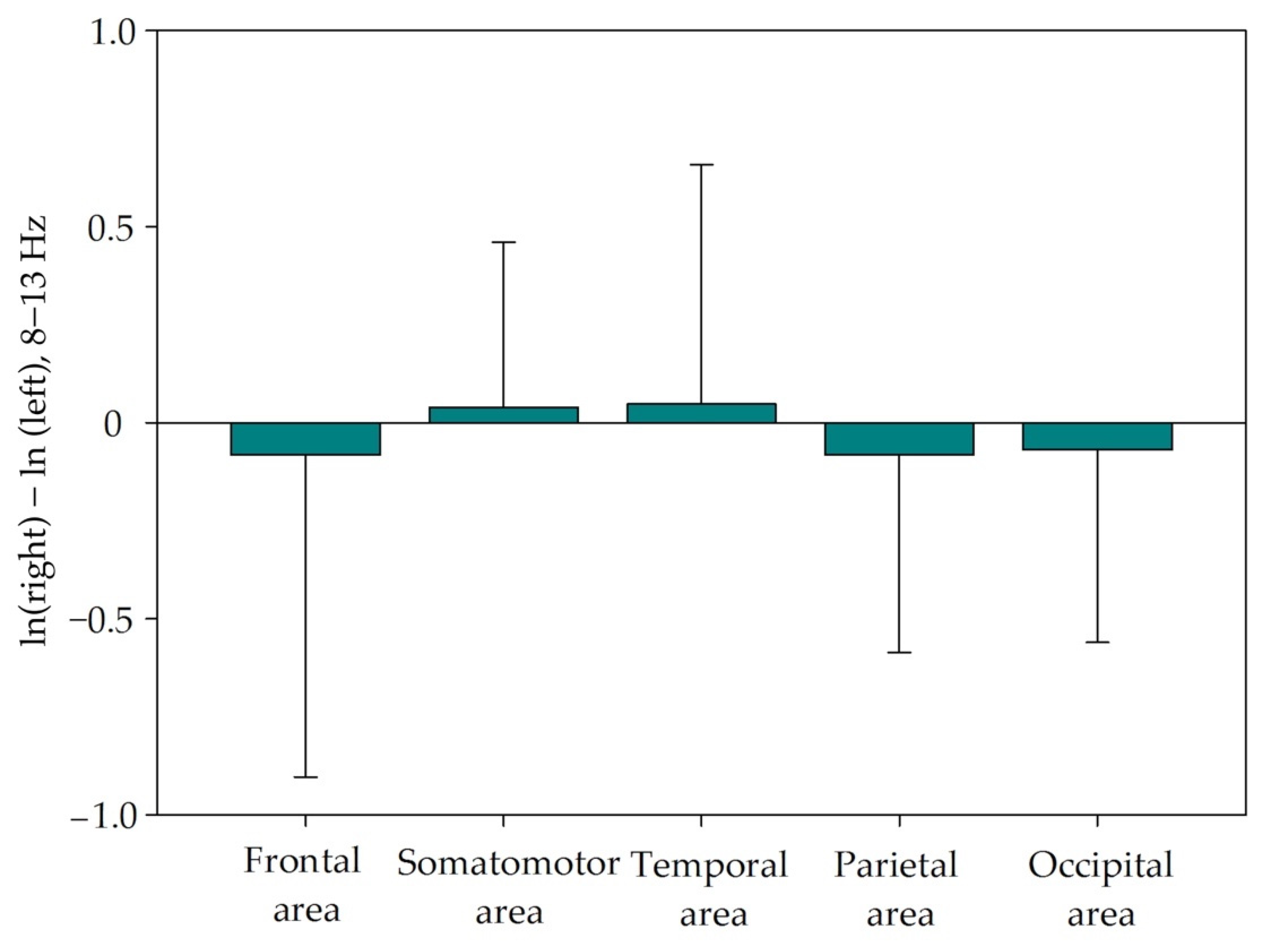
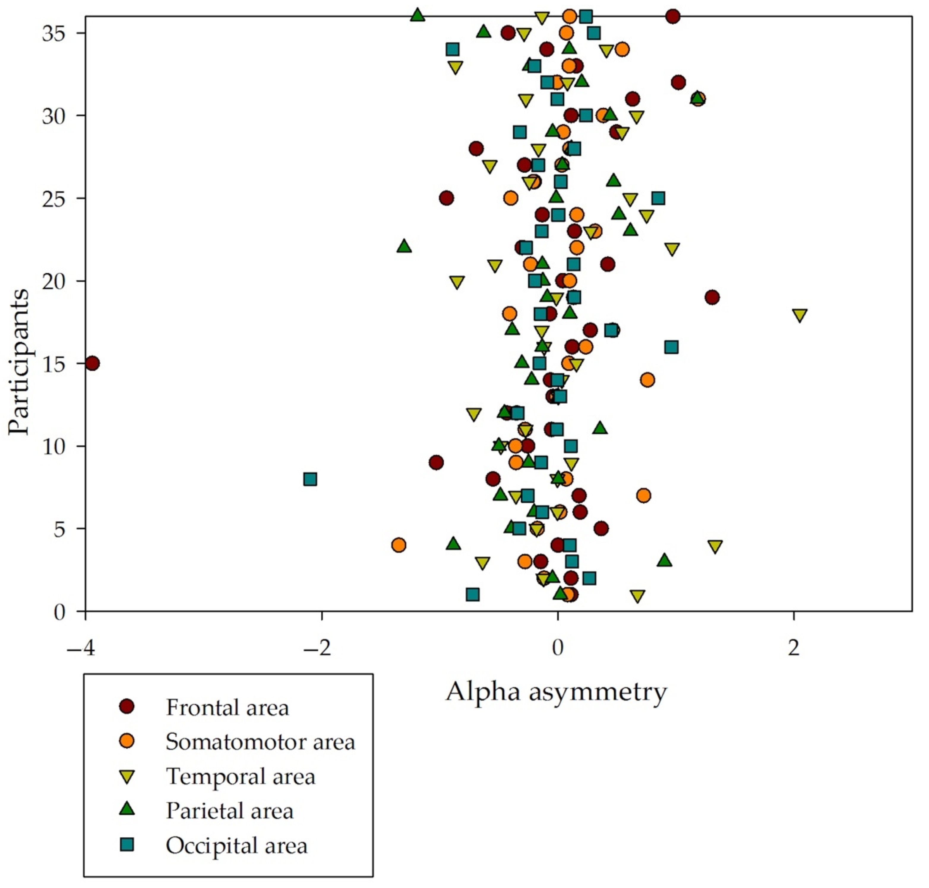
3.2. Alpha Asymmetry
Mean (SD) asymmetry in the analyzed areas was negative in frontal [−0.0822 (0.822)], temporal [−0.0826 (0.504)] and occipital [−0.0826 (0.504)] areas, indicating a greater alpha power in the left hemisphere that would correspond with a greater activity in the right hemisphere of these areas. Asymmetry was found positive in somatomotor [0.0383 (0.422)] and temporal [0.0474 (0.610)] areas indicating a greater alpha power in the right hemisphere and greater activity in the left one (see Table 3). The high deviations indicate a high variability among subjects (Figure 1).
Table 3.
Alpha power and asymmetry in frontal, somatomotor, temporal, parietal and occipital areas, in left and right hemispheres.
Figure 1.
Mean (SD) alpha asymmetry in frontal, somatomotor, temporal, parietal and occipital areas. High SD indicates great variability inter subjects.
We observed that the asymmetry positive or negative trend was not standard among all the participants in each brain area (see Figure 2). In the frontal area, half of participants showed positive asymmetry, and half showed negative. In the somatomotor area, it was 22 (61.1%) positive versus 14 negative. In the temporal area, it was 15 (41.7%) positive versus 21 negative. In the parietal area, it was the same, 15 (41.7%) positive versus 21 negative. And, in the occipital area, we found 16 (44.4%) participants with positive asymmetry and 20 with negative asymmetry.
Figure 2.
Alpha asymmetry from all participants. Data from all participants in this study, distributed in relation to the five areas of this study: frontal, somatomotor, temporal, parietal and occipital.
We computed a Friedman repeated measures ANOVA on ranks with asymmetry of the five studied areas and found no significant difference [X2 (4, N = 36) = 3.044, p = 0.55].
3.3. Alpha Asymmetry and Media Expertise
As explained, half of the participants were media professionals. We computed unpaired t-tests (or the non-parametrical Mann–Whitney rank sum test for non-normally distributed data) in each area with both groups. We found no significant statistical differences between media and non-media professionals in any area: frontal [Mann–Whitney U Statistic = 131.000, T = 302.000, n = 18, p = 0.335; rank sum test, Mann–Whitney rank sum test]; somatomotor [Mann–Whitney U Statistic = 102.000, T = 273.000, n = 18, p = 0.06; rank sum test, Mann–Whitney rank sum test]; temporal [Mann–Whitney U Statistic = 160.000, T = 335.000, n = 18, p = 0.962; rank sum test, Mann–Whitney rank sum test]; parietal [t(34) = −0.919, p = 0.365, unpaired t-test]; and occipital [Mann–Whitney U Statistic = 142.000, T = 313.000, n = 18, p = 0.537; rank sum test, Mann–Whitney rank sum test].
4. Discussion and Conclusions
In this manuscript, we wondered whether there would be brain asymmetry in alpha power when watching cuts in movies, dividing the analysis into five brain areas: frontal, somatomotor, temporal, parietal and occipital. On average, we found positive asymmetry in the somatomotor and the temporal area, as a result of a higher right hemisphere alpha power that corresponds with a higher left hemisphere activity. We also found negative asymmetry in frontal, parietal and occipital areas, suggesting the contrary. However, we found great variability among participants and, when computing the statistics, these asymmetries did not seem to be significant. These results suggest that cuts in audiovisuals do not evoke any specific asymmetrical brain activity in the alpha band in viewers. Note that previous studies analyzing brain asymmetry when watching videos paid attention to the narrative content and the emotions that those contents could elicit in viewers [,]. Here, we did not draw attention to the content but rather to the cut as a specific formal characteristic of videos. These results suggest that brain asymmetry when decoding audiovisual content may be more related with narrative content than with formal style.
We found some differences in alpha power between the non-corresponding brain areas and hemispheres. This could be related to the differences in the rate of flow of brain activity []. We found a significant difference in alpha power in left parietal and right temporal areas. Although it suggests an interesting relation between both hemispheres and areas, further studies should be made to confirm this.
We also looked for differences between media and non-media professionals. Based on previous studies, these two groups perceive audiovisual content differently [,]. Different brain connectivity (functional and effective) has been found between them. Also, there is quite a lot of literature regarding the effect of professional specialization or expertise on the brain (such as in drivers [,], in musicians [], in surgeons [], in athletes [], among others). Here we found that there is no significant difference between media and non-media professionals in terms of alpha asymmetry when watching audiovisual cuts.
To our knowledge no study had looked before at brain asymmetry in relation with media professional expertise. Since brain connectivity differences between media and non-media groups were found significant [], while brain asymmetry was not, we believe further studies connecting these two different manners of approaching brain activity could be of interest for a better understanding of brain processing of local field potentials when watching movies. Furthermore, in the future, a temporal dimension could be added to find possible correlations of brain asymmetry in relation with the narrative content. Moreover, this type of study could be replicated with attention tests [,] in order to look for correlations between asymmetry and attention. Finally, it would be interesting to study correspondences between asymmetry and eye-gaze behaviors when watching audiovisual content.
This study has several limitations: we did not ask participants about their right or left handedness, so this information could not be added to the analysis. Another limitation was the data acquisition approach: a higher number of electrodes (we used a 20-electrode system) could have given a higher volume of conduction of the signal, and an individual MRI could have provided much more specific information for each one of the participants.
In conclusion, cuts in audiovisuals do not evoke asymmetrical brain activities in the alpha band in viewers, suggesting that brain asymmetry, when decoding audiovisual content, may be more related with narrative content than with formal style.
Supplementary Materials
The following supporting information can be downloaded at: https://www.mdpi.com/article/10.3390/sym14101980/s1, Table S1: Alpha power and asymmetry of all participants.
Author Contributions
Conceptualization, C.A.-S., M.Á.M.-P., A.G. and J.M.D.-G.; methodology, C.A.-S. and M.Á.M.-P.; software, C.A.-S. and M.Á.M.-P.; validation, C.A.-S., M.Á.M.-P., A.G. and J.M.D.-G.; formal analysis, C.A.-S. and M.Á.M.-P.; investigation, C.A.-S. and M.Á.M.-P.; resources, C.A.-S., M.Á.M.-P., A.G. and J.M.D.-G.; data curation, C.A.-S.; writing—original draft preparation, C.A.-S. and M.Á.M.-P.; writing—review and editing, J.M.D.-G. and A.G.; visualization, A.G.; supervision, J.M.D.-G.; project administration, C.A.-S. and M.Á.M.-P.; funding acquisition, A.G. and J.M.D.-G. All authors have read and agreed to the published version of the manuscript.
Funding
This research was funded by the Spanish MICINN (grant number PID2021-122446NB-100 to A.G. and J.M.D.-G.) and by the Spanish Junta de Andalucía (grants number PY18-823 to A.G. and BIO-122 to J.M.D.-G.).
Institutional Review Board Statement
The study was conducted according to the guidelines of the Declaration of Helsinki and approved by the Institutional Review Board (or Ethics Committee) of Universitat Autònoma de Barcelona (protocol code CEEAH 2003).
Informed Consent Statement
Informed consent was obtained from all subjects involved in the study.
Data Availability Statement
The data presented in this study are available in Table S1.
Acknowledgments
We acknowledge all the participants of this study.
Conflicts of Interest
The authors declare no conflict of interest. The funders had no role in the design of the study; in the collection, analyses, or interpretation of data; in the writing of the manuscript; or in the decision to publish the results.
References
- Martinez-Conde, S.; Macknik, S.L.; Hubel, D.H. The role of fixational eye movements in visual perception. Nat. Rev. Neurosci. 2004, 5, 229–240. [Google Scholar] [CrossRef]
- Seriès, P.; Seitz, A.R. Learning what to expect (in visual perception). Front. Hum. Neurosci. 2013, 7, 1–14. [Google Scholar] [CrossRef] [PubMed]
- Pylyshyn, Z. Is vision continuous with cognition? The case for cognitive impenetrability of visual perception Zenon. Behav. Brain Sci. 1999, 22, 341–423. [Google Scholar] [CrossRef] [PubMed]
- Yamins, D.L.K.; Di Carlo, J.J. Using goal-driven deep learning models to understand sensory cortex. Nat. Neurosci. 2016, 19, 356–365. [Google Scholar] [CrossRef]
- Groen, I.I.A.; Silson, E.H.; Baker, C.I. Contributions of low- and high-level properties to neural processing of visual scenes in the human brain. Philos. Trans. R. Soc. B Biol. Sci. 2017, 372, 20160102. [Google Scholar] [CrossRef] [PubMed]
- Thorpe, S.; Fize, D.; Marlot, C. Speed of processing in the human visual system. Nature 1996, 381, 520–522. [Google Scholar] [CrossRef]
- Buzsáki, G. How the Brain ‘Constructs’ the Outside World. Sci. Am. 2022, 326, 36–43. [Google Scholar] [CrossRef]
- Hawkins, J.; Blakeslee, S. On Intelligence; Henry Holt and Company: New York, NY, USA, 2004. [Google Scholar]
- Zeki, S.; Bartels, A. Toward a theory of visual consciousness. Conscious. Cogn. 1999, 8, 225–259. [Google Scholar] [CrossRef] [PubMed]
- Pearlman, K. Cutting Rhythms: Shaping the Film Edit; Focal Press: Oxford, UK; Elsevier: Oxford, UK, 2009; ISBN 978-0-240-81014-0. [Google Scholar]
- Zacks, J.M.; Speer, N.K.; Reynolds, J.R. Segmentation in reading and film comprehension. J. Exp. Psychol. Gen. 2009, 138, 307–327. [Google Scholar] [CrossRef]
- Andreu-Sánchez, C.; Martín-Pascual, M.Á.; Gruart, A.; Delgado-García, J.M. Chaotic and Fast Audiovisuals Increase Attentional Scope but Decrease Conscious Processing. Neuroscience 2018, 394, 83–97. [Google Scholar] [CrossRef]
- Andreu-Sánchez, C.; Martín-Pascual, M.Á.; Gruart, A.; Delgado-García, J.M. Eyeblink rate watching classical Hollywood and post-classical MTV editing styles, in media and non-media professionals. Sci. Rep. 2017, 7, 43267. [Google Scholar] [CrossRef]
- Colzato, L.S.; Slagter, H.A.; Spapé, M.M.A.; Hommel, B. Blinks of the eye predict blinks of the mind. Neuropsychologia 2008, 46, 3179–3183. [Google Scholar] [CrossRef] [PubMed]
- Zheng, B.; Jiang, X.; Tien, G.; Meneghetti, A.; Panton, O.N.M.; Atkins, M.S. Workload assessment of surgeons: Correlation between NASA TLX and blinks. Surg. Endosc. Other Interv. Tech. 2012, 26, 2746–2750. [Google Scholar] [CrossRef]
- Andreu-Sánchez, C.; Martín-Pascual, M.Á.; Gruart, A.; Delgado-García, J.M. The Effect of Media Professionalization on Cognitive Neurodynamics During Audiovisual Cuts. Front. Syst. Neurosci. 2021, 1, 598383. [Google Scholar] [CrossRef] [PubMed]
- Bertamini, M.; Makin, A.D.J. Brain activity in response to visual symmetry. Symmetry 2014, 6, 975–996. [Google Scholar] [CrossRef]
- Coan, J.A.; Allen, J.J.B. Frontal EEG asymmetry and the behavioral activation and inhibition systems. Psychophysiology 2003, 40, 106–114. [Google Scholar] [CrossRef] [PubMed]
- Coan, J.A.; Allen, J.J.B. Frontal EEG asymmetry as a moderator and mediator of emotion. Biol. Psychol. 2004, 67, 7–50. [Google Scholar] [CrossRef] [PubMed]
- Fischer, N.L.; Peres, R.; Fiorani, M. Frontal alpha asymmetry and theta oscillations associated with information sharing intention. Front. Behav. Neurosci. 2018, 12, 1–12. [Google Scholar] [CrossRef] [PubMed]
- Jones, N.A.; Fox, N.A. Electroencephalogram asymmetry during emotionally evocative films and its relation to positive and negative affectivity. Brain Cogn. 1992, 20, 280–299. [Google Scholar] [CrossRef]
- Tomarken, A.J.; Davidson, R.J.; Henriques, J.B. Resting Frontal Brain Asymmetry Predicts Affective Responses to Films. J. Pers. Soc. Psychol. 1990, 59, 791–801. [Google Scholar] [CrossRef] [PubMed]
- Ohme, R.; Reykowska, D.; Wiener, D.; Choromanska, A. Application of frontal EEG asymmetry to advertising research. J. Econ. Psychol. 2010, 31, 785–793. [Google Scholar] [CrossRef]
- Germeys, F.; D’Ydewalle, G. The psychology of film: Perceiving beyond the cut. Psychol. Res. 2007, 71, 458–466. [Google Scholar] [CrossRef]
- Magliano, J.P.; Zacks, J.M. The impact of continuity editing in narrative film on event segmentation. Cogn. Sci. 2011, 35, 1489–1517. [Google Scholar] [CrossRef] [PubMed]
- Francuz, P.; Zabielska-Mendyk, E. Does the Brain Differentiate Between Related and Unrelated Cuts When Processing Audiovisual Messages? An ERP Study. Media Psychol. 2013, 16, 461–475. [Google Scholar] [CrossRef]
- Smith, T. The attentional theory of cinematic continuity. Projections 2012, 6, 1–27. [Google Scholar] [CrossRef]
- Jasper, H.H. The ten twenty electrode system of the International Federation. Report of the committee on methods of clinical examination in electroencephalography. Electroencephalogr. Clin. Neurophysiol. 1958, 10, 371–375. [Google Scholar] [CrossRef]
- Delorme, A.; Makeig, S. EEGLAB: An open source toolbox for analysis of single-trial EEG dynamics including independent component analysis. J. Neurosci. Methods 2004, 134, 9–21. [Google Scholar] [CrossRef]
- Mognon, A.; Jovicich, J.; Bruzzone, L.; Buiatti, M. ADJUST: An automatic EEG artifact detector based on the joint use of spatial and temporal features. Psychophysiology 2010, 48, 229–240. [Google Scholar] [CrossRef] [PubMed]
- Mathewson, K.J.; Hashemi, A.; Sheng, B.; Sekuler, A.B.; Bennett, P.J.; Schmidt, L.A. Regional electroencephalogram (EEG) alpha power and asymmetry in older adults: A study of short-term test-retest reliability. Front. Aging Neurosci. 2015, 7, 1–10. [Google Scholar] [CrossRef] [PubMed]
- Davidson, R.J. Emotion and Affective Style: Hemispheric Substrates. Psychol. Sci. 1992, 3, 39–43. [Google Scholar] [CrossRef]
- Andreu-Sánchez, C.; Martín-Pascual, M.Á.; Gruart, A.; Delgado-García, J.M. Looking at reality versus watching screens: Media professionalization effects on the spontaneous eyeblink rate. PLoS ONE 2017, 12, e0176030. [Google Scholar] [CrossRef]
- Maguire, E.A.; Woollett, K.; Spiers, H.J. London taxi drivers and bus drivers: A structural MRI and neuropsychological analysis. Hippocampus 2006, 16, 1091–1101. [Google Scholar] [CrossRef] [PubMed]
- Maguire, E.A.; Gadian, D.G.; Johnsrude, I.S.; Good, C.D.; Ashburner, J.; Frackowiak, R.S.; Frith, C.D. Navigation-related structural change in the hippocampi of taxi drivers. Proc. Natl. Acad. Sci. USA 2000, 97, 4398–4403. [Google Scholar] [CrossRef]
- Lotze, M.; Scheler, G.; Tan, H.-R.; Braun, C.; Birbaumer, N. The musician’s brain: Functional imaging of amateurs and professionals during performance and imagery. Neuroimage 2003, 20, 1817–1829. [Google Scholar] [CrossRef]
- Faubert, J. Professional athletes have extraordinary skills for rapidly learning complex and neutral dynamic visual scenes. Sci. Rep. 2013, 3, 1154. [Google Scholar] [CrossRef] [PubMed]
- Mcgee, R.A.; Clark, S.E.; Symons, D.K. Does the Conners’ Continuous Performance Test aid in ADHD diagnosis? J. Abnorm. Child Psychol. 2000, 28, 415–424. [Google Scholar] [CrossRef] [PubMed]
- Memória, C.M.; Muela, H.C.S.; Moraes, N.C.; Costa-Hong, V.A.; Machado, M.F.; Nitrini, R.; Bortolotto, L.A.; Yassuda, M.S. Applicability of the test of variables of attention—T.O.V.A in Brazilian adults. Dement. Neuropsychol. 2018, 12, 394–401. [Google Scholar] [CrossRef]
Publisher’s Note: MDPI stays neutral with regard to jurisdictional claims in published maps and institutional affiliations. |
© 2022 by the authors. Licensee MDPI, Basel, Switzerland. This article is an open access article distributed under the terms and conditions of the Creative Commons Attribution (CC BY) license (https://creativecommons.org/licenses/by/4.0/).