Article

Functional Activation and Connectivity of the Left Inferior Frontal Gyrus during Lexical and Phonological Retrieval

by
Miguel Ángel Rivas-Fernández
1,
Benxamín Varela-López
1,
Susana Cid-Fernández
2 and
Santiago Galdo-Álvarez
1,*
1
Department of Clinical Psychology and Psychobiology, Universidade de Santiago de Compostela, Rúa Xosé María Suárez Núñez S/N, 15782 Santiago de Compostela, Spain
2
Department of Developmental and Educational Psychology, Universidade de Santiago de Compostela, Rúa Xosé María Suárez Núñez S/N, 15782 Santiago de Compostela, Spain
*
Author to whom correspondence should be addressed.
Symmetry 2021, 13(9), 1655; https://doi.org/10.3390/sym13091655
Submission received: 5 June 2021 / Revised: 1 August 2021 / Accepted: 2 August 2021 / Published: 8 September 2021
(This article belongs to the Special Issue Cognition, Neuroscience and Asymmetry)

Abstract

:
Being language a paradigm of structural and functional asymmetry in cognitive processing, the left Inferior Frontal Gyrus has been consistently related to speech production. In fact, it has been considered a key node in cortical networks responsible for different components of naming. However, isolating these components (e.g., lexical, syntactic, and phonological retrieval) in neuroimaging studies is difficult due to the use of different baselines and tasks. In the present study, functional activation and connectivity of the left inferior frontal gyrus was explored using functional magnetic resonance imaging. Participants performed a covert naming task (pressing a button based on a phonological characteristic). Two conditions were compared: drawings of objects and single letters (baseline condition). Differences in activation and functional connectivity were obtained for objects and letters in different areas of the left Inferior Frontal Gyrus. The pars triangularis was involved in the retrieval of lexical-phonological information, showing a pattern of connectivity with temporal areas in the search for the name of objects and with perisylvanian areas for letters. Selection of phonological information seems to involve the pars opercularis both to letters and objects but recruiting supramarginal and superior temporal areas to letters, probably related to orthographic-phonological conversion. The results support the notion of the left Inferior Frontal Gyrus as a buffer forwarding neural information across cortical networks responsible for different components of speech production.

1. Introduction

Language is probably the most paradigmatic example of structural and functional asymmetry in cognitive processing. In fact, alterations in language lateralization have been linked to language disorders [1], for example in developmental disorders such as autism spectrum disorder [2].
Even in neurotypically developing individuals, and despite the apparent ease with which human beings are able to name, this process involves numerous cognitive functions, making naming a linguistic process particularly sensitive to errors. In fact, errors in naming are a common symptom in different aphasic syndromes [3], semantic dementia [4], or even in the absence of any kind of pathological condition, as in the case of the tip of the tongue phenomenon [5].
It is not surprising, therefore, that naming has been an important object of study in cognitive neuroscience. The development of neuroscientific techniques such as positron emission tomography or functional magnetic resonance imaging (fMRI) has made it possible to associate the brain activity of different neural circuits with the processes involved in naming (although temporal dynamics and the relationship between components is still a matter of debate; see [6,7,8,9]). Thus, a complex network of brain structures involved in object naming has been described (see reviews in [10,11]), including occipital and occipitotemporal areas associated with the extraction of visual characteristics of objects [12], a distributed network of temporal and frontal areas, mainly in the left hemisphere, involved in accessing semantic information [13,14,15,16], left brain regions (including inferior and middle frontal gyri and temporal areas) involved in name retrieval, i.e., access to lexical-phonological information [16,17,18,19], and motor and premotor structures that have been commonly associated with creating a motor plan and articulating the name [13,15].
There is a great consensus in considering the inferior frontal gyrus (IFG) as an essential region in name retrieval and speech production [11]. The importance of the left IFG has been confirmed by numerous studies that have contrasted the participation of different areas of the IFG (pars triangularis, pars opercularis and pars orbitalis; [20]) in the retrieval of single words.
Concretely, the activity of pars triangularis has been associated with processes of retrieval and selection of lexical and syntactic information from stimuli [14,21], while the activity of pars opercularis seems to be more involved in the access and analysis of phonological characteristics [16,22]. On the other hand, pars orbitalis activity has been associated with the retrieval and selection of semantic characteristics [23], especially when access to long-term stored information is required [24]. That is why it has been proposed that the left IFG is a key node in manipulating and forwarding neural information across cortical networks responsible for different components of speech production [20,25].
However, as shown by Price et al. [26], the use of different baselines made it difficult to interpret the role that different neural circuits play in naming objects. This variability between studies has been especially relevant to the task that participants perform in a brain imaging environment. Thus, in order to avoid the muscular artifacts inherent in the articulation of a name, some studies employed covert naming tasks in which there are no observable measures to contrast the performance or the timing of naming [15,19]. Other studies, despite the presence of muscle artifacts and head movements that compromise the signal acquisition and analysis [27], have used tasks that require name articulation compared to conditions that do not require it [28,29] or more standardized forms of articulation [30,31].
In addition, name retrieval (access to lexical and phonological information) is difficult to explore given that studies suggested that it might take place in parallel to semantic processing [32]. In fact, Price et al. [26] designed a covert naming task which, using the same perceptual and motor baselines, compared the activity of the left IFG when participants had to perform a semantic task and a phonological task. Results confirmed the role of the pars opercularis in phonological access, but they could not isolate the role of anterior regions of left IFG in semantic and lexical processing, given that both conditions involved this processing. Moreover, the instructions between conditions to perform the task were different, introducing again differences in the task.
As a result, there are still questions to be resolved regarding how different regions of the left IFG are involved in name retrieval. As far as we are concerned, there are no previous studies comparing a condition requiring name retrieval with one that does not, using the same task, that is, providing the same instructions. Secondly, although there is consensus of the role of left IFG regions in the different components of naming, evidence is scarce on how these regions interact with other structures to retrieve lexical and phonological information.
The present study, therefore, aims to contrast the functional activation and connectivity of the left IFG (pars orbitalis, pars triangularis, and pars opercularis) in the process of name retrieval, using the fMRI technique. To this aim, blood oxygen level-dependent (BOLD) activation and connectivity analyses were combined in two conditions (pictures of objects and letters) requiring the same task (the same instructions). In order to avoid muscular artifacts/head movements within the scanner, an adaptation of Van Turennout et al.’s paradigm [33] has been designed: participants responded by pressing a button based on a phonological feature (the name of the object begins with a vowel or a consonant). Therefore, in order to respond adequately, the name of the stimulus must be retrieved. In addition, an attempt has been made to homogenize as much as possible the perceptual characteristics (different stimuli in each trial) and the task to be carried out from the baseline condition with that of naming objects, using letter drawings.
Based on the previous literature and given that aspects related to the characteristics of the task (absence of articulation, equal phonological decision to perform the task) are matched, a greater involvement of the left IFG to object drawings with respect to the letter drawings is expected to be found. In addition, it is expected to observe a greater connectivity of this region with temporal areas associated with the recovery of lexical information for objects relative to letters; however, as letters require a grapheme-phoneme conversion to perform the task, a greater connectivity of the left IFG with regions associated with the posterior perisilvian regions [34] is expected in the case of letters.

2. Materials and Methods

2.1. Participants

Twenty-two young adults (11 women) between 19 and 37 years old (mean age: 27.14 years old; SD: 5.56) and a mean of 15 years of formal education (SD: 3.24) participated voluntarily in the study. All participants had normal audition and normal or corrected to normal vision, and none had any history of neurological or psychiatric disorders. All participants were right-handed, and all of them gave their written informed consent prior to participation in the study. The research project was approved by the regional Clinical Research Ethics Committee and was performed in accordance with the ethical standards established in the 1964 Declaration of Helsinki.

2.2. Stimuli and Task

The task was designed in the context of a research project (see Funding section) focused on assessment and rehabilitation of aphasic patients.
Sixty black and white images (30 letter drawings and 30 object drawings) were used on a white square with a size of 4.98° × 4.98° visual angle. Table 1 shows the psycholinguistic characteristics (obtained from the EsPAL database; [35]) of the names of the objects used as stimuli. In the case of letters, pictures with different shapes were used in each trial (see Figure 1).
All subjects listed the instructions of the task using a compatible headphone system and stimulus were displayed using an MRI compatible Visual HD System (NordicNeuroLab, Inc., Milwaukee, WI, USA). Responses were collected with a fiber-optic response box (NordicNeuroLab, Inc.,) that participants held in the left hand. Task presentation and recording of behavioral data was handled by a PC running Presentation software (Version 12.2, Neurobehavioral Systems, Inc., Berkeley, CA, USA).
During the task, in order to avoid eye movements, a fixation cross was kept present at all times, except for the period of time when the stimuli were presented. The presentation of each object and each letter had a duration of five seconds. These stimulus trials were completed with one second with the fixation cross to match the repetition time of signal acquisition. The stimulus trials were interspersed with null trials that subserved as baseline trials, in which the fixation cross was presented for a variable length (from 2 to 10 s). The stimuli were pseudorandomly presented, that is, the presentation of each condition and null trials (including their length) was determined to optimize the recovery of BOLD response (using Optseq 2.0 https://surfer.nmr.mgh.harvard.edu/optseq/ accessed on 3 August 2021), as recommended to account for the statistical power in fMRI studies [36]. The task (see Figure 1) consisted in pressing a button with the index finger of the left hand if the presented image corresponded to an object whose name started with a vowel or if the image corresponded to the drawing of a vowel. On the other hand, if the image corresponded to an object whose name started with a consonant or if the image presented was a consonant, the participants had to press a button with their left thumb. Therefore, all the participants performed the different conditions, and the same sample was measured repeatedly.

2.3. Procedure

Magnetic resonance imaging was performed on a Philips 3T Achieva scanner (Philips Medical System, Latham, NY, USA). A sagittal T1-weighted 3D Magnetization Prepared Rapid Acquisition Gradient Echo (MPRAGE) sequence (repetition time/echo time = 7.45 ms/3.40 ms, flip angle = 8°; 180 slices, voxel size = 1 × 1 × 1 mm3, field of view = 240 × 240 mm2, matrix size = 240 × 240 mm2) and functional magnetic resonance images were acquired with a gradient echo-planar imaging (EPI) sequence sensitive to BOLD contrast (repetition time/echo time = 2000 ms/30 ms, flip angle = 87°; 37 interleaved slices, voxel size = 3 × 3 × 3.5 mm3, field of view = 240 × 240 mm2, matrix size = 80 × 80 mm2) during the performance of the naming task. Four dummy scans were automatically discarded before image acquisition to avoid signals arising from progressive saturation. Head movements of the participants were minimized by using a vacuum cushion during the MRI acquisition.

2.4. Data Analysis

2.4.1. Behavioral Data

To analyze the behavioral results, t-tests were performed for related samples by comparing the percentage of hits and reaction times between objects and letters, calculated with SPSS (version 25 for Mac).

2.4.2. Blood Oxygen Level-Dependent (BOLD) Activation Functional Magnetic Resonance Imaging (fMRI) Image Preprocessing

Image preprocessing and statistical analysis of fMRI data was performed in Matlab R2016a (Mathworks, Inc., Natik, MA, USA) using the Statistical Parametric Mapping software SPM12 (Wellcome Centre for Human Neuroimaging, London, UK). Structural and functional EPI images were visually inspected and examined for excessive motion and signal artifacts.
Preprocessing implied, firstly, a reorientation of the structural and functional images to the anterior-posterior commissure. Then, functional images were slice-timed corrected, realigned and a mean realigned functional image was calculated for each subject averaging all realigned functional scans. Then, structural T1-images and the realigned functional images were coregistered. Next, gray matter (GM), white matter (WM) and the cerebrospinal fluid (CSF) tissues were obtained with the application of the unified segmentation algorithm [37] over the structural T1-weighted image of each subject. Finally, functional images were normalized to the Montreal Neurological Institute (MNI) space and spatially smoothed using a Gaussian kernel of 8 mm full-width at half maximum (FWHM).
The spatially normalized and smoothed functional images of each subject were analyzed within the general linear model (GLM) approach implemented in SPM12 in order to model the BOLD signal at each voxel. All statistical analyses were performed only in the brain activity evoked during the presentation of objects and letters using the fixation cross (null trials) as baseline. Thus, onsets and durations (corrected by RTs) of event-related responses evoked during the presentation of objects, letters and the fixation cross were defined into the GLM. Six movement parameters (three translations, three rotations) were included in the model as regressors in order to control residual movement-related effects. The haemodynamic response to the onset of each event type was modelled with the canonical haemodynamic response function (HRF). A high-pass filter with a cut-off of 128 s was applied in order to filter out the low-frequency variations from the time series. Parameter estimates of the model were estimated and then five linear contrasts were calculated using a two-sample t-test: (1) brain activity evoked during the Objects condition (Object > Fixation); (2) brain activity evoked during the Letters condition (Letters > Fixation); (3) significantly higher brain activity in Objects than in Letters (Objects > Letters); (4) significantly higher brain activity in Letters than in Objects (Letters > Objects).
Group analysis was performed using the individual contrast images via one sample t-tests to estimate the brain activity evoked during the Objects condition and Letters conditions as well as the other contrasts, that is, higher brain activity in Objects than in Letters and vice versa. All statistical analyses were performed considering the whole brain as the volume of interest when conditions of the task were compared with the fixation and results were assessed at p < 0.05 family-wise error (FWE) voxel-level corrected for multiple comparisons. However, when comparing both experimental conditions (i.e., Objects > Letters; Letters > Objects) statistical analyses were performed across the entire brain and also adopting a follow-up region of interest (ROI) analyses restricted to three a priori hypothesized regions, including the left IFG subregions (triangular, opercular and orbital part). The ROIs were created by using the Wake Forest Pick Atlas toolbox [38]. The results obtained in the comparison between the Objects and Letters conditions were assessed at p < 0.05 FWE cluster-level corrected for multiple comparisons in a combination with a threshold of p < 0.001 at the uncorrected voxel level. All statistical analyses were performed holding the parametric assumptions of random field theory [39,40].

2.4.3. Functional Connectivity fMRI Image Preprocessing

Both structural and functional images were pre-processed and analysed using the CONN 19c [41] and SPM12 toolboxes implemented in Matlab R2019a (Mathworks, Inc., Natik, MA, USA).
We followed the default preprocessing pipeline of CONN toolbox which included the functional realignment and unwarping, functional centering of the image to (0, 0, 0) coordinates, slice-timing correction, functional outlier detection, functional direct segmentation and normalization to MNI space, structural centering to (0, 0, 0) coordinates, structural segmentation and normalization to MNI space and functional smoothing with a kernel of 8 mm FWHM. Potential confounding effects to the estimated BOLD signal (noise components from white matter and cerebrospinal areas, estimated subject-motion parameters, scrubbing and task effects) were estimated and removed separately for each voxel and for each subject using ordinary least squares (OLS) regression to project each BOLD signal timeseries to the sub-space orthogonal to all potential confounding effects as part of the denoising step [41,42,43,44].
CONN’s default denoising pipeline implements an anatomical component-based noise correction procedure (aCompCor), which can extract white matter and cerebral spinal fluid noise components [42]. This procedure is combined with the quantification of participant motion and the identification of outlier scans through the Artifact Rejection Toolbox (ART) [41,45]. The ART was set to the 97th percentile setting with the mean global-signal deviation threshold set at z = ±5 and the participant-motion threshold set at 0.9 mm. Motion and outliers information were included as covariates in our first-level analyses. Linear regression of the potential confounds and temporal high-pass filtering (0.008-inf) were applied on our data to exclude signal frequencies outside the range of expected BOLD signals, reduce the impact of participant motion, extract white matter and cerebral spinal fluid noise components, account for the common task effects and control for within-participant realignment and scrubbing covariates.
We performed a weighted seed-based connectivity (SBC) analysis to compare functional connectivity between task conditions (Objects vs. Letters). Weighted SBC maps are used to characterize task- or condition-specific functional connectivity strength [41]. Specifically, we applied a GLM using the HRF weighted to the first level using the standard bivariate correlation measure for functional connectivity analyses. Individual seed-to-voxel functional connectivity maps were created for each participant, seed (pars triangularis, pars opercularis and pars orbitalis obtained through WFU Pick Atlas toolbox) and condition (Objects/Letters). The mean BOLD time series was calculated across all voxels within each ROI and Fisher Z transformation was applied to correlations values between each pair of sources. The second-level analysis was composed by the individual seed-to-voxel maps. The results obtained in the comparison between the Objects and Letters conditions were assessed at p < 0.05 FWE cluster-level corrected for multiple comparisons in a combination with a threshold of p < 0.001 at the uncorrected voxel level. All statistical analyses were performed holding the parametric assumptions of random field theory [39,40].

3. Results

3.1. Behavioral Data

The t-tests showed significant differences depending on the type of stimulus both in the percentage of hits (t(21) = −2.806; p = 0.011; Cohen’s d = 0.598) and in reaction times (t(21) = 9.847; p ≤ 0.001; Cohen’s d = 2.099). The participants obtained a higher percentage of hits (98.33 vs. 96.52) and shorter reaction times (894.52 ms vs. 1275.42 ms) for letters than for objects.

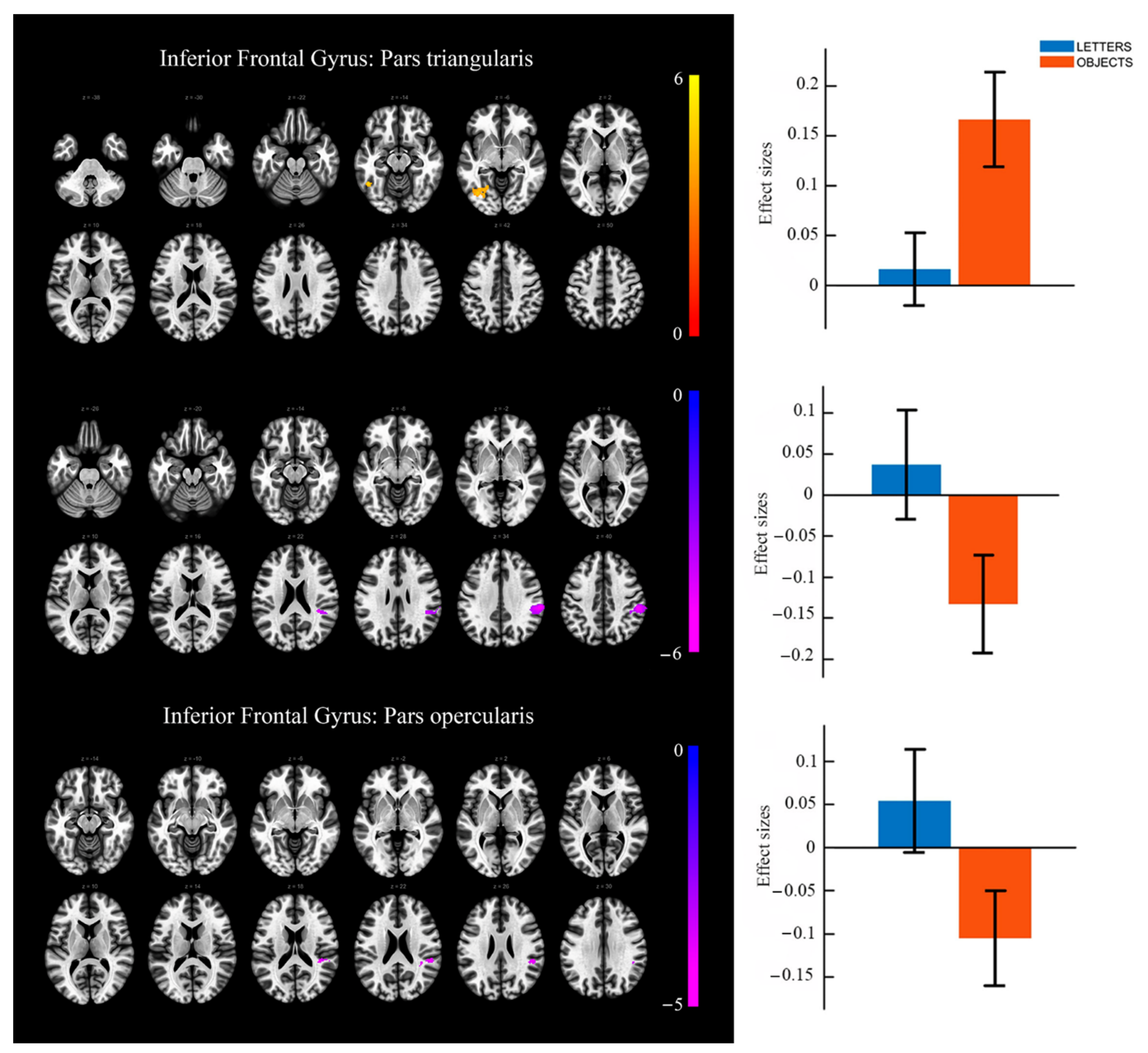
3.2. Task-Based Activation Results

Table 2 shows the significant brain activity obtained in the group analysis for Objects and Letters with respect to the fixation (see Figure 2). Table 3 shows the significant brain activity obtained in the group analysis for Letters with respect to Objects (see Figure 2) and Objects with respect to Letters (see Figure 3).
In the comparison between Letters and Objects conditions (see Table 3 and Figure 2), the whole brain analyses showed that during the Letters condition, relative to the Objects condition, brain activity was significantly higher in frontal brain regions (right middle and superior frontal gyrus), parietal brain regions (right angular gyrus, bilateral precuneus, bilateral supramarginal gyrus), temporal brain regions (left middle and bilateral superior temporal gyrus), sensoriomotor areas (bilateral postcentral gyrus and right precentral gyrus), the insular cortex (left insula and left rolandic operculum) and also in the bilateral midcingulate cortex. The follow-up ROIs analysis did not reveal any significant result in subregion of the left IFG.
The whole brain analyses did not reveal higher activity to Objects than to Letters. However, as can be observed in Table 3 and Figure 3, the follow-up ROI analysis showed that, relative to the Letters condition, during the Objects condition brain activity was significantly higher in the triangular and the orbital part of the left IFG. No significant results were found in the opercular part of the left IFG.

3.3. Task-Based Functional Connectivity Results

Two of the seeds used in the functional connectivity analysis reported significant results between conditions (Object vs. Letters). Specifically, the left pars triangularis and pars opercularis were the regions that showed a significant change in whole brain connectivity between conditions (see Table 4, Figure 4).
For the left pars triangularis seed significant results between conditions were observed in both directions (increase and decrease in functional connectivity). The Objects condition presented an increase in the functional connectivity of the left pars triangularis with left temporo-occipital regions (inferior occipital gyrus, inferior temporal gyrus, lingual gyrus and fusiform gyrus) compared to the Letters condition. On the other hand, a decrease in the functional connectivity between the left pars triangularis and right parietal regions (supramarginal gyrus) was found in Objects relative to the Letters condition.
Connectivity analyses for the left pars opercularis seed only reported significant results in one direction between conditions. Specifically, the analyses showed a decrease in the functional connectivity between the left pars opercularis and right temporo-parietal regions (superior temporal gyrus and supramarginal gyrus) for the Objects condition compared to the Letters condition.

4. Discussion

Since Paul Broca’s 1861 description of patient Tan, showing the relevance of brain asymmetries in proper cognitive performance, numerous studies have corroborated the involvement of the left Inferior Frontal Gyrus in language production [10,11], including naming. However, previous studies have shown the importance of baseline features in the study of the neural bases of naming, making it difficult to understand the role different areas play in the functions required from the perception of the stimulus to the articulation of a name [26]. The aim of this work was to contrast the activation and connectivity of different areas within the left IFG, using a baseline that required performing the same task as in the condition of naming objects, but requiring more effort in name retrieval. This is consistent with the higher percentage of hits and shorter reaction times, although other factors (psycholinguistic characteristics, visual complexity…) may contribute to behavioral differences.
As in numerous previous studies (see reviews in [10,11]), comparing the naming condition with respect to fixation has revealed the activation of a wide distributed network including bilateral frontal brain regions, left parietal brain regions, and occipital/occipito-temporal brain areas, as well as sensorimotor areas. Also, a very similar network has been found to be activated by comparing letter drawings with fixation, suggesting that the characteristics of the control condition (letter drawings) have been largely equated with those of object drawings. Additionally, a greater activation has been observed for letters than for objects in a distributed network including bilateral temporal, parietal and frontal regions, a result similar to that obtained in previous studies when faced with rapid naming tasks of letters compared to objects [34].
Although the whole brain analyses in the comparison Objects > Letters did not reveal significant differences, the ROI analyses revealed a greater activation of the IFG for objects with respect to letters in the left middle (pars triangularis) and left anterior (pars orbitalis) IFG.
Concretely, the activity of pars triangularis has been associated with processes of retrieval and selection of lexical information from stimuli [14]. In the present study, therefore, it is confirmed that, with respect to the baseline, the middle area of the left IFG is more involved when more effort is required in the search for lexical information. Furthermore, the analyses have provided evidence of larger functional connectivity in objects relative to letters between the left pars triangularis and occipitotemporal regions such as the inferior occipital gyrus, the inferior temporal gyrus, the fusiform gyrus and the lingual gyrus. The activity of these areas has been associated to the retrieval of the object features [46,47], and it has been considered as a store for the lexical nodes in common nouns [17], as it is the case for the stimuli of the present study. Furthermore, inferior temporal gyrus and pars triangularis connection is enhanced in tasks when lexical decisions are needed [48]. The result is consistent with the anatomical connections of the left IFG, as well as the functional connectivity observed in resting-state fMRI [49]. Thus, in line with the hypotheses presented by other authors [20,25], the left pars triangularis may be considered as a buffer manipulating and forwarding neural information across large-scale cortical networks to retrieve the required lexical information.
The results have also shown similar differences between conditions in the left pars orbitalis. According to the data observed in the literature, pars orbitalis activity has been associated with the retrieval and selection of semantic characteristics [23], especially when access to long-term stored information is required [24]. However, the lack of significant differences in the functional connectivity analyses make the interpretation of this activation difficult in the present study. On the one hand, it can be considered that there is an automatic retrieval of semantic concepts associated with the stimuli, similar to the presentation of words [50]; in this case, this activation can contribute to the semantic priming effects in naming tasks described in the literature (see review in [51]). On the other hand, in line with serial language production theories [52], the activation of semantic conceptual representations is a requirement for the activation of lexical and phonological nodes. Studies of event-related potential using the paradigm adapted in this study effectively suggested this serial processing [33,53], although later studies, using faces [32], evidence was obtained of parallel access, in line with models of face processing and naming [51]. Future studies, using pictures with words referring to objects instead of letters, could shed light on whether the recovery of semantic representations is automatic or a requirement to access lexical-phonological information in object naming.
Although both Objects and Letters revealed activation of the left pars opercularis when compared to a fixation cross baseline, no differences have been observed in the activation of this area between conditions, which seems to support the role of this region in the retrieval of phonological information [16,22]. Both conditions required access to such information to produce a response. However, the greater connectivity of pars triangularis and pars opercularis with the supramarginal and superior temporal gyri to letters with respect to objects could be related to the processes of orthographic-phonological conversion [15,34,54], which only takes place with letters.
There are some limitations that should be considered in the present work. The use of the covert naming task, despite overcoming the overlap of motor activity that can artefact the BOLD signal, has also been criticized due to its difference to naming in natural environments and the presence of additional cognitive and meta-cognitive processes [6]. However, it is important to highlight that despite this fact, the cortical network observed in the present study corresponds consistently with those regions observed in overt naming studies [11]. In addition, object naming was contrasted with a condition (letters) that shared the cognitive and meta-cognitive processing of covert naming tasks.

5. Conclusions

In summary, this study supported that, using a condition that required the same phonological decision task as a baseline, the asymmetric specialization of the left inferior frontal gyrus plays a role as buffer forwarding neural information across cortical networks responsible for different components of speech production. The activity of pars triangularis, functionally connected to occipitotemporal areas, is required to retrieve lexical information in object naming. However, further research is needed to disentangle the role of the left pars orbitalis in naming, reflecting automatic semantic representations retrieval or a requirement to access lexical-phonological information.

Author Contributions

Conceptualization, S.G.-Á.; methodology, M.Á.R.-F., B.V.-L., S.C.-F. and S.G.-Á.; formal analysis, M.Á.R.-F. and B.V.-L.; investigation, M.Á.R.-F., B.V.-L., S.C.-F. and S.G.-Á.; writing—original draft preparation, S.G.-Á.; writing—review and editing, M.Á.R.-F., B.V.-L., S.C.-F. and S.G.-Á.; project administration, S.G.-Á.; funding acquisition, S.G.-Á. All authors have read and agreed to the published version of the manuscript.

Funding

This research was funded by the Spanish Ministerio de Economía y Competitividad (Grant number: PSI2013-43594-R), Consellería de Cultura, Educación e Ordenación Universitaria, Xunta de Galicia (Grant number: ED431C-2021/04, from the EDRF/FEDER) and by a post-doctoral fellowship from Consellería de Cultura, Educación e Ordenación Universitaria, Xunta de Galicia (Grant number: ED481B2016/078-0). The funding sources were not involved in any aspect of the research or the submission of this study.

Institutional Review Board Statement

The study was conducted according to the guidelines of the Declaration of Helsinki, and approved by Galician Clinical Research Ethics Committee (protocol code 2014/010, date of approval: 26 February 2015).

Informed Consent Statement

Informed consent was obtained from all subjects involved in the study.

Data Availability Statement

The data presented in this study are available on request from the corresponding author. The data are not publicly available due to lack of consent provided by participants on the use of confidential data.

Conflicts of Interest

The authors declare no conflict of interest.

References

  1. Bishop, D.V. Cerebral asymmetry and language development: Cause, correlate, or consequence? Science 2013, 340, 1230531. [Google Scholar] [CrossRef] [Green Version]
  2. Herbert, M.R.; Harris, G.J.; Adrien, K.T.; Ziegler, D.A.; Makris, N.; Kennedy, D.N.; Lange, N.T.; Chabris, C.F.; Bakardjiev, A.; Hodgson, J. Abnormal asymmetry in language association cortex in autism. Ann. Neurol. 2002, 52, 588–596. [Google Scholar] [CrossRef] [PubMed] [Green Version]
  3. Fridriksson, J.; Bonilha, L.; Baker, J.M.; Moser, D.; Rorden, C. Activity in preserved left hemisphere regions predicts anomia severity in aphasia. Cereb. Cortex 2010, 20, 1013–1019. [Google Scholar] [CrossRef]
  4. Woollams, A.M.; Cooper-Pye, E.; Hodges, J.R.; Patterson, K. Anomia: A Doubly typical signature of semantic dementia. Neuropsychologia 2008, 46, 2503–2514. [Google Scholar] [CrossRef] [PubMed]
  5. Brown, A.S. A review of the tip-of-the-tongue experience. Psychol. Bull. 1991, 109, 204. [Google Scholar] [CrossRef]
  6. Strijkers, K.; Costa, A. The cortical dynamics of speaking: Present shortcomings and future avenues. Lang. Cogn. Neurosci. 2016, 31, 484–503. [Google Scholar] [CrossRef] [Green Version]
  7. Indefrey, P. On putative shortcomings and dangerous future avenues: Response to Strijkers & Costa. Lang. Cogn. Neurosci. 2016, 31, 517–520. [Google Scholar] [CrossRef] [Green Version]
  8. Munding, D.; Dubarry, A.-S.; Alario, F.-X. On the cortical dynamics of word production: A review of the MEG evidence. Lang. Cogn. Neurosci. 2016, 31, 441–462. [Google Scholar] [CrossRef] [Green Version]
  9. Schuhmann, T.; Schiller, N.O.; Goebel, R.; Sack, A.T. Speaking of which: Dissecting the neurocognitive network of language production in picture naming. Cereb. Cortex 2012, 22, 701–709. [Google Scholar] [CrossRef] [Green Version]
  10. Indefrey, P.; Levelt, W.J. The spatial and temporal signatures of word production components. Cognition 2004, 92, 101–144. [Google Scholar] [CrossRef] [Green Version]
  11. Price, C.J. The anatomy of language: A review of 100 FMRI studies published in 2009. Ann. N. Y. Acad. Sci. 2010, 1191, 62–88. [Google Scholar] [CrossRef] [PubMed]
  12. Basso, G.; Magon, S.; Reggiani, F.; Capasso, R.; Monittola, G.; Yang, F.-J.; Miceli, G. Distinguishable neurofunctional effects of task practice and item practice in picture naming: A BOLD FMRI study in healthy subjects. Brain Lang. 2013, 126, 302–313. [Google Scholar] [CrossRef]
  13. Birn, R.M.; Kenworthy, L.; Case, L.; Caravella, R.; Jones, T.B.; Bandettini, P.A.; Martin, A. Neural systems supporting lexical search guided by letter and semantic category cues: A self-paced overt response FMRI study of verbal fluency. Neuroimage 2010, 49, 1099–1107. [Google Scholar] [CrossRef] [PubMed] [Green Version]
  14. Chouinard, P.A.; Goodale, M.A. Category-specific neural processing for naming pictures of animals and naming pictures of tools: An ALE meta-analysis. Neuropsychologia 2010, 48, 409–418. [Google Scholar] [CrossRef]
  15. Cummine, J.; Szepesvari, E.; Chouinard, B.; Hanif, W.; Georgiou, G.K. A functional investigation of RAN letters, digits, and objects: How similar are they? Behav. Brain Res. 2014, 275, 157–165. [Google Scholar] [CrossRef] [PubMed]
  16. Heath, S.; McMahon, K.; Nickels, L.; Angwin, A.; MacDonald, A.; van Hees, S.; Johnson, K.; Copland, D. Priming picture naming with a semantic task: An FMRI investigation. PLoS ONE 2012, 7, e32809. [Google Scholar] [CrossRef] [Green Version]
  17. Damasio, H.; Grabowski, T.J.; Tranel, D.; Hichwa, R.D.; Damasio, A.R. A neural basis for lexical retrieval. Nature 1996, 380, 499–505. [Google Scholar] [CrossRef]
  18. Schwartz, M.F.; Faseyitan, O.; Kim, J.; Coslett, H.B. The dorsal stream contribution to phonological retrieval in object naming. Brain 2012, 135, 3799–3814. [Google Scholar] [CrossRef]
  19. Van Turennout, M.; Bielamowicz, L.; Martin, A. Modulation of neural activity during object naming: Effects of time and practice. Cereb. Cortex 2003, 13, 381–391. [Google Scholar] [CrossRef] [Green Version]
  20. Hagoort, P. Nodes and networks in the neural architecture for language: Broca’s region and beyond. Curr. Opin. Neurobiol. 2014, 28, 136–141. [Google Scholar] [CrossRef] [PubMed] [Green Version]
  21. Heim, S.; Friederici, A.D.; Schiller, N.O.; Rüschemeyer, S.-A.; Amunts, K. The Determiner congruency effect in language production investigated with functional MRI. Hum. Brain Mapp. 2009, 30, 928–940. [Google Scholar] [CrossRef]
  22. Nixon, P.; Lazarova, J.; Hodinott-Hill, I.; Gough, P.; Passingham, R. The inferior frontal gyrus and phonological processing: An investigation using RTMS. J. Cogn. Neurosci. 2004, 16, 289–300. [Google Scholar] [CrossRef] [Green Version]
  23. Sabb, F.W.; Bilder, R.M.; Chou, M.; Bookheimer, S.Y. Working memory effects on semantic processing: Priming differences in pars orbitalis. Neuroimage 2007, 37, 311–322. [Google Scholar] [CrossRef]
  24. Badre, D.; Poldrack, R.A.; Paré-Blagoev, E.J.; Insler, R.Z.; Wagner, A.D. Dissociable controlled retrieval and generalized selection mechanisms in ventrolateral prefrontal cortex. Neuron 2005, 47, 907–918. [Google Scholar] [CrossRef] [PubMed] [Green Version]
  25. Flinker, A.; Korzeniewska, A.; Shestyuk, A.Y.; Franaszczuk, P.J.; Dronkers, N.F.; Knight, R.T.; Crone, N.E. Redefining the role of Broca’s area in speech. Proc. Natl. Acad. Sci. USA 2015, 112, 2871–2875. [Google Scholar] [CrossRef] [PubMed] [Green Version]
  26. Price, C.J.; Devlin, J.T.; Moore, C.J.; Morton, C.; Laird, A.R. Meta-analyses of object naming: Effect of baseline. Hum. Brain Mapp. 2005, 25, 70–82. [Google Scholar] [CrossRef] [Green Version]
  27. Ganushchak, L.Y.; Christoffels, I.K.; Schiller, N.O. The use of electroencephalography in language production research: A review. Front. Psychol. 2011, 2, 208. [Google Scholar] [CrossRef] [Green Version]
  28. Etard, O.; Mellet, E.; Papathanassiou, D.; Benali, K.; Houdé, O.; Mazoyer, B.; Tzourio-Mazoyer, N. Picture naming without Broca’s and Wernicke’s area. Neuroreport 2000, 11, 617–622. [Google Scholar] [CrossRef] [PubMed]
  29. Murtha, S.; Chertkow, H.; Beauregard, M.; Evans, A. The neural substrate of picture naming. J. Cogn. Neurosci. 1999, 11, 399–423. [Google Scholar] [CrossRef] [PubMed]
  30. Moore, C.J.; Price, C.J. Three distinct ventral occipitotemporal regions for reading and object naming. Neuroimage 1999, 10, 181–192. [Google Scholar] [CrossRef]
  31. Zelkowicz, B.J.; Herbster, A.N.; Nebes, R.D.; Mintun, M.A.; Becker, J.T. An Examination of regional cerebral blood flow during object naming tasks. J. Int. Neuropsychol. Soc. 1998, 4, 160–166. [Google Scholar] [CrossRef]
  32. Rahman, R.A.; Sommer, W. Does Phonological encoding in speech production always follow the retrieval of semantic knowledge?: Electrophysiological evidence for parallel processing. Cogn. Brain Res. 2003, 16, 372–382. [Google Scholar] [CrossRef]
  33. Van Turennout, M.; Hagoort, P.; Brown, C.M. Electrophysiological evidence on the time course of semantic and phonological processes in speech production. J. Exp. Psychol. Learn. Mem. Cogn. 1997, 23, 787. [Google Scholar] [CrossRef] [PubMed]
  34. Liu, J.; Li, J.; Zhang, H.; Rieth, C.A.; Huber, D.E.; Li, W.; Lee, K.; Tian, J. Neural correlates of top-down letter processing. Neuropsychologia 2010, 48, 636–641. [Google Scholar] [CrossRef] [PubMed] [Green Version]
  35. Duchon, A.; Perea, M.; Sebastián-Gallés, N.; Martí, A.; Carreiras, M. EsPal: One-stop shopping for Spanish word properties. Behav. Res. Methods 2013, 45, 1246–1258. [Google Scholar] [CrossRef] [Green Version]
  36. Durnez, J.; Moerkerke, B.; Nichols, T.E. Post-hoc power estimation for topological inference in FMRI. Neuroimage 2014, 84, 45–64. [Google Scholar] [CrossRef] [PubMed]
  37. Ashburner, J.; Friston, K.J. Unified segmentation. Neuroimage 2005, 26, 839–851. [Google Scholar] [CrossRef]
  38. Maldjian, J.A.; Laurienti, P.J.; Kraft, R.A.; Burdette, J.H. An Automated method for neuroanatomic and cytoarchitectonic atlas-based interrogation of FMRI data sets. Neuroimage 2003, 19, 1233–1239. [Google Scholar] [CrossRef]
  39. Brett, M.; Penny, W.; Kiebel, S. Introduction to random field theory. Hum. Brain Funct. 2003, 2, 867–879. [Google Scholar] [CrossRef] [Green Version]
  40. Roiser, J.P.; Linden, D.E.; Gorno-Tempinin, M.L.; Moran, R.J.; Dickerson, B.C.; Grafton, S.T. Minimum statistical standards for submissions to Neuroimage Clinical. Neuroimage Clin. 2016, 12, 1045–1047. [Google Scholar] [CrossRef] [Green Version]
  41. Whitfield-Gabrieli, S.; Nieto-Castanon, A. Conn: A functional connectivity toolbox for correlated and anticorrelated brain networks. Brain Connect. 2012, 2, 125–141. [Google Scholar] [CrossRef] [Green Version]
  42. Behzadi, Y.; Restom, K.; Liau, J.; Liu, T.T. A component based noise correction method (CompCor) for BOLD and perfusion based FMRI. Neuroimage 2007, 37, 90–101. [Google Scholar] [CrossRef] [PubMed] [Green Version]
  43. Friston, K.J.; Williams, S.; Howard, R.; Frackowiak, R.S.; Turner, R. Movement-related effects in FMRI time-series. Magn. Reson. Med. 1996, 35, 346–355. [Google Scholar] [CrossRef]
  44. Power, J.D.; Mitra, A.; Laumann, T.O.; Snyder, A.Z.; Schlaggar, B.L.; Petersen, S.E. Methods to detect, characterize, and remove motion artifact in resting state FMRI. Neuroimage 2014, 84, 320–341. [Google Scholar] [CrossRef]
  45. Shirer, W.R.; Jiang, H.; Price, C.M.; Ng, B.; Greicius, M.D. Optimization of Rs-FMRI pre-processing for enhanced signal-noise separation, test-retest reliability, and group discrimination. Neuroimage 2015, 117, 67–79. [Google Scholar] [CrossRef]
  46. Büchel, C.; Price, C.; Friston, K. A multimodal language region in the ventral visual pathway. Nature 1998, 394, 274–277. [Google Scholar] [CrossRef]
  47. Martín-Loeches, M.; Hinojosa, J.A.; Fernández-Frías, C.; Rubia, F.J. Functional differences in the semantic processing of concrete and abstract words. Neuropsychologia 2001, 39, 1086–1096. [Google Scholar] [CrossRef]
  48. Heim, S.; Eickhoff, S.B.; Ischebeck, A.K.; Friederici, A.D.; Stephan, K.E.; Amunts, K. Effective connectivity of the left BA 44, BA 45, and inferior temporal gyrus during lexical and phonological decisions identified with DCM. Hum. Brain Mapp. 2009, 30, 392–402. [Google Scholar] [CrossRef] [PubMed]
  49. Xiang, H.-D.; Fonteijn, H.M.; Norris, D.G.; Hagoort, P. Topographical functional connectivity pattern in the perisylvian language networks. Cereb. Cortex 2010, 20, 549–560. [Google Scholar] [CrossRef] [Green Version]
  50. Marí-Beffa, P.; Fuentes, L.J.; Catena, A.; Houghton, G. Semantic priming in the prime task effect: Evidence of automatic semantic processing of distractors. Mem. Cogn. 2000, 28, 635–647. [Google Scholar] [CrossRef] [Green Version]
  51. Valentine, T.; Brennen, T.; Brédart, S. On the Importance of Being Ernest: The Cognitive Psychology of Proper Names; Routledge: London, UK, 1996. [Google Scholar]
  52. Levelt, W.J. Spoken word production: A theory of lexical access. Proc. Natl. Acad. Sci. USA 2001, 98, 13464–13471. [Google Scholar] [CrossRef] [Green Version]
  53. Schmitt, B.M.; Münte, T.F.; Kutas, M. Electrophysiological estimates of the time course of semantic and phonological encoding during implicit picture naming. Psychophysiology 2000, 37, 473–484. [Google Scholar] [CrossRef] [PubMed]
  54. Perrone-Bertolotti, M.; Pichat, C.; Le Bas, J.F.; Baciu, A.; Baciu, M. Functional MRI evidence for modulation of cerebral activity by grapheme-to-phoneme conversion in French, and by the variable of gender. J. Neurolinguist. 2011, 24, 507–520. [Google Scholar] [CrossRef]
Figure 1. Schematic representation of the covert naming task.
Symmetry 13 01655 g001
Figure 2. Significant brain activity obtained in the group analysis for Objects and Letters with respect to the fixation and also Letters compared to Objects.
Symmetry 13 01655 g002
Figure 3. Significant brain activity obtained in the group analysis for Objects with respect to Letters in the pars triangularis (top panel) and the pars orbitalis (lower panel) of the inferior frontal gyrus.
Symmetry 13 01655 g003
Figure 4. Significant differences (left) and effect size based on mean Fisher’s Z scores (right) in functional connectivity analyses for objects with respect to letters. Top panel: functional connectivity increase between the pars triangularis of the left inferior frontal gyrus and left occipito-temporal brain regions; middle panel: functional connectivity decrease between the pars triangularis of the left inferior frontal gyrus and the right supramarginal gyrus; bottom panel: functional connectivity decrease between the pars opercularis of the left inferior frontal gyrus and right temporo-parietal brain regions.
Symmetry 13 01655 g004
Table 1. Psycholinguistic characterization of the object names.
FeatureNMeanStandard Deviation
Word frequency (per million)3030.7646.13
Number of letters306.131.96
Number of substitution neighbors305.977.25
Number of phonemes305.971.85
Number of syllables302.670.76
Number of phonological neighbors3014.3014.87
Familiarity *265.910.75
Imageability *266.170.30
Concreteness *265.950.52
* 4 of the words had no value for this feature.
Table 2. Significant whole brain activated regions during Objects and Letters conditions.
Brain RegionCluster SizeL/RMNI Coordinates (x,y,z)t-Value
OBJECTS > FIXATION
Insula231R4226−112.88
Inferior Frontal Gyrus (triangular part) R3929212.35
Inferior Frontal Gyrus (orbital part) R3329−108.33
Calcarine Sulcus1421L/R−9/15−91/−888/511.50/8.25
Fusiform Gyrus L/R−24/30−52/−58−16/−139.64/11.08
Middle Occipital Gyrus L/R−30/30−82/−852/148.43/10.83
Cerebellum L/R−18/27−64/−61−16/−198.60/10.31
Cerebellum Vermis R3−67−139.98
Inferior Occipital Gyrus R36−79−19.37
Lingual Gyrus R12−76−139.04
Rolandic Operculum30R51−192010.43
Mid Cingulate Cortex550R6233810.40
Supplementary Motor Area R35479.75
Thalamus114L/R−12/15−16/−135/86.44/9.85
Globus Pallidus R18528.22
Putamen R181427.67
Insula233L−3317−19.54
Inferior Frontal Gyrus (orbital part) L−4517−79.52
Superior Occipital Gyrus278L−24−76329.30
Inferior Parietal Lobule L−48−25448.69
Superior Parietal Lobule L−27−58508.67
Postcentral Gyrus L−45−28478.28
Precuneus L−6−73386.42
Precentral Gyrus131L−458358.89
Inferior Frontal Gyrus (opercular part) L−4511298.72
Inferior Frontal Gyrus (triangular part) L−4220176.28
Rolandic Operculum31L−48−25238.64
Globus Pallidus18L−18−427.66
Precentral Gyrus29R36−10537.56
Superior Frontal Gyrus R27−1627.52
Inferior Frontal Gyrus (opercular part)4R5117326.79
Postcentral Gyrus15R42−31506.72
Supplementary Motor Area1L−18−4656.33
Mid Cingulate Cortex2L−12−16476.27
Inferior Parietal Lobule1R30−46506.18
LETTERS > FIXATION
Fusiform Gyrus731R27−55−1311.91
Middle Occipital Gyrus R39−7689.40
Inferior Occipital Gyrus R39−76−18.34
Superior Occipital Gyrus R24−82237.88
Inferior Temporal Gyrus R48−55−137.75
Cuneus R12−79297.60
Lingual Gyrus R15−70−47.50
Precuneus L/R−6/6−73/−6741/416.51/7.14
Lingual Gyrus916L−18−64−711.86
Superior Occipital Gyrus L−18−76268.98
Cerebellum L−12−61−108.96
Middle Occipital Gyrus L−36−7958.95
Superior Parietal Lobule L−24−58478.37
Inferior Temporal Gyrus L−45−61−78.31
Fusiform Gyrus L−42−58−198.00
Postcentral Gyrus L−42−31507.94
Inferior Occipital Gyrus L−39−70−77.92
Keywords: L/R: Left or right hemisphere; Cluster size: numbers of voxels in each cluster; MNI: Montreal Neurological Institute coordinates; Results are significant at p < 0.05 family-wise error (FWE) cluster-corrected in a combination with a threshold of p < 0.001 at the uncorrected voxel level.
Table 3. Significant results in the comparison between Objects and Letters conditions.
WHOLE BRAIN ANALYSESBrain RegionCluster SizeL/RMNI Coordinates (x,y,z)t-Value
LETTERS > OBJECTS
Angular Gyrus1749R45−55329.25
Middle Frontal Gyrus R3023448.17
Superior Frontal Gyrus R2438417.03
Supramarginal Gyrus R57−25295.66
Precentral Gyrus R485415.53
Superior Temporal Gyrus R51−46205.43
Postcentral Gyrus R33−34594.85
Precuneus683L/R−3/12−64/−4947/385.52/6.72
Mid Cingulate Cortex L/R−3/3−28/−2841/414.22/4.05
Supramarginal Gyrus269L−51−19355.51
Postcentral Gyrus L−45−22415.26
Rolandic Operculum L−45−16203.68
Superior Temporal Gyrus77L−57−43204.96
Middle Temporal Gyrus L−51−64174.96
Insula72L−42−1324.27
ROI ANALYSESBrain regionClustersizeL/RMNI Coordinates (x,y,z)t-value
OBJECTS > LETTERS
Inferior Frontal Gyrus (triangular part)15L−483284.24
Inferior Frontal Gyrus (orbital part)6L−3926−103.89
Keywords: L/R: Left or right hemisphere; Cluster size: numbers of voxels in each cluster; MNI: Montreal Neurological Institute coordinates; NS: Not significant. Results are significant at p < 0.05 family-wise error (FWE) cluster-corrected in a combination with a threshold of p < 0.001 at the uncorrected voxel level.
Table 4. Significant differences (increase/decrease) in functional connectivity between left pars triangularis (top panel) or left pars opercularis (bottom panel) and the whole brain. Positive t values implies functional connectivity increases (Objects > Letters), and Negative t values.
Brain RegionCluster SizeL/RMNI Coordinates (x,y,z)t-Value
OBJECTS > LETTERS
Inferior Occipital Gyrus241L−36−72−45.21
Inferior Temporal Gyrus L−48−54−145.10
Lingual Gyrus L−30−64−24.62
Fusiform Gyrus L−28−72−64.49
Supramarginal Gyrus539R54−3040−5.97
Brain regionClustersizeL/RMNI Coordinates (x,y,z)t-value
OBJECTS > LETTERS
Superior Temporal Gyrus117R52−3624−4.40
Supramarginal Gyrus R46−3830−3.74
Keywords: L/R: Left or right hemisphere; Cluster size: numbers of voxels in each cluster; MNI: Montreal Neurological Institute coordinates. Results are significant at p < 0.05 Family-Wise Error (FWE) cluster-corrected in a combination with a threshold of p < 0.001 at the uncorrected voxel level.
Publisher’s Note: MDPI stays neutral with regard to jurisdictional claims in published maps and institutional affiliations.

