Article

Handedness Does Not Impact Inhibitory Control, but Movement Execution and Reactive Inhibition Are More under a Left-Hemisphere Control

by
Christian Mancini
1 and
Giovanni Mirabella
1,2,*
1
Department of Clinical and Experimental Sciences, University of Brescia, Viale Europa 11, 25123 Brescia, BS, Italy
2
IRCCS Neuromed, Via Atinense 18, 86077 Pozzilli, IS, Italy
*
Author to whom correspondence should be addressed.
Symmetry 2021, 13(9), 1602; https://doi.org/10.3390/sym13091602
Submission received: 22 June 2021 / Revised: 3 August 2021 / Accepted: 12 August 2021 / Published: 1 September 2021
(This article belongs to the Special Issue Left-Right Asymmetry in Cell Biology)

Abstract

:
The relationship between handedness, laterality, and inhibitory control is a valuable benchmark for testing the hypothesis of the right-hemispheric specialization of inhibition. According to this theory, and given that to stop a limb movement, it is sufficient to alter the activity of the contralateral hemisphere, then suppressing a left arm movement should be faster than suppressing a right-arm movement. This is because, in the latter case, inhibitory commands produced in the right hemisphere should be sent to the other hemisphere. Further, as lateralization of cognitive functions in left-handers is less pronounced than in right-handers, in the former, the inhibitory control should rely on both hemispheres. We tested these predictions on a medium-large sample of left- and right-handers (n = 52). Each participant completed two sessions of the reaching versions of the stop-signal task, one using the right arm and one using the left arm. We found that reactive and proactive inhibition do not differ according to handedness. However, we found a significant advantage of the right versus the left arm in canceling movements outright. By contrast, there were no differences in proactive inhibition. As we also found that participants performed movements faster with the right than with the left arm, we interpret our results in light of the dominant role of the left hemisphere in some aspects of motor control.

1. Introduction

Flexible behavior critically depends on inhibitory control [1]. Inhibition is a complex executive function with multiple components [2,3]. Motor inhibition has two domains. On the one hand, it is fundamental to stop pending actions when their outcome is likely to be negative. For instance, if a person is about to dive, but he/she suddenly sees a white shark’s fin, the ability to halt the ongoing action is vital. This form of motor inhibition is known as reactive inhibition, i.e., the ability to stop a response outright when a stop instruction is presented. On the other hand, it is also crucial to shaping motor strategies according to the contexts in which an agent operates. Thus, if a person is walking along a trail in a forest inhabited by venomous snakes, he/she must carefully watch each step, slowing down his/her movement. This other form of motor inhibition is termed proactive inhibition. Proactive and reactive inhibitory domains interact continuously with each other and with motor preparation to allow adaptive behavior. Notably, impairments of the interplay between proactive and reactive inhibition often underpin disorders characterized by poor urge control.
The neural bases of inhibitory control are still debated and controversial. One influential theory states that inhibition of limb movements relies upon a small right-lateralized network whose heart lies in the right inferior frontal gyrus (rIFG) [4]. The rIFG would countermand planned movements interacting with the right pre-supplementary motor cortex via a downstream projection to the right subthalamic nucleus (rSTN). In its turn, the rSTN would exert its action by suppressing the premotor activity [5,6] and the primary motor cortex [7,8]. However, such a modular perspective, which assigns a cognitive process to a dedicated brain region and its connection pathways, has been challenged by several findings. For instance, it has been shown that the left IFG has a key role in inhibitory control [9]. Furthermore, it has been shown that bilateral [10,11] but not unilateral STN deep brain stimulation (DBS) [12] restores reactive [10,13,14,15] and proactive [11] inhibition to a near-normal level. Further, Mirabella et al. [16] compared the inhibitory performance of Parkinson’s patients with right- versus left-side motor symptoms predominance (RPD and LPD, respectively) in the middle stage of the disease (Hoehn and Yahr stage 2–3), and they did not find any difference in either reactive or proactive inhibition, even though patients were impaired with respect to healthy controls. This finding has recently been confirmed, testing PD patients in the early stage of the disease (Hoehn and Yahr stage 1) when the disease is unilateral [17]. All in all, these results suggest that inhibitory control does not rely solely on the right hemisphere but also on the cooperation between the two hemispheres.
Thus, this work aims to reassess the right-hemispheric specialization of the inhibitory control investigating whether inhibition differs according to the arm employed and participants’ handedness. When testing this idea, it is crucial to first consider the functional organization of unilateral limb movements.
It is now very well known that unilateral movements are not controlled only by the motor cortices of the hemisphere contralateral to the moving limb. Several studies have shown that the sensorimotor areas of the ipsilateral hemisphere are also activated [18,19]. The role of the ipsilateral hemisphere in unilateral limb movements is not yet clear. The inter-hemispheric competition model assumes that the motor centers of the hemisphere contralateral to the moving limb inhibit the homologous centers in the ipsilateral hemisphere to suppress unwanted mirror movements of the non-moving limb [20,21] (see Carson 2020 for a different interpretation [22]). This theory is mostly based on transcranial magnetic stimulation (TMS)-derived measures, the most classical one being the inter-hemispheric inhibition (IHI). IHI is assessed by applying a TMS suprathreshold conditioning stimulus to the primary motor cortex (M1) ipsilateral to the moving limb either 10 or 40 ms before a test stimulus over the opposite M1. It has been shown that the conditioning stimulus reduces the motor potentials evoked (MEP) from a test stimulus applied on the contralateral hemisphere. Such functional inhibition is thought to be mediated by the fibers of the corpus callosum [23]. Some fMRI studies sustained the inter-hemispheric competition model, showing that neural activity ipsilateral to a moving hand decreases [24,25]. However, the picture is unlikely to be that simple. Electrocorticographic studies demonstrated that signals recorded from the hemisphere ipsilateral to a moving limb allow prediction of the kinematics of the contralateral arm above chance level [26]. In addition, excitatory interactions between the two hemispheres have also been demonstrated with TMS paradigms. First, exploiting the IHI paradigm and applying the conditioning and the test stimuli at short intervals (1–5 ms), Salerno and Georgesco [27] showed an increase of the MEP. Second, a submaximal voluntary contraction of one limb increases the MEP amplitude evoked with TMS in the opposite resting limb [28]. Furthermore, when participants contract a limb at 30% of the maximum voluntary contractions, the IHI measured with the dual-coil paradigm decreased by 50% [29]. Finally, it has also been demonstrated that cooperation between the two motor cortices during skill learning harnesses plasticity in the ipsilateral hemisphere improving motor learning [30]. Taken together, this evidence supports the idea that the ipsilateral M1 activation plays a crucial role in the control of unilateral hand movements [18,19]. It should be underlined that the activation of the two motor cortices during unilateral hand movements is not the same. First, the M1 contralateral to the moving limb shows a stronger activation and occupies a greater cortical surface area than the ipsilateral M1 [31,32]. Second, task complexity and increasing demands for accuracy modulate the strength of ipsilateral motor activity [32,33].
As both hemispheres are active during motor preparation, it is more likely that to suppress a unilateral limb movement, the activities of the motor regions of both hemispheres should be modified. If both hemispheres generate the inhibitory command, there should not be any differences in the length of the stop-signal reaction time (SSRT), i.e., the time it takes to cancel an action [34] for stopping a movement of the right or the left arm. However, given the different activations of the two hemispheres, it could be sufficient to alter the activity of motor regions of the contralateral hemisphere to inhibit a unilateral movement. If this is the case, according to the hypothesis of the right-lateralized control of inhibition, the following scenario can be predicted. If a person has to inhibit the left arm, the inhibitory command produced by the right hemisphere will affect motor activity faster than when the person has to halt the right arm. In fact, in the latter instance, the callosal fibers should send the inhibitory motor command to the other hemisphere. This would increase the SSRT by a constant delay (Figure 1). Predictions on proactive inhibition are less straightforward as strategic planning has a different timing with respect to stopping outright. However, on the ground of our previous results [16,17], we did not expect to find differences between the right and the left arm.
Another way to tackle the issue of lateralization of inhibitory control is to assess the performance of right- versus left-handers. Approximately 90% of humans are right-handers, i.e., they prefer to use the right hand for unimanual tasks, independent of the cultural background [35,36]. Several attempts to identify brain anatomical correlations of handedness have been made, but they have generated mixed results, most likely due to small- to medium-sized samples. Interestingly, the two studies with the largest samples, i.e., Good et al. [37] and Guadalupe et al. [38], enrolling 465 subjects (67 left-handers) and 2066 subjects (106 left-handers), respectively, failed to find significant cortical area correlates of handedness (see also Wiberg et al. [39]). However, when specific cortical features and not the whole cortex were taken into account, e.g., the shape and depth of the central sulcus, some morphological differences between left- and right-handers have been shown [40,41]. In addition, recent research found that the volume of basal ganglia in non-right-handers was larger than those in right-handers [42]. All in all, current evidence indicates that any changes in brain structure associated with handedness are subtle.
By contrast, several studies showed marked differences between left-handers and right-handers in terms of functional asymmetry. The typical finding is that the former is much less lateralized than the latter. For instance, left-handers show stronger functional connectivity between regions belonging to the language networks of the right and left hemispheres [39]. On the same line, imaging studies indicate that left-handers are more likely to show bilateral functional activations in language comprehension and language generation tasks [43,44]. Similarly, a greater degree of bilateral processing has been shown on various motor tasks and with different techniques. A functional magnetic resonance (fMRI) study has shown that during ipsilateral finger movements, left-handers unlike right-handers do not show a clear asymmetry in the activity of the two hemispheres [45]. Furthermore, neural activations of the motor regions underlying complex manual movements are more bilateral in left-handers than right-handers [46]. Besides differences in task performances, other evidence indicates differential hemispheric mechanisms underlying hand motor control in left- and right-handers. For instance, using the resting-state fMRI, Pool et al. [47] showed that the inter-hemispheric functional connectivity between M1 and the dorsolateral premotor cortex (PMd) is higher in right-handers than left-handers. Civardi et al. [48] assessed intracortical inhibition and facilitation via TMS. They found that while right-handers exhibit a functional asymmetry of the motor cortex between the dominant and the non-dominant hand, left-handers did not.
Given that the degree of lateralization of cognitive functions in left-handers is less pronounced than in right-handers, we hypothesize that inhibitory control should rely more on both hemispheres in the former. Thus, we predicted that if inhibitory control is under the control of the right hemisphere, right-handers should inhibit better with the left than with the right arm (Figure 1). In contrast, left-handers should show similar performances with the two arms. Alternatively, if the inhibitory function is right-lateralized even in left-handers, they should have a marked advantage in inhibiting left-hand movements compared to right-handers. Finally, if inhibition is under the control of both hemispheres, we should not find any difference according to handedness. To the best of our knowledge, only one paper has tested the relationship between handedness, laterality, and reactive inhibitory control using the stop-signal task [49]. Contrary to our hypotheses, they found that the SSRT did not differ in right-handers in any experimental condition. By contrast, left-handers showed a better reactive inhibition when they performed a stop-signal task in which the target could appear on the right or the left side, i.e., the SSRT was shorter when participants used the left than the right hand. Interestingly, no differences occurred when the target appeared just on one side. The main limitations of this study are that the sample size is very small (n = 20) and that they did not provide any assessment of proactive inhibition. Serrien and Sovijärvi-Spapé [50] tackled the same issue using a task requiring participants to either move, inhibit, or switch a hand movement while recording electroencephalographic activity. They found that irrespective of hand preference and handedness, the activity of the frontoparietal regions of the left hemisphere correlates with successful response inhibition and switching. Thus, they suggested that the left hemisphere plays a dominant role in higher-order aspects of action planning. Even in this case, the sample size was relatively small (n = 26), and no proactive inhibition measures were provided.
Given such discrepant and partially incomplete results, we decided to reassess inhibitory control in a medium-large sample of left- and right-handers using the reaching arm version of the stop-signal task, which allows measuring both reactive and proactive inhibition [51].

2. Materials and Methods

2.1. Subjects and Apparatus

We tested 26 left- and 26 right-handed subjects (6 males in both groups) with a mean (±SD) age of 22.5 ± 2.9 years (range 19.9–34.9). The average age of the two groups was not significantly different (mean ± SD, right-handers 22.3 ± 2.4, left-handers 22.6 ± 3.4; t-test, t(50) = −0.34; p-value = 0.74). Handedness was determined using the ten-item version of the Edinburgh handedness inventory [52]. Thus, the laterality quotient varied along a continuum of –100 (completely left-handed) to +100 (completely right-handed). As there is no generally accepted rule to set the cutoff score that separates left-handed from right-handed individuals, we used the value of zero, which is more frequently used [53] and also the one indicated by Oldfield [52]. The average value (±SD) of the laterality quotient was 81.1 ± 19.1 and −72 ± 27.0 for right- and left-handers, respectively. The absolute values of the Edinburgh handedness score did not significantly differ (t-test, t(50) = 1.40; p-value = 0.17). Notably, only three left-handers had a laterality quotient <40, and one right-hander had a laterality quotient equal to 40. In other words, our samples were well distinct in terms of handedness.
Subjects were naïve about the purpose of the study, and none had a history of neurological or psychiatric disorder. All had a normal or corrected-to-normal vision. Each subject completed the experiment in one single session. We conducted the study following the ethical guidelines set forth in the Declaration of Helsinki and approved by the Ethics Committee of the University of Brescia, Italy (Prot. NP4452). Informed consent was obtained from all participants. Data will be freely available from the Open Science Framework platform [54].

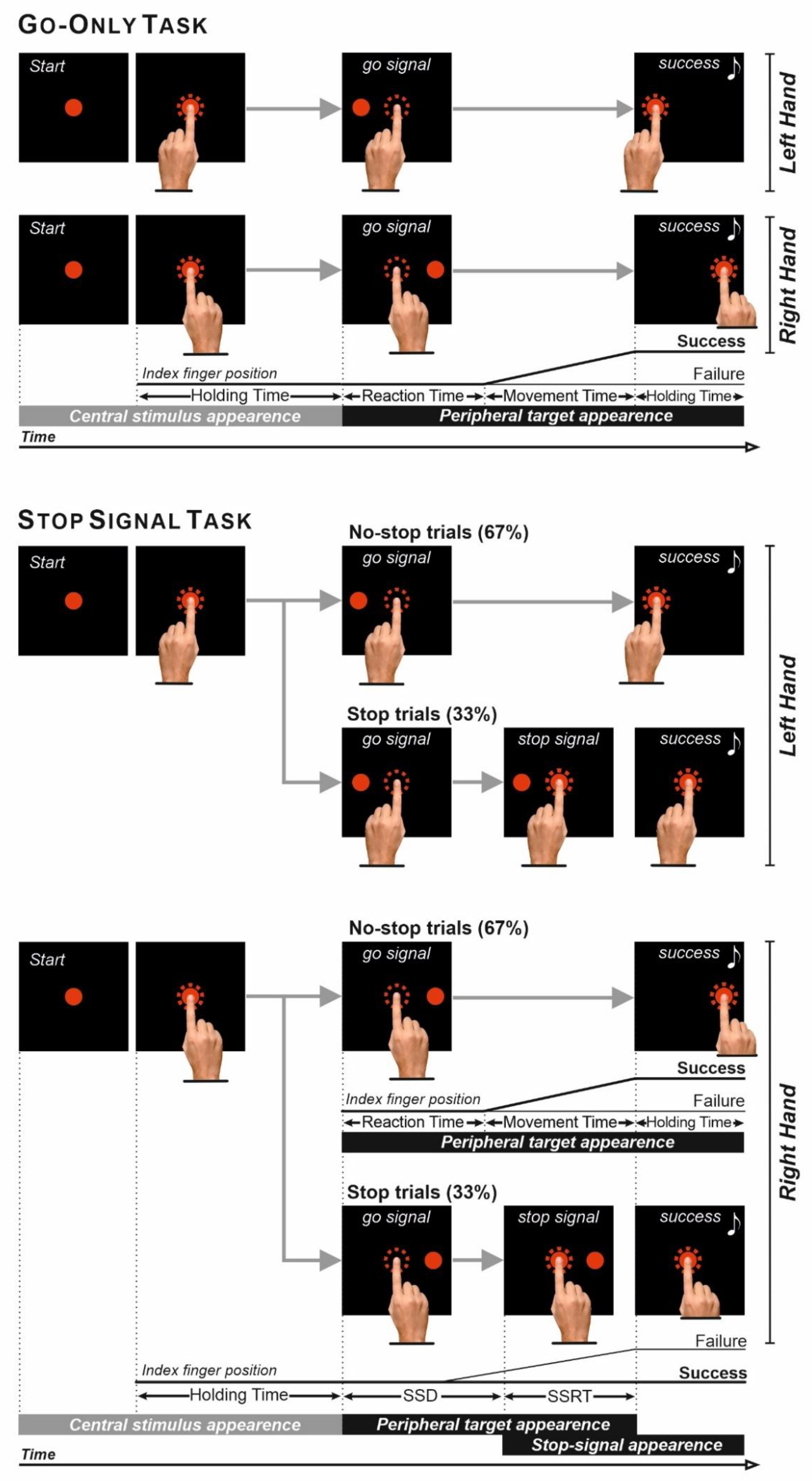
2.2. Experimental Apparatus and Behavioral Tasks

Subjects were seated in a darkened, sound-attenuated room and completed the tasks on a 17-inch liquid crystal display touchscreen monitor (MicroTouch M1700SS, 3M, Saint Paul MN, USA, 1280 × 1024 resolution, 32-bit color depth, refresh rate 75 Hz, sampling rate 200 Hz). Stimuli consisted of red circles (2.434 cd/m2) with a diameter of 4 degrees of visual angle (dva; 2.6 cm) against a dark background of uniform luminance (<0.01 cd/m2). The presentation of stimuli and data acquisition of behavioral responses were controlled by the CORTEX real-time control system, a non-commercial software package [55].
Each participant completed two sessions of the reaching versions of the stop-signal task (SST) [51], one using the right arm (right SST) and one using the left arm (left SST, Figure 2). Participants also performed two simple reaction time tasks (go-only task), again one using the right arm (right go-only) and one using the left arm (left go-only). In all instances, the peripheral visual targets were presented ipsilaterally with respect to the arm used. We decided to present the target on the same side of the responding arm (uncrossed combination) to always put the participants in the most favorable condition in terms of planning speed. In fact, it is well known that subjects have faster reaction times (RT) in the uncrossed than in the crossed combination, i.e., when the target is presented on the opposite side with respect to the responding arm [56,57]. The order of administration of the tasks was counterbalanced across participants. A ten-minute break was interposed between the execution of the tasks if requested.
All trials started with the presentation of a red circle at the center of the monitor. Subjects were instructed to touch and hold the central stimulus with the index finger for a period of time varying from 500 to 800 ms. Subsequently, in the go-only task (Figure 2, Go-Only Task) and the no-stop trials of the SST (Figure 2, Stop-Signal Task), the central stimulus was removed, and, simultaneously, a target appeared (go-signal) on the horizontal plane on the same side of the responding arm at 18.6 degrees (13.5 cm) either to the right (right go-only/no-stop trial) or to the left (left go-only/no-stop trial) of the central stimulus. In these instances, participants had to perform a reaching movement toward the peripheral target, touching and holding it for 300 to 400 ms (in steps of 50 ms) until they heard acoustic feedback signaling a successful trial. By contrast, in the stop trials (34% of all the trials; Figure 2, Stop-Signal Task), after the presentation of the go-signal, the central stimulus reappeared (stop-signal) at a variable delay (stop-signal delay, SSD), and participants were instructed to inhibit their movement towards the peripheral target. The length of the SSD was varied via a staircase algorithm [58] that aimed to allow participants to cancel their responses about 50% of the times. The initial value of the SSD was set to 119.7 ms (9 refresh rates). Then the staircase procedure increased the SSD by 39.9 ms (3 refresh rates) when participants correctly canceled the response, making the stopping more difficult. By contrast, the SSD length was decreased by the same amount of time when the participants did not withhold the movement. Every time the participants successfully withheld the response, keeping the index finger still for 300–400 ms, the trial was successful and signaled by auditory feedback.
As it is known that participants try to delay their response during no-stop trials to increase the probability of stopping their responses [59,60], we told them that the probability of successful stopping would have been around 50%, irrespective of their strategy. To discourage further subjects from slowing down during no-stop trials, we set an upper-RT limit, i.e., whenever RTs were longer than 500 ms, the no-stop trials were signaled as errors and aborted. Nevertheless, to avoid cutting the RTs’ right tail distribution, we gave participants an extra time of an additional 100 ms for releasing the central stimulus. These trials were called overtime-reaching trials [60]. Thus, whenever time participants detached the index finger between 500 and 600 ms after the go-signal, the RT was recorded, but an error was signaled. Trials with RTs longer than 600 ms were aborted without recording the RT. Overreach trials were kept for the final analysis, and on average, they accounted for 10.5% and 10.6% of the total no-stop trials in left-handers and right-handers, respectively. The role of the maximum RT in no-stop trials is to put time pressure on the individuals so that they do not delay their responses too much, waiting for the potential presence of the stop-signal. Overall, participants performed 480 trials in two blocks for the SST and 100 trials of the go-only task in one single block. Due to a technical mistake, three right-handers performed two blocks of 180 for the SST. In addition, one left-hander completed only one single block of 240 trials for the right SST.

2.3. Data Analysis

We used RTs, MTs, and the SSRTs as behavioral parameters. RTs were computed as the time interval between the go-signal presentation and the onset of movement (i.e., the moment in which participants detached their index finger from the touchscreen). MTs were computed as the time interval between the movement onset and the moment in which the peripheral target was touched. Trials with RTs shorter/longer than the mean ± 3 standard deviations (SDs) were considered outliers and discarded. Overall, 0.93% and 0.79% of the data were eliminated in left- and right- handers, respectively.
Reactive inhibition was measured via the SSRT [34,61], which was estimated using the integration method as this method provides the best estimate when proactive slowing occurs [62]. To assess changes in SSRTs, we employed a two-way mixed-design analysis of variance (ANOVA; Between-subject factor: Handedness (Left-handers, Right-handers); Within-subject factor: Reaching Arm (Left arm, Right arm)) using SPSS (Version 26.0. Armonk, NY, USA: IBM Corp).
Proactive inhibition was assessed by comparing the RTs and the MTs of no-stop trials versus those of go-only trials, i.e., by evaluating the presence of the so-called context effect [51]. The context effect is a form of proactive inhibitory control that allows participants to optimize their motor strategies according to the experimental context. Participants have to execute the same reaching movements toward a peripheral target in both no-stop and go-only trials. However, because of the awareness that unexpectedly, on some trials, actions will have to be withheld, when a subject performs a no-stop trial, the RT is lengthened with respect to when he/she performs a go-only trial. This is a common strategy to increase the likelihood of stopping the response on stop-signal trials. Notably, the lengthening of RTs is (partially) compensated by the shortening of MTs, which are significantly longer in go-only trials than in no-stop trials [51]. We checked for the presence of the context effect in two different ways. First, differences in the variances of the mean values of RTs, and MTs, were computed via two three-way mixed-design analyses of variance (ANOVA; Between-subject factor: Handedness (Left-handers, Right-handers); Within-subject factor: Reaching Arm (Left arm, Right arm); Trial Type (no-stop trials; go-only trials)). Second, the cumulative distributions of RTs and MTs in no-stop and go-only trials were compared using the two-sample Kolmogorov–Smirnov tests at individual and population levels.
In all ANOVAs, the assumption of normalcy of the distributions was assessed using the Shapiro–Wilk test, and Bonferroni’s corrections were applied for all multiple comparisons. For each ANOVA, we quantified the effect size in terms of the partial eta-squared (ηp2; values of 0.01, 0.058, and 0.139 indicate small, medium, and large effects, respectively), whereas for the t-test, we quantified it using Cohen’s d (values of 0.2, 0.5, and 0.8 indicate small, medium, and large effects, respectively). We employed the Bayes factors (BF10, r-scale = 0.70727) to quantify the strength of null hypotheses. BF10 values <0.33 and <0.1 provide moderate and robust support, respectively, for a null hypothesis compared to the alternative hypothesis. Conversely, BF10 values >3 and >10 constitutes moderate and strong support for the alternative hypothesis. Finally, to determine whether there were significant differences between the occurrences of the context effects in right- and left-handers, we exploited a χ2-test of independence using the R-Stats package [63].

3. Results

3.1. The Assumptions Underlying the Race Model Were All Satisfied

To check whether collected data allowed computation of a reliable estimate of the SSRT, we assessed whether: (i) the staircase algorithm worked similarly well in the right- and left-handers; and (ii) the main assumption about the stochastic independence between the go process and the stop process was fulfilled, i.e., the RTs of stop-failure trials were shorter RTs than no-stop trials [34]. Both conditions were satisfied (Table 1).
The staircase algorithm worked similarly well for all groups (P(failure), two-tailed t-test; t(50) = −0.79, p = 0.43). Furthermore, in both groups, RTs of stop-failure trials were faster than no-stop trials as revealed a three-way mixed-design analysis of variance (ANOVA; Between-subject factor: Handedness (Left-handers, Right-handers); Within-subject factor: Reaching Arm (Left arm, Right arm); Trial Type (no-stop trials; stop-failure trials); Table 2). The effect of the interaction between Trial Type and Reaching Arm was explained by the fact that the RTs of no-stop trials executed with a given arm were slower than the RTs of the stop-failure trials performed with the opposite arm. Finally, the interaction between Trial Type and Handedness was because the RTs of no-stop trials executed by right-handers were slower than the RTs of the stop-failure trials executed by left-handers, and vice versa. These results are not relevant to the paper’s aim, and we will not discuss them further.

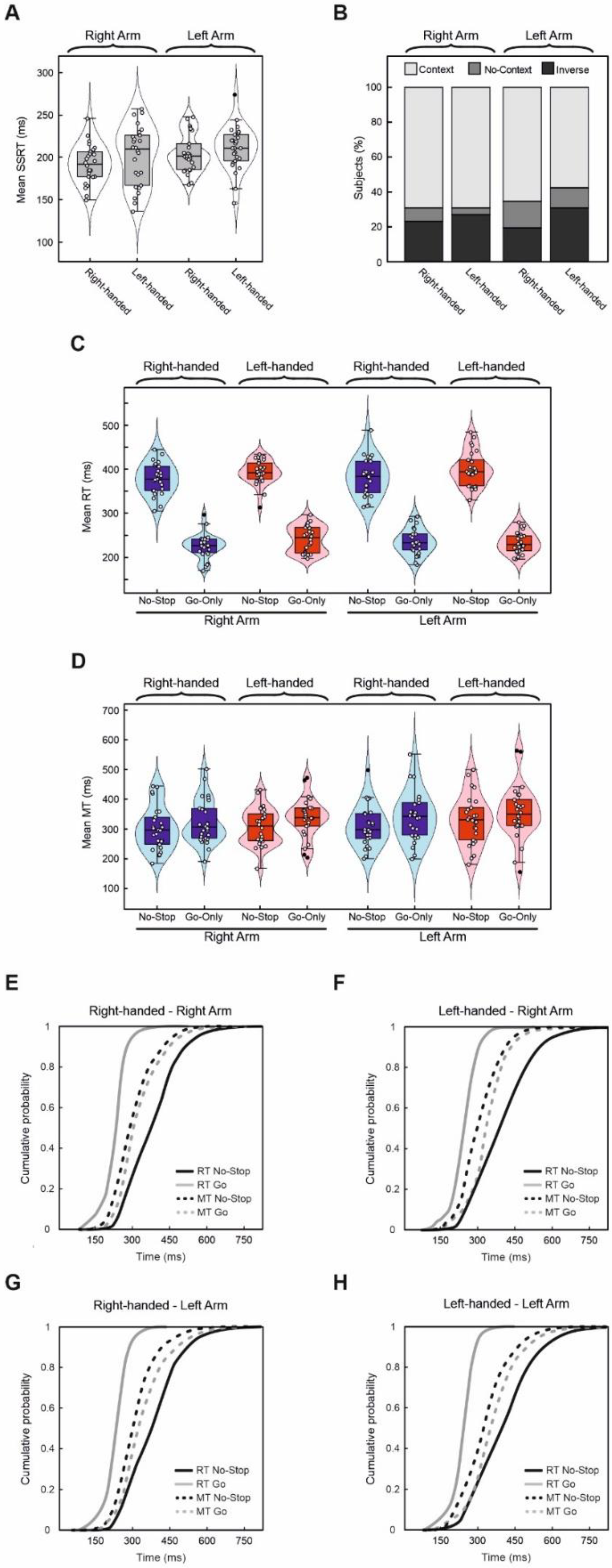
3.2. Reactive and Proactive Inhibitory Control Do Not Differ According to Handedness

The main aim of this work was to assess whether differences in reactive and proactive inhibitory control between right- and left-handers occurred. As far as reactive inhibition is concerned, the two-way ANOVA on SSRTs did not show either a main effect of the factor Handedness or its interaction with the factor Reaching Arm (Figure 3A). However, this analysis unveiled a main effect of the factor Reaching Arm, indicating that the SSRT is shorter for the right arm (M = 196.8 ms, 95% CI [188.6, 205.1]) than for the left arm (M = 207.4 ms, 95% CI (200.7, 214.1), Mdiff = 10.6 ms, 95% CI (2.2, 18.9), see Table 3). The BF10 for Handedness and the interaction were 0.54 and 0.35, respectively, providing moderate support for the null hypothesis. By contrast, the BF10 for Reaching Arm was 3.31, indicating moderate evidence for the alternative hypothesis.
We checked the impact of handedness on proactive inhibition in three different ways [17,51]. First, to evaluate the context effect at the individual level, we measured the context effect following a within-subject approach. We compared the cumulative distributions of RTs and MTs of no-stop versus go-only trials for each participant via the two-sample Kolmogorov–Smirnov test. We defined a participant as having a context effect when he/she showed a significant lengthening of RTs and a significant simultaneous shortening of MTs in no-stop trials with respect to go-only trials. By contrast, we classified a participant as not having a context effect (“no-context effect”) whenever the cumulative distribution of MTs of no-stop trials was not different from that of go-only trials, while RTs of no-stop trials were still longer than those of go-only trials. Finally, we defined a participant as having an “inverse context effect” when he/she showed a significant lengthening of RTs and MTs in no-stop trials with respect to go-only trials. After that, we computed the percentage of participants who exhibited the context effect, and we compared it with the sum of the rate of participants with the no-context effect plus those with the inversed-context effect. We found that 69.2% and 65.4% of right-handers showed a context effect, using the right arm and the left arm, respectively. Left-handers exhibit a context effect in 69.2% of cases using the right arm and 57.7% of cases using the left arm (Figure 3B). The frequency of context effect occurrences across the population and the different experimental conditions, i.e., the use of the right- and left-hand was compared using a χ2 test of independence. These analyses showed that right- and left-handers displayed no different proactive inhibitory control, regardless of whether they used the right or left arm for task execution (χ2(3) = 1.02; p = 0.80).
Second, to assess the context effect at the population level, we compared the means of RTs and MTs, with two three-way mixed-design ANOVAs. As expected, the analysis of RTs revealed a main effect of Trial Type (Mdiff = 154.8 ms, 95% CI (146.1, 163.4)), indicating that overall participants had shorter RTs when executing go-only trials (M = 233.5 ms, 95% CI (228.1, 238.9)) than when executing no-stop trials (Table 4, Figure 3C). In addition, we also found a main effect of the factor Handedness (Mdiff = 12.2 ms, 95% CI (−10.8, 35.2)) because right-handers (M = 304.7 ms, 95% CI (288.7, 320.8)) were faster than left-handers (M = 317.0 ms, 95% CI (300.3, 333.7)).
The analysis of MTs showed two main effects. First, the main effect of Trial Type (Mdiff = 26.6 ms, 95% CI (16.1, 37.0)) was because participants had longer MTs when executing go-only trials (M = 337.7 ms, 95% CI (322.3, 353.1)) than when executing no-stop trials (M = 311.1 ms, 95% CI (297.1, 325.1); Table 5, Figure 3D). Second, the main effect of the Reaching Arm (Mdiff = 16.4 ms, 95% CI (6.7, 26.1)) was given by the fact that participants had shorter MTs when using the right (M = 316.2 ms, 95% CI (302.7, 329.7)) than the left arm (M = 332.6 ms, 95% CI (316.6, 348.6); Table 5, Figure 3D).
Finally, to re-evaluate the context effect at the population level using another approach, we combined data from single participants to create cumulative population distributions of RTs and MTs of go-only trials and no-stop trials (Figure 3E–H). Even when adopting this approach, we found that the two groups of participants behaved similarly, as RTs were longer while MTs were shorter in no-stop trials than in go-only trials (two-sample Kolmogorov–Smirnov test, all p < 0.0001).

3.3. Correlations between the Laterality Quotient Scores and the Behavioral Parameters of the Go-Only and Stop-Signal Tasks

We checked whether behavioral parameters of the go-only task and the SST correlate with the laterality quotient scores. Thus, we computed the values of Spearman’s correlation coefficient (rho) between the laterality quotient score of a given participant and each corresponding behavioral parameter (RT, MT, and SSRT). In none of the instances did we find a significant correlation (not shown).

4. Discussion

Putatively, an asymmetrical organization of cognitive function optimizes processing time. Thus, the idea of a right-lateralized network underpinning (reactive) inhibitory control implicitly exerts attraction [4]. However, experimental evidence supporting this hypothesis is very contradictory. This study investigated whether action planning and motor inhibitory control show any asymmetry according to participants’ handedness and the arm used to respond. We checked the two following hypotheses. First, suppose the right hemisphere would play a crucial role in reactive inhibitory control. In that case, participants should inhibit a planned movement faster with the left arm, as although both hemispheres contribute to unilateral limb movements, the activation of the contralateral hemisphere is more robust, especially for the dominant arm. Second, assuming that as for the other cognitive functions, left-handers have a minor degree of functional asymmetry even for inhibitory control, then, unlike in right-handers, they should exhibit a similar behavioral performance irrespectively of the arm employed. Alternatively, if the right hemisphere specifically underpins inhibition even in left-handers, they should be much more proficient at inhibiting movements with the left arm than right-handers, as this is the dominant arm. By contrast, if the inhibitory control relies on the cooperation of both hemispheres, we predicted not finding differences related to either the arm employed or to handedness. Our results do not support the idea of the existence of a right-hemispheric dominance of inhibitory control, as we did not find any difference in either reactive or proactive inhibition in the right- and left-handers, and we never see a left arm superiority in inhibitory control. However, contrary to our initial hypothesis, we found a small advantage of participants in inhibiting outright movements with the right arm. Notably, there were no differences in proactive inhibitory control. We also found that MTs are faster when performed with the right arm, while right-handers have shorter RTs than left-handers. In the following, we will interpret our data in light of the hypothesis of a left-hemisphere dominance for action planning.

4.1. Inhibition, Handedness, and Laterality

Our results show that left- and right-handers exhibit similar behavioral performances in reactive and proactive inhibition. There are just two studies aimed at assessing the relationship between handedness and reactive inhibitory control in the literature. They provided discrepant results. On the one hand, Hiraoka et al. [49] showed that left-handers had a shorter SSRT when they used the left hand but only in the version of the stop-signal task in which the target could appear on the right or the left side. If the target appeared just on one side, no effects on reactive inhibition appeared. By contrast, right-handers did not show any asymmetry. Serrien and Sovijärvi-Spapé [50] exploited a modified version of a go/no-go task where participants were instructed to either move, inhibit, or switch a hand movement. They did not find differences in any of the behaviors according to handedness. Notably, however, the authors found a significant correlation between EEG coherence, successful inhibition, and switching irrespective of handedness [50]. Thus, they concluded that the left hemisphere might play a dominant role in processing such high-order motor control features. While Hiraoka et al. [49] provide some weak support for the existence of a right-lateralized reactive inhibitory network [4], neither our results nor those by Serrien and Sovijärvi-Spapé [50] are compatible with this idea. In fact, the behavioral results of those two studies did not show either a difference in reactive inhibition between right- and left-handers or a better performance with the left arm in left-handers.
Unexpectedly, we found a significant advantage of the right versus the left arm in canceling movements outright. On the one hand, this result is the opposite of what the right-lateralized hypothesis would predict. However, on the other hand, such a result does not entirely fit with the idea that reactive inhibitory control requires the cooperation of both hemispheres. In two previous works [16,17], we found that neither right- nor left-dominant Parkinson’s patients showed SSRT of different lengths according to the arm employed. Further studies are required to distinguish whether the absence of a laterality effect is due to age or the disease. In any case, the right arm advantage in reactive inhibitory control agrees with Serrien and Sovijärvi-Spapé’s [50] results, suggesting a prevalence of left-hemisphere in reactive inhibition. Notably, proactive inhibition did not show any laterality effect.

4.2. RTs, MTs, Handedness, and Laterality

Most research on the relationship between RTs and handedness did not find differences in a right- versus left-handers [64,65,66]. By contrast, Velay and Benoit-Dubrocard [67], comparing reaching arm movements in left- and right-handers, showed that the latter had shorter RTs with the left than with the right arm. Instead, left-handers were equally fast with both hands. We found right-handers exhibit shorter RTs than left-handers by ~4 ms. As Bayesian factors provide weak support for the alternative hypothesis, we will not make any strong arguments about this result. Even though our sample was homogeneous in terms of age and sex and of medium-large size (n = 52), this result could still be random. Nevertheless, Goodin et al. [68] found a result very similar to ours, but on a very small group of participants (n = 9).
We did not find differences in RTs related to the arm employed. Evidence about a laterality effect on RTs is extremely mixed. Marzi et al. [69] reviewed 16 studies employing simple unimanual RT tasks where keypresses were requested, and participants were right-handed. They found that participants were slightly faster in 13 out of 16 cases with the right than with the left hand. Savage et al. [64] confirmed the occurrence of a right-hand advantage irrespective of participants’ handedness. By contrast, Rabbitt [70], comparing left- and right-handers, did not find any effect of hand dominance on RTs when participants were required to move a given hand. However, he found that when subjects had to choose between hands, they responded faster with the dominant than non-dominant hand. Barthélémy and Boulinguez [71] showed a left-hand advantage in right-handers. By contrast, Hiraoka et al. [49] found that left-handers exhibit shorter RT for the left- than for the right hand but only when targets could appear either on the right or the left side. Whenever the target appeared just on one side, RTs of left- and right-handers were not significantly different. Interestingly, Carson et al. [72] showed that the left arm advantage in right-handers depended upon the experimental context. In particular, such an effect appeared if subjects were not provided with advanced information about target position, but the differences between right and left hand disappeared when such information was available. Moreover, Hodges et al. [73] found that RT advantage changes according to the experimental setting.
Several confounding factors can explain the discrepancies between different findings. Methodological aspects are surely very relevant. Frequently sample sizes are rather small, the composition in terms of sex and age is often not well balanced, and the way of assessing handedness is frequently different and sometimes is not very accurate. Other concerns relate to the task type, i.e., some tasks required keypresses, and others pointing or reaching arm movements. Finally, in the overwhelming majority of previous experiments, the target appeared both in the visual field ipsilateral to the effector (uncrossed condition) and contralaterally (crossed condition). By contrast, in our experiment, targets were always ipsilateral to the arm used, i.e., a condition that favored the fastest movement planning [56,57]. Such an experimental feature indeed has great importance when interpreting the results.
As far as MTs are concerned, we did not find any difference between left- and right-handers. The only other study that assessed the speed of MT found that right-handers move faster with the right arm and left-handers with the left arm [67]. We found, however, a strong effect of the arm on MTs. Participants showed shorter MTs using the right than the left arm independently of handedness (the former was ~16 ms faster than the latter). Furthermore, the value of the Bayesian factors strongly supported the alternative hypothesis. Considering that our result has been obtained in a sample more than twice than that of the previous study (56 vs. 24 participants), we are confident about our results. In addition, it has frequently been found that, in right-handed subjects, mean MT was shorter for the right than for the left hand [71,73,74,75].

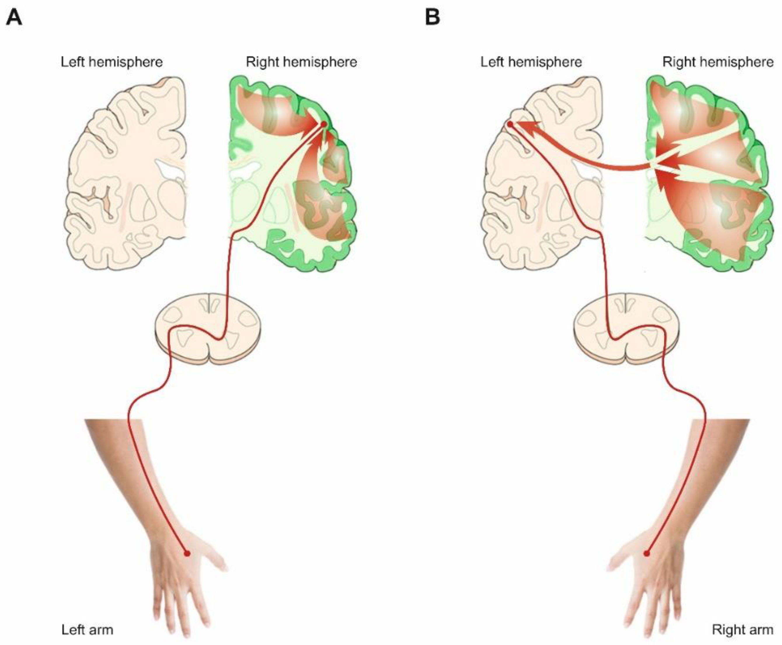
4.3. The Left Hemisphere Role’s in Actions Planning

It has been well established that unimanual arm movements elicit bilateral activity in the sensorimotor areas [18,19]. However, the meaning of ipsilateral hemisphere signals is considerably debated. On the one hand, some maintain that the activation of the hemisphere ipsilateral to the moving limb reflects an active inhibition exerted by the sensorimotor areas of the contralateral hemisphere aimed at avoiding unwanted movements of the other limb [20,21]. By contrast, others believe that the ipsilateral activation contributes to the planning and execution of contralateral limb movements [18,19]. This view is sustained by the fact that kinematic and movement parameters of the contralateral limb can be decoded from signals recorded in the ipsilateral hemisphere in monkeys [76] and humans [26].
However, very little attention has been given to the fact that the two hemispheres seem to have different roles in terms of praxis, i.e., the ability to perform skilled or learned movements. In fact, lesions of the left hemisphere lead to apraxia, i.e., the impairments in the production of complex movement in the absence of muscle deficits, much more often than do right hemisphere damages [77,78]. This evidence agrees with neuroimaging studies that showed a strong left-hemispheric lateralization regardless of the hand used to perform hand pantomimes of tool use in right-handed volunteers [79,80]. In other words, participants’ parietal and frontal regions in the left hemisphere were active during movements of either the left or right hand. By contrast, the activation of the homologous regions in the right hemisphere was more limited to movements with the contralateral hand. Vingerhoets et al. [81] replicated these results in the left-handed group and thus concluded that this leftward asymmetry in praxis was independent of handedness.
Very importantly, a recent study showed that the ipsilateral cortical representation of reaching arm movements strikingly differs between the left and right hemispheres [82]. Merrick et al. [82], using a kinematic encoding model, analyzed six patients’ electrocorticographic activity across the two hemispheres. They found that ipsilateral encoding was much stronger in the left than in the right hemisphere. Thus, the left hemisphere showed a better across-arm generalization, i.e., it has a similar neural representation for right and left arm movements. In contrast, the right hemisphere encodes movement parameters of contralateral movements. This study is very relevant because it shows that left-hemispheric dominance holds not only for complex movements involving tool use or symbolic gesture but also for arm reaching movements, the same used in the present work. This evidence, together with the results of Serrien and Sovijärvi-Spapé [50], showing that brain signals from the left but not from the right hemisphere correlate with inhibitory control irrespective of hand employed and handedness, suggests an explanation for our findings, i.e., the right reaching arm movements have both shorter MTs and SSRTs than left arm reaches irrespective of participants’ handedness. At the same time, proactive inhibitory control did not differ between the two arms.
It is known that right-handers execute movements faster with the right than with the left hand [73,74,75], especially when an online movement correction is required [71,83]. These results have been interpreted in light of a greater specialization of the left hemisphere for controlling the magnitude and timing of muscular forces during movement execution [83,84,85]. According to our data, such functional asymmetry is not linked to participants’ handedness, but it reflects a general function of the left hemisphere. The left hemisphere advantage in temporal processing could explain the shorter SSRTs of the right arm. In fact, canceling pending actions can be considered a special case in which an online movement correction is needed, especially when the SSD is long and the movement that must be halted is an advanced stage of planning. In such a stage, muscles start to be activated, even though no overt movements are yet detected. However, there is a second, not mutually exclusive, explanation for the better reactive inhibition of the right arm. As stated above, the ipsilateral cortical representation of reaching arm movements is stronger in the left than in the right hemisphere [82]. This means that when the left arm is moved, both hemispheres are active, while when the right arm is moved, just the left hemisphere is active. Thus, it is plausible to suggest that whenever a left reach has to be suppressed, the activity of both hemispheres has to be altered. Such processing would require more time with respect to when the activity of one hemisphere has to be changed, as in the case of right reaches. Notably, computations subserving proactive inhibitory control do not show any laterality effect indicating that they are carried on by both hemispheres.

5. Conclusions

To test the hypothesis of the right-hemispheric specialization of inhibition, we measured reactive and proactive inhibitory control in the largest sample of left- and right-handers to date. We found that while handedness does not affect either reactive or proactive inhibition, right arm reaches show shorter SSRT. By contrast, proactive inhibitory control does not show any laterality effect. Such results are incompatible with the idea of a right-hemispheric functional asymmetry of inhibition. However, they are also only partially compatible with the idea that inhibitory control is computed by both hemispheres, at least in young, healthy individuals. In fact, reactive but not proactive inhibition seems to be computed mainly by the left hemisphere. Our findings are in line with evidence showing a left-hemispheric dominance in some aspects of movement control [82,83,84,85], including inhibition [50].

Author Contributions

Conceptualization, G.M.; methodology, G.M. and C.M.; formal analysis, G.M. and C.M.; validation, G.M. and C.M.; investigation, C.M.; data curation, C.M.; writing—original draft preparation, G.M. and C.M.; writing—review and editing, G.M. and C.M.; resources, G.M.; software G.M.; supervision, G.M. All authors have read and agreed to the published version of the manuscript.

Funding

This research was funded by the Antonio Meneghetti Award 2019, and by Erasums+ project 610134-EPP-1-2019-1-JO-EPPKA2-CBHE-JP.

Institutional Review Board Statement

The study was conducted according to the guidelines of the Declaration of Helsinki, and approved by the Ethics Committee of the University of Brescia, Italy (Prot. NP4452, 17 December 2020).

Informed Consent Statement

Informed consent was obtained from all subjects involved in the study.

Data Availability Statement

Data will be freely available from the Open Science Framework platform [54].

Acknowledgments

Special thanks go to Alex Cooper who revised the English.

Conflicts of Interest

The authors declare no conflict of interest.

References

  1. Mirabella, G. Should I stay or should I go? Conceptual underpinnings of goal-directed actions. Front. Syst. Neurosci. 2014, 8, 206. [Google Scholar] [CrossRef] [Green Version]
  2. Mirabella, G. Inhibitory control and impulsive responses in neurodevelopmental disorders. Dev. Med. Child Neurol. 2021, 63, 520–526. [Google Scholar] [CrossRef]
  3. Wostmann, N.M.; Aichert, D.S.; Costa, A.; Rubia, K.; Moller, H.J.; Ettinger, U. Reliability and plasticity of response inhibition and interference control. Brain Cogn. 2013, 81, 82–94. [Google Scholar] [CrossRef] [PubMed]
  4. Aron, A.R.; Robbins, T.W.; Poldrack, R.A. Inhibition and the right inferior frontal cortex: One decade on. Trends Cogn. Sci. 2014, 18, 177–185. [Google Scholar] [CrossRef]
  5. Mirabella, G.; Pani, P.; Ferraina, S. Neural correlates of cognitive control of reaching movements in the dorsal premotor cortex of rhesus monkeys. J. Neurophysiol. 2011, 106, 1454–1466. [Google Scholar] [CrossRef] [PubMed] [Green Version]
  6. Mattia, M.; Pani, P.; Mirabella, G.; Costa, S.; Del Giudice, P.; Ferraina, S. Heterogeneous attractor cell assemblies for motor planning in premotor cortex. J. Neurosci. 2013, 33, 11155–11168. [Google Scholar] [CrossRef] [PubMed]
  7. Mattia, M.; Spadacenta, S.; Pavone, L.; Quarato, P.; Esposito, V.; Sparano, A.; Sebastiano, F.; Di Gennaro, G.; Morace, R.; Cantore, G.; et al. Stop-event-related potentials from intracranial electrodes reveal a key role of premotor and motor cortices in stopping ongoing movements. Front. Neuroeng. 2012, 5, 12. [Google Scholar] [CrossRef] [PubMed] [Green Version]
  8. Coxon, J.P.; Stinear, C.M.; Byblow, W.D. Intracortical inhibition during volitional inhibition of prepared action. J. Neurophysiol. 2006, 95, 3371–3383. [Google Scholar] [CrossRef] [PubMed]
  9. Swick, D.; Ashley, V.; Turken, A.U. Left inferior frontal gyrus is critical for response inhibition. BMC Neurosci. 2008, 9, 102. [Google Scholar] [CrossRef] [Green Version]
  10. Mirabella, G.; Iaconelli, S.; Romanelli, P.; Modugno, N.; Lena, F.; Manfredi, M.; Cantore, G. Deep brain stimulation of subthalamic nuclei affects arm response inhibition in Parkinson’s patients. Cereb. Cortex 2012, 22, 1124–1132. [Google Scholar] [CrossRef] [Green Version]
  11. Mirabella, G.; Iaconelli, S.; Modugno, N.; Giannini, G.; Lena, F.; Cantore, G. Stimulation of subthalamic nuclei restores a near normal planning strategy in Parkinson’s patients. PLoS ONE 2013, 8, e62793. [Google Scholar] [CrossRef] [Green Version]
  12. Mancini, C.; Modugno, N.; Santilli, M.; Pavone, L.; Grillea, G.; Morace, R.; Mirabella, G. Unilateral Stimulation of Subthalamic Nucleus Does Not Affect Inhibitory Control. Front. Neurol. 2018, 9, 1149. [Google Scholar] [CrossRef]
  13. van den Wildenberg, W.P.; van Boxtel, G.J.; van der Molen, M.W.; Bosch, D.A.; Speelman, J.D.; Brunia, C.H. Stimulation of the subthalamic region facilitates the selection and inhibition of motor responses in Parkinson’s disease. J. Cogn. Neurosci. 2006, 18, 626–636. [Google Scholar] [CrossRef] [PubMed] [Green Version]
  14. Swann, N.C.; Cai, W.; Conner, C.R.; Pieters, T.A.; Claffey, M.P.; George, J.S.; Aron, A.R.; Tandon, N. Roles for the pre-supplementary motor area and the right inferior frontal gyrus in stopping action: Electrophysiological responses and functional and structural connectivity. Neuroimage 2012, 59, 2860–2870. [Google Scholar] [CrossRef] [Green Version]
  15. van den Wildenberg, W.P.M.; van Wouwe, N.C.; Ridderinkhof, K.R.; Neimat, J.S.; Elias, W.J.; Bashore, T.R.; Wylie, S.A. Deep-brain stimulation of the subthalamic nucleus improves overriding motor actions in Parkinson’s disease. Behav. Brain Res. 2021, 402, 113124. [Google Scholar] [CrossRef]
  16. Mirabella, G.; Fragola, M.; Giannini, G.; Modugno, N.; Lakens, D. Inhibitory control is not lateralized in Parkinson’s patients. Neuropsychologia 2017, 102, 177–189. [Google Scholar] [CrossRef] [PubMed]
  17. Di Caprio, V.; Modugno, N.; Mancini, C.; Olivola, E.; Mirabella, G. Early-Stage Parkinson’s Patients Show Selective Impairment in Reactive But Not Proactive Inhibition. Mov. Disord. 2020, 35, 409–418. [Google Scholar] [CrossRef]
  18. Bundy, D.T.; Leuthardt, E.C. The Cortical Physiology of Ipsilateral Limb Movements. Trends Neurosci. 2019, 42, 825–839. [Google Scholar] [CrossRef] [PubMed]
  19. Cabibel, V.; Hordacre, B.; Perrey, S. Implication of the ipsilateral motor network in unilateral voluntary muscle contraction: The cross-activation phenomenon. J. Neurophysiol. 2020, 123, 2090–2098. [Google Scholar] [CrossRef] [PubMed]
  20. Duque, J.; Mazzocchio, R.; Dambrosia, J.; Murase, N.; Olivier, E.; Cohen, L.G. Kinematically specific interhemispheric inhibition operating in the process of generation of a voluntary movement. Cereb. Cortex 2005, 15, 588–593. [Google Scholar] [CrossRef] [PubMed]
  21. Di Lazzaro, V.; Oliviero, A.; Profice, P.; Insola, A.; Mazzone, P.; Tonali, P.; Rothwell, J.C. Direct demonstration of interhemispheric inhibition of the human motor cortex produced by transcranial magnetic stimulation. Exp. Brain Res. 1999, 124, 520–524. [Google Scholar] [CrossRef]
  22. Carson, R.G. Inter-hemispheric inhibition sculpts the output of neural circuits by co-opting the two cerebral hemispheres. J. Physiol. 2020, 598, 4781–4802. [Google Scholar] [CrossRef] [PubMed]
  23. Meyer, B.U.; Röricht, S.; Gräfin von Einsiedel, H.; Kruggel, F.; Weindl, A. Inhibitory and excitatory interhemispheric transfers between motor cortical areas in normal humans and patients with abnormalities of the corpus callosum. Brain 1995, 118, 429–440. [Google Scholar] [CrossRef] [PubMed]
  24. Newton, J.M.; Sunderland, A.; Gowland, P.A. fMRI signal decreases in ipsilateral primary motor cortex during unilateral hand movements are related to duration and side of movement. Neuroimage 2005, 24, 1080–1087. [Google Scholar] [CrossRef] [PubMed]
  25. Kobayashi, M.; Hutchinson, S.; Schlaug, G.; Pascual-Leone, A. Ipsilateral motor cortex activation on functional magnetic resonance imaging during unilateral hand movements is related to interhemispheric interactions. Neuroimage 2003, 20, 2259–2270. [Google Scholar] [PubMed]
  26. Bundy, D.T.; Szrama, N.; Pahwa, M.; Leuthardt, E.C. Unilateral, 3D Arm Movement Kinematics Are Encoded in Ipsilateral Human Cortex. J. Neurosci. 2018, 38, 10042–10056. [Google Scholar] [CrossRef] [PubMed] [Green Version]
  27. Salerno, A.; Georgesco, M. Interhemispheric facilitation and inhibition studied in man with double magnetic stimulation. Electroencephalogr. Clin. Neurophysiol. 1996, 101, 395–403. [Google Scholar]
  28. Muellbacher, W.; Facchini, S.; Boroojerdi, B.; Hallett, M. Changes in motor cortex excitability during ipsilateral hand muscle activation in humans. Clin. Neurophysiol. 2000, 111, 344–349. [Google Scholar] [CrossRef]
  29. Perez, M.A.; Cohen, L.G. Mechanisms underlying functional changes in the primary motor cortex ipsilateral to an active hand. J. Neurosci. 2008, 28, 5631–5640. [Google Scholar] [CrossRef]
  30. Waters, S.; Wiestler, T.; Diedrichsen, J. Cooperation Not Competition: Bihemispheric tDCS and fMRI Show Role for Ipsilateral Hemisphere in Motor Learning. J. Neurosci. 2017, 37, 7500–7512. [Google Scholar] [CrossRef] [PubMed] [Green Version]
  31. Kim, S.G.; Ashe, J.; Georgopoulos, A.P.; Merkle, H.; Ellermann, J.M.; Menon, R.S.; Ogawa, S.; Ugurbil, K. Functional imaging of human motor cortex at high magnetic field. J. Neurophysiol. 1993, 69, 297–302. [Google Scholar] [CrossRef] [Green Version]
  32. Grabowska, A.; Gut, M.; Binder, M.; Forsberg, L.; Rymarczyk, K.; Urbanik, A. Switching handedness: fMRI study of hand motor control in right-handers, left-handers and converted left-handers. Acta Neurobiol. Exp. 2012, 72, 439–451. [Google Scholar]
  33. Buetefisch, C.M.; Revill, K.P.; Shuster, L.; Hines, B.; Parsons, M. Motor demand-dependent activation of ipsilateral motor cortex. J. Neurophysiol. 2014, 112, 999–1009. [Google Scholar] [CrossRef] [PubMed] [Green Version]
  34. Logan, G.D.; Cowan, W.B.; Davis, K.A. On the ability to inhibit simple and choice reaction time responses: A model and a method. J. Exp. Psychol. Hum. Percept. Perform. 1984, 10, 276–291. [Google Scholar] [CrossRef]
  35. Faurie, C.; Raymond, M. Handedness frequency over more than ten thousand years. Proc. Biol. Sci. 2004, 271, S43–S45. [Google Scholar] [CrossRef] [PubMed] [Green Version]
  36. Corballis, M.C. From mouth to hand: Gesture, speech, and the evolution of right-handedness. Behav. Brain Sci. 2003, 26, 199–208; discussion 208–160. [Google Scholar] [CrossRef] [PubMed]
  37. Good, C.D.; Johnsrude, I.; Ashburner, J.; Henson, R.N.; Friston, K.J.; Frackowiak, R.S. Cerebral asymmetry and the effects of sex and handedness on brain structure: A voxel-based morphometric analysis of 465 normal adult human brains. Neuroimage 2001, 14, 685–700. [Google Scholar] [CrossRef] [PubMed] [Green Version]
  38. Guadalupe, T.; Willems, R.M.; Zwiers, M.P.; Arias Vasquez, A.; Hoogman, M.; Hagoort, P.; Fernandez, G.; Buitelaar, J.; Franke, B.; Fisher, S.E.; et al. Differences in cerebral cortical anatomy of left- and right-handers. Front. Psychol. 2014, 5, 261. [Google Scholar] [CrossRef] [Green Version]
  39. Wiberg, A.; Ng, M.; Al Omran, Y.; Alfaro-Almagro, F.; McCarthy, P.; Marchini, J.; Bennett, D.L.; Smith, S.; Douaud, G.; Furniss, D. Handedness, language areas and neuropsychiatric diseases: Insights from brain imaging and genetics. Brain 2019, 142, 2938–2947. [Google Scholar] [CrossRef] [Green Version]
  40. Amunts, K.; Schlaug, G.; Schleicher, A.; Steinmetz, H.; Dabringhaus, A.; Roland, P.E.; Zilles, K. Asymmetry in the human motor cortex and handedness. Neuroimage 1996, 4, 216–222. [Google Scholar] [CrossRef]
  41. Sun, Z.Y.; Klöppel, S.; Rivière, D.; Perrot, M.; Frackowiak, R.; Siebner, H.; Mangin, J.-F. The effect of handedness on the shape of the central sulcus. Neuroimage 2012, 60, 332–339. [Google Scholar] [CrossRef] [PubMed]
  42. Jang, H.; Lee, J.Y.; Lee, K.I.; Park, K.M. Are there differences in brain morphology according to handedness? Brain Behav. 2017, 7, e00730. [Google Scholar] [CrossRef] [PubMed]
  43. Pujol, J.; Deus, J.; Losilla, J.M.; Capdevila, A. Cerebral lateralization of language in normal left-handed people studied by functional MRI. Neurology 1999, 52, 1038–1043. [Google Scholar] [CrossRef]
  44. Joliot, M.; Tzourio-Mazoyer, N.; Mazoyer, B. Intra-hemispheric intrinsic connectivity asymmetry and its relationships with handedness and language Lateralization. Neuropsychologia 2016, 93, 437–447. [Google Scholar] [CrossRef] [PubMed]
  45. Kawashima, R.; Yamada, K.; Kinomura, S.; Yamaguchi, T.; Matsui, H.; Yoshioka, S.; Fukuda, H. Regional cerebral blood flow changes of cortical motor areas and prefrontal areas in humans related to ipsilateral and contralateral hand movement. Brain Res. 1993, 623, 33–40. [Google Scholar] [CrossRef]
  46. Solodkin, A.; Hlustik, P.; Noll, D.C.; Small, S.L. Lateralization of motor circuits and handedness during finger movements. Eur. J. Neurol. 2001, 8, 425–434. [Google Scholar] [CrossRef] [PubMed]
  47. Pool, E.M.; Rehme, A.K.; Eickhoff, S.B.; Fink, G.R.; Grefkes, C. Functional resting-state connectivity of the human motor network: Differences between right- and left-handers. Neuroimage 2015, 109, 298–306. [Google Scholar] [CrossRef] [PubMed] [Green Version]
  48. Civardi, C.; Cavalli, A.; Naldi, P.; Varrasi, C.; Cantello, R. Hemispheric asymmetries of cortico-cortical connections in human hand motor areas. Clin. Neurophysiol. 2000, 111, 624–629. [Google Scholar] [CrossRef]
  49. Hiraoka, K.; Igawa, K.; Kashiwagi, M.; Nakahara, C.; Oshima, Y.; Takakura, Y. The laterality of stop and go processes of the motor response in left-handed and right-handed individuals. Laterality 2018, 23, 51–66. [Google Scholar] [CrossRef]
  50. Serrien, D.J.; Sovijarvi-Spape, M.M. Cognitive control of response inhibition and switching: Hemispheric lateralization and hand preference. Brain Cogn. 2013, 82, 283–290. [Google Scholar] [CrossRef]
  51. Mirabella, G.; Pani, P.; Ferraina, S. Context influences on the preparation and execution of reaching movements. Cogn. Neuropsychol. 2008, 25, 996–1010. [Google Scholar] [CrossRef] [PubMed]
  52. Oldfield, R.C. The assessment and analysis of handedness: The Edinburgh inventory. Neuropsychologia 1971, 9, 97–113. [Google Scholar] [CrossRef]
  53. Edlin, J.M.; Leppanen, M.L.; Fain, R.J.; Hackländer, R.P.; Hanaver-Torrez, S.D.; Lyle, K.B. On the use (and misuse?) of the Edinburgh Handedness Inventory. Brain Cogn. 2015, 94, 44–51. [Google Scholar] [CrossRef]
  54. Online Resources: Open Science Framework Platform. Available online: https://osf.io/wrkah/ (accessed on 16 August 2021).
  55. Cortex and Cortex Explorer: Real-Time Software and Data Analysis Tools. Available online: https://www.nimh.nih.gov/research/research-conducted-at-nimh/research-areas/clinics-and-labs/ln/shn/software-projects.shtml (accessed on 16 August 2021).
  56. Poffenberger, A.T. Reaction Time to Retinal Stimulation, with Special Reference to the Time Lost in Conduction through Nerve Centers; The Science Press: New York, NY, USA, 1912. [Google Scholar]
  57. Alberto Marzi, C. Asymmetry of interhemispheric communication. Wiley Interdiscip. Rev. Cogn. Sci. 2010, 1, 433–438. [Google Scholar] [CrossRef] [PubMed]
  58. Levitt, S.; Gutin, B. Multiple choice reaction time and movement time during physical exertion. Res. Q. 1971, 42, 405–410. [Google Scholar] [CrossRef]
  59. Logan, G.D. Attention, automaticity and the ability to stop a speeded choice response. In Attention and Performance IX; Long, J., Baddley, A., Eds.; Erlbaum: Hillsdale, NJ, USA, 1981; pp. 205–222. [Google Scholar]
  60. Mirabella, G.; Pani, P.; Pare, M.; Ferraina, S. Inhibitory control of reaching movements in humans. Exp. Brain Res. 2006, 174, 240–255. [Google Scholar] [CrossRef]
  61. Boucher, L.; Stuphorn, V.; Logan, G.D.; Schall, J.D.; Palmeri, T.J. Stopping eye and hand movements: Are the processes independent? Percept. Psychophys. 2007, 69, 785–801. [Google Scholar] [CrossRef]
  62. Verbruggen, F.; Chambers, C.D.; Logan, G.D. Fictitious inhibitory differences: How skewness and slowing distort the estimation of stopping latencies. Psychol. Sci. 2013, 24, 352–362. [Google Scholar] [CrossRef] [Green Version]
  63. RC Team. R: A Language and Environment for Statistical Computing; R Foundation for Statistical Computing: Vienna, Austria, 2020; Available online: http://www.R-project.org/ (accessed on 16 August 2021).
  64. Savage, C.R.; Thomas, D.G. Information processing and interhemispheric transfer in left- and right-handed adults. Int. J. Neurosci. 1993, 71, 201–219. [Google Scholar] [CrossRef]
  65. Klöppel, S.; Vongerichten, A.; van Eimeren, T.; Frackowiak, R.S.; Siebner, H.R. Can left-handedness be switched? Insights from an early switch of handwriting. J. Neurosci. 2007, 27, 7847–7853. [Google Scholar] [CrossRef] [PubMed] [Green Version]
  66. Woods, D.L.; Wyma, J.M.; Yund, E.W.; Herron, T.J.; Reed, B. Factors influencing the latency of simple reaction time. Front. Hum. Neurosci. 2015, 9, 131. [Google Scholar] [CrossRef] [Green Version]
  67. Velay, J.L.; Benoit-Dubrocard, S. Hemispheric asymmetry and interhemispheric transfer in reaching programming. Neuropsychologia 1999, 37, 895–903. [Google Scholar] [CrossRef]
  68. Goodin, D.S.; Aminoff, M.J.; Ortiz, T.A.; Chequer, R.S. Response times and handedness in simple reaction-time tasks. Exp. Brain Res. 1996, 109, 117–126. [Google Scholar] [CrossRef] [PubMed]
  69. Marzi, C.A.; Bisiacchi, P.; Nicoletti, R. Is interhemispheric transfer of visuomotor information asymmetric? Evidence from a meta-analysis. Neuropsychologia 1991, 29, 1163–1177. [Google Scholar] [CrossRef]
  70. Rabbitt, P. Hand dominance, attention, and the choice between responses. Q. J. Exp. Psychol. 1978, 30, 407–416. [Google Scholar] [CrossRef] [PubMed]
  71. Barthélémy, S.; Boulinguez, P. Manual asymmetries in the directional coding of reaching: Further evidence for hemispatial effects and right hemisphere dominance for movement planning. Exp. Brain Res. 2002, 147, 305–312. [Google Scholar] [CrossRef] [PubMed]
  72. Carson, R.G.; Chua, R.; Goodman, D.; Byblow, W.D.; Elliott, D. The preparation of aiming movements. Brain Cogn. 1995, 28, 133–154. [Google Scholar] [CrossRef] [Green Version]
  73. Hodges, N.J.; Lyons, J.; Cockell, D.; Reed, A.; Elliott, D. Hand, space and attentional asymmetries in goal-directed manual aiming. Cortex 1997, 33, 251–269. [Google Scholar] [CrossRef]
  74. Carey, D.P.; Hargreaves, E.L.; Goodale, M.A. Reaching to ipsilateral or contralateral targets: Within-hemisphere visuomotor processing cannot explain hemispatial differences in motor control. Exp. Brain Res. 1996, 112, 496–504. [Google Scholar] [CrossRef]
  75. Mieschke, P.E.; Elliott, D.; Helsen, W.F.; Carson, R.G.; Coull, J.A. Manual asymmetries in the preparation and control of goal-directed movements. Brain Cogn. 2001, 45, 129–140. [Google Scholar] [CrossRef] [PubMed] [Green Version]
  76. Ames, K.C.; Churchland, M.M. Motor cortex signals for each arm are mixed across hemispheres and neurons yet partitioned within the population response. Elife 2019, 8, e46159. [Google Scholar] [CrossRef] [PubMed]
  77. Haaland, K.Y. Left hemisphere dominance for movement. Clin. Neuropsychol. 2006, 20, 609–622. [Google Scholar] [CrossRef]
  78. De Renzi, E.; Lucchelli, F. Ideational Apraxia. Brain 1988, 111, 1173–1185. [Google Scholar] [CrossRef] [PubMed]
  79. Choi, S.H.; Na, D.L.; Kang, E.; Lee, K.M.; Lee, S.W.; Na, D.G. Functional magnetic resonance imaging during pantomiming tool-use gestures. Exp. Brain Res. 2001, 139, 311–317. [Google Scholar] [CrossRef]
  80. Johnson-Frey, S.H.; Newman-Norlund, R.; Grafton, S.T. A distributed left hemisphere network active during planning of everyday tool use skills. Cereb. Cortex 2005, 15, 681–695. [Google Scholar] [CrossRef] [Green Version]
  81. Vingerhoets, G.; Acke, F.; Alderweireldt, A.S.; Nys, J.; Vandemaele, P.; Achten, E. Cerebral lateralization of praxis in right- and left-handedness: Same pattern, different strength. Hum. Brain Mapp. 2012, 33, 763–777. [Google Scholar] [CrossRef]
  82. Merrick, C.M.; Dixon, T.C.; Breska, A.; Lin, J.J.; Edward, F.C.; King-Stephens, D.; Laxer, K.D.; Weber, P.B.; Carmena, J.M.; Knight, R.T.; et al. Left Hemisphere Dominance for Bilateral Kinematic Encoding in the Human Brain. bioRxiv 2021. [Google Scholar] [CrossRef]
  83. Elliott, D.; Lyons, J.; Chua, R.; Goodman, D.; Carson, R.G. The influence of target perturbation on manual aiming asymmetries in right-handers. Cortex 1995, 31, 685–697. [Google Scholar] [CrossRef]
  84. Goodale, M.A. Hemispheric differences in motor control. Behav. Brain Res. 1988, 30, 203–214. [Google Scholar] [CrossRef]
  85. Floegel, M.; Kell, C.A. Functional hemispheric asymmetries during the planning and manual control of virtual avatar movements. PLoS ONE 2017, 12, e0185152. [Google Scholar] [CrossRef] [PubMed]
Figure 1. Schematic representation of the consequences of right-hemispheric lateralization of inhibition on unilateral upper limb movements. (A) Suppose inhibitory commands (translucent red arrows) are generated in the right hemisphere (marked in green). In that case, people should suppress their movement faster when moving the left arm because inhibitory commands act on the same hemisphere, which primarily contributes to the planned movement. (B) In contrast, when moving the right arm, people should suppress their movement more slowly because inhibitory commands need to be sent to the opposite hemisphere through the callosal fibers (solid red arrow), increasing by a constant delay the time it takes to cancel an action outright.
Symmetry 13 01602 g001
Figure 2. Schematic representation of the go-only task and the stop-signal task. Go-only task. At the start of a trial, subjects were instructed to reach and hold a central stimulus with the index finger for a variable period of 500–800 milliseconds (ms). After that, a peripheral target appeared 18.6 degrees of visual angle (dva) to the right or the left, and subjects had to reach and hold it for 300–400 ms. We administered two different versions of the go-only task: one for left arm reaching movements to left-sided target, the other with right arm movements reaching to a right target. Stop-signal task. This task consisted of a pseudorandom mix of no-stop trials and stop trials. No-stop trials were the same as the go-trials in the go-only task and accounted for two-thirds (67%) of the total. In contrast, in the stop trials (33%), after the presentation of the peripheral target (go-signal), the central stimulus (stop-signal) reappeared at a variable delay (stop-signal delay; SSD). Participants were instructed to inhibit the incipient movement before lifting the index finger from the touchscreen. Whenever subjects canceled the ongoing movement and kept the index finger on the stop-signal for 300–400 ms, the trial was scored as a stop-success trial. Otherwise, the trial was scored as a stop-failure trial. Correct responses were signaled by auditory feedback (depicted as a white musical note). The dotted circle (blind to the subjects) indicates the size of the tolerance window for the touches (5 dva of diameter). All visual stimuli (central target/stop-signal and go-signal) consisted of red circles of 4 dva presented against a dark background.
Symmetry 13 01602 g002
Figure 3. (A) Mean stop-signal reaction times (SSRTs) as a measure of reactive inhibitory control for right- and left-handed subjects using the right and left arm. (B) Percentage of subjects showing context, no-context, and inverse context effect as a measure of proactive inhibition for each handedness group and arm used. (C) Mean reaction times (RTs) of the no-stop trials during the stop-signal task (no-stop) and the go-only task (go-only) in each condition. (D) Mean movement times (MTs) for each condition (as in C). Milliseconds (ms). The violin plots depict kernel probability density, i.e., the width of the areas represents the relative frequency of the data. Box plots are shown inside the violin plots. The lower box’s boundary indicates the first quartile, the median is marked with a black line, and the upper box’s boundary indicates the third quartile. Whiskers indicate values 1.5 times the interquartile range below the first quartile and above the third quartile. Single data points are pictured as jittered white dots. Outliers are represented as full black dots. (E) Cumulative distributions of RTs (solid lines) and MTs (dotted lines) of right-handed subjects (n = 26) using the right arm for go-only trials (gray) and no-stop trials (black). These cumulative distributions were obtained by collapsing together the cumulative distributions of RTs and MTs of no-stop and go-only trials of single subjects. Two-sample Kolmogorov–Smirnov test: (i) RTs, D = 0.71, p < 0.0001; (ii) MTs, D = 0.12, p < 0.0001. (F) Cumulative distributions of RTs and MTs of left-handers subjects using the right arm (n = 26) for go-only and no-stop trials. Two-sample Kolmogorov–Smirnov test: (i) RTs, D = 0.65, p < 0.0001; (ii) MTs, D = 0.24, p < 0.0001. (G) Same Right-handers subjects as in C, but using the left arm. Two-sample Kolmogorov–Smirnov test: (i) RTs, D = 0.67, p < 0.0001; (ii) MTs, D = 0.13, p < 0.0001. (H) Same left-handers subjects as in D but using the left arm. Two-sample Kolmogorov–Smirnov test: (i) RTs, D = 0.69, p < 0.0001; (ii) MTs, D = 0.15, p < 0.0001.
Symmetry 13 01602 g003
Table 1. Summary of behavioral values for right- and left-handed participants using the right and left reaching arm during the stop-signal task and the go-only. Task performance accuracy is defined as the ratio between the number of go-trials correctly executed and the total number of go-trials delivered, given by the sum of trials correctly executed, trials in which participants missed the target, trials in participants remained still on the central stimulus for more than two seconds, and trials in which they did not hold the central stimulus or the target for the requested amount of time. In all cases the average value across the samples (±SD) is reported.
Right-HandersLeft-Handers
Right Reaching ArmLeft Reaching ArmRight Reaching ArmLeft Reaching Arm
Mean SSD180.9 ± 47.5176.5 ± 51.9189.8 ± 41.1192.4 ± 47.8
P(failure)0.51 ± 0.020.52 ± 0.030.52 ± 0.030.52 ± 0.03
SSRT192.1 ± 22.6204.3 ± 22.4201.6 ± 35.0210.5 ± 25.8
RT No-Stop trials377.5 ± 35.4382.0 ± 42.5392.8 ± 27.3400.7 ± 39.2
RT Stop-Failure trials328.2 ± 34.6330.7 ± 35.3330.4 ± 37.5327.7 ± 38.6
RT Go-Only trials225.1 ± 27.7234.4 ± 28.4241.2 ± 30.1233.2 ± 23.5
MT No-Stop trials300.4 ± 72.8308.7 ± 68.6306.1 ± 62.9329.3 ± 82.5
MT Go-Only trials322.8 ± 74.5339.2 ± 83.7335.5 ± 66.0353.2 ± 91.5
Accuracy Go-Only trials0.95 ± 0.060.95 ± 0.050.94 ± 0.050.95 ± 0.04
Accuracy No-Stop trials0.97 ± 0.020.96 ± 0.040.95 ± 0.040.96 ± 0.03
Table 2. Results of the mixed-design three-way ANOVA on reaction times (RT). Post hoc tests (pairwise comparisons) had an adjusted alpha level corrected according to Bonferroni. Statistically significant results are reported in bold. Bayes factors report the ratio of likelihood of the alternative hypothesis to the likelihood of the null hypothesis (BF10). ANOVA, analysis of variance. Measures of size effects: ηp2 for ANOVAs and Cohen’s d for post hoc tests. Differences in the estimated marginal means (Mdiff) are reported along with their 95% confidence interval (CI).
Three-Way ANOVA of RTs: Trial Type (No-Stop Trials, Stop-Failure Trials); Handedness (Left-Handers, Right-Handers); Reaching Arm (Left Arm, Right Arm)
Value of Parametersp-ValuesMdiff95% CIEffect SizeBF10
Main effect: Trial TypeF[1, 50] = 350.84p < 0.00158.99[48.50, 69.48]ηp2 = 0.88>100
Interaction: Trial TypexReaching ArmF[1, 50] = 5.53p = 0.023 ηp2 = 0.540.34
Post hoc Tests
Right Arm
No-Stop vs. Stop-Failure
t(67.3) = 16.33p < 0.00155.86[43.02, 68.70]d = 2.26>100
Left Arm
No-Stop vs. Stop-Failure
t(67.3) = 18.17p < 0.00162.12[47.55, 76.69]d = 2.52>100
Right Arm No-Stop vs. Left Arm Stop-Failuret(99.2) = 11.97p < 0.00155.95[42.52, 69.38]d = 1.66>100
Left Arm No-Stop vs. Right Arm Stop-Failuret(99.2) = 13.27p < 0.00162.03[47.11, 76.95]d = 1.84>100
No-Stop
Right vs. Left Arm
t(64.5) = −1.66p = 0.602−6.17[−14.40, 2.07]d = 0.230.44
Stop-Failure
Right vs. Left Arm
t(64.5) = 0.02p = 10.09[−6.45, 6.62]d < 0.010.15
Interaction: Trial TypexHandednessF[1, 50] = 7.67p = 0.008 ηp2 = 0.1314.8
Post hoc Tests
Right-handers
No-Stop vs. Stop-Failure
t(50) = 11.29p < 0.00150.27[36.10, 64.43]d = 1.57>100
Left-handers
No-Stop vs. Stop-Failure
t(50) = 15.20p < 0.00167.71[53.54, 81.88]d = 2.11>100
Right-handers No-Stop vs. Left-handers Stop-Failuret(62.4) = 5.38p < 0.00150.72[35.75, 65.68]d = 0.75>100
Left-handers No-Stop vs. Right-handers Stop-Failuret(62.4) = 7.13p < 0.00167.26[54.53, 80.00]d = 0.99>100
No-Stop
Left- vs. Right-handers
t(62.4) = 1.80p = 0.45816.99[3.20, 30.79]d = 0.252.53
Stop-Failure
Left- vs. Right-handers
t(62.4) = −0.05p = 1−0.45[−14.90, 14.01]d < 0.010.21
Table 3. Results of the statistical analysis on stop-signal reaction times (SSRT). Statistically significant results are reported in bold. Bayes factors report the ratio of likelihood of the alternative hypothesis to the likelihood of the null hypothesis (BF10). ANOVA, analysis of variance. Measures of size effects: ηp2.
Two-Way ANOVA of SSRT:
Handedness (Left-Handers, Right-Handers); Reaching Arm (Left Arm, Right Arm)
Value of Parametersp-ValuesEffect SizeBF10
Main effect: Reaching ArmF[1, 50] = 6.33p = 0.015ηp2 = 0.113.31
Main effect: HandednessF[1, 50] = 1.62p = 0.209ηp2 = 0.030.54
Interaction: Reaching Arm xHandednessF[1, 50] = 0.17p = 0.685ηp2 < 0.010.35
Table 4. Results of the statistical analysis on reaction times (RT). Statistically significant results are reported in bold. Bayes factors report the ratio of likelihood of the alternative hypothesis to the likelihood of the null hypothesis (BF10). ANOVA, analysis of variance. Measures of size effects: ηp2.
Three-Way ANOVA of RTs:
Handedness (Left-Handers, Right-Handers); Reaching Arm (Left Arm, Right Arm); Trial Type (No-Stop, Go-Only)
Value of Parametersp-ValuesEffect SizeBF10
Main effect: Trial TypeF[1, 50] = 777.45p < 0.001ηp2 = 0.94>100
Main effect: HandednessF[1, 50] = 4.07p = 0.049ηp2 = 0.081.29
Main effect: Reaching ArmF[1, 50] = 2.10p = 0.154ηp2 = 0.040.22
Interaction: Reaching ArmxHandednessF[1, 50] = 2.15p = 0.149ηp2 = 0.040.33
Interaction: Trial TypexHandednessF[1, 50] = 0.73p = 0.396ηp2 = 0.010.41
Interaction: Trial TypexReaching ArmF[1, 50] = 1.05p = 0.311ηp2 = 0.020.28
Interaction: Trial TypexReaching ArmxHandednessF[1, 50] = 3.65p = 0.062ηp2 = 0.070.77
Table 5. Results of the statistical analysis of movement times (MT). Statistically significant results are reported in bold. Bayes factors report the ratio of likelihood of the alternative hypothesis to the likelihood of the null hypothesis (BF10). ANOVA, analysis of variance. Measures of size effects: ηp2.
Three-Way ANOVA of MTs:
Handedness (Left-Handers, Right-Handers); Reaching Arm (Left Arm, Right Arm); Trial Type (No-Stop, Go-Only)
Value of Parametersp-ValuesEffect SizeBF10
Main effect: Trial TypeF[1, 50] = 16.15p < 0.001ηp2 = 0.24>100
Main effect: Reaching ArmF[1, 50] = 7.56p = 0.008ηp2 = 0.138.67
Main effect: HandednessF[1, 50] = 0.50p = 0.484ηp2 = 0.010.54
Interaction: Reaching ArmxHandednessF[1, 50] = 0.47p = 0.498ηp2 = 0.010.28
Interaction: Trial TypexHandednessF[1, 50] < 0.01p = 0.989ηp2 < 0.010.19
Interaction: Trial TypexReaching ArmF[1, 50] = 0.03p = 0.866ηp2 < 0.010.19
Interaction: Trial TypexReaching ArmxHandednessF[1, 50] = 0.89p = 0.350ηp2 = 0.020.31
Publisher’s Note: MDPI stays neutral with regard to jurisdictional claims in published maps and institutional affiliations.

