Abstract
In recent years, tremendous efforts have been made in the engineering of bispecific or multi-specific antibody-based therapeutics by combining two or more functional antigen-recognizing elements into a single construct. However, to the best of our knowledge there has been no reported cases of effective antiviral antibody-peptide bispecific fusion proteins. We previously developed potent fully human monoclonal antibodies and inhibitory peptides against Middle East Respiratory Syndrome Coronavirus (MERS-CoV), a novel coronavirus that causes severe acute respiratory illness with high mortality. Here, we describe the generation of antibody-peptide bispecific fusion proteins, each of which contains an anti-MERS-CoV single-chain antibody m336 (or normal human IgG1 CH3 domain as a control) linked with, or without, a MERS-CoV fusion inhibitory peptide HR2P. We found that one of these fusion proteins, designated as m336 diabody-pep, exhibited more potent inhibitory activity than the antibody or the peptide alone against pseudotyped MERS-CoV infection and MERS-CoV S protein-mediated cell-cell fusion, suggesting its potential to be developed as an effective bispecific immunotherapeutic for clinical use.
1. Introduction
The antibody-based therapeutic modalities have shown clinical success in the treatment of many diseases [1,2,3,4]. In recent years, tremendous efforts have been made in the engineering of bispecific or multi-specific antibodies by combining two or more functional antigen-recognizing elements into a single construct [5,6]. Such novel antibodies, or antibody-based fusion proteins, could be particularly beneficial for the treatment of viral infections, which typically require potent and multi-functional therapeutics to prevent the frequent incidence of viral escape mutants [7]. For instance, we previously engineered a bispecific and multivalent anti-HIV-1 fusion protein, by incorporating the HIV-1 neutralizing antibody and the engineered single-domain CD4 into a single antibody-like molecule, and found that it was able to neutralize all tested HIV-1 isolates, mediate potent antibody-dependent cellular cytotoxicity (ADCC) against HIV-1-infected cells, and effectively suppress HIV-1 or SHIV infection in humanized mice and chronically infected macaques [8,9,10]. Notably, in addition to antibody-based therapeutics, the polypeptides-based fusion inhibitors represent another type of effective antivirals, that could inhibit the entry of viruses by inhibiting virus-mediated cell-cell fusion [11]. However, due to the vast differences in activity, bioavailability, and biophysical properties between polypeptides and monoclonal antibodies, there has been no reported case of antibody-peptide bispecific fusion protein that is able to effectively neutralize and inhibit cell-cell fusion mediated by viruses.
The Middle East respiratory syndrome coronavirus (MERS-CoV) is a novel coronavirus first isolated in September 2012 from a patient in Saudi Arabia [12,13]. It causes SARS-like symptoms, including fever, cough, shortness of breath, and can lead to respiratory or renal failure [14,15]. Bats are natural reservoirs of MERS-CoV, but it is predominantly transmitted via dromedary camels and humans [16,17,18,19,20,21,22,23]. At the end of May 2019, 27 countries have reported 2442 laboratory-confirmed cases of MERS-CoV infections with at least 842 related deaths since September 2012 (http://www.who.int/emergencies/mers-cov/en/). The effective therapeutics and vaccines are urgently needed, considering the possibility of evolution and pandemic potential of MERS-CoV [24,25,26,27].
Like SARS-CoV, MERS-CoV is an enveloped virus using its spike (S) protein to enter target cells. The S protein can be cleaved into two subunits, S1 and S2 whereby the S1 subunit binds to the cellular receptor DPP4 and S2 subunit mediates membrane fusion [28,29,30,31,32]. Therefore, both S1 and S2 subunits could be targets for the development of prophylactic and therapeutic agents against MERS-CoV infection [33]. In previous studies, by screening a large phage-displayed antibody Fab library, we have identified a panel of human neutralizing monoclonal antibodies (mAbs) targeting the receptor binding domain (RBD) of the MERS-CoV S protein S1 subunit. Among these antibodies, the mAbs m336 showed the most potent virus neutralization activity at low nanomolar concentrations [34,35,36,37]. Further structural study indicated that the binding epitope of m336 on MERS-CoV almost completely overlapped with the viral receptor-binding site, revealing the mechanism for its high neutralizing potency [38]. Meanwhile, fusion inhibitory peptides derived from heptad repeat 2 domain (HR2) of MERS-CoV S protein S1 subunit can inhibit the formation of six helix bundles (6-HB), which are required for fusion of the virus with its target cell [39,40]. Two of the peptides, P1 and HR2P, were reported to interact with heptad repeat 1 domain (HR1) of S protein S2 subunit, to form a 6-HB complex and block viral fusion and replication [40]. It was recently found that the combination of m336 antibody and HR2P peptide exhibited potent synergism in inhibiting MERS-CoV S protein-mediated cell-cell fusion and pseudovirus infections [41]. However, the peptide was mixed with antibody at a molar concentration ratio of 10,000:1, and no synergic effect was observed when mixing the peptide with antibody at a ratio of 1:1, probably due to the significant difference between the peptide and antibody in their bioactivities and biophysical properties.
2. Materials and Methods
2.1. Gene Construction
Four constructs, m336 scFv, m336 scFv-pep, m336 diabody-pep, and CH3-pep were generated. The m336 scFv was generated by linking the m336 variable region heavy and variable region light chains with a 15 amino acids (G4S)3 linker. Fusion protein m336 scFv-pep was prepared by linking m336 scFv with MERS-CoV-derived HR2P peptide (LTQINTTLLDLTYEMLSLQQVVKALNESYIDLKEL) using another (G4S)3 linker as the spacer. Fusion protein m336 diabody-pep was generated by replacing the (G4S)3 linker between VH and VL of m336 scFv-pep with a 5 amino acids GGGGS linker, so that two VHs and VLs can pair together to form the dimeric diabody. The CH3-pep was also generated by fusing HR2P peptide with IgG1 CH3 domain. Coding fragments were synthesized by Genewiz Biotech Co., and inserted between two SfiI restriction sites in the pComb3x vector with Flag-tag (DYKDDDDK) and His6-tag on C-termini. The sequences of the constructs were verified by direct DNA sequencing.
2.2. Protein Expression and Purification
All four constructs were expressed as soluble proteins in E. coli and purified on Ni-NTA column. Briefly, the expression vectors were transformed into E. coli strain HB2151 competent cells. A single fresh colony was inoculated into 3 mL of 2YT medium containing 100 μg/mL ampicillin and incubated at 37 °C, 250 rpm overnight. The incubated culture was transferred to 200 mL of fresh 2YT medium with 100 μg/mL ampicillin for large-scale protein production and 4–6 h growth at 37 °C. Then, 1 mM isopropyl-β-d-thiogalactoside (IPTG) was applied to induce protein overexpression, and the cells were grown overnight at 30 °C before harvesting by centrifugation. Protein fragments were harvested from the bacterial cell pellet and the precipitate was re-suspended with 50 mL PBS with 0.5 M NaCl. Then, 0.2 mL 1 M polymyxin B was added to lyse bacteria and the sample was rotated 30 min at room temperature, 250 rpm. The cultures were centrifuged at 12,000 rpm for 15 min at 4 °C. Supernatant was collected and loaded onto a Ni-NTA Superflow (Qiagen, Redwood City, CA, USA). After the impurities were removed, target proteins were eluted with elution buffer (250 mM imidazole in PBS). The proteins were resolved with SDS-PAGE and the purity was estimated as >90%. The protein purity was further confirmed by size exclusion chromatography using an FPLC AKTA system (GE Healthcare, Chicago, IL, USA) with a Superdex 200 10/300 GL column (GE Healthcare). The protein concentration was measured spectrophotometrically (NanoVue, GE Healthcare).
2.3. MERS-CoV S Protein Binding
The experiments were performed using the ProteOn XRP36 system (Bio-Rad, Hercules, CA, USA) to measure the binding kinetics of m336 scFv, m336 scFv-pep, m336 diabody-pep, and CH3-pep to MERS-CoV S protein (amino acid 18–725). The S protein was immobilized on a ProteOn GLM biosensor chip using standard amine coupling chemistry (300 nM in 10 mM sodium acetate buffer, pH 5.0), and ~3000 resonance units were immobilized. The surface of the sensor chip was activated with 200 mM 1-ethyl-3-dimethyl aminopropylcarbodiimide hydrochloride and 50 mM N-hydroxysulfosuccinimide. m336 scFv, m336 scFv-pep, m336 diabody-pep, and CH3-pep were prepared in PBS, pH 7.4, containing 0.005% Tween-20 (PBS-T) and injected (50 μL/min for 120 s, 1:3 dilution from 200 nM). The dissociation phase was followed for 600 s and chip surfaces were regenerated by injecting 10 mM glycine HCl, pH2.0, 100 μL/min for 18 s. Data were analyzed using ProteOn Manager 3.1 software and fitted to the 1:1 interaction model [42].
2.4. MERS-CoV Neutralization Assay
MERS-CoV neutralization assay was performed as previously described [43,44]. Briefly, 293T cells in 10 cm2 dishes were transiently co-transfected with a pcDNA3.1-MERS-CoV-S plasmid and a PNL4-3.luc.RE plasmid encoding an Env-defective luciferase-expressing HIV-1genome. After 48 h post-transfection, the produced pseudovirus was harvested from the supernatant, and filtered through 0.45 μm sterilized membrane. The MERS-CoV pseudovirus was incubated with four inhibitors at 37 °C for 30 min, and then pseudovirus and inhibitors were added to DPP4-expressing Huh-7 cells (104/well) preplated in 96 well tissue culture plates for 6 h. After 12 h, fresh medium was added to the plates and incubated for another 48 h. Cells were lysed with lysis reagent (Promega, Madison, WI, USA) and lysates were transferred into 96-well Costar flat-bottom luminometer plates (Corning, Corning, NY, USA). Luciferase substrate was added and the readings were recorded with an Ultra 384 Microplate Reader (Tecan, Männedorf, Switzerland).
2.5. Cell-Cell Fusion Assay
To further compare the inhibitory effects of fusion proteins, the cell-cell fusion assay was performed as MERS-CoV S protein, which was expressed on the cell surface, can mediate cell fusion with neighboring cells [43]. First, 293T cells were transiently transfected with pAA-IRES-MERS-EGFP (293T/MERS/EGFP, effector cells) encoding the MERS-CoV proteins or pAA-IRES-EGFP (293T/EGFP) as a negative control. Cells were cultured in DMEM containing 10% FBS at 37 °C for 48 h. Preplated Huh-7 cells (104, target cells), which express the DPP4 MERS-CoV receptor, were cultured in 96 well plates at 37 °C for 5 h. After addition of 293T/MERS/EGFP or 293T/EGFP cells with 2-fold diluted m336 scFv, m336 scFv-pep, m336 diabody-pep, or CH3-pep from 2.5 μg/mL, the mixture and Huh-7 cells (target cells) were further cultures at 37 °C for 4 h, and fused or unfused cells were visualized under an inverted fluorescent microscope (Nikon Eclipse Ti-S100, Nikon, Tokyo, Japan) [45].
3. Results
3.1. Generation of Anti-MERS-CoV Fusion Proteins
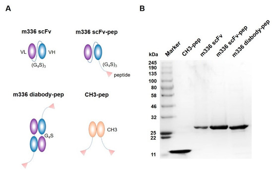
To generate the bispecific antibody-peptide inhibitor, the MERS-CoV S2-derived peptide HR2P was fused to m336 scFv using a flexible (G4S)3 linker. Furthermore, a dimeric format, namely m336 diabody-pep, was also generated by replacing the (G4S)3 linker between variable region heavy (VH) and variable region light (VL) of m336 scFv with a 5 amino acids GGGGS linker. The shorter linker can result in the pairing of VH and VL from two different chains and thus the formation of dimeric diabody with two VHs and VLs. A dimeric peptide-only control, namely CH3-pep, was also generated by linking HR2P peptide and IgG1 CH3 domain with the flexible (G4S)3 linker. This fusion protein possesses two peptides and comparable molecular weight compared to other constructs (Figure 1A).

Figure 1.
Construction and characterization of anti-MERS-CoV inhibitors. (A) Schematic structure of proteins m336 scFv, m336 scFv-pep, m336 diabody-pep, and CH3-pep. (B) SDS-PAGE of the anti-MERS-CoV inhibitors.
The four proteins, m336 scFv, m336 scFv-pep, m336 diabody-pep, and CH3-pep were soluble expressed in E. coli cells with high efficiency. Purified proteins were obtained with yields of 15–30 mg/L bacterial culture. The m336 diabody-pep migrated as a monomer under the denaturing conditions of the SDS-PAGE, and thus displayed similar molecular weight (~30 kDa) with m336 scFv and m336 scFv-pep (Figure 1B). All proteins were monomeric as demonstrated by size exclusion chromatography (data not shown).
3.2. Interactions between Fusion Proteins and MERS-CoV S Protein
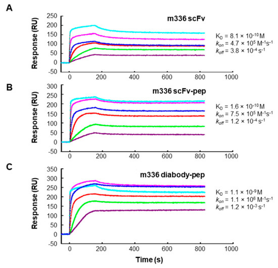
To measure the binding kinetics of the fusion proteins with MERS-CoV S protein, the surface plasmon resonance (SPR) assay was performed by using a ProteOn system SPR biosensor instrument (Figure 2). We have previously demonstrated that m336 bound specifically to MERS-CoV S protein, but not other unrelated proteins [35]. As shown in Table 1, all three m336-based fusion proteins (m336 scFv, m336 scFv-pep, and m336 diabody-pep) exhibited potent binding to S protein with very similar binding pattern. The equilibrium dissociation constant (KD) of m336 scFv for S protein was 0.8 nM with on-rate (kon) of 4.7 × 10⁵ M−1s−1 and off-rate (koff) of 3.8 × 10⁻⁴ s−1. The m336 diabody-pep displayed similar binding kinetics to that of m336 scFv (kon 1.1 × 10⁶ M−1s−1, koff 1.2 × 10⁻³ s−1, KD 1.1 nM). The m336 scFv-pep had slightly more potent binding affinity (KD 0.2 nM) with slower dissociation rate constant (koff 1.2 × 10⁻⁴ s−1), compared to the other two fusion proteins. In contrast, the CH3-peptide displayed much lower affinity, compared to the antibody m336-based fusion proteins (KD 1.1 M).

Figure 2.
Interactions between m336 scFv (A), m336 scFv-pep (B), and m336 diabody-pep (C) with MERS-CoV S protein measured using the ProteOn XRP36 SPR system.

Table 1.
Binding kinetics analysis of fusion proteins to MERS-CoV S protein.
3.3. Neutralization of MERS-CoV Pseudovirus Infection
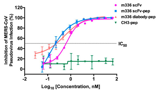
Next, we measured the neutralization activity of m336 scFv, m336 scFv-pep, m336 diabody-pep, and CH3-pep at graded concentrations against pseudotyped MERS-CoV infection. As shown in Figure 3, m336-scFv, m336 scFv-pep and m336 diabody-pep exhibited potent neutralization activity. Among them, m336 scFv-pep exhibited the most potent neutralization capability, with 50% inhibitory concentration (IC50) of 0.21 ± 0.06 nM. Although, displaying comparable IC50 (m336 diabody-pep, 0.25 ± 0.07 nM; m336 scFv, 0.69 ± 0.03 nM), interestingly, m336 diabody-pep inhibited the infection more potent than that of m336 scFv at low protein concentrations (<0.03 nM). In a previous study, IgG1 m336 displayed exceptional neutralization against pseudotyped MERS-CoV infection with IC50 of 0.03 nM [35]. The control fusion protein CH3-pep could not inhibit the infection.

Figure 3.
Potent neutralization of MERS-CoV by anti-MERS-CoV inhibitors. Pseudotyped virus was incubated with proteins before infection of DPP4-expressing Huh-7 cells. Luciferase activities were measured, and percent neutralization was calculated.
3.4. Inhibition of MERS S Protein-Mediated Cell-Cell Fusion
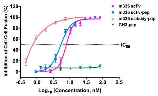
We used a well-established MERS-CoV S protein-mediated cell-cell fusion assay to determine whether the fusion proteins have the ability to inhibit MERS-CoV fusion with the target cells [40,46]. In such assay, the 293T cells that express MERS-CoV S protein and EGFP were used as the effector cells, and Huh-7 cells that express DPP4 receptor were used as the target cells. The CH3-pep showed no appreciable activity at a concentration of 83 nM, which is consistent with the previous report that the HR2P peptide did not have significant inhibitory effect at concentrations lower than 100 nM [40]. Notably, we found that m336 diabody-pep was able to block the fusion between 293T/MERS/EGFP cells and Huh-7 cells at a concentration as low as 0.5 nM (Figure 4). The calculated IC50 for m336 diabody-pep was 0.81 ± 0.02 nM. As shown in Figure 4, the m336 diabody-pep was significantly more potent than m336 scFv in inhibiting cell-cell fusion. The m336 scFv-pep also showed evidently more potent inhibitory activity than m336 scFv at low concentrations, with slightly lower IC50 (m336 scFv-pep, 5.25 ± 0.03 nM; m336 scFv, 7.76 ± 0.02 nM). These results indicate that antibody-peptide bispecific fusion proteins, especially the m336 diabody-pep, have the potential to be developed as potent MERS-CoV inhibitors that could, not only neutralize MERS-CoV infection, but also inhibit MERS-CoV S protein-mediated cell-cell fusion.

Figure 4.
Inhibitory activity of anti-MERS-CoV inhibitors on MERS-CoV S protein-mediated cell-cell fusion. The number of 293T/MERS/EGFP cells fused or unfused with Huh-7 cells were counted, and the percentage of inhibition was calculated.
4. Discussion
Monoclonal antibodies represent one of the most promising immunotherapeutic agents for the treatment of cancers and infectious diseases [7,47,48,49,50,51,52]. Previously, we used RBD of MERS-CoV S protein to screen a non-immune phage-displayed Fab library and identified a highly potent human neutralizing mAb m336 and a panel of other mAbs against MERS-CoV [34,36,37]. Jiang et al. also used MERS-CoV S RBD to screen a non-immune yeast-displayed scFv library and identified two potent MERS-CoV neutralizing mAbs, MERS-4 and MERS-27 [53]. Tang et al. used a full-length S protein to screen a non-immune phage-display scFv library and identified a potent MERS-CoV neutralizing mAb 3B11 [54]. All these reported mAbs inhibit the binding of virus to cellular reporter DPP4 by recognizing the different epitopes of RBD, thus having the potential to be developed as anti-MERS-CoV therapeutics. However, further engineering is still needed to address the concerns in using such mAbs in clinics, e.g., the high production costs, etc.
In this study, we generated two bispecific anti-MERS-CoV fusion proteins, m336 scFv-pep and m336 diabody-pep. Compared with the antibody or peptide inhibitors that block only one site of the spike protein, the bispecific inhibitors showed greater potency. The m336 scFv-pep showed the highest binding affinity to MERS-CoV S protein, as well as the most potent neutralizing activity, while the m336 diabody-pep was the most potent in blocking MERS-CoV S-mediated cell-cell fusion. These results confirmed the improved potency of bispecific antibody-peptide inhibitors compared to antibody or peptide alone. The m336-scFv interferes with viral attachment to the DPP4 receptor on human cells [35], while the HR2P peptide inhibits the formation of the 6-helix-bundle fusion core and thus interrupts viral fusion with the target cell membrane [40]. It is probably that the antibody-peptide fusion proteins, which are composed of both antiviral components, linked by flexible (G4S)3 linker, can act at both steps during viral infection.
Interestingly, the m336 diabody-pep was found to potently block MERS-CoV S-mediated cell-cell fusion even at concentrations lower than 0.5 nM. This phenomenon may be attributed to the difference in steric hindrance of the fusion proteins. Compared to the m336 scFv-pep, the m336 diabody-pep has a much larger (twice) molecular weight and size, and thus, inhibits the viral fusion to target cells by competitive inhibition and steric hindrance, leading to the much greater inhibitory activity than that of m336 scFv-pep. Although, having largely improved potency in inhibiting cell-cell fusion, m336 diabody-pep did not show evidently increased neutralization activity compared to m336 scFv. Further investigations are required to explore the mechanism for the disparate performance of m336 diabody-pep in different assays. As shown in Figure 3 and Figure 4, the IC50 of m336 diabody-pep was 0.25 ± 0.07 nM in the neutralization assay, and 0.81 ± 0.02 nM in the cell-cell fusion assay. Importantly, m336 diabody-pep was much more potent than m336 scFv at concentrations higher than 5 nM in the cell-cell fusion assay. At these concentrations, all the fusion proteins can result in high percentage of inhibition (>70%) in the neutralization assay, and therefore, may explain the little difference among difference groups.
All the m336-based fusion proteins could be easily produced in E. coli in large amount and low production cost. Their sizes are larger than m336 scFv or HR2P peptides, suggesting that they would possess longer in vivo half-life. Still, their half-life would be shorter than full-size IgG due to the lack of Fc region. Therefore, their prophylactic and therapeutic efficacy against MERS-CoV should be assessed carefully using different approaches and doses in vivo. However, the RBD of MERS-CoV specifically targets on human DPP4 [29], and most small animal models are not susceptible to MERS-CoV infection [55], which pose a significant barrier to the development of anti-MERS-CoV inhibitors. Fortunately, researchers have constructed several animal models recently that simulate the morbidity and mortality of human infections, of which nonhuman primates (NHP) models and human DPP4-expressing mouse model are considered to be ideal candidates [56] and the latter is promising to be utilized in our following studies.
5. Conclusions
In conclusion, our study indicated that the bispecific inhibitors have increased the efficacy against MERS-CoV, compared to the neutralizing antibody or polypeptide alone. Such inhibitors with the advantage of multiple biologics have the potential to be further developed as effective prophylactic and therapeutic agents, and may find wide application in treating viral diseases.
Author Contributions
L.W., Y.W., F.Y., and T.Y. conceived and designed the experiments; L.W., J.X., Y.K., R.L., W.L., J.L. (Jinyao Li), J.L. (Jun Lu), and D.S.D. performed the experiments; L.W., J.X., and Y.W. analyzed the data; L.W., F.Y., and T.Y. wrote the paper. All authors discussed the results and contributed to the final manuscript.
Funding
This work was supported by grants from the National Natural Science Foundation of China (81601761, 31570936, 81630090, 81822027 and 81501735), the grant from Chinese Academy of Medical Sciences (2019PT350002), Fudan’s Undergraduate Research Opportunities Program (15088), Starting Grant from Hebei Agricultural University (YJ201843), National Fundamental Fund of Training Basic Science (J1210041-1629), Program for Talents Introduction from studying overseas of Hebei Province (CN201707), Program for Youth Talent of Higher Learning Institutions of Hebei Province (BJ2018045), National Megaprojects of China for Major Infectious Diseases (2018ZX10301403), and the New Zealand Ministry of Education’s New Zealand-China Tripartite Partnership Fund.
Acknowledgments
We are grateful to Shibo Jiang, Lu Lu and Cong Wang at Fudan University for providing cell-cell fusion system.
Conflicts of Interest
The authors declare no conflicts of interest. The funding sponsors had no role in the writing of the manuscript or the decision to publish this article.
References
- Zhu, Z.; Dimitrov, A.S.; Bossart, K.N.; Crameri, G.; Bishop, K.A.; Choudhry, V.; Mungall, B.A.; Feng, Y.; Choudhary, A.; Zhang, M.; et al. Potent Neutralization of Hendra and Nipah Viruses by Human Monoclonal Antibodies. J. Virol. 2006, 80, 891–899. [Google Scholar] [CrossRef] [PubMed]
- Bossart, K.N.; Geisbert, T.W.; Feldmann, H.; Zhu, Z.; Feldmann, F.; Geisbert, J.B.; Yan, L.; Feng, Y.; Brining, D.; Scott, D.P.; et al. A Neutralizing Human Monoclonal Antibody Protects African Green Monkeys from Hendra Virus Challenge. Sci. Transl. Med. 2011, 3, 105ra103. [Google Scholar] [CrossRef] [PubMed]
- Qiu, X.; Wong, G.; Audet, J.; Bello, A.; Fernando, L.; Alimonti, J.B.; Faustherbovendo, H.; Wei, H.; Aviles, J.; Hiatt, E.; et al. Reversion of advanced Ebola virus disease in nonhuman primates with ZMapp. Nature 2014, 514, 47–53. [Google Scholar] [CrossRef] [PubMed]
- Hu, D.; Zhu, Z.; Li, S.; Deng, Y.; Wu, Y.; Zhang, N.; Puri, V.; Wang, C.; Zou, P.; Lei, C.; et al. A broadly neutralizing germline-like human monoclonal antibody against dengue virus envelope domain III. PLoS Pathog. 2019, 15, e1007836. [Google Scholar] [CrossRef]
- Brinkmann, U.; Kontermann, R.E. The making of bispecific antibodies. mAbs 2017, 9, 182–212. [Google Scholar] [CrossRef]
- Xu, T.; Ying, T.; Wang, L.; Zhang, X.D.; Wang, Y.; Kang, L.; Huang, T.; Cheng, L.; Wang, L.; Zhao, Q. A native-like bispecific antibody suppresses the inflammatory cytokine response by simultaneously neutralizing tumor necrosis factor-alpha and interleukin-17A. Oncotarget 2017, 8, 81860–81872. [Google Scholar] [CrossRef]
- Jin, Y.; Lei, C.; Hu, D.; Dimitrov, D.S.; Ying, T. Human monoclonal antibodies as candidate therapeutics against emerging viruses. Front. Med. 2017, 11, 462–470. [Google Scholar] [CrossRef]
- Bardhi, A.; Wu, Y.; Chen, W.; Li, W.; Zhu, Z.; Zheng, J.H.; Wong, H.; Jeng, E.; Jones, J.; Ochsenbauer, C.; et al. Potent In Vivo NK Cell-Mediated Elimination of HIV-1-Infected Cells Mobilized by a gp120-Bispecific and Hexavalent Broadly Neutralizing Fusion Protein. J. Virol. 2017, 91, e00937-17. [Google Scholar] [CrossRef]
- Kong, D.; Wang, Y.; Ji, P.; Li, W.; Ying, T.; Huang, J.; Wang, C.; Wu, Y.; Wang, Y.; Chen, W.; et al. A defucosylated bispecific multivalent molecule exhibits broad HIV-1-neutralizing activity and enhanced antibody-dependent cellular cytotoxicity against reactivated HIV-1 latently infected cells. AIDS 2018, 32, 1749–1761. [Google Scholar] [CrossRef]
- Wu, Y.; Xue, J.; Wang, C.; Li, W.; Wang, L.; Chen, W.; Prabakaran, P.; Kong, D.; Jin, Y.; Hu, D.; et al. Rapid elimination of broadly neutralizing antibodies correlates with treatment failure in the acute phase of SHIV infection. J. Virol. 2019, 93, e01077-19. [Google Scholar] [CrossRef]
- Pu, J.; Wang, Q.; Xu, W.; Lu, L.; Jiang, S. Development of Protein- and Peptide-Based HIV Entry Inhibitors Targeting gp120 or gp41. Viruses 2019, 11, 705. [Google Scholar] [CrossRef] [PubMed]
- Zaki, A.M.; van Boheemen, S.; Bestebroer, T.M.; Osterhaus, A.D.; Fouchier, R.A. Isolation of a novel coronavirus from a man with pneumonia in Saudi Arabia. N. Engl. J. Med. 2012, 367, 1814–1820. [Google Scholar] [CrossRef]
- Chen, X.; Chughtai, A.A.; Dyda, A.; MacIntyre, C.R. Comparative epidemiology of Middle East respiratory syndrome coronavirus (MERS-CoV) in Saudi Arabia and South Korea. Emerg. Microbes Infect. 2017, 6, 1–6. [Google Scholar] [CrossRef] [PubMed]
- Chan, J.F.; Lau, S.K.; To, K.K.; Cheng, V.C.; Woo, P.C.; Yuen, K.Y. Middle East respiratory syndrome coronavirus: Another zoonotic betacoronavirus causing SARS-like disease. Clin. Microbiol. Rev. 2015, 28, 465–522. [Google Scholar] [CrossRef] [PubMed]
- Chan, J.F.W.; Li, K.S.M.; To, K.K.W.; Cheng, V.C.C.; Chen, H.; Yuen, K. Is the discovery of the novel human betacoronavirus 2c EMC/2012 (HCoV-EMC) the beginning of another SARS-like pandemic? J. Infect. 2012, 65, 477–489. [Google Scholar] [CrossRef]
- Alqahtani, A.S.; Wiley, K.E.; Tashani, M.; Heywood, A.E.; Willaby, H.W.; BinDhim, N.F.; Booy, R.; Rashid, H. Camel exposure and knowledge about MERS-CoV among Australian Hajj pilgrims in 2014. Virol. Sin. 2016, 31, 89–93. [Google Scholar] [CrossRef]
- Woo, P.C.Y.; Lau, S.K.P.; Huang, Y.; Yuen, K. Coronavirus Diversity, Phylogeny and Interspecies Jumping. Exp. Biol. Med. 2009, 234, 1117–1127. [Google Scholar] [CrossRef]
- Weiss, S.R.; Navas-Martin, S. Coronavirus pathogenesis and the emerging pathogen severe acute respiratory syndrome coronavirus. Microbiol. Mol. Biol. Rev. 2005, 69, 635–664. [Google Scholar] [CrossRef]
- Azhar, E.I.; El-Kafrawy, S.A.; Farraj, S.A.; Hassan, A.M.; Al-Saeed, M.S.; Hashem, A.M.; Madani, T.A. Evidence for camel-to-human transmission of MERS coronavirus. N. Engl. J. Med. 2014, 370, 2499–2505. [Google Scholar] [CrossRef]
- Vijaykrishna, D.; Smith, G.J.; Zhang, J.X.; Peiris, J.S.; Chen, H.; Guan, Y. Evolutionary insights into the ecology of coronaviruses. J. Virol. 2007, 81, 4012–4020. [Google Scholar] [CrossRef]
- Annan, A.; Baldwin, H.J.; Corman, V.M.; Klose, S.M.; Owusu, M.; Nkrumah, E.E.; Badu, E.K.; Anti, P.; Agbenyega, O.; Meyer, B.; et al. Human betacoronavirus 2c EMC/2012-related viruses in bats, Ghana and Europe. Emerg. Infect. Dis. 2013, 19, 456–459. [Google Scholar] [CrossRef] [PubMed]
- Reusken, C.; Haagmans, B.L.; Muller, M.A.; Gutierrez, C.; Godeke, G.; Meyer, B.; Muth, D.; Raj, V.S.; De Vries, L.S.; Corman, V.M.; et al. Middle East respiratory syndrome coronavirus neutralising serum antibodies in dromedary camels: A comparative serological study. Lancet Infect. Dis. 2013, 13, 859–866. [Google Scholar] [CrossRef]
- Lau, S.K.; Wernery, R.; Wong, E.Y.; Joseph, S.; Tsang, A.K.; Patteril, N.A.; Elizabeth, S.K.; Chan, K.H.; Muhammed, R.; Kinne, J.; et al. Polyphyletic origin of MERS coronaviruses and isolation of a novel clade A strain from dromedary camels in the United Arab Emirates. Emerg. Microbes Infect. 2016, 5, 1–9. [Google Scholar] [CrossRef] [PubMed]
- Milne-Price, S.; Miazgowicz, K.L.; Munster, V.J. The emergence of the Middle East respiratory syndrome coronavirus. Pathog. Dis. 2014, 71, 121–136. [Google Scholar] [CrossRef] [PubMed]
- Assiri, A.; Altawfiq, J.A.; Alrabeeah, A.A.; Alrabiah, F.; Alhajjar, S.; Albarrak, A.; Flemban, H.; Alnassir, W.N.; Balkhy, H.H.; Alhakeem, R.F.; et al. Epidemiological, demographic, and clinical characteristics of 47 cases of Middle East respiratory syndrome coronavirus disease from Saudi Arabia: A descriptive study. Lancet Infect. Dis. 2013, 13, 752–761. [Google Scholar] [CrossRef]
- Memish, Z.A.; Mishra, N.; Olival, K.J.; Fagbo, S.F.; Kapoor, V.; Epstein, J.H.; Alhakeem, R.; Durosinloun, A.; Al Asmari, M.; Islam, A.; et al. Middle East respiratory syndrome coronavirus in bats, Saudi Arabia. Emerg. Infect. Dis. 2013, 19, 1819–1823. [Google Scholar] [CrossRef] [PubMed]
- Lu, L.; Xia, S.; Ying, T.; Jiang, S. Urgent development of effective therapeutic and prophylactic agents to control the emerging threat of Middle East respiratory syndrome (MERS). Emerg. Microbes Infect. 2015, 4, e37. [Google Scholar] [CrossRef] [PubMed]
- Cui, J.; Eden, J.; Holmes, E.C.; Wang, L. Adaptive evolution of bat dipeptidyl peptidase 4 (dpp4): Implications for the origin and emergence of Middle East respiratory syndrome coronavirus. Virol. J. 2013, 10, 304. [Google Scholar] [CrossRef]
- Raj, V.S.; Mou, H.; Smits, S.L.; Dekkers, D.H.W.; Muller, M.A.; Dijkman, R.; Muth, D.; Demmers, J.; Zaki, A.M.; Fouchier, R.A.M.; et al. Dipeptidyl peptidase 4 is a functional receptor for the emerging human coronavirus-EMC. Nature 2013, 495, 251–254. [Google Scholar] [CrossRef]
- Ying, T.; Chen, W.; Feng, Y.; Wang, Y.; Gong, R.; Dimitrov, D.S. Engineered Soluble Monomeric IgG1 CH3 Domain: Generation, mechanisms of function, and implications for design of biological therapeutics*. J. Biol. Chem. 2013, 288, 25154–25164. [Google Scholar] [CrossRef]
- Boonacker, E.; Van Noorden, C.J.F. The multifunctional or moonlighting protein CD26/DPPIV. Eur. J. Cell Biol. 2003, 82, 53–73. [Google Scholar] [CrossRef] [PubMed]
- Jiang, S.; Lu, L.; Du, L.; Debnath, A.K. A predicted receptor-binding and critical neutralizing domain in S protein of the novel human coronavirus HCoV-EMC. J. Infect. 2013, 66, 464–466. [Google Scholar] [CrossRef] [PubMed]
- Jiang, S.; Lu, L.; Liu, Q.; Xu, W.; Du, L. Receptor-binding domains of spike proteins of emerging or re-emerging viruses as targets for development of antiviral vaccines. Emerg. Microbes Infect. 2012, 1, e13. [Google Scholar] [CrossRef] [PubMed]
- Van Doremalen, N.; Falzarano, D.; Ying, T.; de Wit, E.; Bushmaker, T.; Feldmann, F.; Okumura, A.; Wang, Y.; Scott, D.P.; Hanley, P.W.; et al. Efficacy of antibody-based therapies against Middle East respiratory syndrome coronavirus (MERS-CoV) in common marmosets. Antivir. Res. 2017, 143, 30–37. [Google Scholar] [CrossRef] [PubMed]
- Ying, T.; Du, L.; Ju, T.W.; Prabakaran, P.; Lau, C.C.; Lu, L.; Liu, Q.; Wang, L.; Feng, Y.; Wang, Y.; et al. Exceptionally potent neutralization of Middle East respiratory syndrome coronavirus by human monoclonal antibodies. J. Virol. 2014, 88, 7796–7805. [Google Scholar] [CrossRef] [PubMed]
- Agrawal, A.S.; Ying, T.; Tao, X.; Garron, T.; Algaissi, A.; Wang, Y.; Wang, L.; Peng, B.H.; Jiang, S.; Dimitrov, D.S.; et al. Passive transfer of a germline-like neutralizing human monoclonal antibody protects transgenic mice against lethal Middle East respiratory syndrome coronavirus infection. Sci. Rep. 2016, 6, 31629. [Google Scholar] [CrossRef]
- Houser, K.V.; Gretebeck, L.; Ying, T.; Wang, Y.; Vogel, L.; Lamirande, E.W.; Bock, K.W.; Moore, I.N.; Dimitrov, D.S.; Subbarao, K. Prophylaxis with a Middle East respiratory syndrome coronavirus (MERS-CoV)-specific human monoclonal antibody protects rabbits from MERS-CoV infection. J. Infect. Dis. 2016, 213, 1557–1561. [Google Scholar] [CrossRef]
- Ying, T.; Prabakaran, P.; Du, L.; Shi, W.; Feng, Y.; Wang, Y.; Wang, L.; Li, W.; Jiang, S.; Dimitrov, D.S.; et al. Junctional and allele-specific residues are critical for MERS-CoV neutralization by an exceptionally potent germline-like antibody. Nat. Commun. 2015, 6, 8223. [Google Scholar] [CrossRef]
- Channappanavar, R.; Lu, L.; Xia, S.; Du, L.; Meyerholz, D.K.; Perlman, S.; Jiang, S. Protective Effect of Intranasal Regimens Containing Peptidic Middle East Respiratory Syndrome Coronavirus Fusion Inhibitor Against MERS-CoV Infection. J. Infect. Dis. 2015, 212, 1894–1903. [Google Scholar] [CrossRef]
- Lu, L.; Liu, Q.; Zhu, Y.; Chan, K.H.; Qin, L.; Li, Y.; Wang, Q.; Chan, J.F.; Du, L.; Yu, F.; et al. Structure-based discovery of Middle East respiratory syndrome coronavirus fusion inhibitor. Nat. Commun. 2014, 5, 3067. [Google Scholar] [CrossRef]
- Wang, C.; Hua, C.; Xia, S.; Li, W.; Lu, L.; Jiang, S. Combining a Fusion Inhibitory Peptide Targeting the MERS-CoV S2 Protein HR1 Domain and a Neutralizing Antibody Specific for the S1 Protein Receptor-Binding Domain (RBD) Showed Potent Synergism against Pseudotyped MERS-CoV with or without Mutations in RBD. Viruses 2019, 11, 31. [Google Scholar] [CrossRef] [PubMed]
- Screening, Ranking, and Epitope Mapping of Anti-Human IL-9 Supernatants. Available online: http://www.bio-rad.com/webroot/web/pdf/lsr/literature/Bulletin_5540.pdf (accessed on 28 October 2019).
- Du, L.; Zhao, G.; Yang, Y.; Qiu, H.; Wang, L.; Kou, Z.; Tao, X.; Yu, H.; Sun, S.; Tseng, C.T.K.; et al. A Conformation-Dependent Neutralizing Monoclonal Antibody Specifically Targeting Receptor-Binding Domain in Middle East Respiratory Syndrome Coronavirus Spike Protein. J. Virol. 2014, 88, 7045–7053. [Google Scholar] [CrossRef] [PubMed]
- Du, L.; Kou, Z.; Ma, C.; Tao, X.; Wang, L.; Zhao, G.; Chen, Y.; Yu, F.; Tseng, C.T.K.; Zhou, Y.; et al. A Truncated Receptor-Binding Domain of MERS-CoV Spike Protein Potently Inhibits MERS-CoV Infection and Induces Strong Neutralizing Antibody Responses: Implication for Developing Therapeutics and Vaccines. PLoS ONE 2013, 8, e81587. [Google Scholar] [CrossRef] [PubMed]
- Xia, S.; Xu, W.; Wang, Q.; Wang, C.; Hua, C.; Li, W.; Lu, L.; Jiang, S. Peptide-Based Membrane Fusion Inhibitors Targeting HCoV-229E Spike Protein HR1 and HR2 Domains. Int. J. Mol. Sci. 2018, 19, 487. [Google Scholar] [CrossRef] [PubMed]
- Xia, S.; Liu, Q.; Wang, Q.; Sun, Z.; Su, S.; Du, L.; Ying, T.; Lu, L.; Jiang, S. Middle East respiratory syndrome coronavirus (MERS-CoV) entry inhibitors targeting spike protein. Virus Res. 2014, 194, 200–210. [Google Scholar] [CrossRef]
- Ying, T.; Wang, Y.; Feng, Y.; Prabakaran, P.; Gong, R.; Wang, L.; Crowder, K.; Dimitrov, D.S. Engineered antibody domains with significantly increased transcytosis and half-life in macaques mediated by FcRn. mAbs 2015, 7, 922–930. [Google Scholar] [CrossRef]
- Ying, T.; Gong, R.; Ju, T.W.; Prabakaran, P.; Dimitrov, D.S. Engineered Fc based antibody domains and fragments as novel scaffolds. Biochim. Biophys. Acta 2014, 1844, 1977–1982. [Google Scholar] [CrossRef]
- Ying, T.; Feng, Y.; Wang, Y.; Chen, W.; Dimitrov, D.S. Monomeric IgG1 Fc molecules displaying unique Fc receptor interactions that are exploitable to treat inflammation-mediated diseases. mAbs 2014, 6, 1201–1210. [Google Scholar] [CrossRef]
- Yu, F.; Song, H.; Wu, Y.; Chang, S.Y.; Wang, L.; Li, W.; Hong, B.; Xia, S.; Wang, C.; Khurana, S.; et al. A potent germline-like human monoclonal antibody targets a pH-sensitive epitope on H7N9 influenza Hemagglutinin. Cell Host Microbe 2017, 22, 471–483. [Google Scholar] [CrossRef]
- Hong, B.; Wen, Y.; Ying, T. Recent Progress on Neutralizing Antibodies against Hepatitis B Virus and its Implications. Infect. Disord. Drug Targets 2019, 19, 213–223. [Google Scholar] [CrossRef]
- Wu, Y.; Jiang, S.; Ying, T. Single-Domain Antibodies as Therapeutics against Human Viral Diseases. Front. Immuno. 2017, 8, 1802. [Google Scholar] [CrossRef] [PubMed]
- Jiang, L.; Wang, N.; Zuo, T.; Shi, X.; Poon, K.V.; Wu, Y.; Gao, F.; Li, D.; Wang, R.; Guo, J.; et al. Potent Neutralization of MERS-CoV by Human Neutralizing Monoclonal Antibodies to the Viral Spike Glycoprotein. Sci. Transl. Med. 2014, 6, 234ra59. [Google Scholar] [CrossRef] [PubMed]
- Tang, X.; Agnihothram, S.; Jiao, Y.; Stanhope, J.; Graham, R.L.; Peterson, E.C.; Avnir, Y.; Tallarico, A.S.C.; Sheehan, J.; Zhu, Q.; et al. Identification of human neutralizing antibodies against MERS-CoV and their role in virus adaptive evolution. Proc. Natl. Acad. Sci. USA 2014, 111, E2018–E2026. [Google Scholar] [CrossRef]
- Cockrell, A.S.; Peck, K.M.; Yount, B.; Agnihothram, S.; Scobey, T.; Curnes, N.R.; Baric, R.S.; Heise, M.T. Mouse Dipeptidyl Peptidase 4 Is Not a Functional Receptor for Middle East Respiratory Syndrome Coronavirus Infection. J. Virol. 2014, 88, 5195–5199. [Google Scholar] [CrossRef] [PubMed]
- Van Doremalen, N.; Munster, V.J. Animal models of Middle East respiratory syndrome coronavirus infection. Antivir. Res. 2015, 122, 28–38. [Google Scholar] [CrossRef]
© 2019 by the authors. Licensee MDPI, Basel, Switzerland. This article is an open access article distributed under the terms and conditions of the Creative Commons Attribution (CC BY) license (http://creativecommons.org/licenses/by/4.0/).