Review

Targeting sTNF/TNFR1 Signaling as a New Therapeutic Strategy

Institute of Cell Biology and Immunology, University of Stuttgart, Allmandring 31, 70569 Stuttgart, Germany
*
Author to whom correspondence should be addressed.
Antibodies 2015, 4(1), 48-70; https://doi.org/10.3390/antib4010048
Submission received: 29 January 2015 / Revised: 23 February 2015 / Accepted: 27 February 2015 / Published: 6 March 2015
(This article belongs to the Special Issue TNF in the Regulation of Immune Cells)

Abstract

:
Deregulation of the tumor necrosis factor (TNF) plays an important role in the initiation and perpetuation of chronic inflammation and has been implicated in the development of various autoimmune diseases. Accordingly, TNF-inhibitors are successfully used for the treatment of several diseases, such as rheumatoid arthritis, inflammatory bowel disease, and psoriasis. However, total inhibition of TNF can cause severe side effects such as an increased risk of inflammation and reactivation of tuberculosis. This is likely due to the different actions of the two TNF receptors. Whereas TNFR1 predominantly promotes inflammatory signaling pathways, TNFR2 mediates immune modulatory functions and promotes tissue homeostasis and regeneration. Therefore, the specific blockage of TNFR1 signaling, either by direct inhibition with TNFR1-selective antagonists or by targeting soluble TNF, which predominantly activates TNFR1, may prevent the detrimental effects associated with total TNF-inhibitors and constitute a next-generation approach to interfere with TNF.

1. Introduction

The tumor necrosis factor (TNF) is the prototypic member of a large family of cytokines that play an important role in the regulation of the innate and adaptive immune system [1,2]. TNF itself is a key player in the initiation and orchestration of inflammation and immunity and is a potent proinflammatory cytokine, which is predominantly produced by activated immune cells in response to infections and tissue damage. Controlled expression of TNF is therefore essential to fight infections and to promote tissue repair. However, deregulation of TNF expression and signaling can cause chronic inflammation, which may result in the development of autoimmune diseases and tissue damage [3,4,5]. Indeed, elevated TNF levels have been associated with several diseases, such as rheumatoid arthritis (RA), psoriasis, inflammatory bowel disease (IBD), and multiple sclerosis (MS), and TNF therapeutics are successfully used in several of these diseases, including RA, juvenile RA (JRA), psoriasis, ankylosing spondylitis (AS), Crohn’s disease (CD), and ulcerative colitis (UC) [3,6,7,8,9]. Surprisingly, however, clinical trials with TNF-neutralizing reagents in MS patients resulted in disease exacerbation [10,11]. Moreover, TNF inhibitors can cause side effects including opportunistic infections, reactivation of tuberculosis, development of autoimmune disease, and an increased risk for lymphoma [12,13,14,15].
TNF is expressed as a transmembrane protein (memTNF) that can be processed into a soluble form (sTNF) [2,16] and exerts its functions via two receptors, TNF receptor 1 (TNFR1) and TNFR2 (Figure 1). The distinct roles of these receptors in immune regulation and tissue regeneration may provide a mechanistic explanation for the failure of TNF inhibitors in MS, as well as some of the side effects associated with anti-TNF treatment. While TNFR1 is predominantly associated with inflammation and tissue degeneration, TNFR2 is mainly implicated in tissue regeneration and immune modulation. Moreover, sTNF and memTNF differ in their capability to stimulate signaling via TNFR1 and TNFR2 (Figure 1). Whereas TNFR1 can be activated by binding of either sTNF or memTNF, TNFR2 is only fully activated by memTNF [17]. Accordingly, the proinflammatory effects of TNF have been associated with signaling of sTNF via TNFR1, while signaling of memTNF, predominantly via TNFR2, is associated with tissue regeneration and immune modulation. A further level of complexity is added by proteolytic cleavage of the extracellular domains of both receptors [18,19], which is increased upon TNF activation [20]. The shed receptor ectodomains still bind to TNF, albeit with low efficiency, and can thus act as natural inhibitors of TNF.
Although severe side effects of anti-TNF treatment are relatively rare, there is an obvious demand to minimize the risk associated with long-term total TNF inhibition. This may be achieved by the use of more specific anti-TNF therapeutics. Based on the role of sTNF and TNFR1 in promoting inflammation, the two major strategies are the development of reagents that exclusively target sTNF, leaving signaling via memTNF intact, and the development of TNFR1-specific antagonists, thus preserving TNFR2 signaling. Along with the possibility of reducing the side effects associated with total TNF inhibition, these more specific next-generation therapeutics may open the path for the treatment of additional diseases in which total TNF inhibition is detrimental, such as MS [7,21,22,23].
Figure 1. Schematic representation of TNF processing and signaling via its receptors TNFR1 and TNFR2 as well as the mechanisms of action of currently approved total TNF inhibitors and novel inhibitors selective for either soluble TNF (sTNF) or TNFR1.
Antibodies 04 00048 g001

2. TNF Signaling

2.1. General Aspects

TNF is synthesized as a type II transmembrane protein that self-assembles into non-covalently linked homotrimers. Proteolytic cleavage of the ectodomain of this initial membrane-bound form of TNF (memTNF) by the TNF-α converting enzyme (TACE/ADAM17), a member of the a disintegrin and metalloprotease (ADAM) protein family, results in the release of soluble TNF (sTNF) [16]. TNF can bind to two structurally distinct membrane receptors, TNFR1 (TNFRSF1, CD120a) and TNFR2 (TNFRSF2, CD120b). Whereas TNFR1 is constitutively expressed on virtually all tissue cells, expression of TNFR2 is restricted to cells of the immune system, especially regulatory T cells, endothelial, and neuronal tissues, and its expression can be highly regulated by the cellular activation status. Both receptors are typical type I transmembrane proteins with extracellular and intracellular domains of about equal sizes and single transmembrane domains [2,5].

2.2. TNF Receptors

The extracellular domains of both receptors are quite similar, comprising four cysteine-rich domains (CRD), which are required for ligand binding. In addition, the most distal CRD carries a homophilic interaction motif, the so-called preligand-binding assembly domain (PLAD) [24]. The structure of the intracellular signaling domains of both receptors is highly distinct. The intracellular domains of the TNF receptors, which do not possess any enzymatic activity, define them as representatives of the two main subgroups of the TNFR family, the death domain (DD)-containing receptors (TNFR1) and the TRAF-interacting receptors (TNFR2), respectively [5].
Binding of TNF to TNFR1 leads to the activation of two major, well-understood signaling pathways. One leads to the induction of anti-apoptotic genes, mainly through activation of the transcription factor NF-κB. The second signaling pathway results in the engagement of cellular suicide programs, including the prototype of programmed cell death, apoptosis, but also the execution of programmed necrosis (necroptosis) [5,25]. In normal functional cells, TNF does not induce cell death unless transcription, translation, or specifically the NF-κB pathway are blocked. This is caused by the primarily induced anti-apoptotic program leading to the expression of anti-apoptotic proteins such as cFLIP, cIAP1, cIAP2, XIAP, A20 and TRAF proteins, causing inhibition of apoptosis induction and/or execution [5].
Upon TNF binding, TNFR1 can interact via its DD with other DD-containing proteins, such as TRADD (TNF receptor 1 associated death domain protein). This in turn results in recruitment of a signaling complex consisting of TNF receptor-associated factor 2 (TRAF2), cellular inhibitor of apoptosis proteins (cIAPs), the receptor interacting protein kinase 1 (RIP1), the IκBα kinase (IKK) complex, and the linear ubiquitin chain assembly complex (LUBAC) [26]. This membrane-associated primary “TNFR1 signaling complex I” can induce several signaling pathways, with the NF-κB signaling pathway being the most common and best characterized pathway [27]. NF-κB in turn can induce the transcription of many genes that promote inflammation. These include IL-6, IL-8, and, importantly, TNF itself [28], potentially resulting in an amplification loop of proinflammatory TNF signaling. Next to the NF-κB pathway, complex I can initiate the activation of the stress-activated MAP kinases p38 and c-jun N-terminal kinase (JNK), important promotors of inflammation, which can also induce TNF expression [29,30,31]. After complex I internalization, a secondary pro-apoptotic signaling complex II is formed by binding of the adaptor protein FADD (Fas-associated protein with death domain) and the procaspase 8 to the receptor complex. This results in the formation of the death-inducing signaling complex (DISC) and the induction of apoptosis [5]. Besides induction of classical apoptosis via complex II, TNFR1 is also capable of initiating cell death by necroptosis, which is activated especially under conditions of inhibited apoptosis by the kinase activities of RIP1 and RIP3 [25].
In comparison with TNFR1, knowledge of TNFR2 signaling pathways is scarce. Because TNFR2 is only fully activated by memTNF, it is most likely only activated in terms of direct cell–cell interaction and thus plays an important role in localized signaling. In contrast to TNFR1, TNFR2 does not contain a DD and therefore cannot directly induce cell death. However, TNFR2 can directly interact with TRAF2 and TNFR2 stimulation is causing recruitment of TRAF2 to the plasma membrane, thereby affecting TNFR1 signaling [32]. Recruitment of TRAF2 to TNFR2 activates, in a cell context-dependent manner, both the canonical and non-canonical NF-κB signaling pathways [33,34]. Moreover, by an as yet not fully elucidated mechanism, TNFR2 can activate the phosphatidyl inositol (PI) 3-kinase / Akt pathway, thus promoting cell survival and proliferation [35,36,37,38].

2.3. TNF Signaling in Immune Regulation

TNF is a pleiotropic cytokine that is a master regulator of the immune system [3,5]. Generally, TNF is known as a powerful proinflammatory molecule with stimulatory activities for most cells of the immune system. Monocytes and macrophages are the major sources of TNF synthesis in vivo, although many other cell types are also capable of producing TNF under certain circumstances. TNF acts as a co-stimulator for natural killer cells and activated B and T lymphocytes, and it enhances the pathogen-directed cytotoxicity of monocytes, neutrophils, and eosinophils. As a stimulatory agent for phagocytes, TNF is of special importance for the killing of bacteria, yeast, parasites, and virus-infected cells.
The use of transgenic mice in animal disease models has greatly enhanced our knowledge of the importance of TNF in fighting infections. In contrast to wild-type mice, TNF-deficient mice are highly sensitive to infections [39,40]. Moreover, TNF is essential for the maturation of the humoral immune response and for the formation of granuloma, organized accumulations of (infected) macrophages and lymphocytes, which are required to prevent the spreading of pathogens such as mycobacteria in the organism [40,41].
Furthermore, TNF is of great importance for the penetration of immune cells into the tissue due to the stimulatory effects of TNF on endothelial cells, resulting in enhanced surface expression of adhesion molecules and an increased permeability of the endothelium. This is a central step for the recruitment of immune cells, e.g., neutrophils, lymphocytes, and monocytes, which is followed by transendothelial cell migration into the tissue [42].
Most of the described proinflammatory functions of TNF are predominantly mediated by TNFR1, since TNFR1 knockout animals are highly susceptible to bacterial infections [43]. Moreover, loss of function due to TNF knockout is largely mimicked by TNFR1 deficiency [44]. However, a role of TNFR2 in T-cell activation and in the interaction of dendritic cells and NK cells has also been described [45]. Importantly, mice expressing a non-cleavable TNF, thus preventing the generation of sTNF, were at least partially protected against various pathogens, such as Mycobacterium tuberculosum and Listeria monocytogenes. This demonstrates that signaling of memTNF, presumably mainly via TNFR2, can at least partially convey immunity against these pathogens [46,47].
While the proinflammatory functions of TNF, via TNFR1, are very well established, quite recently an important role of TNFR2 in the modulation of the immune system has been discovered. In comparison with other T-cell populations, TNFR2 is predominantly expressed by CD4+CD25+FoxP3+ regulatory T-cells (Tregs), which are important in regulating and suppressing T-cell effector functions [48,49]. TNFR2 is expressed especially in a maximally suppressive subset of Tregs and activation of TNFR2 is important for the proliferation and function of Tregs, indicating an important role of TNFR2 in the regulation and suppression of the immune response [50,51,52,53]. Importantly, inhibition of Treg function leads to a highly increased risk for the development of autoimmune diseases [54]. Accordingly, complete TNF inhibition may compromise Treg function, which may explain the development of novel autoimmune diseases in patients treated with TNF inhibitors.

2.4. Pathophysiology of TNF Deregulation

The pleiotropic functions of TNF in regulation of the adaptive and innate immune response explain why deregulated TNF production due to overreaction of the host or expression in an inappropriate location can lead to major pathogenic consequences. An important mechanism of the organism to prevent TNF toxicity is the increased shedding of TNFR ectodomains [20]. On the one hand, this reduces the number of signal competent receptors on the cell surface and, on the other hand, the soluble receptors can bind to TNF and act as intrinsic TNF inhibitors. Accordingly, mice expressing non-cleavable TNFR1 develop spontaneous liver pathology and enhanced susceptibility to inflammation and autoimmunity, indicating that TNFR1 receptor shedding regulates TNF activity in vivo [55].
A role for non-cleavable TNFR1 had been proposed for the development of TRAPS (tumor necrosis factor receptor-associated periodic syndrome), an autosomal dominantly inherited disease characterized by unprovoked, often prolonged, attacks of fever and inflammation of multiple organs, which is caused by mutations in the TNFR1 gene [56]. However, although mutations that affect TNFR1 shedding have been reported, most mutations relevant for TRAPS development affect the structure of TNFR1, thereby rendering the receptor non-functional or causing its retention in the endoplasmic reticulum [57]. Accordingly, functional TNFR1 at the cell surface should be downregulated, resulting in the paradoxical situation that reduced levels of TNFR1 can cause an inflammatory disease. The systemic inflammation might be induced by ER stress, which may also explain the autosomal dominant character of the disease.
TNF is induced after bacterial or fungal infections and after tissue damage such as in hepatitis, pancreatitis, acute respiratory distress syndrome (ARDS), and myocardial ischemia / reperfusion injury, resulting in acute inflammation of the affected tissue [58,59,60]. Importantly, TNF induces the recruitment of immune cells into the tissue due to increased expression of chemokines and adhesion molecules [42], thereby further promoting inflammation. Sustained elevated TNF levels can lead to a systemic inflammatory response syndrome (SIRS), which can, when excessive, result in multiple organ failure [58].
Persistently elevated levels of TNF have been implicated in chronic inflammation and have been associated with a variety of diseases including autoimmune diseases such as RA, IBD, and MS and neurodegenerative diseases, e.g., Alzheimer’s disease (AD) and Parkinson’s disease (PD). Again, the use of transgenic animals has been an invaluable tool for establishing the role of TNF in the pathology of various diseases and syndromes. Important evidence for the involvement of TNF in autoimmune diseases came from studies in mice overexpressing TNF due to stabilization of its mRNA. These mice develop severe chronic inflammatory arthritis [61]. Importantly, these effects are largely abolished in TNFR1-deficient mice, whereas TNFR2 knockout exacerbates the disease [61]. Moreover, in an induced animal model of RA, collagen-induced arthritis (CIA), treatment with TNF inhibitors strongly ameliorates disease symptoms, which is largely mimicked by TNFR1 deficiency [62,63]. These results indicate that TNF signaling via TNFR1 is pathogenic in RA, whereas TNFR2 might have an anti-inflammatory role [61,62,63].
Crohn’s disease (CD) and ulcerative colitis (UC) are inflammatory diseases of the intestine and the colon, generally referred to as IBD. Mice overexpressing TNF also develop intestinal inflammation resembling CD [61]. TNFR1 deficiency protects against this disease, again confirming the proinflammatory role of TNFR1. Interestingly, however, TNFR2-deficient mice show a marked attenuation of inflammation, indicating a pathogenic role of TNFR2 in this disease [61].
TNF levels are elevated in affected areas of the central nervous system (CNS) in various neurodegenerative diseases such as AD, PD, and MS, indicating a pathogenic role of TNF in these diseases [64,65,66]. CNS-specific overexpression of TNF in transgenic mice results in spontaneous demyelination [67], providing additional evidence for a role of TNF in MS, which is characterized by focal demyelination in the CNS. A particular role of TNFR1 in MS is suggested by genome-wide association studies, which found a polymorphism in the TNFR1 gene that is linked to increased susceptibility for MS but not to other autoimmune diseases [68]. Moreover, TNFR1 is essential for the disease induction of experimental autoimmune encephalomyelitis (EAE), the animal model of MS, whereas TNFR2 deficiency exacerbates the disease [69,70,71,72]. Of interest for the development of novel anti-TNF therapeutics is also the finding that mice expressing non-cleavable memTNF are protected against EAE, suggesting that the interaction of sTNF with TNFR1 is responsible for the pathology [73].
Because of the pathogenic role of TNF in inflammatory and degenerative diseases, blocking of TNF signaling has been evaluated in various inflammatory diseases and is successfully used for treatment of several autoimmune diseases.

3. TNF Inhibition in Disease

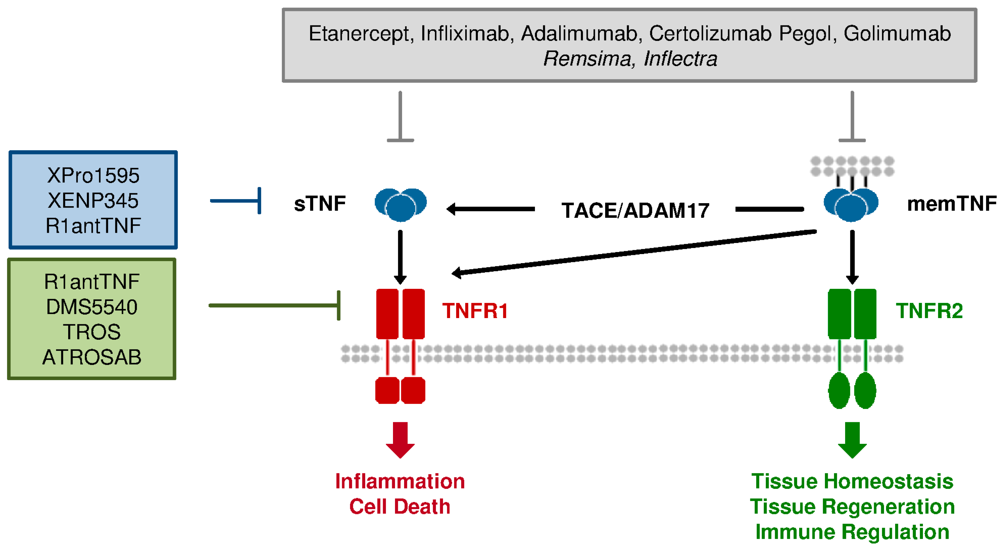
3.1. Current Anti-TNF Therapeutics: Application and Limitation

Conceptually, neutralization of TNF signaling can be achieved at different levels, the level of the ligand, of receptors and of the intracellular signal pathway(s) [7,8,74]. The currently approved anti-TNF therapies all interfere at the level of the ligand by directly binding to TNF, thereby preventing its interaction with both TNF receptors (Figure 1). Five structurally different anti-TNF drugs are currently approved for the treatment of various inflammatory diseases [7,9,22,75,76] (see Table 1): (1) Infliximab (Remicade) is a humanized chimeric monoclonal antibody against human TNF of the subtype IgG1, which is approved for the treatment of RA, psoriatic arthritis (PA), plaque psoriasis (PP), ankylosing spondylitis (AS), CD, and UC; (2) Adalimumab (Humira) is a fully human monoclonal antibody of the IgG1/k isotype that is indicated for the treatment of RA, PA, PP, AS, CD, and juvenile idiopathic arthritis (JIA); (3) Certolizumab pegol (Cimzia) is a PEGylated, humanized anti-TNF Fab’ fragment with a molecular mass of 91 kDa, which is approved for the treatment of CD, RA, AS, and PA; (4) Golimumab (Simponi) is a fully human monoclonal antibody of the IgG1/k isotype that binds and neutralizes human TNF and is approved for the treatment of RA, PA, AS, CD, and UC (5) Etanercept (Enbrel) is a dimeric fully human fusion protein consisting of the extracellular receptor region, which contains the ligand-binding site of human TNFR2 (TNFR2exc), fused to the Fc part of human IgG1 to increase neutralizing activity and plasma half-life. Enbrel binds with high affinity to TNF and is approved for the treatment of RA, JIA, PA, PP, and AS. Several other anti-TNF therapeutics and as of late biosimilars are currently being tested in clinical studies [75,76]. Indeed, the first biosimilar antibodies approved for use in the European Union are the infliximab biosimilars Remsima and Inflectra (Table 1) [77].
In spite of the success of anti-TNF-therapeutics, there are, however, some setbacks. One example is lenercept, similar to etanercept a soluble TNFR-Fc fusion protein, which consists of the extracellular region of TNFR1 directly fused to the hinge region of the IgG1 heavy chain Fc region [78,79]. Lenercept had entered clinical trials for severe sepsis, early septic shock, RA, and MS [7]. However, a Phase II randomized, multi-center, placebo-controlled study for the treatment of relapsing remitting MS had to be stopped since there were no significant differences on magnetic resonance imaging. Furthermore, symptoms of lenercept-treated patients were significantly increased compared with patients receiving placebo, and neurologic deficits tended to be more severe in the lenercept treatment groups [11], indicating that anti-TNF therapies might aggravate demyelinating diseases. The observation that some patients, subsequent to anti-TNF therapies of juvenile rheumatoid arthritis, developed MS-like exacerbations and demyelinating lesions further supports this assumption [13].
Table 1. Currently approved anti-TNF therapeutics and selective inhibitors of sTNF and TNFR1 under development.
Approved anti-TNF therapeutics
Drug name (brand name)StructureApproved disease indications
Etanercept (Enbrel)Human TNFR2exc:IgG1-FcRA, JIA, PA, PP, AS
Infliximab (Remicade)humanized chimeric IgG/k mAbRA, PA, PP, AS, CD, UC
Adalimumab (Humira)fully human IgG1/k mAbRA, PA, PP, AS, CD, JIA
Certolizumab Pegol (Cimzia)PEGylated Fab’ fragmentCD, RA, AS, PA
Golimumab (Simponi)fully human IgG1/k mAbRA, PA, AS, UC
Approved anti-TNF biosimilars
Drug nameBiosimilar ofDisease indication
RemsimaInfliximabRA, PA, PP, AS, CD, UC
InflectraInfliximabRA, PA, PP, AS, CD, UC
Current sTNF/TNFR1 targeting agents under development
NameStructureMechanism
XPro1595Dominant-negative TNF muteinsTNF inhibitor
XENP345Dominant-negative TNF muteinsTNF inhibitor
R1antTNFTNFR1-selective antagonistic mutant TNFTNFR1 antagonist (sTNF inhibitor?)
DMS5540TNFR1-selective monovalent domain antibody fused to albumin specific domain antibody TNFR1 antagonist
TROSTwo TNFR1-selective single domain nanobodies fused to anti-albumin nanobody TNFR1 antagonist
ATROSABHumanized TNFR1-selective IgG1TNFR1 antagonist
AS: ankylosing spondylitis; CD: Crohn’s disease; JIA: Juvenile idiopathic arthritis; PA: Psoriatic arthritis; PP: Plaque psoriasis; RA: Rheumatoid arthritis; UC: Ulcerative colitis.
Next to induction or aggravation of demyelinating diseases, all anti-TNF therapeutics may induce severe side effects including serious infections, reactivation of tuberculosis, invasive fungal infections, and other opportunistic infections. Even an increased susceptibility to develop additional autoimmune diseases and lymphomas has been reported. Moreover, not all patients respond to the anti-TNF treatment, further illustrating the limitations of current anti-TNF therapeutics [7,22,75].

3.2. Differences between TNFR1 & TNFR2: Requirement for Selective Therapeutics

The failure of the lenercept study and the severe side effects of anti-TNF treatment might be explained by the pleiotropic functions of TNF, including proinflammatory and anti-inflammatory but also immune regulatory signals. Blocking all effects of TNF can therefore be counter-productive. Because most of the proinflammatory effects of TNF are mediated by TNFR1, a more effective therapeutic approach seems to be the selective blocking of sTNF/TNFR1 signaling. This would leave memTNF/TNFR2 signaling intact, which has been implicated in protective TNF-mediated responses, e.g., neuroprotection and regeneration, anti-apoptotic signaling, and immune modulation.
The group of Denise Faustman demonstrated that insulin-specific autoreactive CD8+ T cells, which cause the destruction of insulin-producing pancreatic islet beta cells, display several defects in the activation of NF-κB and therefore favor cell death via apoptosis instead of anti-apoptotic effects [80,81]. In animal models, administration of exogenous TNF effectively killed these autoreactive T cells, leading to the inhibition or reversal of type I diabetes [82]. In a subsequent study, the group demonstrated that the insulin-specific autoreactive CD8+ T cells can be selectively destroyed by activation of TNFR2 [83]. In addition to these data, TNFR2 stimulation has been shown to result in the generation and expansion of a subpopulation of protective Tregs [51,84,85] that may control the destruction of excessive CD8+ T cells at sites of inflammation by inhibition of proinflammatory cytokines, leading to suppression of inflammatory responses and thereby supporting the regeneration of the inflamed tissue [84].
Regulatory T-cells constitutively express high levels of TNFR2 [86] and the expression of TNFR2 defines a unique subtype of Tregs with highly potent suppressive activity [51]. Further studies demonstrated that activation of TNFR2 results in the generation and expansion of a subpopulation of protective regulatory T-cells that may suppress autoimmunity [84,85]. Whereas TNFR2 seems not to be necessary to maintain Treg activity, recent results suggest that TNFR2 mediates the activation of Tregs [50] and plays a functional role in their expansion [52] and stabilization [53].
Next to its immune regulatory role, TNFR2 critically contributes to neuronal survival and regeneration. In contrast to TNFR1, which promotes neuronal tissue destruction, TNFR2 was protective in a mouse model of retinal ischemia via activation of the PKB/Akt pathway [87]. Similar results were observed in the cuprizone-induced mouse model of demyelination and remyelination, where genomic ablation of TNF resulted in delayed remyelination and a reduction in the pool of proliferating oligodendrocyte progenitors followed by a reduced number of mature oligodendrocytes. Analysis of TNFR1−/− and TNFR2−/− mice indicated that TNFR2 is critical for TNF-mediated oligodendrocyte regeneration [88], suggesting that TNFR1 signaling mediates axon demyelination, whereas signaling via TNFR2 appears to be responsible for remyelination. Further, different articles emphasize the protective role of TNF in the CNS, demonstrating that TNFR2 protects neurons against excitotoxic insults [35,36] and oxidative stress [37], protects oligodendrocyte progenitor cells against oxidative stress [89], and promotes oligodendrocyte maturation and myelination [38,90].
This dual role of TNF signaling may provide an explanation for the failure of the lenercept study [11], indicating that the inhibition of memTNF/TNFR2 signaling was the molecular basis for the aggravated symptoms in the MS patients of this trial. Accordingly, EAE disease onset and progression is suppressed in transgenic animals exclusively expressing physiologically regulated levels of memTNF, whereas the autoimmune suppressive properties of wild-type TNF were retained [73].
The ability of memTNF/TNFR2 to preserve a subset of beneficial activities, such as immune functions like self-tolerance and resistance to infection, while lacking detrimental effects indicates that selective targeting of sTNF/TNFR1 as a therapeutic strategy may offer several advantages over complete blocking of TNF in the treatment of chronic inflammatory, autoimmune, and demyelinating diseases.

4. Novel sTNF and TNFR1-Specific Antagonists

Next-generation therapeutics targeting the TNF pathway should ideally inhibit the undesirable signals of TNF mainly mediated via sTNF/TNFR1 signaling while preserving its homeostatic functions in the CNS and immune system by sparing memTNF/TNFR2 signaling. Two approaches for specifically targeting the inflammatory effects of TNF have emerged: (1) exclusive inhibition of sTNF and (2) blocking of TNFR1 signaling [7,21,23].
One strategy to generate selective TNF inhibitors is the formation of signaling-incompetent dominant-negative TNF (dnTNF) derivatives that specifically inhibit sTNF, thus leaving signaling via memTNF intact (Table 1). Indeed, there is good evidence that memTNF is sufficient to support TNF-dependent formation of secondary lymphoid organ structure and granulomas [91,92] and to provide at least a partial protection against infections [47,73,92,93] without causing autoimmune diseases [73,91]. Another approach is the use of TNFR1-specific antagonists to neutralize the proinflammatory activity of TNF while maintaining the advantageous responses mediated by TNFR2, including immune regulation, tissue homeostasis, and neuroprotection [94]. Here either antagonistic TNFR1-selective TNF derivatives or TNFR1-specific antagonistic antibodies can be applied (Table 1).

4.1. sTNF-Selective Dominant-Negative TNF Derivatives

In 2005, a novel class of TNF inhibitors was described by Steed et al. [95] that are based on mutations in sTNF, which block binding of the TNF mutein to either TNFR. These TNF muteins can rapidly exchange subunits with endogenous TNF, thereby forming mixed TNF heterotrimers inactivating endogenous TNF in a dominant-negative fashion [96]. The exchange of subunits with native TNF is only efficient for sTNF, thus leaving memTNF unaffected. Several of these muteins had been generated and the therapeutic effect of one of them, XPro1595, which contains the mutations C31H, Y87H, and A145R, has been extensively studied in various animal disease models after PEGylation to improve its in vivo half-life [96].
Importantly, while the ameliorating effect of XPro1595 in various animal disease models, such as acute hepatitis and inflammatory arthritis [96,97], is comparable to total TNF inhibition via etanercept, the normal immune functions are only preserved in the case of XPro1595 treatment. Thus, whereas etanercept-treated mice are highly susceptible to infection with Listeria monocytogenes, XPro1595-treated animals are largely unaffected [96]. As expected, XPro1595 treatment therefore reflects the overexpression of non-cleavable TNF in transgenic mice, which are also protected against bacterial infections, chronic inflammation, and autoimmune disease [73].
The main focus for the application of dnTNFs is their use in animal models of neurodegenerative diseases with elevated TNF levels such as spinal cord injury (SCI), MS, and PD. In an animal model of PD (striatal injection of 6-hydroxydopamine (6-OHDA)), the injection of XENP345, a PEGylated dnTNF variant with the mutations A145R/I97T, into the substantia nigra reduced the loss of dopaminergic neurons by 50% [98]. In a follow-up study, it was shown that a single injection of lentivirus expressing XENP345 into the rat substantia nigra, is sufficient to reduce neuronal loss and behavioral deficits after 6-OHDA-induced degeneration of dopaminergic neurons [99].
As mentioned above, TNF inhibition is detrimental in MS in spite of the elevated levels of TNF in MS lesions and the important role of the peripheral immune system in this disease [10,11]. The potential of treating MS with a selective inhibitor of sTNF was addressed in the EAE model by comparing total TNF inhibition by etanercept with inhibition of sTNF by XPro1595. In these studies, both reagents were injected subcutaneously twice weekly with a dosage of 10 mg/kg. When starting the treatment at the time of disease onset, etanercept had no therapeutic effect, whereas XPro1595 treatment significantly ameliorated EAE symptoms [100]. This effect was not due to changes in the antigen-specific immune response, but was associated with a reduced overall immunoreactivity and an increased expression of neuroprotective molecules in the CNS.
Even when treatment was started at the peak of disease, i.e., when marked demyelination was already in progress, treatment with XPro1595 improved EAE symptoms, whereas etanercept had no effect [101]. Here the therapeutic effect of dnTNF treatment was associated with axonal preservation, an increased number of oligodendrocytes, improved myelin compaction, and remyelination approximately 40 days after starting the treatment. These results indicate that sTNF promotes CNS inflammation while memTNF is neuroprotective, and suggest that selective inhibition of sTNF may provide a new strategy for the treatment of multiple sclerosis [100,101].
TNF is also elevated in the injured spinal cord and it has been suggested that this contributes to impaired healing after SCI [102]. In contrast to other tissues, the sequential activation of distinct macrophage types that promote either inflammation or tissue repair is perturbed in SCI, resulting in a prolonged inflammatory phase wherein repair and remodeling are not properly initiated [103]. This is reminiscent of the inflammation in chronic, non-healing wounds and may at least partially be due to an imbalance of TNFR1 vs. TNFR2 signaling. To test the role of sTNF in SCI, the effect of XPro1595 was compared with etanercept in a mouse model of SCI. Injection of XPro1595 into the spinal cord lesion resulted in improved locomotor function, decreased anxiety-related behavior, and reduced damage to the lesioned spinal cord, whereas etanercept had no therapeutic effects [104]. Interestingly, this beneficial effect of XPro1595 treatment was accompanied by an increased level of TNFR2 protein levels in the lesion area, indicating that signaling of memTNF via TNFR2 may contribute to the improved neuroregeneration [104].
Finally, since TNF is known to sustain the complex inflammatory responses associated with strokes, the effect of XPro1595 was compared to etanercept in an animal model of focal cerebral ischemia. Although infarct volume was not changed, both compounds significantly reduced the peripheral inflammatory response and improved motor function and motor learning skills after focal cerebral ischemia [105]. This indicates that sTNF is predominantly involved in the inflammatory response after a stroke and supports the notion that selective targeting of sTNF might be sufficient for the treatment of most TNF-mediated diseases, thus potentially reducing the risk of severe side effects for the patient by preserving memTNF signaling [105].

4.2. TNFR1-Selective Antagonistic TNF

R1antTNF, a TNFR1-selective antagonistic mutant TNF with substitution of the amino acids at positions 84–89, was isolated from a phage display library [106]. Whereas the affinity of R1antTNF to TNFR1 is almost similar to wild-type TNF (3.3 nM vs. 1.4 nM), its affinity to TNFR2 is greatly impaired (92.9 µM vs. 2.1 nM). The therapeutic effect of R1antTNF was initially evaluated in two models for acute hepatitis. After hepatitis induction with both carbon tetrachloride and concanavalin A, R1antTNF significantly ameliorated liver injury as assessed by serum levels of alanine aminotransferase. Moreover, it reduced serum levels of the proinflammatory cytokines IL-2 and IL-6. This therapeutic effect of R1antTNF was superior to that obtained with an antagonistic anti-TNF antibody [107], indicating that blocking of TNFR1 may be preferable to complete TNF blocking in acute hepatitis.
After mono-PEGylation of R1antTNF to increase the in vivo half-life of the molecule, the therapeutic effect of PEG-R1antTNF was evaluated in animal models of autoimmune diseases, namely CIA and EAE [108,109]. PEG-R1antTNF treatment significantly delayed and ameliorated CIA symptoms both in a prophylactic and a therapeutic setting. Interestingly, when treatment was started when CIA was already established, PEG-R1antTNF treatment (3 µg intraperitoneal, twice daily) was slightly more effective than complete TNF blocking by etanercept (25 µg intraperitoneal, twice weekly) [108]. Moreover, PEG-R1antTNF did not affect the clearance of injected adenovirus, whereas viral load strongly increased after etanercept treatment [108]. In EAE, PEG-R1antTNF significantly reduced the disease incidence, improved the clinical score and reduced demyelination when treatment (10 µg intraperitoneal, twice daily) was initiated at the time of disease induction, i.e., in a prophylactic treatment regime [109]. This was accompanied by significant suppression of T-cell activation, in particular the activation of Th1 and Th17 cells, and a strongly reduced infiltration of inflammatory T-cells into the spinal cord [109].
Interestingly, it was recently described that R1antTNF forms heterotrimers with endogenous TNF [110]. It therefore resembles dnTNFs such as XPRO1595 in its mode of action and may predominantly act as an inhibitor of sTNF signaling. It is therefore unclear whether the observed therapeutic effects are due to specific TNFR1 or sTNF neutralization (Table 1).

4.3. TNFR1-Specific Antibodies

A second strategy to specifically inhibit TNFR1 is the generation of TNFR1-specific antagonistic antibodies. Recently, a monovalent domain antibody (DMS5540; Table 1) specific for mouse TNFR1 was compared to etanercept treatment in the CIA mouse model of rheumatoid arthritis to evaluate the use of antibody-mediated TNFR1 inhibition in autoimmune diseases [111]. To increase the in vivo half-life of the domain antibody, it was fused to a second domain antibody specific for mouse albumin. Specific inhibition of TNFR1 and complete TNF blockage were similarly effective in suppressing CIA progression. However, treatment with etanercept resulted in increased effector T-cell activity, which was not observed after selective TNFR1 blockade, suggesting an immunoregulatory role for TNFR2 that is suppressed by etanercept. Indeed, only TNFR1-specific inhibition promoted the expansion and activation of Tregs, in particular in joints undergoing remission. This supports the concept that a therapeutic strategy that targets TNFR1, but does not affect TNFR2, has the potential to both inhibit inflammation and promote Treg activity and might thus be superior to TNF blockade [111].
A similar approach was taken by Steeland et al. [112], who generated a human TNFR1 selective inhibitor based on nanobody technology. Here two anti-human TNFR1 selective single domain antibodies (nanobodies) were fused to an anti-albumin nanobody to increase the in vivo half-life. The resulting protein, TROS (TNF receptor one silencer), competes with TNF for binding to TNFR1 and strongly inhibits TNF-induced gene expression (Table 1). Importantly, TROS inhibits acute TNF-induced liver inflammation in mice expressing human TNFR1 [112].
In comparison to single domain antibodies, full-length antibodies in the IgG format have the advantage of a highly increased in vivo half-life. Therefore, Kontermann et al. humanized the neutralizing mouse anti-human TNFR1 monoclonal antibody H398 [94,113]. The resulting IgG1, ATROSAB (antagonistic tumor necrosis factor receptor one-specific antibody), retained the receptor selectivity and TNFR1-neutralizing ability of the parental mouse antibody (Table 1). Importantly, ATROSAB binds with similar efficacy to human and rhesus TNFR1-Fc fusion proteins, which opens the way to further analyze the therapeutic activity of ATROSAB in non-human primates [114]. Moreover, the Fc region of ATROSAB has been modified to abrogate complement fixation and antibody-mediated cellular effector functions, thus preventing the unwanted activation of the immune system [114,115].
Since TNFR1 is essential for the development of EAE, we recently applied a commercial hamster IgG specific for mouse TNFR1 [116] to evaluate the potential of antibody-mediated anti-TNFR1 treatment in EAE [72]. Interestingly, a single dose of the antibody (5 mg/kg) was sufficient to ameliorate the disease when given at the time of disease induction. Moreover, administration at day 1 and 4 after disease onset (20 mg/kg), i.e., in a therapeutic treatment regime, resulted in a stable, even slightly reduced disease course for at least seven additional days and thus led to a strongly reduced cumulative EAE score. These results demonstrate that antagonistic TNFR1-specific antibodies may represent a therapeutic approach for the treatment of MS [72].

4.4. Comparison of sTNF Inhibitors and TNFR1 Antagonists

Although both sTNF inhibitors and TNFR1 antagonists block responses mediated by the sTNF/TNFR1 pathway, there are also important differences in their mode of action that may affect their therapeutic use in different diseases.
First, exclusive inhibition of sTNF leaves the local signaling of memTNF via TNFR1 intact. Whether this is detrimental or beneficial for treatment is currently unclear and may depend on the disease context. However, memTNF signaling via TNFR1 may contribute to the observation that mice treated with sTNF inhibitors such as dnTNF have a largely intact immune system that can protect them against microbial and viral infections [96]. Given the uncertainty of the mode of action of R1antTNF, this aspect still needs to be verified for TNFR1-selective antagonists. Moreover, under certain circumstances signaling of memTNF via TNFR1 can promote cell survival, e.g., in neurons [117]. Accordingly, TNFR1 antagonists may prevent these potentially beneficial aspects of TNFR1 signaling. On the other hand, there is some evidence that sTNF can activate TNFR2 in cells with high TNFR2 content, such as Tregs [50,51,52]. Whether activation of Tregs by memTNF/TNFR2 is sufficient for Treg activation or if additional activation of TNFR2 by sTNF can modulate Treg activity is currently unclear, but blocking of sTNF might compromise the immune modulatory functions of TNFR2.
In comparison to inhibition of sTNF, the specific neutralization of TNFR1 may have additional consequences that might be therapeutically relevant. In particular, TNFR1 antagonists can bind to soluble TNFR1 and thus reduce the suspected TNF neutralizing capacity of soluble TNFR1. Importantly, since signaling competent TNFR1 at the cell surface is inhibited by the TNFR1 antagonists as well, the excess TNF would preferentially bind to TNFR2, which may promote the immune regulatory and tissue regenerative properties of this receptor. Moreover, a polymorphism in the TNFR1 gene that results in release of soluble TNFR1 has been associated with an increased risk of developing MS [68]. This is presumably due to an altered state of monocytes, which show increased expression of CXCL10 and a more robust interferon type I response upon TNF stimulation compared to cells with the wild-type allele [118]. Accordingly, neutralization of soluble TNFR1 may have beneficial consequences, which may, however, depend on the disease context. Finally, in contrast to sTNF inhibitors, TNFR1 antagonists can block the binding of lymphotoxin-α (LT-α), another member of the TNF superfamily, to TNFR1. Since a proinflammatory role of LT-α has been described in RA and in animal disease models, such as CIA and EAE [119,120,121,122,123], simultaneous blocking of TNF and LT-α binding to TNFR1 by TNFR1 antagonists may have advantages compared to sTNF inhibition in acute and chronic inflammatory diseases.
Importantly, the above studies show that both sTNF inhibitors and TNFR1 antagonists are capable of reducing symptoms in animal models of acute and chronic inflammation. Thus, both sTNF inhibitors and TNFR1 antagonists can reduce the severity in animal models of inflammatory diseases, namely EAE and CIA [72,100,101,111]. At the moment one can therefore only speculate whether the different modes of actions of sTNF inhibitors and TNFR1 antagonists may ultimately result in one strategy being preferred as a therapeutic. This will obviously be one of the major aspects of future research and may well depend on the type of disease or the target tissue.

5. Conclusions

In the past approximately 15 years, TNF blockers have demonstrated their effectiveness in the treatment of several autoimmune diseases and belong now to the top-selling drugs worldwide. Nevertheless, the development of serious side effects during long-lasting TNF inhibition revealed the risks that are associated with complete blocking of TNF. Moreover, total inhibition of TNF has been detrimental for the treatment of several diseases in which TNF has been implicated, such as MS. While some of the described side effects are due to TNF inhibition, e.g., the increased risk of infections, many of the side effects are most likely due to the role of TNF in immune regulation and tissue regeneration, which has been revealed recently.
Importantly, a more specific targeting of pathological TNF can be achieved because some of the pathological and beneficial signals diverge at the level of ligand (sTNF or memTNF) and at the level of receptor (TNFR1 or TNFR2). Accordingly, specific inhibition of sTNF or TNFR1 might be sufficient to inhibit the pathological consequences of deregulated TNF signaling while keeping memTNF/TNFR2 signaling intact. This might greatly reduce the side effects associated with total TNF blockade and may enable the treatment of other diseases such as MS, where total TNF inhibition is detrimental. Whether selective inhibition of TNFR1 will be sufficient for redirection of the available TNF to induce activation of TNFR2, thus promoting immune modulation and tissue regeneration, remains to be investigated. Importantly, selective TNFR2 agonists are currently evaluated to boost the activation of TNFR2 and the beneficial actions of these agonists have been evaluated in several in vitro [37,38,89] and ex vivo [124] applications. Therefore, a combination therapy, i.e., the concomitant use of sTNF/TNFR1 antagonists and a TNFR2 agonist, might present a novel therapeutic concept to treat diverse inflammatory and degenerative diseases.

Acknowledgements

This work was partially funded by grants from the Federal Ministry of Education and Research of Germany (to Roman Fischer and Roland E. Kontermann), the Carl Zeiss Foundation (to Roman Fischer) and the Hertie Foundation (to Olaf Maier).

Author Contributions

Roman Fischer and Olaf Maier wrote the review. Roland E. Kontermann contributed to Section 3 and Section 4.

Conflicts of Interest

Roman Fischer and Olaf Maier declare no conflict of interest. Roland E. Kontermann is the named inventor on patents covering the ATROSAB technology.

References

  1. Aggarwal, B.B. Signalling pathways of the TNF superfamily: A double-edged sword. Nat. Rev. Immunol. 2003, 3, 745–756. [Google Scholar] [CrossRef] [PubMed]
  2. Locksley, R.M.; Killeen, N.; Lenardo, M.J. The TNF and TNF receptor superfamilies: Integrating mammalian biology. Cell 2001, 104, 487–501. [Google Scholar] [CrossRef] [PubMed]
  3. Apostolaki, M.; Armaka, M.; Victoratos, P.; Kollias, G. Cellular mechanisms of TNF function in models of inflammation and autoimmunity. Curr. Dir. Autoimmun. 2010, 11, 1–26. [Google Scholar] [PubMed]
  4. Kollias, G. TNF pathophysiology in murine models of chronic inflammation and autoimmunity. Semin. Arthritis Rheum. 2005, 34, 3–6. [Google Scholar] [CrossRef] [PubMed]
  5. Wajant, H.; Pfizenmaier, K.; Scheurich, P. Tumor necrosis factor signaling. Cell Death Diff. 2003, 10, 45–65. [Google Scholar] [CrossRef]
  6. Wong, M.; Ziring, D.; Korin, Y.; Desai, S.; Kim, S.; Lin, J.; Gjertson, D.; Braun, J.; Reed, E.; Singh, R.R. TNFα blockade in human diseases: Mechanisms and future directions. Clin. Immunol. 2008, 126, 121–136. [Google Scholar] [CrossRef] [PubMed]
  7. Kontermann, R.E.; Scheurich, P.; Pfizenmaier, K. Antagonists of TNF action: Clinical experience and new developments. Exp. Opin. Drug Discov. 2009, 4, 279–292. [Google Scholar] [CrossRef]
  8. Tracey, D.; Klareskog, L.; Sasso, E.H.; Salfeld, J.G.; Tak, P.P. Tumor necrosis factor antagonist mechanisms of action: A comprehensive review. Pharmacol. Ther. 2008, 177, 244–279. [Google Scholar] [CrossRef]
  9. Cessak, G.; Kuzawińska, O.; Burda, A.; Lis, K.; Wojnar, M.; Mirowska-Guzel, D.; Bałkowiec-Iskra, E. TNF inhibitors—Mechanisms of action, approved and off-label indications. Pharmacol. Rep. 2014, 66, 836–844. [Google Scholar] [CrossRef] [PubMed]
  10. van Oosten, B.W.; Barkhof, F.; Truyen, L.; Boringa, J.B.; Bertelsmann, F.W.; von Blomberg, B.M.; Woody, J.N.; Hartung, H.P.; Polman, C.H. Increased MRI activity and immune activation in two multiple sclerosis patients treated with the monoclonal anti-tumor necrosis factor antibody cA2. Neurology 1996, 47, 1531–1534. [Google Scholar] [CrossRef] [PubMed]
  11. The Lenercept Study Group. TNF neutralization in MS: results of a randomized, placebo-controlled multicenter study. The Lenercept Multiple Sclerosis Study Group and The University of British Columbia MS/MRI Analysis Group. Neurology 1999, 53, 457–465. [Google Scholar]
  12. Slifman, N.R.; Gershon, S.K.; Lee, J.H.; Edwards, E.T.; Braun, M.M. Listeria monocytogenes infection as a complication of treatment with tumor necrosis actor alpha-neutralizing agents. Arthritis Rheum. 2003, 48, 319–324. [Google Scholar]
  13. Sicotte, N.L.; Voskuhl, R.R. Onset of multiple sclerosis associated with anti-TNF therapy. Neurology 2001, 57, 1885–1888. [Google Scholar] [CrossRef] [PubMed]
  14. Shakoor, N.; Michalska, M.; Harris, C.A.; Block, J.A. Drug-induced systemic lupus erythematosus associated with etanercept therapy. Lancet 2002, 359, 579–580. [Google Scholar] [CrossRef] [PubMed]
  15. Brown, S.L.; Greene, M.H.; Gershon, S.K.; Edwards, E.T.; Braun, M.M. Tumor necrosis factor antagonist therapy and lymphoma development: Twenty-six cases reported to the Food and Drug Administration. Arthritis Rheum. 2002, 46, 3151–3158. [Google Scholar] [CrossRef] [PubMed]
  16. Black, R.A.; Rauch, C.T.; Kozlosky, C.J.; Peschon, J.J.; Slack, J.L.; Wolfson, M.F.; Castner, B.J.; Stocking, K.L.; Reddy, P.; Srinivasan, S.; et al. A metalloproteinase disintegrin that releases tumour-necrosis factor-α from cells. Nature 1997, 385, 729–733. [Google Scholar] [CrossRef] [PubMed]
  17. Grell, M.; Douni, E.; Wajant, H.; Löhden, M.; Clauss, M.; Maxeiner, B.; Georgopoulos, S.; Lesslauer, W.; Kollias, G.; Pfizenmaier, K.; et al. The transmembrane form of tumor necrosis factor is the prime activating ligand of the 80 kDa tumor necrosis factor receptor. Cell 1995, 83, 793–802. [Google Scholar] [CrossRef] [PubMed]
  18. Wallach, D.; Engelmann, H.; Nophar, Y.; Aderka, D.; Kemper, O.; Hornik, V.; Holtmann, H.; Brakebusch, C. Soluble and cell surface receptors for tumor necrosis factor. Agents Actions Suppl. 1991, 35, 51–57. [Google Scholar] [PubMed]
  19. Solomon, K.A.; Pesti, N.; Wu, G.; Newton, R.C. Cutting edge: A dominant negative form of TNF-alpha converting enzyme inhibits proTNF and TNFRII secretion. J. Immunol. 1999, 163, 4105–4108. [Google Scholar] [PubMed]
  20. Aderka, D. The potential biological and clinical significance of the soluble tumor necrosis factor receptors. Cytokine Growth Factor Rev. 1996, 7, 231–240. [Google Scholar] [CrossRef] [PubMed]
  21. Tansey, M.G.; Szymkowski, D.E. The TNF superfamily in 2009: New pathways, new indications, and new drugs. Drug Discov. Today 2009, 14, 1082–1088. [Google Scholar] [CrossRef] [PubMed]
  22. Pfizenmaier, K.; Szymkowski, D.E. Workshop Summary: Introduction to rational design of new means for therapeutic modulation of function of the TNF family. Adv. Exp. Med. Biol. 2011, 691, 487–491. [Google Scholar] [PubMed]
  23. van Hauwermeiren, F.; Vandenbroucke, R.E.; Libert, C. Treatment of TNF mediated diseases by selective inhibition of soluble TNF or TNFR1. Cytokine Growth Factor Rev. 2011, 22, 311–319. [Google Scholar] [CrossRef] [PubMed]
  24. Chan, F.K.; Chun, H.J.; Zheng, L.; Siegel, R.M.; Bui, K.L.; Lenardo, M.J. A domain in TNF receptors that mediates ligand-independent receptor assembly and signaling. Science 2000, 288, 2351–2354. [Google Scholar] [CrossRef] [PubMed]
  25. vanden Berghe, T.; Linkermann, A.; Jouan-Lanhouet, S.; Walczak, H.; Vandenabeele, P. Regulated necrosis: The expanding network of non-apoptotic cell death pathways. Nat. Rev. Mol. Cell Biol. 2014, 15, 135–147. [Google Scholar] [CrossRef] [PubMed]
  26. Walczak, H. TNF and ubiquitin at the crossroads of gene activation, cell death, inflammation, and cancer. Immunol. Rev. 2011, 244, 9–28. [Google Scholar] [CrossRef] [PubMed]
  27. Wajant, H.; Scheurich, P. TNFR1-induced activation of the classical NF-κB pathway. FEBS J. 2011, 278, 862–876. [Google Scholar] [CrossRef] [PubMed]
  28. Kaltschmidt, B.; Baeuerle, P.A.; Kaltschmidt, C. Potential involvement of the transcription factor NF-kappa B in neurological disorders. Mol. Aspects Med. 1993, 14, 171–190. [Google Scholar] [CrossRef] [PubMed]
  29. Sabio, G.; Davis, R.J. TNF and MAP kinase signalling pathways. Semin. Immunol. 2014, 26, 237–245. [Google Scholar] [CrossRef] [PubMed]
  30. Guma, M.; Firestein, G.S. c-Jun N-Terminal Kinase in inflammation and rheumatic diseases. Open Rheumatol. J. 2012, 6, 220–231. [Google Scholar] [CrossRef] [PubMed]
  31. Yang, Y.; Kim, S.C.; Yu, T.; Yi, Y.S.; Rhee, M.H.; Sung, G.H.; Yoo, B.C.; Cho, J.Y. Functional roles of p38 mitogen-activated protein kinase in macrophage-mediated inflammatory responses. Mediators Inflamm. 2014, 352371. [Google Scholar]
  32. Naudé, P.J.; den Boer, J.A.; Luiten, P.G.; Eisel, U.L. Tumor necrosis factor receptor cross-talk. FEBS J. 2011, 278, 888–898. [Google Scholar] [CrossRef] [PubMed]
  33. Rauert, H.; Wicovsky, A.; Müller, N.; Siegmund, D.; Spindler, V.; Waschke, J.; Kneitz, C.; Wajant, H. Membrane tumor necrosis factor (TNF) induces p100 processing via TNF receptor-2 (TNFR2). J. Biol. Chem. 2010, 285, 7394–7404. [Google Scholar] [CrossRef] [PubMed]
  34. Fischer, R.; Maier, O.; Naumer, M.; Krippner-Heidenreich, A.; Scheurich, P.; Pfizenmaier, K. Ligand-induced internalization of TNF receptor 2 mediated by a di-leucin motif is dispensable for activation of the NFκB pathway. Cell Signal. 2011, 23, 161–170. [Google Scholar] [CrossRef] [PubMed]
  35. Marchetti, L.; Klein, M.; Schlett, K.; Pfizenmaier, K.; Eisel, U.L. Tumor necrosis factor (TNF)-mediated neuroprotection against glutamate-induced excitotoxicity is enhanced by N-methyl-D-aspartate receptor activation. Essential role of a TNF receptor 2-mediated phosphatidylinositol 3-kinase-dependent NF-kappa B pathway. J. Biol. Chem. 2004, 279, 32869–32881. [Google Scholar] [CrossRef] [PubMed]
  36. Dolga, A.M.; Nijholt, I.M.; Ostroveanu, A.; Ten Bosch, Q.; Luiten, P.G.; Eisel, U.L. Lovastatin induces neuroprotection through tumor necrosis factor receptor 2 signaling pathways. J. Alz. Dis. 2008, 13, 111–122. [Google Scholar]
  37. Fischer, R.; Maier, O.; Siegemund, M.; Wajant, H.; Scheurich, P.; Pfizenmaier, K. A TNF Receptor 2 selective agonist rescues neurons from oxidative stress-induced cell death. PLoS One 2011, 6, e27621. [Google Scholar] [CrossRef]
  38. Fischer, R.; Wajant, H.; Kontermann, R.; Pfizenmaier, K.; Maier, O. Astrocyte-specific activation of TNFR2 promotes oligodendrocyte maturation by secretion of leukemia inhibitory factor. Glia 2014, 62, 272–283. [Google Scholar] [CrossRef] [PubMed]
  39. Wilhelm, P.; Ritter, U.; Labbow, S.; Donhauser, N.; Rollinghoff, M.; Bogdan, C.; Korner, H. Rapidly fatal leishmaniasis in resistant C57BL/6 mice lacking TNF. J. Immunol. 2001, 166, 4012–4019. [Google Scholar] [CrossRef] [PubMed]
  40. Pasparakis, M.; Alexopoulou, L.; Episkopou, V.; Kollias, G. Immune and inflammatory responses in TNFα-deficient mice: A critical requirement for TNFα in the formation of primary B cell follicles, follicular dendritic cell networks and germinal centers, and in the maturation of the humoral immune response. J. Exp. Med. 1996, 184, 1397–1411. [Google Scholar] [CrossRef] [PubMed]
  41. Mielke, M.E.; Peters, C.; Hahn, H. Cytokines in the induction and expression of T-cell-mediated granuloma formation and protection in the murine model of listeriosis. Immunol. Rev. 1997, 158, 79–93. [Google Scholar] [CrossRef] [PubMed]
  42. Mackay, F.; Loetscher, H.; Stueber, D.; Gehr, G.; Lesslauer, W. Tumor necrosis factor alpha (TNF-alpha)-induced cell adhesion to human endothelial cells is under dominant control of one TNF receptor type, TNF-R55. J. Exp. Med. 1993, 177, 1277–1286. [Google Scholar] [CrossRef] [PubMed]
  43. Rothe, J.; Lesslauer, W.; Lötscher, H.; Lang, Y.; Koebel, P.; Köntgen, F.; Althage, A.; Zinkernagel, R.; Steinmetz, M.; Bluethmann, H. Mice lacking the tumour necrosis factor receptor 1 are resistant to TNF-mediated toxicity but highly susceptible to infection by Listeria monocytogenes. Nature 1993, 364, 798–802. [Google Scholar] [CrossRef] [PubMed]
  44. Rothe, J.; Mackay, F.; Bluethmann, H.; Zinkernagel, R.; Lesslauer, W. Phenotypic analysis of TNFR1-deficient mice and characterization of TNFR1-deficient fibroblasts in vitro. Circ. Shock 1994, 44, 51–56. [Google Scholar] [PubMed]
  45. Xu, J.; Chakrabarti, A.K.; Tan, J.L.; Ge, L.; Gambotto, A.; Vujanovic, N.L. Essential role of the TNF–TNFR2 cognate interaction in mouse dendritic cell-natural killer cell crosstalk. Blood 2007, 109, 333–3341. [Google Scholar]
  46. Musicki, K.; Briscoe, H.; Tran, S.M.; Britton, W.J.; Saunders, B.M. Differential requirements for soluble and transmembrane tumor necrosis factor in the immunological control of primary and secondary Listeria monocytogenes infection. Infect. Immun. 2006, 74, 3180–3189. [Google Scholar] [CrossRef] [PubMed]
  47. Torres, D.; Janot, L.; Quesniaux, V.F.; Grivennikov, S.I.; Maillet, I.; Sedgwick, J.D.; Ryffel, B.; Erard, F. Membrane tumor necrosis factor confers partial protection to Listeria infection. Am. J. Pathol. 2005, 167, 1677–1687. [Google Scholar] [CrossRef] [PubMed]
  48. Sakaguchi, S. Regulatory T cells: Key controllers of immunologic self-tolerance. Cell 2000, 101, 455–458. [Google Scholar] [CrossRef] [PubMed]
  49. Sakaguchi, S.; Yamaguchi, T.; Nomura, T.; Ono, M. Regulatory T cells and immune tolerance. Cell 2008, 133, 775–787. [Google Scholar] [CrossRef] [PubMed]
  50. Chen, X.; Baumel, M.; Männel, D.N.; Howard, O.M.; Oppenheim, J.J. Interaction of TNF with TNF receptor type 2 promotes expansion and function of mouse CD4+CD25+ T regulatory cells. J. Immunol. 2007, 179, 154–161. [Google Scholar] [CrossRef] [PubMed]
  51. Chen, X.; Subleski, J.J.; Kopf, H.; Howard, O.M.; Männel, D.N.; Oppenheim, J.J. Cutting edge: expression of TNFR2 defines a maximally suppressive subset of mouse CD4+ CD25+ FoxP3+ T regulatory cells: Applicability to tumor-infiltrating T regulatory cells. J. Immunol. 2008, 180, 6467–6471. [Google Scholar] [CrossRef] [PubMed]
  52. Chen, X.; Oppenheim, J.J. TNF-alpha: An activator of CD4+FoxP3+TNFR2+ regulatory T cells. Curr. Dir. Autoimmun. 2010, 11, 119–134. [Google Scholar] [PubMed]
  53. Chen, X.; Wu, X.; Zhou, Q.; Howard, O.M.; Netea, M.G.; Oppenheim, J.J. TNFR2 is critical for the stabilization of the CD4+Foxp3+ regulatory T cell phenotype in the inflammatory environment. J. Immunol. 2013, 190, 1076–1084. [Google Scholar] [CrossRef] [PubMed]
  54. Kim, J.M.; Rasmussen, J.P.; Rudensky, A.Y. Regulatory T cells prevent catastrophic autoimmunity throughout the lifespan of mice. Nat. Immunol. 2007, 8, 191–197. [Google Scholar] [CrossRef] [PubMed]
  55. Xanthoulea, S.; Pasparakis, M.; Kousteni, S.; Brakebusch, C.; Wallach, D.; Bauer, J.; Lassmann, H.; Kollias, G. Tumor necrosis factor (TNF) receptor shedding controls thresholds of innate immune activation that balance opposing TNF functions in infectious and inflammatory diseases. J. Exp. Med. 2004, 200, 367–376. [Google Scholar] [CrossRef] [PubMed]
  56. McDermott, M.F.; Aksentijevich, I.; Galon, J.; McDermott, E.M.; Ogunkolade, B.W.; Centola, M.; Mansfield, E.; Gadina, M.; Karenko, L.; Pettersson, T.; et al. Germline mutations in the extracellular domains of the 55 kDa TNF receptor, TNFR1, define a family of dominantly inherited autoinflammatory syndromes. Cell 1999, 97, 133–144. [Google Scholar] [CrossRef] [PubMed]
  57. Kimberley, F.C.; Lobito, A.A.; Siegel, R.M.; Screaton, G.R. Falling into TRAPS—Receptor misfolding in the TNF receptor 1-associated periodic fever syndrome. Arthritis Res. Ther. 2007, 9, 217. [Google Scholar] [CrossRef] [PubMed]
  58. Bhatia, M. Acute pancreatitis as a model of SIRS. Front. Biosci. 2009, 14, 2042–2050. [Google Scholar] [CrossRef]
  59. Bhatia, M.; Moochhala, S. Role of inflammatory mediators in the pathophysiology of acute respiratory distress syndrome. J. Path. 2004, 202, 145–156. [Google Scholar] [CrossRef] [PubMed]
  60. Kleinbongard, P.; Schulz, R.; Heusch, G. TNFα in myocardial ischemia/reperfusion, remodeling and heart failure. Heart Fail. Rev. 2011, 16, 49–69. [Google Scholar] [CrossRef] [PubMed]
  61. Kontoyiannis, D.; Pasparakis, M.; Pizarro, T.T.; Cominelli, F.; Kollias, G. Impaired on/off regulation of TNF biosynthesis in mice lacking TNF AU-rich elements: Implications for joint and gut-associated immunopathologies. Immunity 1999, 10, 387–398. [Google Scholar] [CrossRef] [PubMed]
  62. Piguet, P.F.; Grau, G.E.; Vesin, C.; Loetscher, H.; Gentz, R.; Lesslauer, W. Evolution of collagen arthritis in mice is arrested by treatment with anti-tumour necrosis factor (TNF) antibody or a recombinant soluble TNF receptor. Immunology 1992, 77, 510–514. [Google Scholar] [PubMed]
  63. Mori, L.; Iselin, S.; De Libero, G.; Lesslauer, W. Attenuation of collagen-induced arthritis in 55-kDa TNF receptor type 1 (TNFR1)-IgG1-treated and TNFR1-deficient mice. J. Immunol. 1996, 157, 3178–3182. [Google Scholar] [PubMed]
  64. Dickson, D.W. The pathogenesis of senile plaques. J. Neuropathol. Exp. Neurol. 1997, 56, 321–339. [Google Scholar] [CrossRef] [PubMed]
  65. Mogi, M.; Harada, M.; Riederer, P.; Narabayashi, H.; Fujita, K.; Nagatsu, T. Tumor necrosis factor-alpha (TNF-alpha) increases both in the brain and in the cerebrospinal fluid from parkinsonian patients. Neurosci. Lett. 1994, 165, 208–210. [Google Scholar] [CrossRef] [PubMed]
  66. Hofman, F.M.; Hinton, D.R.; Johnson, K.; Merrill, J.E. Tumor necrosis factor identified in multiple sclerosis brain. J. Exp. Med. 1989, 170, 607–612. [Google Scholar] [CrossRef] [PubMed]
  67. Probert, L.; Akassoglou, K.; Pasparakis, M.; Kontogeorgos, G.; Kollias, G. Spontaneous inflammatory demyelinating disease in transgenic mice showing central nervous system-specific expression of tumor necrosis factor alpha. Proc. Natl. Acad. Sci. USA 1995, 92, 11294–11298. [Google Scholar] [CrossRef] [PubMed]
  68. Gregory, A.P.; Dendrou, C.A.; Attfield, K.E.; Haghikia, A.; Xifara, D.K.; Butter, F.; Poschmann, G.; Kaur, G.; Lambert, L.; Leach, O.A.; et al. TNF receptor 1 genetic risk mirrors outcome of anti-TNF therapy in multiple sclerosis. Nature 2012, 488, 508–511. [Google Scholar] [CrossRef] [PubMed]
  69. Eugster, H.P.; Frei, K.; Bachmann, R.; Bluethmann, H.; Lassmann, H.; Fontana, A. Severity of symptoms and demyelination in MOG-induced EAE depends on TNFR1. Eur. J. Immunol. 1999, 29, 626–632. [Google Scholar] [CrossRef] [PubMed]
  70. Suvannavejh, G.C.; Lee, H.O.; Padilla, J.; Dal Canto, M.C.; Barrett, T.A.; Miller, S.D. Divergent roles for p55 and p75 tumor necrosis factor receptors in the pathogenesis of MOG(35-55)-induced experimental autoimmune encephalomyelitis. Cell. Immunol. 2000, 205, 24–33. [Google Scholar] [CrossRef] [PubMed]
  71. Kassiotis, G.; Kollias, G. Uncoupling the proinflammatory from the immunosuppressive properties of tumor necrosis factor (TNF) at the p55 TNF receptor level: Implications for pathogenesis and therapy of autoimmune demyelination. J. Exp. Med. 2001, 193, 427–434. [Google Scholar] [CrossRef] [PubMed]
  72. Williams, S.K.; Maier, O.; Fischer, R.; Fairless, R.; Hochmeister, S.; Stojic, A.; Pick, L.; Haar, D.; Musiol, S.; Storch, M.K.; et al. Antibody-mediated inhibition of TNFR1 attenuates disease in a mouse model of multiple sclerosis. PLoS One 2014, 9, e90117. [Google Scholar] [CrossRef]
  73. Alexopoulou, L.; Kranidioti, K.; Xanthoulea, S.; Denis, M.; Kotanidou, A.; Douni, E.; Blackshear, P.J.; Kontoyiannis, D.L.; Kollias, G. Transmembrane TNF protects mutant mice against intracellular bacterial infections, chronic inflammation and autoimmunity. Eur. J. Immunol. 2006, 36, 2768–2780. [Google Scholar] [CrossRef] [PubMed]
  74. Palladino, M.A.; Bahjat, F.R.; Theodorakis, E.A.; Moldawer, L.L. Anti-TNF-α therapies: The next generation. Nat. Rev. Drug Discov. 2003, 2, 736–746. [Google Scholar] [CrossRef] [PubMed]
  75. Sedger, L.M.; McDermott, M.F. TNF and TNF-receptors: From mediators of cell death and inflammation to therapeutic giants—Past, present and future. Cytokine Growth Factor Rev. 2014, 25, 453–472. [Google Scholar] [CrossRef] [PubMed]
  76. Monaco, C.; Nanchahal, J.; Taylor, P.; Feldmann, M. Anti-TNF therapy: Past, present and future. Int. Immunol. 2015, 27, 55–62. [Google Scholar] [CrossRef] [PubMed]
  77. Beck, A.; Reichert, J.M. Approval of the first biosimilar antibodies in Europe: A major landmark for the biopharmaceutical industry. MAbs 2013, 5, 621–623. [Google Scholar] [CrossRef] [PubMed]
  78. Christen, U.; Thuerkauf, R.; Stevens, R.; Lesslauer, W. Immune response to a recombinant human TNFR55-IgG1 fusion protein: auto-antibodies in rheumatoid arthritis (RA) and multiple sclerosis (MS) patients have neither neutralizing nor agonist activities. Hum. Immunol. 1999, 60, 774–790. [Google Scholar] [CrossRef] [PubMed]
  79. Abraham, E.; Laterre, P.F.; Garbino, J.; Pingleton, S.; Butler, T.; Dugernier, T.; Margolis, B.; Kudsk, K.; Zimmerli, W.; Anderson, P.; et al. Lenercept (p55 tumor necrosis factor fusion protein) in severe sepsis and early septic shock: A randomized, double-blind, placebo-controlled, multicenter phase III trial with 1,342 patients. Crit. Care Med. 2001, 29, 503–570. [Google Scholar] [CrossRef] [PubMed]
  80. Hayashi, T.; Faustman, D. NOD mice are defective in proteasome production and activation of NF-kappaB. Mol. Cell. Biol. 1999, 19, 8646–8659. [Google Scholar] [PubMed]
  81. Faustman, D.; Davis, M. TNF receptor 2 pathway: Drug target for autoimmune diseases. Nat. Rev. Drug Discov. 2010, 9, 482–493. [Google Scholar] [CrossRef] [PubMed]
  82. Kodama, S.; Davis, M.; Faustman, D.L. The therapeutic potential of tumor necrosis factor for autoimmune disease: a mechanistically based hypothesis. Cell. Mol. Life Sci. 2005, 62, 1850–1862. [Google Scholar] [CrossRef] [PubMed]
  83. Ban, L.; Zhang, J.; Wang, L.; Kuhtreiber, W.; Burger, D.; Faustman, D.L. Selective death of autoreactive T cells in human diabetes by TNF or TNF receptor 2 agonism. Proc. Natl. Acad. Sci. USA 2008, 105, 13644–13649. [Google Scholar] [CrossRef] [PubMed]
  84. Askenasy, N.; Kaminitz, A.; Yarkoni, S. Mechanisms of T regulatory cell function. Autoimmun. Rev. 2008, 7, 370–375. [Google Scholar] [CrossRef]
  85. Nagar, M.; Jacob-Hirsch, J.; Vernitsky, H.; Berkun, Y.; Ben-Horin, S.; Amariglio, N.; Bank, I.; Kloog, Y.; Rechavi, G.; Goldstein, I. TNF activates a NF-kappaB-regulated cellular program in human CD45RA- regulatory T cells that modulates their suppressive function. J. Immunol. 2010, 184, 3570–3581. [Google Scholar] [CrossRef] [PubMed]
  86. Annunziato, F.; Cosmi, L.; Liotta, F.; Lazzeri, E.; Manetti, R.; Vanini, V.; Romagnani, P.; Maggi, E.; Romagnani, S. Phenotype, localization, and mechanism of suppression of CD4(+)CD25(+) human thymocytes. J. Exp. Med. 2002, 196, 379–387. [Google Scholar] [CrossRef] [PubMed]
  87. Fontaine, V.; Mohand-Said, S.; Hanoteau, N.; Fuchs, C.; Pfizenmaier, K.; Eisel, U. Neurodegenerative and neuroprotective effects of tumor necrosis factor (TNF) in retinal ischemia: opposite roles of TNF receptor 1 and TNF receptor 2. J. Neurosci. 2002, 22, RC216. [Google Scholar] [PubMed]
  88. Arnett, H.A.; Mason, J.; Marino, M.; Suzuki, K.; Matsushima, G.K.; Ting, J.P. TNFα promotes proliferation of oligodendrocyte progenitors and remyelination. Nat. Neurosci. 2001, 4, 1116–1122. [Google Scholar] [CrossRef] [PubMed]
  89. Maier, O.; Fischer, R.; Agresti, C.; Pfizenmaier, K. TNFR2 activation protects oligodendrocyte progenitor cells against oxidative stress, Bioch. Biophys. Res. Comm. 2013, 440, 336–341. [Google Scholar] [CrossRef]
  90. Patel, J.R.; Williams, J.L.; Muccigrosso, M.M.; Liu, L.; Sun, T.; Rubin, J.B.; Klein, R.S. Astrocyte TNFR2 is required for CXCL12-mediated regulation of oligodendrocyte progenitor proliferation and differentiation within the adult CNS. Acta Neuropathol. 2012, 124, 847–860. [Google Scholar] [CrossRef] [PubMed]
  91. Ruuls, S.R.; Hoek, R.M.; Ngo, V.N.; McNeil, T.; Lucian, L.A.; Janatpour, M.J.; Körner, H.; Scheerens, H.; Hessel, E.M.; Cyster, J.G.; et al. Membrane-bound TNF supports secondary lymphoid organ structure but is subservient to secreted TNF in driving autoimmune inflammation. Immunity 2001, 15, 533–543. [Google Scholar] [CrossRef] [PubMed]
  92. Saunders, B.M.; Tran, S.; Ruuls, S.; Sedgwick, J.D.; Briscoe, H.; Britton, W.J. Transmembrane TNF is sufficient to initiate cell migration and granuloma formation and provide acute, but not long-term, control of Mycobacterium tuberculosis infection. J. Immunol. 2005, 174, 4852–4859. [Google Scholar] [CrossRef] [PubMed]
  93. Fremond, C.; Allie, N.; Dambuza, I.; Grivennikov, S.I.; Yeremeev, V.; Quesniaux, V.F.; Jacobs, M.; Ryffel, B. Membrane TNF confers protection to acute mycobacterial infection. Respir. Res. 2005, 6, 136. [Google Scholar] [CrossRef] [PubMed] [Green Version]
  94. Kontermann, R.E.; Münkel, S.; Neumeyer, J.; Müller, D.; Branschädel, M.; Scheurich, P.; Pfizenmaier, K. A humanized tumor necrosis factor receptor 1 (TNFR1)-specific antagonistic antibody for selective inhibition of tumor necrosis factor (TNF) action. J. Immunother. 2008, 31, 225–234. [Google Scholar] [CrossRef] [PubMed]
  95. Steed, P.M.; Tansey, M.G.; Zalevsky, J.; Zhukovsky, E.A.; Desjarlais, J.R.; Szymkowski, D.E.; Abbott, C.; Carmichael, D.; Chan, C.; Cherry, L.; et al. Inactivation of TNF signaling by rationally designed dominant-negative TNF variants. Science 2003, 301, 1895–1898. [Google Scholar] [CrossRef] [PubMed]
  96. Zalevsky, J.; Secher, T.; Ezhevsky, S.A.; Janot, L.; Steed, P.M.; O'Brien, C.; Eivazi, A.; Kung, J.; Nguyen, D.H.; Doberstein, S.K.; et al. Dominant-negative inhibitors of soluble TNF attenuate experimental arthritis without suppressing innate immunity to infection. J. Immunol. 2007, 179, 1872–1883. [Google Scholar] [CrossRef] [PubMed]
  97. Olleros, M.L.; Vesin, D.; Fotio, A.L.; Santiago-Raber, M.L.; Tauzin, S.; Szymkowski, D.E.; Garcia, I. Soluble TNF, but not membrane TNF, is critical in LPS-induced hepatitis. J. Hepatol. 2010, 53, 1059–1068. [Google Scholar] [CrossRef] [PubMed]
  98. McCoy, M.K.; Martinez, T.N.; Ruhn, K.A.; Szymkowski, D.E.; Smith, C.G.; Botterman, B.R.; Tansey, K.E.; Tansey, M.G. Blocking soluble tumor necrosis factor signaling with dominant-negative tumor necrosis factor inhibitor attenuates loss of dopaminergic neurons in models of Parkinson’s disease. J. Neurosci. 2006, 26, 9365–9375. [Google Scholar] [CrossRef] [PubMed]
  99. McCoy, M.K.; Ruhn, K.A.; Martinez, T.N.; McAlpine, F.E.; Blesch, A.; Tansey, M.G. Intranigral lentiviral delivery of dominant-negative TNF attenuates neurodegeneration and behavioral deficits in Hemiparkinsonian rats. Mol. Ther. 2008, 16, 1572–1579. [Google Scholar] [CrossRef] [PubMed]
  100. Taoufik, E.; Tseveleki, V.; Chu, S.Y.; Tselios, T.; Karin, M.; Lassmann, H.; Szymkowski, D.E.; Probert, L. Transmembrane tumour necrosis factor is neuroprotective and regulates experimental autoimmune encephalomyelitis via neuronal nuclear factor-κB. Brain 2011, 134, 2722–2735. [Google Scholar] [CrossRef] [PubMed]
  101. Brambilla, R.; Ashbaugh, J.J.; Magliozzi, R.; Dellarole, A.; Karmally, S.; Szymkowski, D.E.; Bethea, J.R. Inhibition of soluble tumour necrosis factor is therapeutic in experimental autoimmune encephalomyelitis and promotes axon preservation and remyelination. Brain 2011, 134, 2736–2754. [Google Scholar] [CrossRef] [PubMed]
  102. Donnelly, D.J.; Popovich, P.G. Inflammation and its role in neuroprotection, axonal regeneration and functional recovery after spinal cord injury. Exp. Neurol. 2008, 209, 378–388. [Google Scholar] [CrossRef] [PubMed]
  103. Gensel, J.C.; Zhang, B. Macrophage activation and its role in repair and pathology after spinal cord injury. Brain Res. 2015, in press. [Google Scholar]
  104. Novrup, H.G.; Bracchi-Ricard, V.; Ellman, D.G.; Ricard, J.; Jain, A.; Runko, E.; Lyck, L.; Yli-Karjanmaa, M.; Szymkowski, D.E.; Pearse, D.D.; et al. Central but not systemic administration of XPro1595 is therapeutic following moderate spinal cord injury in mice. J. Neuroinflamm. 2014, 11, 159. [Google Scholar] [CrossRef] [Green Version]
  105. Clausen, B.; Degn, M.; Martin, N.; Couch, Y.; Karimi, L.; Ormhøj, M.; Mortensen, M.L.; Gredal, H.; Gardiner, C.; Sargent, I.I.; et al. Systemically administered anti-TNF therapy ameliorates functional outcomes after focal cerebral ischemia. J. Neuroinflamm. 2014, 11, 203. [Google Scholar] [CrossRef]
  106. Shibata, H.; Yoshioka, Y.; Ohkawa, A.; Minowa, K.; Mukai, Y.; Abe, Y.; Taniai, M.; Nomura, T.; Kayamuro, H.; Nabeshi, H.; et al. Creation and X-ray structure analysis of the tumor necrosis factor receptor-1-selective mutant of a tumor necrosis factor-alpha antagonist. J. Biol. Chem. 2008, 283, 998–1007. [Google Scholar] [CrossRef] [PubMed]
  107. Shibata, H.; Yoshioka, Y.; Ohkawa, A.; Abe, Y.; Nomura, T.; Mukai, Y.; Nakagawa, S.; Taniai, M.; Ohta, T.; Mayumi, T.; et al. The therapeutic effect of TNFR1-selective antagonistic mutant TNF-alpha in murine hepatitis models. Cytokine 2008, 44, 229–233. [Google Scholar] [CrossRef] [PubMed]
  108. Shibata, H.; Yoshioka, Y.; Abe, Y.; Ohkawa, A.; Nomura, T.; Minowa, K.; Mukai, Y.; Nakagawa, S.; Taniai, M.; Ohta, T.; et al. The treatment of established murine collagen-induced arthritis with a TNFR1-selective antagonistic mutant TNF. Biomaterials 2009, 30, 6638–6647. [Google Scholar] [CrossRef] [PubMed]
  109. Nomura, T.; Abe, Y.; Kamada, H.; Shibata, H.; Kayamuro, H.; Inoue, M.; Kawara, T.; Arita, S.; Furuya, T.; Yamashita, T.; et al. Therapeutic effect of PEGylated TNFR1-selective antagonistic mutant TNF in experimental autoimmune encephalomyelitis mice. J. Control. Release 2011, 149, 8–14. [Google Scholar] [CrossRef] [PubMed]
  110. Kitagaki, M.; Isoda, K.; Kamada, H.; Kobayashi, T.; Tsunoda, S.; Tsutsumi, Y.; Niida, T.; Kujiraoka, T.; Ishigami, N.; Ishihara, M.; et al. Novel TNFα receptor 1 antagonist treatment attenuates arterial inflammation and intimal hyperplasia in mice. J. Atheroscler. Thromb. 2012, 19, 36–46. [Google Scholar] [CrossRef] [PubMed]
  111. McCann, F.E.; Perocheau, D.P.; Ruspi, G.; Blazek, K.; Davies, M.L.; Feldmann, M.; Dean, J.L.; Stoop, A.A.; Williams, R.O. Selective tumor necrosis factor receptor I blockade is antiinflammatory and reveals immunoregulatory role of tumor necrosis factor receptor II in collagen-induced arthritis. Arthritis Rheumatol. 2014, 66, 2728–2738. [Google Scholar] [CrossRef] [PubMed]
  112. Steeland, S.; Puimège, L.; Vandenbroucke, R.E.; Van Hauwermeiren, F.; Haustraete, J.; Devoogdt, N.; Hulpiau, P.; Leroux-Roels, G.; Laukens, D.; Meuleman, P.; et al. Generation and characterization of small single domain antibodies inhibiting human TNF receptor 1. J. Biol. Chem. 2014, 290, 4022–4037. [Google Scholar] [CrossRef] [PubMed]
  113. Thoma, B.; Grell, M.; Pfizenmaier, K.; Scheurich, P. Identification of a 60-kD tumor necrosis factor (TNF) receptor as the major signal transducing component in TNF responses. J. Exp. Med. 1990, 172, 1019–1023. [Google Scholar] [CrossRef] [PubMed]
  114. Zettlitz, K.A.; Lorenz, V.; Landauer, K.H.; Münkel, S.; Herrmann, A.; Scheurich, P.; Pfizenmaier, K.; Kontermann, R.E. ATROSAB, a humanized antagonistic anti-tumor necrosis factor receptor one-specific antibody. MAbs 2010, 2, 639–647. [Google Scholar] [CrossRef] [PubMed]
  115. Richter, F.; Liebig, T.; Guenzi, E.; Herrmann, A.; Scheurich, P.; Pfizenmaier, K.; Kontermann, R.E. Antagonistic TNF receptor one-specific antibody (ATROSAB): Receptor binding and in vitro bioactivity. PLoS One 2013, 8, e72156. [Google Scholar] [CrossRef] [PubMed]
  116. Sheehan, K.C.; Pinckard, J.K.; Arthur, C.D.; Dehner, L.P.; Goeddel, D.V.; Schreiber, R.D. Monoclonal antibodies specific for murine p55 and p75 tumor necrosis factor receptors: Identification of a novel in vivo role for p75. J. Exp. Med. 1995, 181, 607–617. [Google Scholar] [CrossRef] [PubMed]
  117. Taoufik, E.; Petit, E.; Divoux, D.; Tseveleki, V.; Mengozzi, M.; Roberts, M.L.; Valable, S.; Ghezzi, P.; Quackenbush, J.; Brines, M.; et al. TNF receptor I sensitizes neurons to erythropoietin- and VEGF-mediated neuroprotection after ischemic and excitotoxic injury. Proc. Natl. Acad. Sci. USA 2008, 105, 6185–6190. [Google Scholar] [CrossRef] [PubMed]
  118. Ottoboni, L.; Frohlich, I.Y.; Lee, M.; Healy, B.C.; Keenan, B.T.; Xia, Z.; Chitnis, T.; Guttmann, C.R.; Khoury, S.J.; Weiner, H.L.; et al. Clinical relevance and functional consequences of the TNFRSF1A multiple sclerosis locus. Neurology 2013, 81, 1891–1899. [Google Scholar] [CrossRef] [PubMed]
  119. Ruddle, N.H.; Steinman, L. Lymphotoxin and tumor necrosis factor-alpha production by myelin basic protein-specific T cell clones correlates with encephalitogenicity. Int. Immunol. 1990, 2, 539–544. [Google Scholar] [CrossRef] [PubMed]
  120. Suen, W.E.; Bergman, C.M.; Hjelmström, P.; Ruddle, N.H. A critical role of lymphotoxin in experimental allergic encephalomyelitis. J. Exp. Med. 1997, 186, 1233–1240. [Google Scholar] [CrossRef] [PubMed]
  121. Chiang, E.Y.; Kolumam, G.A.; Yu, X.; Francesco, M.; Ivelja, S.; Peng, I.; Gribling, P.; Shu, J.; Lee, W.P.; Refino, C.J.; et al. Targeted depletion of lymphotoxin-a-expressing TH1 and TH17 cells inhibits autoimmune disease. Nat. Med. 2009, 15, 766–773. [Google Scholar] [CrossRef] [PubMed]
  122. Buch, M.H.; Conaghan, P.G.; Quinn, M.A.; Bingham, S.J.; Veale, D.; Emery, P. True infliximab resistance in rheumatoid arthritis: A role for lymphotoxin a? Ann. Rheum. Dis. 2004, 63, 1344–1346. [Google Scholar] [CrossRef] [PubMed]
  123. Calmon-Hamaty, F.; Combe, B.; Hahne, M.; Morel, J. Lymphotoxin a revisted: General features and implications in rheumatoid arthritis. Arthritis Res. 2011, 13, 232. [Google Scholar] [CrossRef]
  124. Okubo, Y.; Mera, T.; Wang, L.; Faustman, D.L. Homogeneous expansion of human T-regulatory cells via tumor necrosis factor receptor 2. Sci. Rep. 2013, 3, 3153. [Google Scholar] [CrossRef] [PubMed]

