Review

Engineered Bovine Antibodies in the Development of Novel Therapeutics, Immunomodulators and Vaccines

1
Department of Biomedical and Molecular Sciences, Queen's University, Kingston, ON K7L3N6, Canada
2
Meat Hygiene Division, Canadian Food Inspection Agency, 1400 Merivale Road, Ottawa, ON K1A 0Y9, Canada
3
Department of Molecular and Cellular Biology, University of Guelph, Guelph, ON N1G 2W1, Canada
*
Author to whom correspondence should be addressed.
Antibodies 2014, 3(2), 205-214; https://doi.org/10.3390/antib3020205
Submission received: 24 February 2014 / Revised: 5 April 2014 / Accepted: 25 April 2014 / Published: 9 May 2014
(This article belongs to the Special Issue Antibody Engineering)

Abstract

:
Some bovine antibodies across all classes are unique, such as the CDR3 of the variable heavy-domain (VH CDR3), which is exceptionally long (up to 66 amino acids), unlike most conventional antibodies where the VH CDR3 loops range from 10 to 25 amino acids. The exceptionally long VH CDR3 is encoded by unusually long germline IGHD genes together with insertion of novel “a” nucleotide rich conserved short nucleotide sequence (CSNS) specifically at the IGH V-D junction. Such an exceptionally long VH CDR3 confers unique “knob and stalk” structural architecture where the knob, formed by intra-VH CDR3 disulfide bridges, is separated by 20 Å solvent exposed stalk composed of anti-parallel beta strands. The substitution of the knob with cytokines, such as, erythropoietin and granulocyte colony stimulating factor 3 (granulocyte colony stimulating factor), results in expression of functional fusion proteins with enhanced pharmacokinetics. The beta stranded stalk can be substituted with other rigid structures, for example, repeat alpha helices to form coiled-coil that mimics the beta-stranded stalk and, thus, opens opportunities for insertion of this structure in the CDRs of antibodies across species. Given the versatility of such a structural platform in bovine antibody VH CDR3, it provides the opportunity for the development of new generation of diagnostics, therapeutics, vaccines and immunomodulating drugs.

Graphical Abstract

1. Introduction

Two identical polypeptide chains of an immunoglobulin (IG), heavy (H)- and light (L)-chain, pair together and fold into domains where amino terminal comprises two antigen-binding sites of the same specificity [1]. The constant region domains constitute the carboxyl terminal responsible for a variety of biological effector functions, including the half-life of IG. Unlike other species, for example mice and humans, the bovine antibody repertoire is restrained by limited germline sequence divergence of genetic elements, both at the heavy (IGH)- and light kappa (IGK) or lambda (IGL) loci [2,3,4]. Based on the number of functional genetic elements, restricted combinatorial diversity (10IGHV × 13IGHD × 2IGHJ × (8IGKV × 3IGKJ + 17IGLV × 2IGLJ = 1.5 × 10⁴) is evident in the bovine antibody repertoire, far below the needed ability to recognize possible 108–1011 epitopes. Our laboratory discovered that, in addition to known mechanisms of antibody diversification (e.g., somatic hypermutations, junctional flexibility including N or P additions), generation of an exceptionally long (≥50–61 amino acids) complementarity-determining region 3 (CDR3) of the variable heavy-domain (VH CDR3) in some cattle antibodies provides a new mechanism of antibody diversification [5,6,7,8,9,10,11,12] that seems to compensate for limited genetic combinatorial diversity for acquisition of required functional antibody-mediated humoral immunity. Such an exceptionally long VH CDR3, with multiple cysteines, expressed in 8%–10% of circulating bovine B cells [6], is the first to be documented for an antibody in a species. The antibodies with such atypical VH CDR3 require restricted and specific V-LAMBDA and VH pairings where IG lambda chain does not make contact with the target epitope but provides the structural support to heavy chain variable domain that principally recognizes the antigen [7].
The exceptionally long VH CDR3 is found in all bovine antibody isotypes [13,14] and appears early during B cell ontogeny [8]. A recombination of germline IGHV(BF4E9), single longest IGHD2 and IGHJ(pB7S2) genes, together with novel conserved short nucleotide sequence (CSNS; 13–18 nucleotides; distinct from N- or P-additions) insertion specifically at IGH V-D junction (non-IGHV and non-IGHD origin) results in VH CDR3 size beyond germline recombination potential [6,8,10]. The mechanism for generation of such CSNS insertion that enhances VH CDR3 size considerably is yet to be understood. The unique structural features of the bovine antibodies with massive VH CDR3 and corresponding novel configuration of the antigen-combining site offer the potential for developing new generation of antibody-based therapeutics and vaccines [4,15]. Indeed, recent crystallization and X-ray diffraction analysis of bovine antibodies, BLV5B8 and BLV1H12 with exceptionally long VH CDR3 [6] developed in our laboratory, revealed a unique “stalk and knob” structure where the knob is formed by mini-domains created by intra-VH CDR3 disulfide bridges between multiple cysteine amino acids [16]. These VH CDR3 have an unusual architecture since beta strand “stalk” supports a disulfide-bonded “knob” resulting in mini-domains that provide functionally significant configurational diversity. Since antibodies or antibody-fragment related products are among the fastest growing new therapeutics [15], unique genetic, structural and configurational properties of bovine antibodies with exceptionally long VH CDR3 offer opportunities for the development of new antibody-based diagnostics, immunomodulators, therapeutics and vaccines.

2. Designing Antibody Fragments of Desired Function
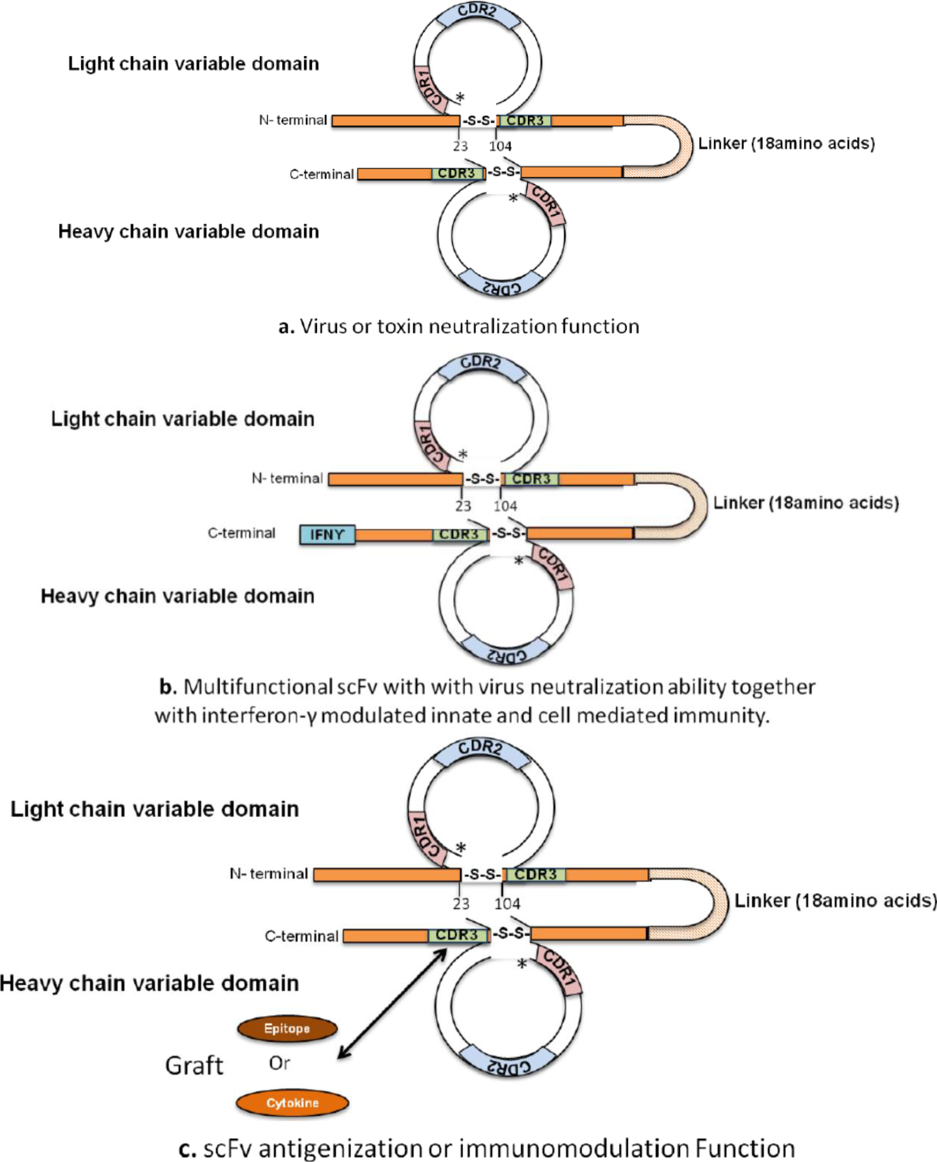
The utilization of antibodies in clinical- or immuno-diagnosis, immunomodulation and disease prevention necessitates specific design and construction of antibody fragments or their derivatives to confer a desired function; for example, virus or toxin neutralization (Figure 1a), microbial agglutination and opsonization, complement-dependent cytotoxicity (CDC) and antibody-dependent cellular cytotoxicity (ADCC). The development of such tailor-designed antibodies is feasible because specific antibody effector functions are confined to a particular structural domain or specific sequence in the modular structure of an antibody. In this context, antibody fragments (reviewed elsewhere [15,17,18,19,20]) comprising VH and VL domains linked together, referred as single chain fragment variable (scFv), are capable of generating a functional antigen-combing site that can recognize an antigen and, also, neutralize a virus or toxin. Given the small molecular size of scFvs, these have a short half-life and are rapidly cleared in vivo. This characteristic is advantageous for clinical diagnostic purposes where radioisotope labeled antibody detects antigen via gamma imaging. Alternatively, the serum half-life of the scFv may be enhanced, if desired, through chemical (e.g., PEGylation) or genetic (expression as scFV-FCGRT (FcRn) fusion protein [21]) manipulation. Similarly the opsonization and phagocytic function can be tailored into an scFv by incorporating the relevant Fc domain with the desired target sequence. An insertion of the specific Fc domain at an appropriate location in scFv will also provide ADCC function, e.g., modifications aimed at enhancing FcγRIIIA binding. Similarly, IGHG CH2 or IGHM CH3 domains could be incorporated into scFv to insert complement-binding site with an objective to confer CDC function. The antibody fragments could also be designed to survive in the hostile gut environment, where these could be made resistant to the proteolytic degradation by including the polymeric immunoglobulin receptor (PIGR). From therapeutic perspective, a variety of molecules, e.g., drugs, immuno-pharmaceuticals, toxins, cytokines, etc. can be conjugated to the desired Fc subsequent to its expression as fusion protein together with antigen binding function of scFv (Figure 1b) [17,20]. Such variations aimed at conferring a desired effector function would provide useful diagnostics and therapeutics against infectious agents [22,23,24,25], cancers [26,27,28] and immune mediated diseases [29,30].
A unique structural feature of IG V-DOMAIN is that it is composed of hypervariable regions or CDR, three each on VH and VL domains, within relatively conserved four framework regions or FR [1,31,32]. A remarkable characteristic of the FR is that these provide the required molecular microenvironment that permit the CDR to extend into solvent in an immunologically accessible manner while retaining the required configurational structural support. This provides the structural basis of IG variable domain antigenicity that could reside in the FR (public) or CDR (private), termed as idiotype. Indeed, an idiotype (id) acts as IG variable domain epitope present on H and/or L chains forming a regulatory “id-anti-id” network [33,34,35]. In this context, it is important to note that genetic imprints of all antigens present in the universe (108−1011) are enshrined within the framework of “internal mirror image” as idiotype in the antibody repertoire. Given this intrinsic characteristic of the IG CDR, these could be genetically antigenized by grafting desired T and B cell epitopes (Figure 1c) to generate immune response upon immunization [36]. Indeed, viral T and B cell epitopes [37,38], parasitic B cell epitope [33] and CD4 molecule [39] have been used to antigenize IG that induced specific immune response subsequent to immunization. Unlike shorter VH CDR3 of mouse or human IG, exceptionally long VH CDR3 (>50 codons) with multiple cysteine amino acids of bovine IG is better suited for antigenization with configurational B cell epitopes for inducing humoral immunity. The properties of selected configurational epitope grafted onto VH CDR3 of bovine scFv would confer upon it the dominant immunogenicity. Since idiotype of an IG is known to be antigenic [33,34,36,37,38,39,40,41,42], grafted epitope(s) onto the CDR will antigenize the IG fragment. Similarly, immunoregulatory molecules such as cytokines could be grafted onto a CDR (Figure 1c) with an objective to enhance their half-life in vivo and/or sustained desired regulatory functions. Given the “knob and stalk” structural configuration of the bovine VH CDR3, it would provide the molecular solvent exposed environment sustained by the FR to display desired epitopes or regulatory cytokines in an immunologically-accessible and configurationally-suitable way to achieve the desired function. The antigenized IG or its fragment variable (Fv), by desired epitope grafting, will upon immunization elicit the targeted immune response. Similarly, grafting of an immunomodulating cytokine would engage in effector function with desired enhanced half-life in vivo.
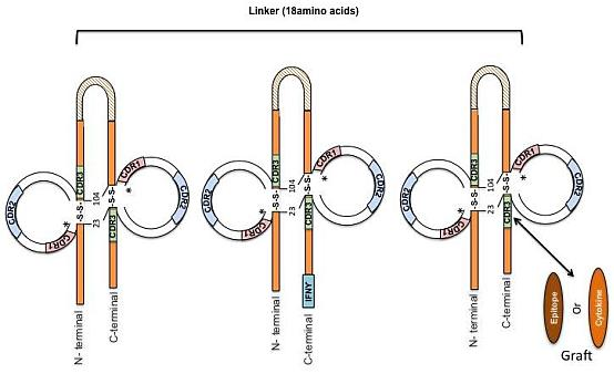
Figure 1. Diagrammatic representation of antibody fragment, scFv, design for virus or toxin neutralization function (a); expression as fusion protein with cytokine, e.g., interferon-ϒ for multiple combined functions, such as, virus neutralization, innate and cell mediated immunity against a virus (b); and epitope or cytokine grafting in the CDR3H of bovine scFv to achieve antigenization or immunomodulation function (c). * Indicates three FR amino acids before VH CDR1.
Antibodies 03 00205 g001

3. Development of Bovine Antiviral Antibodies

The development of engineered bovine antibody fragments is yet to be fully realized given its unique structural characteristics, especially the VH CDR3 architecture. A wide variety of scFvs or Fab, originating from hybridomas or phage libraries, for diagnostic and therapeutic purposes, have been constructed against various bacterial and viral pathogens using different expression systems [15]. An scFv with a (Gly4Ser)3 linker, against foot and mouth disease virus (FMDV) in cattle, was expressed in E. coli as inclusion bodies where the VH and VL were amplified from a murine monoclonal antibody, 1C7, specific for Type O FMDV. As expected, the scFv conserved the same binding specificity as the parent monoclonal antibody [43]. Other similar scFv developed against various bacterial and viral antigens for diagnosis and treatment of human diseases include anti-hepatitis B surface antigen (HBsAg), Listeria monocytogenes [44] and Mycobacterium avium subsp. paratuberculosis [45]. Single chain Fvs have been recently produced against cellular prion protein (PrPc), one of which inhibits PrPc translocation to the cell surface [46,47,48]. Engineered scFv against bovine pathogens hold a strong clinical potential in immunodiagnostics and therapeutics, however, not many of these engineered antibodies have been developed against veterinary pathogens. We previously reported the construction of scFv against bovine herpesvirus type-1 (BoHV-1) [24,25,49]. The anti-BoHV-1 scFv were produced in the Pichia pastoris yeast expression system to allow linker-influenced multimerization and glycosylation. Significant in vitro virus neutralization was achieved by both the monomeric (18 amino acid linker) and dimerized (7 amino acid linker) scFv [24], though two-fold concentration of monomeric scFv was needed for virus neutralization as compared to dimerized scFv (diabody). A single amino acid change (Asp to Gly) at position 98 according to IMGT unique numbering [31,32] in the VH FR3 of scFv affected the viral neutralization dynamics in a dose-dependent manner [49]. Further, enhancement in the potency of virus neutralization function due to non-covalent multimerization was noted in scFv with two amino acid linker (tri- or tetra-body) [25]. These mono- or multi-meric scFv produced in the Pichia pastoris had extensive glycosylation heterogeneity (discussed in [25]) but it did not affect their viral antigen recognition and neutralization function.
Earlier, phage display technology was exploited to generate bovine antibodies against a model antigen glutathione S-transferase [50]. This was followed by the development of a bovine-anti-FMDV Fab phage library from vaccinated cattle [51]. Single VH-CH1 from Fab were obtained, which showed properties of antigen binding in in vitro assays, such as ELISA, western blotting and viral neutralization. It was suggested that the absence of VL domains in these single chain Fabs did not affect their virus neutralizing function. This finding was attributed to the main role of extended VH CDR3 (24 and 27 amino acids) [51] to virus neutralization. The bovine antibodies, especially those with exceptionally long VH CDR3 [6], provide opportunities for developing novel immunodiagnostics and therapeutics against infectious diseases across various species.

4. Development of Immunomodulating Drugs and Vaccines

Recent X-ray crystal structure analysis of the largest existing antibody (BLV1H12) with an exceptional VH CDR3 size [6,8], revealed a unique “stalk and knob” structure of the antigen-combining site [16]. The knob region was shown to play a crucial role in antigen-binding and, therefore, as suggested previously [6], was exploited for generating fusion proteins with desired pharmacologic properties [52,53,54]. Since the knob region of BLV1H12 antibody constitutes the antigen-binding site, it was substituted with bovine granulocyte colony-stimulating factor 3 (CSF3; GCSF) and expressed as a stable fusion protein in mammalian expression system. The bovine antibody-bovine GCSF showed potency similar to bovine GCSF, stimulated granulocyte progenitors in dose-dependent manner, significantly increased the serum half life and sustained neutrophil populations for more than three weeks in rodent system [53]. These observations suggested that long-acting therapeutics could be developed using bovine antibody VH CDR3 for fusion of cytokines. Another recent study substituted the knob of BLV1H12 antibody by human erythropoietin protein (hEPO) [52]. The bovine antibody-hEPO fusion protein had potency comparable to the original hEPO, extended plasma half-life and sustained erythropoiesis stimulating activity in mouse model. Thus, generality of knob domain of bovine VH CDR3 for producing biologically active fusion proteins with enhanced pharmacokinetics opens opportunities for developing new generation of immunomodulating drugs. The stalk region of the bovine antibody (BLV1H12) VH CDR3 is a unique and rare protein motif that is seven-residue long solvent exposed beta-strand. Recent studies demonstrate that substitution of beta strand stalk in bovine antibody (BLV1H12) with another rigid structure with a superhelix composed of two or more alpha-helices with a repeated pattern, called coiled-coil, permits generation of functional antibody-bGCSF fusion protein [54]. This opens the opportunity for inserting these unique structures in the CDR of human or other antibodies for fusion of cytokines and growth factors. Such fusion proteins hold promise in developing new therapeutic drugs to combat a variety of infectious diseases, cancers and immune mediated diseases in humans.
Given the size (>50 codons), structural and configurational characteristics of the exceptionally long VH CDR3 in bovine antibodies, these provide suitable platform for antigenization to develop new generation of vaccines. Overall, these observations suggest that the unique structural properties of the VH CDR3 of bovine antibody can be exploited for development of novel next generation of diagnostics, therapeutics, immunomodulators and vaccines.

5. Conclusions and Future Directions

Unlike conventional antibodies where the VH CDR3 loops across species usually range from 10 to 25 amino acids, the bovine VH CDR3 can be massive in size given its extensive heterogeneity extending from 3 to 66 amino acids. The exceptionally long VH CDR3 is encoded by unusually long germline D genes (49 codons) together with insertion of CSNS specifically at the IGH V-D junction. The mechanism of insertion of “a” nucleotide rich CSNS of non-IGHV and non-IGHD origin is yet to be understood. Interestingly, such an exceptionally long VH CDR3 confers unique “knob and stalk” architecture where the knob is separated by 20 Å solvent exposed rigid stalk composed of anti-parallel beta-strands. The substitution of knob with cytokines, such as, erythropoietin and granulocyte colony stimulating factor, resulted in functional fusion proteins with enhanced pharmacokinetics. Further, the ability to replace the beta-stranded stalk with other rigid structures, for example, repeat alpha-helices to form coiled-coil, without loss of function that mimicked the beta-stranded stalk opens opportunities for insertion of this structure in the CDR of antibodies of other species. Given the versatility of such a structural architecture in bovine antibody VH CDR3, it sets new future directions for the development of novel diagnostics, therapeutics, vaccines and immunomodulating drugs.

Acknowledgments

This research was supported by NSERC Canada Discovery Grant to Azad K. Kaushik.

Author Contributions

Ashish Sachan and Surinder Saini prepared the background information while Madhuri Koti reviewed the development of engineered anti-microbial antibodies. The conceptual aspects of desgining antibody fragments for desired function was provided by Azad Kaushik.

Conflicts of Interest

The authors declare no conflict of interest.

References

  1. Lefranc, M.-P.; Lefranc, G. The Immunoglobulin Factsbook; Academic Press: San Diego, CA, USA, 2001. [Google Scholar]
  2. Saini, S.S.; Hein, W.R.; Kaushik, A. A single predominantly expressed polymorphic immunoglobulin VH gene family, related to mammalian group, I, clan, II, is identified in cattle. Mol. Immunol. 1997, 34, 641–651. [Google Scholar] [CrossRef]
  3. Pasman, Y.; Saini, S.S.; Smith, E.; Kaushik, A.K. Organization and genomic complexity of bovine lambda-light chain gene locus. Vet. Immunol. Immunopathol. 2010, 135, 306–313. [Google Scholar] [CrossRef]
  4. Pasman, Y.; Bhogal, R.; Kaushik, A.K. Novel perspective on antibody diversification from bovine immunoglobulin genetics. In Cattle: Domestication, Diseases and Environment; Liu, G., Ed.; Nova Science Publishers, Inc.: New York, NY, USA, 2013; pp. 1–23. [Google Scholar]
  5. Kaushik, A.K.; Kehrli, M.E., Jr.; Kurtz, A.; Ng, S.; Koti, M.; Shojaei, F.; Saini, S.S. Somatic hypermutations and isotype restricted exceptionally long CDR3H contribute to antibody diversification in cattle. Vet. Immunol. Immunopathol. 2009, 127, 106–113. [Google Scholar] [CrossRef]
  6. Saini, S.S.; Allore, B.; Jacobs, R.M.; Kaushik, A. Exceptionally long CDR3H region with multiple cysteine residues in functional bovine IgM antibodies. Eur. J. Immunol. 1999, 29, 2420–2426. [Google Scholar]
  7. Saini, S.S.; Farrugia, W.; Ramsland, P.A.; Kaushik, A.K. Bovine IgM antibodies with exceptionally long complementarity-determining region 3 of the heavy chain share unique structural properties conferring restricted VH + Vlambda pairings. Int. Immunol. 2003, 15, 845–853. [Google Scholar] [CrossRef]
  8. Saini, S.S.; Kaushik, A. Extensive CDR3H length heterogeneity exists in bovine fetal VDJ rearrangements. Scand.J. Immunol. 2002, 55, 140–148. [Google Scholar] [CrossRef]
  9. Shojaei, F.; Saini, S.S.; Kaushik, A.K. Unusually long germline DH genes contribute to large sized CDR3H in bovine antibodies. Mol. Immunol. 2003, 40, 61–67. [Google Scholar]
  10. Koti, M.; Kataeva, G.; Kaushik, A.K. Novel atypical nucleotide insertions specifically at VH-DH junction generate exceptionally long CDR3H in cattle antibodies. Mol. Immunol. 2010, 47, 2119–2128. [Google Scholar]
  11. Kaushik, A.; Shojaei, F.; Saini, S.S. Novel insight into antibody diversification from cattle. Vet. Immunol. Immunopathol. 2002, 87, 347–350. [Google Scholar] [CrossRef]
  12. Koti, M.; Kataeva, G.; Kaushik, A.K. Organization of D(H)-gene locus is distinct in cattle. Dev. Biol. (Basel) 2008, 132, 307–313. [Google Scholar]
  13. Larsen, P.A.; Smith, P.L. Application of circular consensus sequencing and network analysis to characterize the bovine IgG repertoire. BMC Immunol. 2012, 13, 52. [Google Scholar] [CrossRef]
  14. Walther, S.; Czerny, C.P.; Diesterbeck, U.S. Exceptionally long CDR3H are not isotype restricted in bovine immunoglobulins. PLoS One 2013, 8, e64234. [Google Scholar]
  15. Dubel, S. Handbook of Therapeutic Antibodies; Weiley-Blackwell: Weinheim, Germany, 2010; p. 1204. [Google Scholar]
  16. Wang, F.; Ekiert, D.C.; Ahmad, I.; Yu, W.; Zhang, Y.; Bazirgan, O.; Torkamani, A.; Raudsepp, T.; Mwangi, W.; Criscitiello, M.F.; et al. Reshaping antibody diversity. Cell 2013, 153, 1379–1393. [Google Scholar] [CrossRef]
  17. Beck, A.; Wurch, T.; Bailly, C.; Corvaia, N. Strategies and challenges for the next generation of therapeutic antibodies. Nat. Rev. Immunol. 2010, 10, 345–352. [Google Scholar] [CrossRef]
  18. Nelson, A.L.; Dhimolea, E.; Reichert, J.M. Development trends for human monoclonal antibody therapeutics. Nat. Rev. Drug Discov. 2010, 9, 767–774. [Google Scholar] [CrossRef]
  19. Brekke, O.H.; Sandlie, I. Therapeutic antibodies for human diseases at the dawn of the twenty-first century. Nat. Rev. Drug Discov. 2003, 2, 52–62. [Google Scholar] [CrossRef]
  20. Nelson, A.L. Antibody fragments: Hope and hype. MAbs 2010, 2, 77–83. [Google Scholar] [CrossRef]
  21. Kontermann, R.E. Strategies for extended serum half-life of protein therapeutics. Curr. Opin. Biotechnol. 2011, 22, 868–876. [Google Scholar] [CrossRef]
  22. Chen, W.; Dimitrov, D.S. Human monoclonal antibodies and engineered antibody domains as HIV-1 entry inhibitors. Curr. Opin. HIV AIDS 2009, 4, 112–117. [Google Scholar] [CrossRef]
  23. Smith, K.G.; Clatworthy, M.R. FcgammaRIIB in autoimmunity and infection: Evolutionary and therapeutic implications. Nat. Rev. Immunol. 2010, 10, 328–343. [Google Scholar] [CrossRef]
  24. Koti, M.; Farrugia, W.; Nagy, E.; Ramsland, P.A.; Kaushik, A.K. Construction of single-chain Fv with two possible CDR3H conformations but similar inter-molecular forces that neutralize bovine herpesvirus 1. Mol. Immunol. 2010, 47, 953–960. [Google Scholar]
  25. Pasman, Y.; Nagy, E.; Kaushik, A.K. Enhanced bovine herpesvirus type 1 neutralization by multimerized single-chain variable antibody fragments regardless of differential glycosylation. Clin. Vaccine Immunol. 2012, 19, 1150–1157. [Google Scholar] [CrossRef]
  26. Weiner, L.M.; Surana, R.; Wang, S. Monoclonal antibodies: Versatile platforms for cancer immunotherapy. Nat. Rev. Immunol. 2010, 10, 317–327. [Google Scholar] [CrossRef]
  27. Weiner, L.M.; Murray, J.C.; Shuptrine, C.W. Antibody-based immunotherapy of cancer. Cell 2012, 148, 1081–1084. [Google Scholar] [CrossRef]
  28. De Bruyn, M.; Bremer, E.; Helfrich, W. Antibody-based fusion proteins to target death receptors in cancer. Cancer Lett. 2013, 332, 175–183. [Google Scholar]
  29. Chan, A.C.; Carter, P.J. Therapeutic antibodies for autoimmunity and inflammation. Nat. Rev. Immunol. 2010, 10, 301–316. [Google Scholar] [CrossRef]
  30. Townsend, M.J.; Monroe, J.G.; Chan, A.C. B-cell targeted therapies in human autoimmune diseases: An updated perspective. Immunol. Rev. 2010, 237, 264–283. [Google Scholar] [CrossRef]
  31. Lefranc, M.P.; Pommié, C.; Ruiz, M.; Giudicelli, V.; Foulquier, E.; Truong, L.; Thouvenin-Contet, V.; Lefranc, G. IMGT unique numbering for immunoglobulin and T cell receptor variable domains and Ig superfamily V-like domains. Dev. Comp. Immunol. 2003, 27, 55–77. [Google Scholar]
  32. Kaas, Q.; Ehrenmann, F.; Lefranc, M.P. IG, TR and IgSF, MHC and MhcSF: What do we learn from the IMGT Colliers de Perles? Brief. Funct. Genomics Proteomics 2007, 6, 253–264. [Google Scholar]
  33. Billetta, R.; Hollingdale, M.R.; Zanetti, M. Immunogenicity of an engineered internal image antibody. Proc. Natl. Acad. Sci. USA 1991, 88, 4713–4717. [Google Scholar] [CrossRef]
  34. Zanetti, M.; Rossi, F.; Lanza, P.; Filaci, G.; Lee, R.H.; Billetta, R. Theoretical and practical aspects of antigenized antibodies. Immunol. Rev. 1992, 130, 125–150. [Google Scholar] [CrossRef]
  35. Jerne, N.K. The Nobel Lectures in Immunology. The Nobel Prize for Physiology or Medicine, 1984. The generative grammar of the immune system. Scand. J. Immunol. 1993, 38, 1–9. [Google Scholar]
  36. Zanetti, M. Antigenized antibodies. Nature 1992, 355, 476–477. [Google Scholar] [CrossRef]
  37. Brumeanu, T.D.; Bot, A.; Bona, C.A.; Dehazya, P.; Wolf, I.; Zaghouani, H. Engineering of doubly antigenized immunoglobulins expressing T and B viral epitopes. Immunotechnology 1996, 2, 85–95. [Google Scholar] [CrossRef]
  38. Musselli, C.; Daverio-Zanetti, S.; Zanetti, M. Antigenized antibodies expressing Vbeta8.2 TCR peptides immunize against rat experimental allergic encephalomyelitis. J. Immune Based Ther. Vaccines 2004, 2, 9. [Google Scholar] [CrossRef] [Green Version]
  39. Lanza, P.; Billetta, R.; Antonenko, S.; Zanetti, M. Active immunity against the CD4 receptor by using an antibody antigenized with residues 41–55 of the first extracellular domain. Proc. Natl. Acad. Sci. USA 1993, 90, 11683–11687. [Google Scholar] [CrossRef]
  40. Billetta, R.; Zanetti, M. Antigenicity and immunogenicity of antigenized antibodies. Studies on B and T cells. Int. Rev. Immunol. 1993, 10, 251–263. [Google Scholar] [CrossRef]
  41. Kumagai, Y. Epitope-grafted and antigenized antibodies can be used for versatile vaccination strategies to induce epitope-specific immune responses. J. Nippon Med. Sch. 2011, 78, 66–67. [Google Scholar] [CrossRef]
  42. Prechl, J.; Molnár, E.; Szekeres, Z.; Isaák, A.; Papp, K.; Balogh, P.; Erdei, A. Murine CR1/2 targeted antigenized single-chain antibody fragments induce transient low affinity antibodies and negatively influence an ongoing immune response. Adv. Exp. Med. Biol. 2007, 598, 214–225. [Google Scholar] [CrossRef]
  43. Cao, S.F.; Lu, P.; Suen, T.; Wang, X.; Wang, G.F. Construction, expression, purification, refold and activity assay of a specific scFv fragment against foot and mouth disease virus. Vet. Res. Commun. 2003, 27, 243–256. [Google Scholar] [CrossRef]
  44. Paoli, G.C.; Chen, C.Y.; Brewster, J.D. Single-chain Fv antibody with specificity for Listeria monocytogenes. J. Immunol. Methods 2004, 289, 147–155. [Google Scholar] [CrossRef]
  45. Berger, S.; Hinz, D.; Bannantine, J.P.; Griffin, J.F.T. Isolation of high-affinity single-chain antibodies against Mycobacterium avium subsp. paratuberculosis surface proteins from sheep with Johne’s disease. Clin. Vaccine Immunol. 2006, 13, 1022–1029. [Google Scholar]
  46. Cardinale, A.; Filesi, I.; Vetrugno, V.; Pocchiari, M.; Sy, M.S.; Biocca, S. Trapping prion protein in the endoplasmic reticulum impairs PrPC maturation and prevents PrPSc accumulation. J. Biol. Chem. 2005, 280, 685–694. [Google Scholar]
  47. Donofrio, G.; Heppner, F.L.; Polymenidou, M.; Musahl, C.; Aguzzi, A. Paracrine inhibition of prion propagation by anti-PrP single-chain Fv miniantibodies. J. Virol. 2005, 79, 8330–8338. [Google Scholar] [CrossRef]
  48. Padiolleau-Lefevre, S.; Alexandrenne, C.; Dkhissi, F.; Clement, G.; Essono, S.; Blache, C.; Couraud, J.Y.; Wijkhuisen, A.; Boquet, D. Expression and detection strategies for an scFv fragment retaining the same high affinity than Fab and whole antibody: Implications for therapeutic use in prion diseases. Mol. Immunol. 2007, 44, 1888–1896. [Google Scholar] [CrossRef]
  49. Koti, M.; Nagy, E.; Kaushik, A.K. A single point mutation in framework region 3 of heavy chain affects viral neutralization dynamics of single-chain Fv against bovine herpes virus type 1. Vaccine 2011, 29, 7905–7912. [Google Scholar] [CrossRef]
  50. O’Brien, P.M.; Aitken, R.; O’Neil, B.W.; Campo, M.S. Generation of native bovine mAbs by phage display. Proc. Natl. Acad. Sci. USA 1999, 96, 640–645. [Google Scholar]
  51. Kim, Y.J.; Lebreton, F.; Kaiser, C.; Crucière, C.; Rémond, M. Isolation of foot-and-mouth disease virus specific bovine antibody fragments from phage display libraries. J. Immunol. Methods 2004, 286, 155–166. [Google Scholar] [CrossRef]
  52. Zhang, Y.; Wang, D.-L.; Welzel, G.; Wang, Y.; Schultz, P.G.; Wang, F. An antibody CDR3-erythropoietin fusion protein. ACS Chem. Biol. 2013, 8, 2117–2121. [Google Scholar] [CrossRef]
  53. Zhang, Y.; Wang, D.; de Lichtervelde, L.; Sun, S.B.; Smider, V.V.; Schultz, P.G.; Wang, F. Functional antibody CDR3 fusion proteins with enhanced pharmacological properties. Angew. Chem. Int. Ed. Engl. 2013, 52, 8295–8298. [Google Scholar]
  54. Zhang, Y.; Goswami, D.; Wang, D.; Wang, T.-S.A.; Sen, S.; Magliery, T.J.; Griffin, P.R.; Schultz, P.G.; Wang, F. An antibody with a variable-region coiled-coil “knob” domain. Angew. Chem. Int.Ed. Engl. 2014, 53, 132–135. [Google Scholar] [CrossRef]