Article

Microbiological Risk Assessment of Drinking Water Using Integrated Phenotypic and Molecular Approaches in Guaranda

by
Darwin Alberto Núñez Torres
1,*,
E. Fabián Rivera
1,
Stefani Vanesa Vega Reinel
2 and
José Luis Altuna Vásquez
1
1
Carrera de Ingeniería Agroindustrial, Universidad Estatal de Bolivar, Guaranda 020150, Ecuador
2
Carrera de Ingeniería Agronómica, Universidad Estatal de Bolivar, Guaranda 020150, Ecuador
*
Author to whom correspondence should be addressed.
Water 2026, 18(12), 1491; https://doi.org/10.3390/w18121491
Submission received: 22 April 2026 / Revised: 28 May 2026 / Accepted: 5 June 2026 / Published: 17 June 2026
(This article belongs to the Special Issue Drinking Water Quality: Monitoring, Assessment and Management)

Abstract

This study evaluates the microbiological quality of drinking water in the urban area of Guaranda through an integrated approach combining culture-based methods, biochemical characterization, and polymerase chain reaction (PCR) analysis. A total of 50 drinking water samples were collected from strategically selected points within the urban distribution system following Ecuadorian technical standards. Microbiological analyses included the detection of total and fecal coliforms, as well as the isolation and identification of Escherichia coli O157:H7, Salmonella spp., and Listeria monocytogenes. Culture-based analyses revealed that 22% of samples were positive for total coliforms and 4% for fecal coliforms. In selective culture media, contamination rates reached 18% for E. coli O157:H7, 8% for Salmonella spp., and 46% for Listeria monocytogenes. However, biochemical profiling showed substantial inconsistencies with the expected phenotypic characteristics of these pathogens, particularly in oxidase and citrate tests, suggesting possible false-positive identifications in complex environmental matrices. PCR assays confirmed lower detection frequencies, identifying E. coli O157:H7 and Salmonella spp. in 2% of samples each, and Listeria monocytogenes in 10% of samples. Agarose gel electrophoresis validated the amplification of specific DNA fragments of 212 bp, 244 bp, and 388 bp, respectively. The findings demonstrate significant discrepancies between conventional phenotypic methods and molecular techniques, highlighting the limitations of culture-based identification when used alone. This study emphasizes the importance of integrating molecular diagnostics into routine water quality monitoring programs to improve the reliability of pathogen detection and support more effective public health risk management in urban drinking water systems.

1. Introduction

The quality of water intended for human consumption constitutes one of the primary determinants of public health, environmental sustainability, and socioeconomic development, particularly in developing countries where water supply and distribution systems often present structural and operational limitations [1,2]. At the global level, the World Health Organization estimates that millions of people continue to consume microbiologically unsafe water, especially in regions with limited infrastructure, aging distribution networks, or insufficient disinfection systems. In this context, microbiological contamination remains one of the most critical threats to water security, as it facilitates the transmission of gastrointestinal diseases, systemic infections, and epidemic outbreaks associated with pathogenic microorganisms of fecal origin [3]. The presence of total and fecal coliforms continues to serve as the principal indicator of microbiological deterioration in drinking water, while the detection of specific pathogens is particularly relevant due to their direct association with severe clinical diseases [4]. Among the main microorganisms of sanitary concern are Escherichia coli, Salmonella spp., and Listeria monocytogenes, whose presence in drinking water indicates contamination, deficiencies in water treatment processes, and vulnerabilities within distribution systems.
At the global level, the World Health Organization estimates that millions of people consume microbiologically unsafe water, particularly in regions with limited infrastructure, aging distribution networks, or insufficient disinfection systems [1]. The presence of total and fecal coliforms continues to be the primary indicator of microbiological deterioration in water, while the detection of specific pathogens is of particular importance due to their direct association with severe clinical diseases [5]. Specifically, Escherichia coli O157:H7 has been associated with hemorrhagic colitis and hemolytic uremic syndrome; Salmonella spp. with gastroenteritis, enteric fever, and septicemia; and Listeria monocytogenes with listeriosis, a potentially life-threatening disease affecting pregnant women, elderly individuals, and immunocompromised populations [6].
Traditionally, microbiological water quality assessment has relied on culture-based methods using selective and differential media, complemented by biochemical tests for the phenotypic identification of isolated microorganisms. Media such as Eosin Methylene Blue (EMB) agar are commonly used for the selective isolation of Escherichia coli and other coliform bacteria, while Xylose Lysine Deoxycholate (XLD) agar is widely employed for the detection of Salmonella spp. due to its selective properties for enteric pathogens. Likewise, Agar Listeria according to Ottaviani and Agosti (ALOA) is frequently used for the selective isolation and differentiation of Listeria monocytogenes based on enzymatic activity. In addition, biochemical assays including catalase, oxidase, and IMViC tests constitute widely used tools because of their low cost and relative ease of implementation [7]. However, numerous studies have demonstrated that conventional methods present important limitations in sensitivity and specificity when applied to complex environmental samples.
In recent years, the development of molecular techniques has overcome many of these limitations. Among them, polymerase chain reaction (PCR) has emerged as a highly sensitive, specific, and rapid tool for the detection of pathogens in drinking water [8]. PCR enables the identification of specific genetic sequences even when microorganisms are present in low concentrations or exhibit reduced viability, significantly reducing the time required for microbiological diagnosis. Consequently, the integration of culture techniques, biochemical characterization, and molecular analysis currently represents one of the most robust approaches for monitoring microbiological water quality [9].
In Ecuador, national technical regulations, particularly the NTE INEN 1108 standard [10], establish that water intended for human consumption must be free of fecal coliforms, Escherichia coli, and other pathogenic microorganisms. However, several studies conducted in Andean regions of the country have reported the persistence of microbiological contamination in both urban and rural water supply systems, attributed to deficiencies in water capture, storage, disinfection, and distribution processes [11]. These issues are especially relevant in inter-Andean cities, where topography, aging hydraulic infrastructure, and rapid urban growth increase the risk of infiltration and secondary contamination [12].
Despite the implementation of microbiological monitoring programs in many urban drinking water systems, several regions in developing countries still present limited information regarding the occurrence of specific waterborne pathogens and the reliability of conventional detection methods. Most previous studies have primarily focused on traditional microbiological indicators without incorporating molecular approaches capable of confirming pathogen presence and evaluating the diagnostic accuracy of phenotypic identification techniques. This limitation restricts the ability to accurately assess microbiological risks and may lead to underestimation or misinterpretation of contamination events in urban water distribution systems.
In this context, the present study aims to evaluate the microbiological quality of drinking water in the urban area of Guaranda through an integrated approach based on culture techniques, biochemical characterization, and molecular testing using PCR. Specifically, this study seeks to determine the presence of total and fecal coliforms, as well as to identify Escherichia coli O157:H7, Salmonella spp., and Listeria monocytogenes at different points within the distribution system. Additionally, it aims to compare the diagnostic performance of conventional and molecular methods in order to establish their strengths, limitations, and complementarity. The results obtained will provide relevant scientific evidence for water quality monitoring, health risk management, and the design of more precise and effective surveillance strategies in urban water supply systems.

2. Related Work

The study of microbiological water quality has evolved significantly over recent decades, transitioning from traditional culture-based approaches to more sensitive and specific molecular methodologies. In this context, one of the earliest relevant advances corresponds to the development of PCR-based detection protocols for enteric pathogens in surface water. In particular, study [13] proposed a method based on a single enrichment step followed by PCR, achieving detection limits below 3 CFU/L for Escherichia coli O157:H7 and Salmonella spp., demonstrating high analytical sensitivity. However, this study also revealed an important limitation: the detection of virulence genes in the absence of the target microorganism, suggesting potential interference from other bacteria harboring such genes. Complementarily, studies such as [14] introduced a risk-based approach by integrating PCR data with human exposure models, demonstrating that molecular detection alone does not guarantee an accurate assessment of health risk due to factors such as DNA inhibitors and limitations in filtration processes.
Subsequently, several studies applied comparative approaches between conventional and molecular methods across different water matrices. In this line, studies [15,16] evaluated microbiological quality in bottled and tap water, showing that PCR detects a higher proportion of pathogens compared to culture-based methods. For instance, study reported a significant difference in E. coli detection (26.38% by PCR versus 7.58% by culture), confirming the higher sensitivity of molecular techniques. Nevertheless, both studies highlight that PCR may overestimate pathogen presence due to the detection of DNA from non-viable cells, which constitutes a major limitation in terms of sanitary interpretation.
In the context of environmental and production systems, study [17] reported high levels of microbiological contamination in water used in dairy farming, including elevated concentrations of coliforms, Salmonella, and E. coli, as well as the detection of parasites using real-time PCR. These findings underscore the complexity of environmental matrices and the need for robust analytical methods. Similarly, study [18] evaluated the detection of Salmonella enterica and Listeria monocytogenes in irrigation water using qPCR and culture methods, demonstrating that factors such as water type, seasonality, and sample volume significantly influence sensitivity and agreement between methods. This study highlights a critical limitation: variability in methodological accuracy depending on environmental conditions.
At a more local scale, study [19] conducted in the canton of Chambo reported high levels of microbiological contamination in drinking water, with prevalence rates of up to 80% for E. coli O157:H7 and 40% for Listeria monocytogenes. These results are particularly relevant as they reveal similar issues in geographically comparable contexts. However, the study presents methodological limitations, as it does not extensively address validation through multiple complementary techniques nor the analysis of inconsistencies between methods.
On the other hand, more recent research has explored advanced technologies for microbiological water monitoring. Study [20] compared traditional methods with rapid techniques such as flow cytometry and luminometry, demonstrating the need for faster tools to support real-time decision-making. Likewise, study [21] incorporated concentration techniques and 16S rRNA gene sequencing alongside digital PCR, revealing changes in microbial communities and the presence of potentially pathogenic bacteria influenced by anthropogenic activities. These approaches represent significant progress, although their application remains limited due to cost and technical complexity.
Additionally, study [22] addressed the spatial and temporal variability of E. coli in irrigation water, demonstrating that factors such as sampling depth and timing significantly affect bacterial concentration. This finding highlights a recurrent limitation in water quality studies: the lack of standardization in sampling designs, which can compromise the comparability of results.
Finally, the review study [23] synthesizes recent advances in molecular tools for microbiological water quality assessment, highlighting the growing application of quantitative PCR (qPCR), droplet digital PCR (ddPCR), and next-generation sequencing (NGS). Quantitative PCR enables real-time amplification and quantification of pathogen-specific DNA, offering high sensitivity and reduced analysis time compared with conventional PCR. In contrast, ddPCR provides absolute quantification of nucleic acids through sample partitioning into thousands of microdroplets, significantly improving detection accuracy in low-concentration environmental samples and reducing susceptibility to amplification inhibitors. Additionally, next-generation sequencing technologies allow comprehensive characterization of microbial communities by simultaneously analyzing large numbers of DNA sequences, enabling the identification of pathogenic and non-culturable microorganisms as well as shifts in microbial diversity associated with anthropogenic contamination. Although these molecular approaches have substantially improved pathogen detection and environmental microbiological surveillance, important challenges remain regarding standardization, cost, data interpretation, and integration with conventional microbiological methods.
Overall, the reviewed studies demonstrate significant advances in pathogen detection in water, but also reveal persistent limitations: (i) discrepancies between culture-based and molecular methods, (ii) challenges in the interpretation of molecular results, (iii) the influence of environmental factors on detection, and (iv) the lack of integrated approaches that systematically combine multiple techniques. In this context, the present study addresses these gaps by integrating culture-based methods, biochemical testing, and PCR, enabling not only pathogen detection but also a critical evaluation of the reliability of each technique under real conditions of an urban water supply system. This approach contributes to strengthening microbiological monitoring systems and improving decision-making in water quality management.

3. Materials and Methods

3.1. Study Area

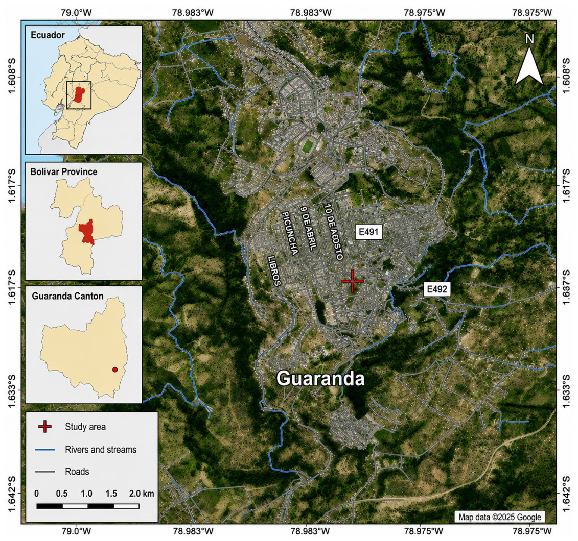
Guaranda, the capital of Bolívar Province, is located in the central-western region of Ecuador within the western Andean mountain range (1.5912° S, 78.9990° W) shown in Figure 1.

3.2. Sampling

Following the recommendations established in the Ecuadorian Technical Standard NTE INEN 1108, which specifies the collection of 12 samples for every 5000 consumers, a total of 50 drinking water samples were collected from strategically selected points within the urban area of Guaranda, Ecuador. It is important to note that the sampling points were selected based on the five main drinking water distribution routes established by the Municipal Public Water Supply and Sewerage Company of Guaranda (EP-EMAP-G), ensuring representative spatial coverage of the urban distribution network. Consequently, 10 samples were collected from each distribution route using a spatially distributed sampling design. The collection, preservation, storage, and transportation of the samples were carried out in accordance with NTE INEN 2169:2013 to maintain their physicochemical and microbiological integrity until processing at the laboratories of the Research Department of the Universidad Estatal de Bolívar. Microbiological analyses were performed in triplicate using selective and differential culture media for the detection of Escherichia coli, Listeria monocytogenes, Salmonella spp., as well as total and fecal coliforms. Analytical procedures were conducted in accordance with NTE INEN 1105:1983, and the results were interpreted based on the microbiological quality limits established in NTE INEN 1108:2011.

3.3. Microbiological Analysis Method

For the detection of total and fecal coliforms, Compact Dry plates were inoculated with 10 µL of each water sample, allowing homogeneous distribution by capillary action, and subsequently incubated at 35–37 °C for 24 h. Microbial identification was based on colony coloration, where pink to red colonies indicated total coliforms, while blue to purple colonies indicated fecal coliforms. Following microbiological analysis, all culture plates were sterilized by autoclaving in accordance with established biosafety regulations [24].

3.3.1. Preparation of Culture Media

The preparation of ALOA, EMB, and XLD culture media was carried out under aseptic laboratory conditions following the manufacturer’s recommendations to ensure the accuracy and reproducibility of the microbiological analyses. ALOA Listeria Agar (Titan Biotech Ltd., A-902 A, RIICO Industrial Area, Phase-III, Bhiwadi-301019, Rajasthad, India; Lot No. M1EE3BZ01) was prepared by dissolving 36.02 g of dehydrated medium in 460 mL of type II distilled water and sterilized by autoclaving at 121 °C and 15 psi for 15 min. This medium was used for the selective isolation and differentiation of Listeria spp. Likewise, Eosin Methylene Blue (EMB) Agar (Titan Biotech Ltd., A-902 A, RIICO Industrial Area, Phase-III, Bhiwadi-301019, Rajasthad, India; Lot No. M3D7AY01) was prepared by dissolving 51.55 g of dehydrated medium in 1000 mL of distilled water and sterilized under the same conditions for the detection and differentiation of coliform bacteria and Escherichia coli. In contrast, Xylose Lysine Deoxycholate (XLD) Agar (Neogen, 602 Lesher Place, Lansing, MI 48912, USA; Lot No. 108350B) was prepared at a concentration of 55 g/L and heated with constant agitation until complete dissolution was achieved. Due to the presence of heat-sensitive selective compounds, the XLD medium was not autoclaved in order to preserve its selective and differential properties for enteric pathogen detection. After preparation, approximately 15–20 mL of each culture medium was aseptically poured into sterile Petri dishes inside a biosafety cabinet and allowed to solidify prior to microbiological analyses [9].

3.3.2. Membrane Filtration

Bacterial concentration was performed using membrane filtration under vacuum, processing 150 mL of each sample through sterile cellulose ester membranes, where microorganisms were retained. Under aseptic conditions, each membrane was transferred using flame-sterilized forceps onto selective culture media in Petri dishes, which were then sealed, labeled, and incubated at 35 °C ± 0.5 °C for 48 h to promote the growth and differentiation of colony-forming units (CFU). The procedure was conducted in triplicate to ensure reproducibility and reliability of the results [25].

3.4. Biochemical Tests Method

3.4.1. Oxidase Test

An oxidase reagent strip was used, onto which a bacterial colony was placed. A color change to blue or purple indicated a positive result, confirming the presence of cytochrome c oxidase. The absence of color change indicated a negative result [12].

3.4.2. Catalase Test

A drop of 3% hydrogen peroxide (H2O2) was placed on a glass slide, and a bacterial colony was added using a sterile loop. The formation of bubbles indicated a positive result, confirming the presence of the catalase enzyme. The absence of effervescence indicated a negative result [3,26].

3.4.3. IMViC Tests

The methyl red test was performed by inoculating a bacterial colony into 5 mL of MR-VP broth using a sterile inoculation loop, followed by gentle homogenization and incubation at 35 °C for 24 h. After incubation, three drops of methyl red reagent were added to each tube. The development of a red coloration was interpreted as a positive result, indicating the production of stable acidic metabolites derived from glucose fermentation, whereas a yellow coloration indicated a negative reaction [8].
The Voges–Proskauer test was conducted by inoculating a colony into 5 mL of MR-VP broth, followed by incubation at 35 °C for 24 h. After incubation, 12 drops of α-naphthol and 4 drops of KOH were added. The development of a red color indicated a positive result, confirming the production of acetoin, while no color change indicated a negative result.
The citrate test was performed using medium prepared in type II distilled water, sterilized in an autoclave, and distributed into slanted tubes. A colony was inoculated and incubated at 35 °C for 24 h. A color change from green to blue indicated a positive result, confirming citrate utilization as the sole carbon source, whereas no color change indicated a negative result.

3.5. DNA Extraction

Bacterial DNA extraction was performed using a commercial Invitrogen™ kit (Invitrogen™, Thermo Fisher Scientific, Waltham, MA, USA; No. K1820-00), following the manufacturer’s specifications. Briefly, bacterial cells were recovered by centrifugation and subjected to enzymatic lysis to release genomic DNA. Subsequently, the extracted DNA was purified through organic phase separation, alcohol precipitation, and sequential washing steps prior to final resuspension in a preservation buffer. DNA concentration and purity were assessed using a NanoDrop spectrophotometer. Only samples exhibiting an A260/A280 absorbance ratio between 1.8 and 2.0 were considered suitable for molecular analyses and PCR amplification. For agarose gel analysis, the base pair sizes indicated in Table 1 were considered.

3.6. Master Mix Preparation

For DNA amplification by PCR, a reaction mixture was prepared including thermostable GoTaq DNA polymerase, forward and reverse primers specific to the target region, dNTPs, reaction buffer with magnesium, and nuclease-free water to adjust the final volume. Once the DNA template was added at an appropriate concentration, the mixture was distributed into PCR tubes and processed in a thermocycler under defined temperature cycles to ensure efficient and specific amplification. As shown in Table 2.

3.7. Conventional PCR

DNA amplification was performed using a thermocycler under cyclic conditions comprising three sequential stages: initial denaturation to separate double-stranded DNA, primer annealing based on complementary base pairing, and extension at 72 °C, during which DNA polymerase synthesized new complementary strands. Successive amplification cycles enabled the exponential replication of the target DNA fragment, generating sufficient genetic material for subsequent molecular analyses. As shown in Table 3.

3.8. Agarose Gel Electrophoresis

PCR products were analyzed by electrophoresis on 1.5% agarose gels prepared in TAE buffer and stained with Cyber Safe nucleic acid dye prior to gel solidification. Amplified products were loaded into the gel wells alongside a molecular weight marker (Blue Orange), including both positive and negative controls to validate amplification specificity. Electrophoretic separation was performed under a constant electric field, allowing DNA fragments to migrate according to their molecular size. Subsequently, amplified bands were visualized using a UV transillumination gel documentation system, enabling the identification of pathogen-specific amplification products based on their expected fragment sizes.

4. Results and Discussion

4.1. Georeferencing

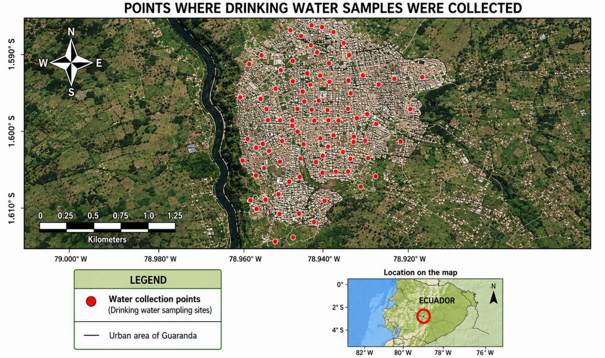
The water collection points considered in this study are presented in Figure 2.
Figure 2 presents the georeferenced water collection points identified using a Garmin GPS receiver, which enabled the precise recording of the geographic coordinates of each sampling site and supported the development of a detailed digital map of the urban area of Guaranda, Ecuador.
Table 4 summarizes the georeferenced sampling locations and collection information for the drinking water samples obtained from different sectors of the urban distribution system of Guaranda. The table includes the sampling code, collection site, sampling time, and associated geographic information used for spatial analysis and microbiological assessment.

4.2. Microbiological Analysis Results

4.2.1. Identification of Total and Fecal Coliforms

The results presented in Table 5 show that, out of 50 drinking water samples analyzed, 8 were positive for total coliforms and 2 for fecal coliforms, corresponding to 22% and 4% contamination, respectively. These findings indicate the presence of microbiological contamination in specific sampling routes, particularly routes 18 and 22.
Overall, the detection of total coliforms in 22% of samples reflects microbiological deterioration in several areas of the urban distribution system. These results are consistent with recent studies that identify coliforms as reliable indicators of fecal contamination in water, representing a potential public health risk [27].
In addition, the Compact Dry method used in this study is a rapid microbiological technique that has been internationally validated and certified under the official AOAC 110401 method for the detection and quantification of total coliforms, fecal coliforms, and Escherichia coli [27]. Several studies have demonstrated that this methodology exhibits a high analytical correlation with ISO reference methods, achieving correlation coefficients greater than r2 = 0.93 for the enumeration of coliforms and E. coli, indicating high levels of sensitivity, specificity, and analytical reproducibility [28]. In this context, the detection of total and fecal coliforms using Compact Dry plates confirmed the presence of microbiological contamination at several of the sampling points evaluated, in accordance with the microbiological quality criteria established in the Ecuadorian technical standard NTE INEN 1108 for drinking water.

4.2.2. Identification of Escherichia coli, Salmonella spp., and Listeria monocytogenes

The culture-based microbiological analysis revealed the presence of pathogenic bacteria in several drinking water samples collected from the urban distribution system of Guaranda. As shown in Table 6, Listeria monocytogenes exhibited the highest detection frequency, being identified in 23 out of 50 samples (46%), followed by Escherichia coli in 9 samples (18%) and Salmonella spp. in 4 samples (8%). The predominance of Listeria monocytogenes suggests substantial microbiological deterioration within portions of the distribution system and indicates the potential persistence of environmental pathogens under unfavorable sanitary conditions.

4.3. Biochemical Tests

Biochemical tests were performed on contaminated samples to confirm the identity of isolated microorganisms.

Results for Escherichia coli

The biochemical and IMViC test results applied to the nine contaminated samples revealed a heterogeneous microbiological profile, with partially compatible responses for E. coli. Catalase (55.56%), methyl red (66.67%), and indole (44.44%) showed moderate positivity, consistent with the expected phenotypic behavior of this species. However, oxidase (88.89%) and citrate (77.78%) tests yielded positive results, which are contradictory since E. coli is typically negative for both tests. These inconsistencies suggest the possible presence of non-target microorganisms, potentially belonging to genera outside the Enterobacteriaceae family. This highlights a key limitation of conventional phenotypic methods: their susceptibility to ambiguous identification when applied to complex environmental samples, reinforcing the need for molecular confirmation using PCR. As shown in Table 7.
The biochemical and IMViC results for the four samples indicated in Table 8 showed notable inconsistencies relative to the expected profile of Salmonella. While citrate positivity (100%) aligns with its metabolic characteristics and catalase showed partial agreement (50%), methyl red positivity was low (25%), and both indole (25%) and Voges–Proskauer (50%) tests showed unexpected positive results, despite being typically negative for this genus. Most critically, oxidase was positive in 100% of samples, which is incompatible with Salmonella spp., as it lacks cytochrome c oxidase. These findings suggest possible cross-contamination, degraded reagents, or the presence of oxidase-positive microorganisms such as Pseudomonas spp. in mixed cultures. Overall, these results demonstrate that phenotypic methods alone are insufficient for reliable identification, making PCR confirmation essential.
The biochemical results for the 23 samples showed a mixed profile combining expected and inconsistent responses. Positive results for catalase (47.83%), methyl red (52.17%), and Voges–Proskauer (39.13%) are consistent with known metabolic behavior, although moderate percentages reflect phenotypic variability among isolates. However, indole (47.83%), citrate (69.57%), and oxidase (56.52%) showed unexpected positivity, contradicting the established biochemical profile of Listeria monocytogenes, which is typically negative for these tests as shown in Table 9. These inconsistencies suggest sample contamination, methodological errors, or the coexistence of multiple microbial species. Under these conditions, molecular confirmation through PCR becomes essential to ensure reliable identification.

4.4. PCR Results for Molecular Identification

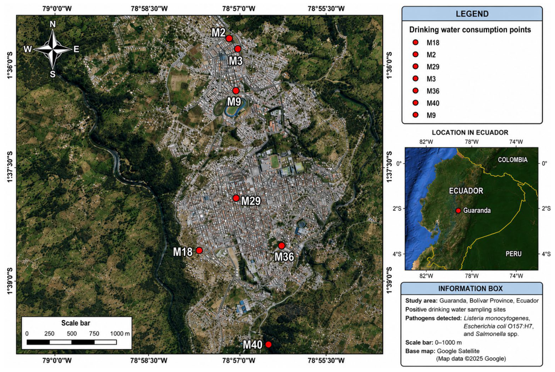
PCR analysis was performed on selected contaminated samples, including those positive for Escherichia coli O157:H7, Salmonella spp., and Listeria monocytogenes. The selected sampling points were M2, M3, M9, M18, M29, M36, and M40, as shown in Figure 3.
As shown in Table 10, the results revealed the presence of Escherichia coli (2%), Salmonella spp. (2%), and Listeria monocytogenes (10%) among the analyzed samples. These values were significantly lower than those obtained through conventional culture-based microbiological methods, suggesting possible overestimation associated with phenotypic analyses. The results were validated through comparison with characteristic biochemical profiles, allowing confirmation of the detected pathogens. However, one of the main limitations of the study is related to the sampling period and sample size, since seasonal variations associated with factors such as precipitation, surface runoff, and contaminant transport may significantly influence the microbiological load of water sources. In addition, the possible presence of viable but non-culturable bacteria (VBNC) could limit microbiological detection using conventional methods. Therefore, future studies should incorporate multi-temporal monitoring during dry and rainy seasons, increase the number of samples analyzed, and complement the evaluation using quantitative PCR (qPCR), genetic sequencing, and the assessment of additional pathogenic Escherichia coli serotypes, including ETEC, EPEC, EIEC, EAEC, and STEC, in order to strengthen the microbiological risk assessment associated with water consumption.

4.5. Agarose Gel Electrophoresis Results

PCR results were further confirmed through agarose gel electrophoresis, allowing visualization of amplified DNA fragments.

4.5.1. Detection of Escherichia coli O157:H7

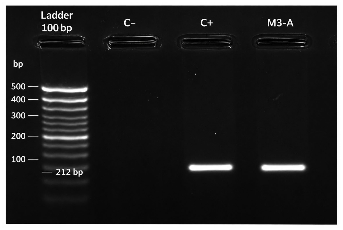
Figure 4 shows the agarose gel electrophoresis corresponding to the PCR performed for the detection of E. coli O157:H7. For amplification, Invitrogen primers were used with the following sequences: E. coli (F, 5′-CCAGGCAAAGAGTTTATGTTGA-3′) and E. coli (R, 5′-GCTATTTCCTGCCGATAAGAGA-3′), which target the DNA of the E. coli genus, amplifying fragments of approximately 212 base pairs (bp).
In the post-PCR gel, a clear DNA band of approximately 212 bp is observed in the lane corresponding to sample M3-A, identified as E. coli (serotype O157:H7). This band exhibits migration similar to that of the positive control (C+), while the negative control (C−) shows no evidence of amplification, thereby validating the specificity of the PCR assay.
This banding pattern confirms the presence of the specific E. coli DNA sequence in sample M3-A, which is interpreted as a positive result for the detection of this microorganism using PCR. Through PCR analysis and agarose gel electrophoresis, and following visualization under ultraviolet light using a gel documentation system, it was determined that the isolates correspond to E. coli O157:H7, using specific positive controls for this serotype as a reference.
This finding is consistent with recent studies conducted in Ecuador that report similar issues in water quality. For example, in Airón (Chimborazo), the presence of Escherichia coli O157:H7 has been identified in water systems, reflecting a serious problem of microbiological contamination in water intended for human consumption [27]. This type of contamination is particularly concerning, as pathogenic strains such as E. coli O157:H7 can cause severe gastrointestinal diseases, especially in vulnerable populations.

4.5.2. Detection of Salmonella spp. By Agarose Gel Electrophoresis

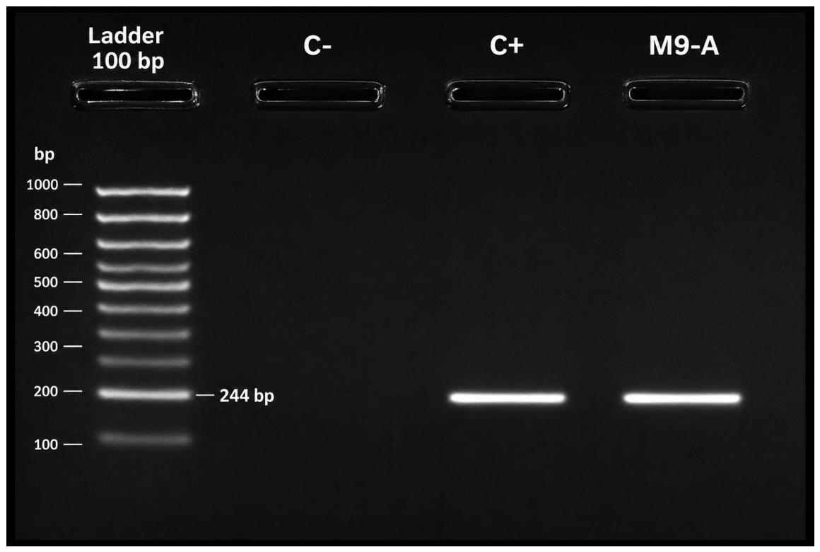
Figure 5 shows the agarose gel electrophoresis corresponding to the PCR performed for the detection of Salmonella spp. For amplification, Invitrogen primers were used with the following sequences: invaA3R (R, 5′-TCCATCAAATTAGCGGAGGC-3′) and inva3F (F, 5′-AACGTGTTTCCGTCGTAAT-3′), which target a DNA sequence specific to the Salmonella genus, amplifying fragments of approximately 244 base pairs (bp).
In the post-PCR gel, a DNA band of approximately 244 bp was observed in the lane corresponding to sample M9-A, previously identified as Salmonella. This band exhibited migration similar to that of the positive control (C+), indicating a positive result for the presence of this pathogen.
The negative control (C−) showed no bands, confirming the absence of contamination and the specificity of the amplification. In sample M9-A, a distinct band corresponding to Salmonella spp. was observed, validating its presence through PCR. The electrophoretic analysis, together with the use of positive controls, confirmed the molecular identification of the species.

4.5.3. Detection of Salmonella spp. And Listeria monocytogenes by Agarose Gel Electrophoresis

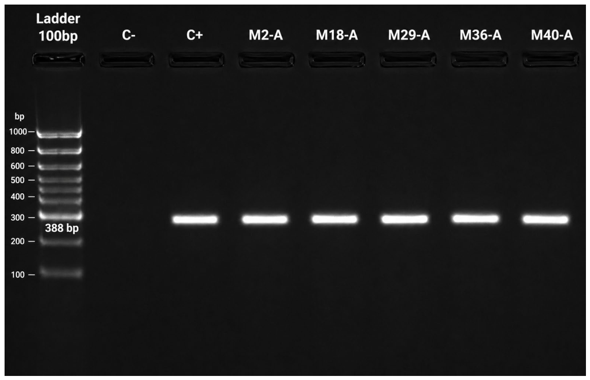
Figure 6 shows the agarose gel electrophoresis corresponding to the PCR performed for the detection of Listeria monocytogenes. For amplification, Invitrogen primers were used with the following sequences: Listeria (F, 5′-GAATGTAAACTTCGGCGCAATCAG-3′) and Listeria (R, 5′-GCCGTCGATGATTTGAACTTCATC-3′), which target DNA sequences of the Listeria genus and amplify fragments of approximately 388 base pairs (bp).
In the post-PCR agarose gel, DNA bands of approximately 388 bp were observed in the lanes corresponding to samples M2-A, M8-A, M29-A, M36-A, and M40-A. These bands exhibited migration patterns similar to that of the positive control (C+), while the negative control (C−) showed no bands, thereby validating the specificity of the amplification process.
Through PCR analysis and agarose gel electrophoresis, and after visualization under ultraviolet light using a gel documentation system, it was determined that the isolates correspond to Listeria monocytogenes, using specific positive controls as reference. This banding pattern confirms the successful amplification of the specific DNA fragment of Listeria monocytogenes in the five samples analyzed, which is interpreted as a positive result for the detection of this bacterium by PCR.
The confirmation of Listeria monocytogenes in the analyzed water samples reflects a high level of microbiological contamination, considering that this bacterium can cause listeriosis, a severe disease particularly affecting immunocompromised individuals, pregnant women, and the elderly. In a study conducted in the canton of Chambo, Listeria monocytogenes was detected in 40% of drinking water samples, indicating that this issue is not isolated. Furthermore, this bacterium is capable of surviving under extreme conditions, such as low temperatures and nutrient-limited environments, facilitating its persistence within water distribution systems.
The discrepancies observed between biochemical assays and PCR results highlight the limitations of conventional phenotypic identification when applied to complex environmental water matrices. While selective culture media and biochemical profiling suggested relatively high contamination rates for Escherichia coli, Salmonella spp., and Listeria monocytogenes, molecular confirmation by PCR revealed substantially lower detection frequencies. This divergence may be associated with the coexistence of heterogeneous microbial populations capable of producing overlapping biochemical responses, particularly in oxidase and citrate assays, which generated atypical positive reactions for microorganisms that are conventionally negative for these tests. Such findings suggest the presence of mixed bacterial communities or non-target environmental microorganisms interfering with phenotypic characterization.
Furthermore, the occurrence of total and fecal coliforms in contaminated samples indicates a broader deterioration of microbiological water quality, potentially favoring the persistence and proliferation of opportunistic and pathogenic microorganisms within the distribution system. The higher prevalence detected through culture-based methods may also reflect the detection of viable but non-specific microorganisms sharing similar colony morphology on selective media. In contrast, PCR demonstrated greater specificity by targeting pathogen-specific genetic sequences, reducing the probability of false-positive identifications. These results emphasize that microbial abundance in environmental water systems can directly influence the reliability of biochemical assays, particularly under conditions of high microbial diversity and mixed contamination.
Although conventional PCR is not a recent methodology, several studies have demonstrated that this technique exhibits sensitivity and specificity levels exceeding 90% for the detection of pathogenic microorganisms in environmental and water samples [29,30]. In the present study, conventional PCR allowed the molecular confirmation of Escherichia coli in 1 out of 50 analyzed samples (2%), Salmonella spp. in 1 sample (2%), and Listeria monocytogenes in 5 samples (10%). These results were lower than those obtained through conventional phenotypic and biochemical methods, highlighting the higher reliability and specificity of PCR in reducing possible false-positive identifications associated with microorganisms presenting similar biochemical responses [31]. Furthermore, previous studies have reported that the integration of molecular and microbiological techniques can increase diagnostic accuracy by up to 30% compared to methods exclusively based on culture techniques [32,33]. In this context, visualization of amplified products through agarose gel electrophoresis provided robust molecular evidence confirming the presence of pathogens of sanitary importance and strengthened the microbiological risk assessment associated with water consumption.
The results obtained revealed important differences between conventional microbiological methods and molecular confirmation by PCR for the detection of pathogenic microorganisms in the analyzed drinking water samples. These findings are consistent with those reported by Khant et al. [34] and Park et al. [29], who indicated that conventional phenotypic methods may overestimate the presence of pathogenic microorganisms due to biochemical and metabolic similarities between environmental bacteria and target pathogens, particularly in complex water matrices. In contrast, conventional PCR has demonstrated sensitivity and specificity values exceeding 90% for the detection of waterborne pathogens [31], enabling more accurate identification through the amplification of pathogen-specific genetic sequences while reducing the probability of false-positive results. In this context, the integration of conventional microbiological methods and molecular tools improved the analytical reliability of the results, strengthening microbiological risk assessment and providing a more robust characterization of the microbiological quality of the drinking water distribution system.

5. Conclusions

The implementation of a georeferenced sampling strategy within the urban area of Guaranda enabled a spatially representative assessment of the drinking water distribution system, facilitating the identification of areas with potential microbiological contamination risk. Strict compliance with Ecuadorian technical standards (NTE INEN 2169:2013, NTE INEN 2176:2013, and NTE INEN 2176:1998) ensured methodological validity in sampling, preservation, and transport procedures, thereby strengthening the reliability of the results.
Microbiological analyses based on culture techniques revealed the presence of total coliforms, fecal coliforms, and pathogenic microorganisms of public health concern, including Escherichia coli, Salmonella spp., and Listeria monocytogenes, in multiple sampling points. These findings confirm the existence of microbiological contamination in water intended for human consumption, representing a significant public health risk and indicating deficiencies in treatment and/or distribution processes.
Although biochemical tests enabled preliminary phenotypic characterization of the isolated microorganisms, the results exhibited notable inconsistencies with expected biochemical profiles, highlighting limitations in specificity and reliability when applied to complex environmental matrices. These discrepancies underscore the necessity of complementing conventional methods with molecular techniques to avoid misinterpretation.
In this context, the application of polymerase chain reaction (PCR) allowed for specific and reliable identification of the analyzed pathogens, confirming the presence of Escherichia coli, Salmonella spp., and Listeria monocytogenes in the evaluated samples. The observed differences between culture-based and PCR results suggest a potential overestimation by phenotypic methods, emphasizing the superior diagnostic specificity of molecular techniques.
Future research should incorporate advanced molecular and quantitative approaches, including real-time PCR (qPCR), droplet digital PCR (ddPCR), and next-generation sequencing (NGS), to improve pathogen quantification and characterization of microbial communities within drinking water distribution systems. Additionally, longitudinal monitoring studies considering seasonal variability, hydraulic conditions, and biofilm formation are necessary to better understand the dynamics of microbiological contamination in urban water networks. Future investigations should also evaluate antimicrobial resistance profiles and the occurrence of non-culturable microorganisms to support the development of more comprehensive water quality surveillance strategies and public health protection programs.


Author Contributions

Conceptualization, E.F.R. and D.A.N.T.; methodology, E.F.R. and D.A.N.T.; software, S.V.V.R. and D.A.N.T.; validation, J.L.A.V. and E.F.R.; formal analysis D.A.N.T.; investigation, D.A.N.T. and S.V.V.R.; resources, D.A.N.T.; data curation E.F.R.; writing—original draft preparation, D.A.N.T.; writing—review and editing, E.F.R.; visualization, S.V.V.R.; supervision, J.L.A.V.; project administration, J.L.A.V.; funding acquisition, J.L.A.V. All authors have read and agreed to the published version of the manuscript.

Funding

This research was funded by the research project approved by the University Council under Resolution No. RCU-011-2024-152, entitled: “Identificación de microorganismos patógenos mediante la técnica m-pcr (reacción en cadena de la polimerasa) en agua de consumo humano y productos hortofrutícolas de los mercados de Guaranda”.

Data Availability Statement

The original contributions presented in this study are included in the article. Further inquiries can be directed to the corresponding author.

Acknowledgments

The authors gratefully acknowledge the support provided by the Universidad Estatal de Bolívar through the research project approved under Resolution No. RCU-011-2024-152, entitled “Identificación de microorganismos patógenos mediante la técnica m-PCR (reacción en cadena de la polimerasa) en agua de consumo humano y productos hortofrutícolas de los mercados de Guaranda”. The authors also thank the research department and laboratory staff for their technical assistance during sample collection, microbiological analysis, and molecular processing.

Conflicts of Interest

The authors declare no conflicts of interest.

Abbreviations

The following abbreviations are used in this manuscript:
ALOAAgar Listeria according to Ottaviani and Agosti
bpBase pairs
CFUColony-forming units
DNADeoxyribonucleic acid
ddPCRDroplet digital polymerase chain reaction
EMBEosin methylene blue agar
GPSGlobal positioning system
IMViCIndole, methyl red, Voges–Proskauer, and citrate tests
LAMPLoop-mediated isothermal amplification
NGSNext-generation sequencing
PCRPolymerase chain reaction
qPCRQuantitative polymerase chain reaction
TAETris–Acetate–EDTA buffer
UVUltraviolet
WHOWorld Health Organization
XLDXylose lysine deoxycholate agar

References

  1. Abu-Sini, M.K.; Maharmah, R.A.; Abulebdah, D.H.; Al-Sabi, M.N.S. Isolation and Identification of Coliform Bacteria and Multidrug-Resistant Escherichia coli from Water Intended for Drug Compounding in Community Pharmacies in Jordan. Healthcare 2023, 11, 299. [Google Scholar] [CrossRef] [PubMed]
  2. Paredes, D.S.; Rivera, E.F.; Baldeón-Egas, P.; Toasa, R.M. The Multiscale Assessment of Infrastructure Vulnerability to River Floods in Andean Areas: A Case Study of the Chibunga River in the Parish of San Luis, Ecuador. Sustainability 2025, 17, 7915. [Google Scholar] [CrossRef]
  3. Ilsan, N.; Inggraini, M.; Elsa, L.P.; Alfath, S.D. Identification and characterization of bacteria from feces and river in East Bekasi using biochemical test. Bioma 2025, 21, 52–63. [Google Scholar] [CrossRef]
  4. Arafa, A.A.; Abd El-Razik, K.A.; Kandil, M.M.; Abdalhamed, A.M. Phenotypic and Genotypic Characterization of Escherichia coli Isolates Recovered from Equines with Respiratory Problems in Egypt. Int. J. Vet. Sci. 2025, 14, 260–267. [Google Scholar] [CrossRef]
  5. Habib, M.B.; Ashraf, A.; Tahir, A.; Qaiser, A.; Jabeen, S.; Akbar, N.S.; Raza, A.; Kamran, M. A Comparative Study of Filter Water and Bore Water Samples for Assessment of Fecal Contamination of Total Coliform and Fecal Coliform. J. Health Rehabil. Res. 2024, 4, 338–342. [Google Scholar] [CrossRef]
  6. Ma, M.; Rjr, M.; Rt, T.; Hossain; Mm, R.; Parvin, A. Investigating the presence of multidrug-resistant Escherichia coliin raw mutton from open marketplaces. J. Agric. Food Environ. 2024, 5, 1–6. [Google Scholar] [CrossRef]
  7. Hafezi, A.; Khamar, Z. The Method and Analysis of Some Biochemical Tests Commonly Used for Microbial Identification: A Review. Compr. Health Biomed. Stud. 2024, 3, e160199. [Google Scholar] [CrossRef]
  8. Al-Hashemi, A.N.; Al-Janabi, N.M.S. Screening of Bacterial Isolates Producing Diacetyl and Selecting the Most Efficient Isolate. IOP Conf. Ser. Earth Environ. Sci. 2023, 1259, 012079. [Google Scholar] [CrossRef]
  9. Terrones-Fernandez, I.; Casino, P.; López, A.; Peiró, S.; Ríos, S.; Nardi-Ricart, A.; García-Montoya, E.; Asensio, D.; Marqués, A.M.; Castilla, R.; et al. Improvement of the Pour Plate Method by Separate Sterilization of Agar and Other Medium Components and Reduction of the Agar Concentration. Microbiol. Spectr. 2023, 11, e0316122. [Google Scholar] [CrossRef] [PubMed]
  10. NTE INEN 1 108:2011; Norma Técnica Ecuatoriana NTE INEN 1 108:2011. Instituto Ecuatoriano de Normalización: Cuenca, Ecuador, 2011.
  11. Dinatale, F.; Cardozo, L.; Vera, A.; Sandoval, A. Determination of Listeria monocytogenes, Salmonella spp. and coliforms in Paraguay cheese commercialized in different sales points in the cities; José Falcón, Benjamín Aceval and Villa Hayes 2019. Compend. Cienc. Vet. 2020, 10, 12–20. [Google Scholar] [CrossRef]
  12. Coila Añasco, P.U.; Aparicio Armas, R.L.; Oros Butrón, O.D.; Sánchez Herencia, D.; Zapata Coacalla, C. Aislamiento y caracterización convencional de bacterias anaerobias del compartimento 1 de la alpaca (Vicugna pacos). Cienc. Lat. Rev. Cient. Multidiscip. 2023, 7, 2131–2140. [Google Scholar] [CrossRef]
  13. Bonetta, S.; Borelli, E.; Bonetta, S.; Conio, O.; Palumbo, F.; Carraro, E. Development of a PCR protocol for the detection of Escherichia coli O157:H7 and Salmonella spp. in surface water. Environ. Monit. Assess. 2011, 177, 493–503. [Google Scholar] [CrossRef] [PubMed]
  14. Loge, F.J.; Thompson, D.E.; Call, D.R. PCR detection of specific pathogens in water: A risk-based analysis. Environ. Sci. Technol. 2002, 36, 2754–2759. [Google Scholar] [CrossRef] [PubMed]
  15. Bonyadian, M.; Moshtghi, H.; Nadi, H. PCR detection of Vibrio cholerae, Escherichia coli, and Salmonella sp. from bottled drinking water in Iran. J. Infect. Dev. Ctries. 2018, 12, 700–705. [Google Scholar] [CrossRef] [PubMed]
  16. Momtaz, H.; Dehkordi, F.S.; Rahimi, E.; Asgarifar, A. Detection of Escherichia coli, Salmonella species, and Vibrio cholerae in tap water and bottled drinking water in Isfahan, Iran. BMC Public Health 2013, 13, 556. [Google Scholar] [CrossRef] [PubMed]
  17. Francy, D.S.; Bushon, R.N.; Brady, A.M.G.; Bertke, E.E.; Kephart, C.M.; Likirdopulos, C.A.; Mailot, B.E.; Schaefer, F.W., III; Alan Lindquist, H.D. Comparison of traditional and molecular analytical methods for detecting biological agents in raw and drinking water following ultrafiltration. J. Appl. Microbiol. 2009, 107, 1479–1491. [Google Scholar] [CrossRef] [PubMed]
  18. Acheamfour, C.L.; Parveen, S.; Gutierrez, A.; Handy, E.T.; Behal, S.; Kim, D.; Kim, S.; East, C.; Xiong, R.; Haymaker, J.R.; et al. Detection of Salmonella enterica and Listeria monocytogenes in alternative irrigation water by culture and qPCR-based methods in the Mid-Atlantic U.S. Microbiol. Spectr. 2024, 12, e0353623. [Google Scholar] [CrossRef] [PubMed]
  19. Núñez, D. Recognition of Pathogenic Bacteria in Drinking Water Through PCR Analysis. J. Inf. Syst. Eng. Manag. 2025, 10, 795–808. [Google Scholar] [CrossRef]
  20. Zamorska, J.; Karwowska, E.; Przystaś, W. Assessment of Microbiological Quality of Water Using Culture Methods, Flow Cytometry and Luminometry. Water 2023, 15, 4077. [Google Scholar] [CrossRef]
  21. Suzuki, Y.; Jikumaru, A.; Tamai, S.; Nukazawa, K.; Masago, Y.; Ishii, S. Application of Coagulation and Foam Concentration Method to Quantify Waterborne Pathogens in River Water Samples. Water 2022, 14, 3642. [Google Scholar] [CrossRef]
  22. Stocker, M.D.; Smith, J.E.; Pachepsky, Y.A. Depth-Dependent Concentrations of E. coli in Agricultural Irrigation Ponds. Water 2022, 14, 2276. [Google Scholar] [CrossRef]
  23. Paruch, L. Molecular Diagnostic Tools Applied for Assessing Microbial Water Quality. Int. J. Environ. Res. Public Health 2022, 19, 5128. [Google Scholar] [CrossRef] [PubMed]
  24. Toledo Sarango, J.C.; Torres Mañay, K.D.; Torres Segarra, S.M. Determinación de Escherichia coli/coliformes en lechugas expendidas en el mercado 10 de Agosto de la ciudad de Cuenca. Tesla Rev. Cient. 2023, 3, e257. [Google Scholar] [CrossRef]
  25. Togănel, R.O.; Ciurea, C.N.; Cighir, A.; Mare, A.D.; Coșeriu, R.L.; Vintilă, C.; Cucoranu, D.C.; Man, A. The Latest Practices in Culture-Free Detection of Bacteria in Water, From Sampling to Membrane Filtration and DNA Extraction: A Systematic Review. Microbiologyopen 2025, 14, e70119. [Google Scholar] [CrossRef] [PubMed]
  26. Alcívar Muñoz, W.A.; Vanoni Fernandez, E.J.; Medrano Ortiz, N.A.; González Alcivar, A.R. Potencial probiótico de bacterias ácido lácticas presentes en el mucílago de cacao (Theobroma cacao L.). Cienc. Lat. Rev. Cient. Multidiscip. 2025, 9, 9135–9148. [Google Scholar] [CrossRef]
  27. Mudau, M.; Ngobeni-Nyambi, R.; Momba, M.N.B. The Fascinating Cross-Paths of Pathogenic Bacteria, Human and Animal Faecal Sources in Water-Stressed Communities of Vhembe District, South Africa. Pathogens 2023, 12, 1085. [Google Scholar] [CrossRef] [PubMed]
  28. Dabrowska, A.; Lewis, G.R.; Atlabachew, M.; Salter, S.J.; Henderson, C.; Ji, C.; Ehlers, A.; Stirling, J.; Mower, S.; Allen, L.; et al. Expanding access to water quality monitoring with the open-source WaterScope testing platform. npj Clean Water 2024, 7, 68. [Google Scholar] [CrossRef]
  29. Park, M.; Park, C.; Kim, S.B.; Kim, S. Optimized Viability-ddPCR with Triton X-100 Enhancement for Selective Detection of Live Salmonella enterica and Cronobacter sakazakii. J. Microbiol. Biotechnol. 2026, 36, e2508007. [Google Scholar] [CrossRef] [PubMed]
  30. Aleruchi, O. Impact of Agricultural and Urban Runoff on Waterborne Pathogen Contamination. Eur. J. Sci. Res. Rev. 2025, 2, 104. [Google Scholar] [CrossRef]
  31. Di Bella, C.; Costa, A.; Sciortino, S.; Oliveri, G.; Cammilleri, G.; Geraci, F.; Monaco, D.L.; Carpintieri, D.; Bue, G.L.; Bongiorno, C.; et al. Validation of a commercial loop-mediated isothermal amplification (Lamp)-based kit for the detection of Salmonella spp. according to iso 16140:2016. Appl. Sci. 2021, 11, 6669. [Google Scholar] [CrossRef]
  32. Salza, S.; Piras, G.; Melillo, R.; Molotzu, M.; Giagnoni, L.; Doneddu, L.; Tondello, A.; Cecchinato, A.; Stevanato, P.; Squartini, A.; et al. Environmental monitoring of Listeria monocytogenes contamination in dairy processing facilities combining culturing technique and molecular methods. LWT 2024, 211, 116870. [Google Scholar] [CrossRef]
  33. Bird, P.; Bastin, B.; Klass, N.; Crowley, E.; Agin, J.; Goins, D.; Bakken, H.; Lingle, C.; Schumacher, A. Evaluation of the 3MTM PetrifilmTM Rapid E. coli/ Coliform Count Plate for the Enumeration of E. coli and Coliforms: Collaborative Study, First Action: 2018.13. J. AOAC Int. 2021, 103, 513–522. [Google Scholar] [CrossRef]
  34. Khant, N.A.; Lumongsod, R.M.; San, A.; Moon, J.; Namkoong, S.; Kim, H. Navigating the complex landscape of waterborne disease research. J. Water Health 2025, 23, 168–189. [Google Scholar] [CrossRef] [PubMed]
Figure 1. Geographic location of the study area in Guaranda Canton, Bolívar Province, Ecuador. The red cross identifies the urban sector selected for water sampling. The map includes hydrographic features, road networks, and administrative boundaries to provide spatial context for the study area. Base map derived from Google Satellite imagery (Map data ©2025 Google). Source: Authors’ elaboration, 2025.
Water 18 01491 g001
Figure 2. Geographic distribution of drinking water sampling sites in the urban area of Guaranda, Ecuador. Red circles represent the sampling locations used for physicochemical and microbiological analyses. The inset map indicates the location of the study area within Ecuador. Base map: Google Satellite (Map data ©2025 Google). Source: Authors’ elaboration, 2025.
Water 18 01491 g002
Figure 3. Geographic location of drinking water sampling sites identified as positive or of interest for microbiological assessment in Guaranda, Ecuador. The selected points (M2, M3, M9, M18, M29, M36, and M40) correspond to sites included in the molecular and phenotypic detection of bacterial pathogens. The inset map shows the location of Guaranda within Ecuador. Base map derived from Google Satellite imagery (Map data ©2025 Google). Source: Authors’ elaboration, 2025.
Water 18 01491 g003
Figure 4. Agarose gel electrophoresis for PCR detection of Escherichia coli O157:H7. Lane M: 100 bp DNA ladder; C−: negative control; C+: positive control; M3-A: positive drinking water sample. Expected amplicon size: 212 bp.
Water 18 01491 g004
Figure 5. Agarose gel electrophoresis for PCR detection of Salmonella spp. Lane M: 100 bp DNA ladder; C−: negative control; C+: positive control; M9-A: positive drinking water sample. Expected amplicon size: 244 bp.
Water 18 01491 g005
Figure 6. Agarose gel electrophoresis for PCR detection of Listeria monocytogenes. Lane M: 100 bp DNA ladder; C−: negative control; C+: positive control; M2-A, M18-A, M29-A, M36-A, and M40-A: positive drinking water samples. Expected amplicon size: 388 bp.
Water 18 01491 g006
Table 1. Molecular sequences of bacterial strains analyzed.
BacteriaSequencesBase Pairs
Listeria monocytogenes(R,5′GCCGTCGATGATTTGAACTTCATC-3′) (F,5′GAATGTAAACTTCGGCGCAATCAG3′)388 bp
Escherichia coli con su serotipo O157:H7(R,5′-GCTATTTCCTGCCGATAAGAGA-3′) (F,5′-CCAGGCAAAGAGTTTATGTTGA-3′)212 bp
Salmonella spp.(R,5′TCCATCAAATTAGCGGAGGC-3′) inva3F (F,5′AACGTGTTTCCGTCGTAAT-3′)244 bp
Table 2. Specific reagents used for the detection of Listeria monocytogenes, Escherichia coli O157:H7, and Salmonella spp. by conventional PCR.
ReagentsVolume (μL)Listeria: No. of SamplesE. coli: No. of SamplesSalmonella spp.: No. of Samples
GoTaq®Green Master Mix 2×12.5
Forward Primer, 10 μM0.25–2.5
611
Reverse Primer, 10 μM0.25–2.5
DNA Template1–5
Nuclease-Free Water25
Table 3. Thermal cycling conditions for PCR amplification.
Number of CyclesT (°C)/T Listeria monocytogenesT (°C)/T Escherichia coliT (°C)/T Salmonella spp.Phases
195/2 min95/5 min95/5 minDenaturation
95/1 min95/1 min95/30 sDenaturation
3053/45 s63/45 min56/30 sInitiator annealing
72/1 min72/1 min72/30 sExtension
172/1 min72/1 min72/30 sExtension
Table 4. Georeferenced drinking water sampling points and collection information from the urban distribution system of Guaranda.
CodeCity Address QuantityHour
M1Guaranda 10 de agosto1.5 L7:00 a.m.
M2Guaranda 7 de mayo1.5 L7:20 a.m.
M3Guaranda 9 de abril1.5 L7:40 a.m.
M4Guaranda A. Dávila1.5 L8:00 a.m.
M5Guaranda Antigua Colombia1.5 L8:48 a.m.
M6Guaranda Av. García Moreno1.5 L8:52 a.m.
M7Guaranda Av. Cándido Rada1.5 L8:40 a.m.
M8Guaranda Av. General Enríquez1.5 L8:59 a.m.
M9Guaranda Av. Elisa Mariño de Carvajal1.5 L9:20 a.m.
M10Guaranda García Moreno1.5 L9:36 a.m.
M11Guaranda Cándido Rada1.5 L6:40 a.m.
M12Guaranda Rocafuerte1.5 L7:00 a.m.
M13Guaranda Sucre1.5 L7:10 a.m.
M14Guaranda Avenida Amazonas 1.5 L7:20 a.m.
M15Guaranda Bellavista1.5 L7:40 a.m.
M16Guaranda Gustavo Lemos1.5 L8:00 a.m.
M17Guaranda Juan Montalvo1.5 L8:48 a.m.
M18Guaranda Convención 18841.5 L8:52 a.m.
M19Guaranda Isidro Ayora1.5 L8:40 a.m.
M20Guaranda Lirios1.5 L8:59 a.m.
M21Guaranda Eloy Alfaro1.5 L9:20 a.m.
M22Guaranda Manuela Cañizares1.5 L9:36 a.m.
M23Guaranda Montufar1.5 L6:40 a.m.
M24Guaranda Espejo1.5 L7:20 a.m.
M25Guaranda Simón Bolívar1.5 L7:40 a.m.
M26Guaranda Pichincha1.5 L8:00 a.m.
M27Guaranda Solanda1.5 L8:48 a.m.
M28Guaranda V Noboa1.5 L8:52 a.m.
M29Guaranda Plaza Roja1.5 L8:40 a.m.
M30Guaranda Complejo 1.5 L8:59 a.m.
M31Guaranda Terminal Terrestre1.5 L9:20 a.m.
M32Guaranda Cárcel1.5 L8:00 a.m.
M33Guaranda Primero de Mayo1.5 L8:48 a.m.
M34Guaranda Verbo Divino1.5 L8:52 a.m.
M35Guaranda 15 de mayo1.5 L8:40 a.m.
M36Guaranda Pileta1.5 L8:59 a.m.
M37Guaranda Parque Central1.5 L9:20 a.m.
M38Guaranda La Rueda1.5 L7:00 a.m.
M39Guaranda Mercado 10 de noviembre1.5 L7:20 a.m.
M40Guaranda Plaza de Carnaval1.5 L7:40 a.m.
M41Guaranda Coloma Román Sur1.5 L8:00 a.m.
M42Guaranda Colegio Técnico Guaranda1.5 L8:48 a.m.
M43Guaranda Bomberos 1.5 L8:52 a.m.
M44Guaranda 5 de junio1.5 L8:40 a.m.
M45Guaranda 9 de octubre 1.5 L8:59 a.m.
M46Guaranda Guitarra1.5 L9:20 a.m.
M47Guaranda Indio Guaranga1.5 L9:36 a.m.
M48Guaranda Parque Echeandía1.5 L7:00 a.m.
M49Guaranda Federación Deportiva1.5 L7:20 a.m.
M50Guaranda Hospital del IES1.5 L7:40 a.m.
Table 5. Microbiological results obtained from the analysis of 50 water samples using Compact Dry plates.
Total Samples AnalyzedTarget PathogenPositive Samples (n) Detection Rate (%)
50Total coliforms1122
50Fecal coliforms24
Table 6. Prevalence of pathogenic bacteria detected in drinking water samples using culture-based selective media.
Total Samples AnalyzedTarget PathogenPositive Samples (n) Detection Rate (%) Total Samples Analyzed
50Escherichia coli OH:1579 18
50Salmonella spp.4 8
50Listeria monocytogenes23 46
Table 7. Biochemical test results for Escherichia coli.
Biochemical TestsPositive Samples (n) Detection Rate (%)
Catalase Test555.56
Oxidase Test888.89
Methyl Red Test666.67
Indole Test444.44
Voges–Proskauer Test333.33
Citrate Utilization Test777.78
Table 8. Biochemical test results for Salmonella spp.
Biochemical TestsPositive Samples (n)Detection Rate (%)
Catalase Test250
Oxidase Test4100
Methyl Red Test125
Indole Test125
Voges–Proskauer Test250
Citrate Utilization Test4100
Table 9. Biochemical test results for Listeria monocytogenes.
Biochemical TestsPositive Samples (n)Detection Rate (%)
Catalase Test1147.83
Oxidase Test1356.52
Methyl Red Test1252.17
Indole Test1147.83
Voges–Proskauer Test939.13
Citrate Utilization Test1669.57
Table 10. Molecular confirmation of pathogens in water samples.
Total Samples AnalyzedTarget PathogenPositive Samples (n)Detection Rate (%)
50Escherichia coli OH:15712
50Salmonella spp.12
50Listeria monocytogenes510
Disclaimer/Publisher’s Note: The statements, opinions and data contained in all publications are solely those of the individual author(s) and contributor(s) and not of MDPI and/or the editor(s). MDPI and/or the editor(s) disclaim responsibility for any injury to people or property resulting from any ideas, methods, instructions or products referred to in the content.