Article

Snapshot of the Bacterial Composition of Two Invertebrates, Peltodoris atromaculata and Petrosia ficiformis, from a Shallow Hydrothermal Spring on the West Coast of Sicily

1
Dipartimento Scienze e Tecnologie Biologiche, Chimiche e Farmaceutiche, University of Palermo, Viale delle Scienze, 90133 Palermo, Italy
2
NBFC, National Biodiversity Future Center, Piazza Marina 61, 90133 Palermo, Italy
*
Authors to whom correspondence should be addressed.
Water 2025, 17(7), 1036; https://doi.org/10.3390/w17071036
Submission received: 27 January 2025 / Revised: 13 March 2025 / Accepted: 29 March 2025 / Published: 31 March 2025
(This article belongs to the Special Issue Aquatic Environment and Ecosystems)

Abstract

:
Hydrothermal springs (HTSs) are unique environments characterized by the emergence of geothermally heated groundwater that often releases large amounts of dissolved minerals. Despite the interest in HTSs, the microbial composition of these sites remains largely under-explored, particularly concerning the interactions between marine invertebrates and microorganisms. The shallow HTSs near the west coast of Sicily (Italy), with a constant temperature of 31 °C throughout the year, host two invertebrates: the nudibranch Peltodoris atromaculata (P. atromaculata) and the sponge Petrosia ficiformis (P. ficiformis). Using Next-Generation Sequencing (NGS) of the bacterial 16S rRNA gene marker, the bacterial communities of these invertebrates were analyzed. Microbial diversity was higher in the P. atromaculata mantle and in P. ficiformis than in the P. atromaculata gut, with notable differences in families such as Caldilineaceae, Endozoicomonadaceae, Alteromonadaceae, and Enterobacteriaceae, showing abundance variations among the samples. Unique bacterial signatures, including Mycoplasmataceae, Endozoicomonadaceae, and Alteromonadaceae in the gut and Enterobacteriaceae in the mantle of P. atromaculata, were also identified. These findings provide valuable insights into the bacterial diversity of these two marine invertebrates, which are recognized as bioindicators of environmental conditions.

1. Introduction

Hydrothermal springs (HTSs) are fissures on the seafloor where fluid migrates through sediments, moving from the subsurface to the seabed and into the water column. They are typically found near volcanic or active tectonic areas [1]. Different fissures occurring within a few square kilometers constitute a hydrothermal spring ecosystem. Each fissure has unique chemical and physical parameters, such as temperatures ranging from 20 °C to 135 °C and varying concentrations of specific chemical elements. Consequently, the flora, fauna, and these parameters change following the gradients of temperature and chemical elements [2]. Such gradients shape distinct biological communities and foster specialized microbial populations, making HTSs prime sites for studying microbial-driven ecological processes [3].
Microbial symbioses are ubiquitous in nature and critically influence the biology and ecology of diverse host organisms. From plants and nitrogen-fixing bacteria to mammals and their gut microbiota, these associations are fundamental in shaping nutrient cycling, immunity, and host fitness [4]. Studying host–microbiota relationships in extreme environments provides insights into the origin, evolution, structure, and function of communities and ecosystems. The mechanisms of inorganic carbon metabolism used by hydrothermal-vent microorganisms for primary production are highly diverse, which may reflect the range of physical and chemical microhabitats that they occupy. Indeed, the drastically different chemical conditions found above and below the seafloor at hydrothermal springs create a wide range of geochemical niches and possible energy sources for microbes. Here, symbiotic chemolithoautotroph microorganisms contribute to primary production and detoxification, forming the base of complex food webs [5,6]. Despite extensive research on microbial communities in hydrothermal environments, little is known about the microbiota of marine invertebrates inhabiting shallow-water hydrothermal springs, particularly nudibranchs (including Rostanga alisae, first reported by Bergh in 1879, and Pteraeolidia semperi, first reported by Bergh in 1870) [7,8]. Nudibranchs are a group of mollusks easily identifiable by their striking coloring and body shape. These invertebrates have lost their shells during evolution and have an annual life cycle [7]. Moreover, nudibranchs provide critical ecological functions in the marine food web as both predators (feeding on corals, sponges, anemones, barnacles, and fish) and prey (for fish, sea spiders, turtles, and sea stars). Moreover, due to their sensitivity to environmental changes, nudibranchs are considered to be bioindicators of marine ecosystem health. These mechanisms, along with trophic interactions between organisms, significantly affect species distribution on the regional, local, and even microhabitat scales, as seen in other invertebrates [9].
Peltodoris atromaculata, first reported by Bergh (1880), is a nudibranch of the family Discodorididae (Bergh, 1891) with an oval body shape, reaching up to 15 cm in length, a white mantle, and dark patches ranging from brown to black. It can be found in the Mediterranean and Atlantic seas, inhabiting shallow (<200 m) rocky-bottom, dendritic, pre-coralligenous, and coralligenous communities [10,11,12]. The P. atromaculata species has an annual life cycle with faster growth (6–7 months), and eggs are laid on a surface, where they develop and hatch into a planktonic vestigial veliger larval stage, eventually maturing into adults [13]. Unlike other nudibranchs, P. atromaculata feeds on sponges such as Petrosia ficiformis and Haliclona fulva (reported for the first time by Topsent, 1893) [14]. Moreover, P. atromaculata shares much of its life cycle with its prey, P. ficiformis [15]. Although numerous studies have reported the presence of this nudibranch in the Mediterranean Sea, this is the first study specifically focused on the microbial communities associated with P. atromaculata.
Petrosia ficiformis, reported for the first time by Poiret (1789), is a benthic marine sponge belonging to the Demospongiae order, living in the Mediterranean sublittoral rocky ecosystem [16,17]. This sponge plays significant ecological roles in the aquatic environment by filtering vast amounts of water (liters/kilogram of sponge per day), removing food particles and other mineral elements from the water column. It provides shelter and food for various organisms (e.g., polychaetes, shrimps, and amphipods), offering protection from predators [18,19]. Many studies have reported bacterial symbiosis with marine sponges and the importance of these symbionts in nutrition and defensive mechanisms [20,21]. Petrosia ficiformis hosts a diverse and abundant community of symbiotic bacteria, such as Chloroflexi, Gammaproteobacteria, Alphaproteobacteria, and Acidobacteria and the cyanobacterial symbiont Synechococcus feldmannii [19,22,23,24,25], which is phylogenetically related to free-living Synechococcus/Prochlorococcus species.
Given their close ecological interaction, P. atromaculata and P. ficiformis represent an ideal system to explore host-associated microbial communities within HTS ecosystems. However, no study to date has characterized the microbiota of P. atromaculata or compared them with its prey sponge under the influence of HTS conditions. Using Next-Generation Sequencing (NGS) of the bacterial 16S rRNA gene, we aimed to achieve a better understanding of host–microbe interactions in extreme environments, and to provide new insights into the microbiota of nudibranchs and their sponge prey in HTS ecosystems. This work also highlights the potential role of nudibranchs as bioindicators of environmental conditions and microbial community shifts in marine hydrothermal habitats.

2. Materials and Methods

2.1. Study Area

Sampling was conducted by SCUBA diving at Lat. 38°1′18.3″ N-Long. 12°29′23.28″ E, near the west coast of Sicily, Italy (Figure 1). The site presents an underwater canyon that reaches a maximum depth of 18 m and is 10 m wide between two main rocky structures. The deepest area (18 m) is characterized by a benthic community developed in association with coralline algae in a shaded area. This area is characterized by numerous fissures on the seafloor, through which water is discharged. Additionally, hydrothermal springs located along the canyon emit a continuous flow of water at a temperature of 31 °C. The surrounding region is defined by a temperate Mediterranean climate.
During a sampling campaign, three adults of P. ficiformis and P. atromaculata (Figure 2) were collected between 2 and 10 m from the hydrothermal spring on the same day and in the same area, to understand whether the three adults of P. atromaculata and P. ficiformis shared the same microbiota. All of the samples were collected using sterile tubes and stored at −20 °C. Given the size of P. atromaculata, to understand the differences between the tissues, it was cut aseptically into two different tissues (gut and mantle), and each tissue was analyzed separately.

2.2. Genomic DNA Extraction, PCR Amplification, and Sequencing

Before the DNA extraction, three adults of P. atromaculata (11–50 mm long) were aseptically dissected using a sterile scalpel. The mantle and gut were separated with sterilized pliers and transferred into sterile tubes. The total DNA was extracted using the QIAamp DNA Microbiome Kit (QIAGEN, Milano, Italy), following the suggested protocol, from three small pieces of the gut and mantle of each nudibranch (approximately 20 mg) and from three whole sponges (approximately 20 mg) while homogenizing the tissue. Also, the DNeasy PowerWater Kit (QIAGEN, Milano, Italy) was used to extract total DNA from seawater. Metagenomic DNA was verified by electrophoresis on 1% w/v agarose gel and quantified by a NanoDrop 2000c spectrophotometer (Thermo Fisher Scientific, Waltham, MA, USA). The DNA extracted was used to amplify the V3-V4 region of the 16S rDNA using the primers Pro341F: 5′-TCGTCGGCAGCGTCAGATGTGTATAAGAGACAGCCTACGGGNBGCASCAG-3′ and Pro805R: 5′-GTCTCGTGGGCTCGGAGATGTGTATAAGAGACAGGACTACNVGGGTATCTAATCC-3′, as previously described in [26]. Amplification products were verified by electrophoresis on 1% w/v agarose gel and were sequenced in one 300 bp paired-end run on an Illumina MiSeq platform at IGA Technology Services s.r.l. (Udine, Italy). The sequence dataset was deposited in the database GenBank (BioProject n. PRJNA1156989).

2.3. Raw Data Processing and Statistical Analyses

An internal pipeline was established to analyze metabarcoding sequences. Read pairs were overlapped using flash v. 1.2.11 [27], with the parameter minimum overlap set to 15, generating consensus pseudo-reads, while non-overlapping reads were kept as separate pairs. Both overlapping and non-overlapping reads were retained. The primer sequences used in amplification were removed using cutadapt v. 2.7 [28] with the following parameters: “discard-untrimmed minimum length 200 overlap 10 times 2 error-rate 0.15”. Low-quality bases at the 3′ ends of the reads were trimmed with erne-filter v. 1.4.3 [29] using the parameter “min-size 200”. Subsequently, the QIIME2 pipeline was executed. The library was scanned for chimeras using the VSEARCH algorithm v. 2.14.1 [30]. The Operational Taxonomic Unit (OTU) picking process was performed in “open-reference” mode against the SILVA v.138 database [31]. Taxonomy was assigned to OTUs using the RDP classifier v. 2.2 [32]. Only OTUs with a minimum identity threshold of 97% and a minimum confidence threshold of 0.50 were retained for further classification. Principal Coordinate Analysis (PCoA) was conducted in order to assess the variations among samples, based on the Bray–Curtis distance matrix. The expression-based heatmap of correlation was generated using the average-linkage Euclidean test through the Heatmapper web server (http://www.heatmapper.ca/expression/—accessed on 12 June 2024). Venn diagrams were constructed to highlight the bacterial communities shared among the samples using the bioinformatics tools for genomics and transcriptomics analyses (https://www.biotools.fr/misc/venny, accessed on 5 March 2025). The distribution of microbiota abundance was depicted using GraphPad Prism software (version 10.3.1, GraphPad Software, San Diego, CA, USA).

3. Results

3.1. Sequencing Output and Analysis

Metagenomic DNA extracted from P. ficiformis and the mantle and gut of P. atromaculata were examined through Next-Generation Sequencing (NGS) analysis. In total, 357,386 high-quality reads were successfully identified and classified at the genus level using a 97% sequence similarity threshold against the “SILVA” database (Table 1). The 1276 OTUs that were not assigned were categorized as “Unclassified”. Excluding the unclassified taxa, 54 phyla, 102 classes, 211 orders, 324 families, and 540 genera were detected.
A large number of families were associated with the invertebrate tissues, which were higher in the P. atromaculata mantle (181) and in P. ficiformis (180) than the P. atromaculata gut (134). The rarefaction curves showed a good level of diversity sampling, as confirmed by Good’s coverage index for all of the samples (Table 1 and Supplementary Figure S1). Furthermore, the Shannon–Wiener diversity index was, on average, 2.17. The Simpson’s index ranged between 0.19 and 0.4, while evenness ranged between 0.35 and 0.46 (Table 1). These results suggest a higher diversity in the P. atromaculata mantle and P. ficiformis than in the P. atromaculata gut (Table 1).
The PCoA plot, based on Bray–Curtis dissimilarity, illustrates the distribution of bacterial communities across the analyzed samples. The P. atromaculata gut (blue) and mantle (red) samples form partially overlapping but distinguishable clusters, suggesting both shared and specific microbial components between these tissues. Interestingly, the P. atromaculata gut samples cluster closely with seawater samples (green), indicating a significant overlap in microbial community composition. In contrast, the P. ficiformis samples (yellow) are more dispersed along the axes, reflecting higher intra-group variability in their associated bacterial communities (Figure 3).

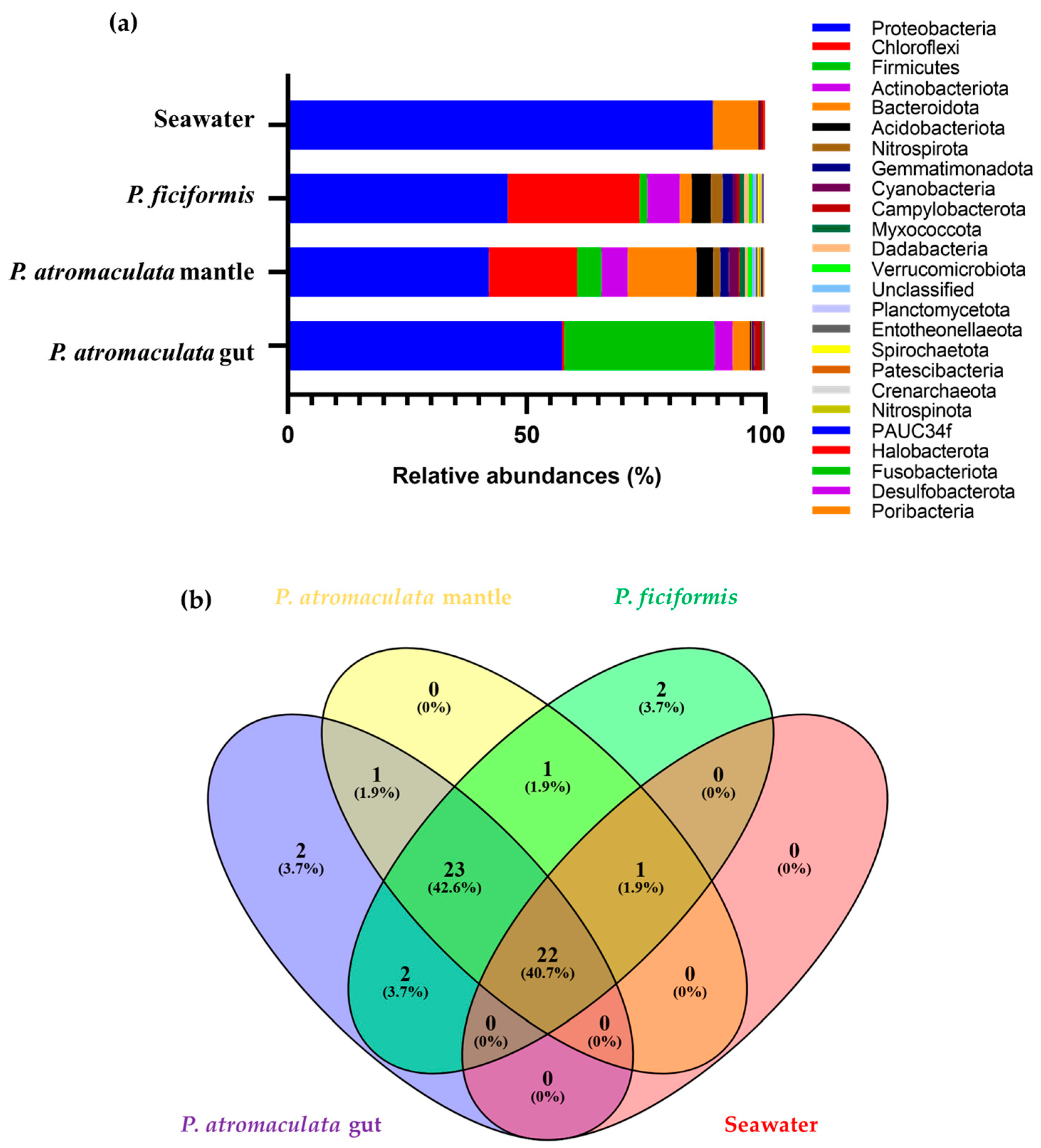
3.2. The Bacterial Composition of P. atromaculata and P. ficiformis Collected at the Phylum Level

Proteobacteria emerged as the most prevalent phylum across all samples, underscoring its dominance in the bacterial composition (Figure 3). The Peltodoris atromaculata gut revealed a more complex and diverse microbial community than the other samples, with a large abundance of Proteobacteria (57.4%) and Firmicutes (31.5%). Actinobacteriota (3.8%), Bacteroidota (3.5%), and other phyla were less represented. In contrast, the bacterial community of the P. atromaculata mantle presented a significant abundance of Proteobacteria (42%) and Chloroflexi (18.5%). Moreover, Bacteroidota (14%), Actinobacteriota (5.5%), and Acidobacteriota (3.4%) increased, and Firmicutes (5%) decreased. Another 25 phyla were represented, such as Cyanobacteria (1.9%) (Figure 4a).
The P. ficiformis bacterial community was rich in Proteobacteria (46%) and Chloroflexi (27.6%). Actinobacteriota (6.8%) and Acidobacteriota (4%) were also represented, along with other less represented phyla, such as Nitrospirota (2.5%) and Bacteroidota (2.5%; Figure 4a).
Additionally, the prevailing phyla in the surrounding seawater were Proteobacteria (88.9%) and Bacteroidota (9.5%).
The Venn diagram highlighted two unique phyla—the Latescibacterota and Euryarchaeota—in the P. atromaculata gut, and two—Sva0485 and Calditrichota—in P. ficiformis. On the other hand, this analysis demonstrated the co-occurrence of 22 phyla in all of the analyzed samples, indicating they could be derived from the environment (Figure 4b).

3.3. The Bacterial Composition of P. atromaculata and P. ficiformis at the Class Level

At the class level, the bacterial community of the P. atromaculata gut was rich in Gammaproteobacteria (47.7%) and Bacilli (31.4%), followed by Alphaproteobacteria (9.7%). Another 25 classes were less represented, such as Actinobacteria (3.6%) (Figure 4). Conversely, the bacterial community of the P. atromaculata mantle was highly abundant in Gammaproteobacteria (35.0%), followed by Bacteroidia (13%). Moreover, Anaerolineae, Dehalococcoida, Alphaproteobacteria, and Bacilli were also present, with relative abundances of 9.5%, 7.8%, 7%, and 5%, respectively. Another 25 classes were less represented, such as Acidimicrobia (2.2%) (Figure 5a).
The bacterial community of P. ficiformis was notably enriched in Gammaproteobacteria (27%), Alphaproteobacteria (19%), Anaerolineae (17.8%), and Dehalococcoidia (7.8%). The remaining classes, including Acidimicrobiia, Actinobacteria, and Nitrospiria, were less represented, with relative abundances of 4.0%, 3%, and 2.4%, respectively (Figure 5a).
Finally, the most abundant classes in the surrounding seawater were Gammaproteobacteria (71.4%), Alphaproteobacteria (17.5%), and Bacteroidota (9.5%).
The Venn diagram highlighted one unique class (the Thermococci) in the P. atromaculata gut and on (Calditrichia) in P. ficiformis respectively. Moreover, 10 classes—Dojkabacteria, Kryptonia, Vampirivibrionia, ABY1, Pla4 lineage, Dicrogenomatia, Desulfobacteria, Marinimicrobia bacterium, OM190, and Brevinematia—were found exclusively in the P. atromaculata mantle. On the other hand, this analysis demonstrated the co-occurrence of 34 classes in all of the analyzed samples, indicating that they could be derived from the environment (Figure 5b).

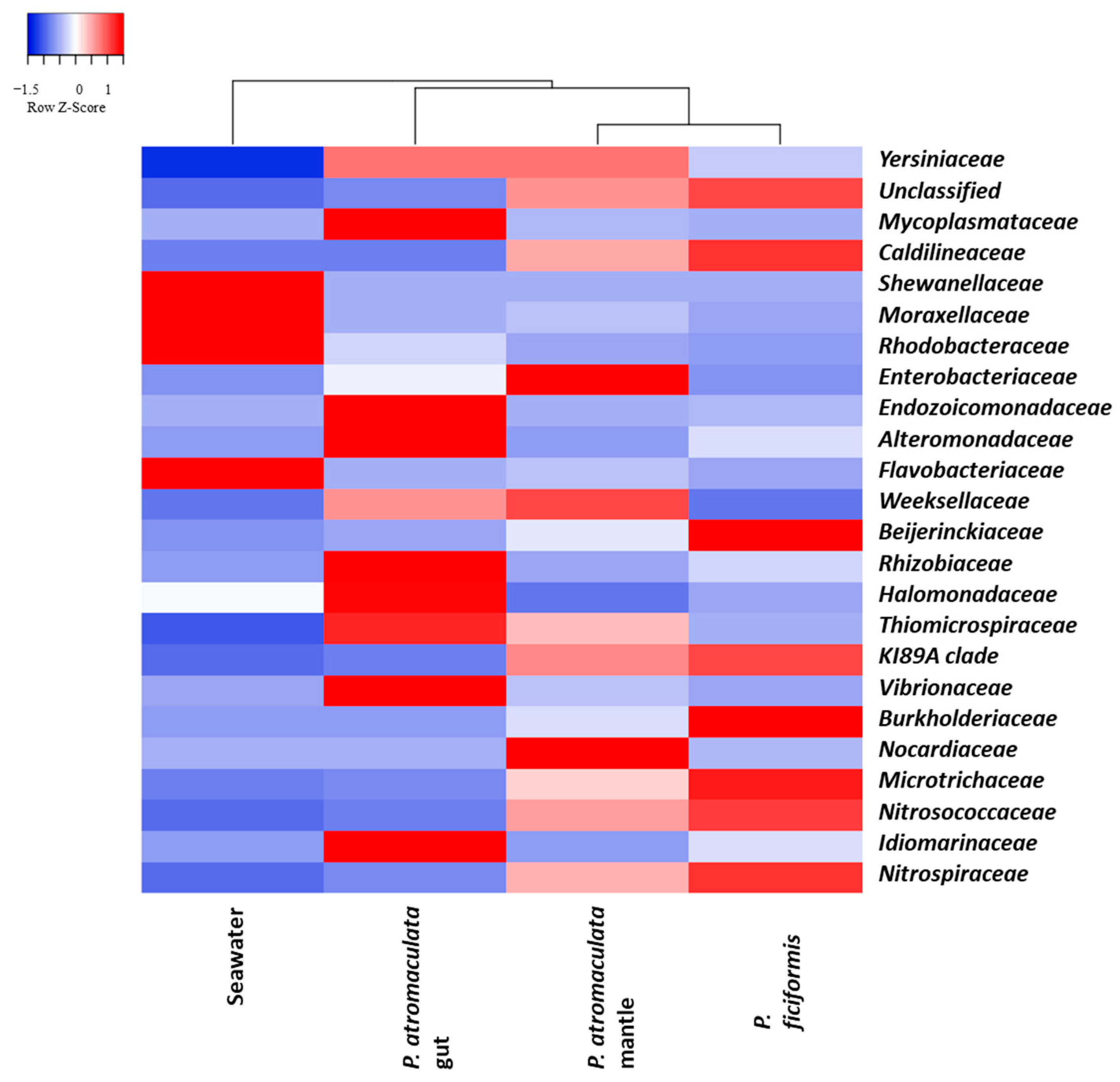
3.4. The Bacterial Composition of P. atromaculata and P. ficiformis at the Family Level

At the family level, the bacterial communities of the P. atromaculata gut were much more diverse than those of the P. atromaculata mantle and P. ficiformis samples. The P. atromaculata gut was highly abundant in Mycoplasmataceae, Endozoicomonadaceae, and Alteromonadaceae, with relative abundances of 29.5%, 26.1%, and 4.6%, respectively. Other families, such as Rhizobiaceae (3.6%), were less represented. The P. atromaculata mantle was enriched in unclassified bacteria (23.8%), followed by Enterobacteriaceae and Weeksellaceae, with relative abundances of 18.5% and 11%, respectively. Other families, such as Caldilineaceae (9%) and Nocardiaceae (2.8%), were abundant. Moreover, other families were less represented, such as the Mycoplasmataceae (2.3%) and the KI89A clade (2.97%) (Figure 6a).
The bacterial community of P. ficiformis, excluding the unclassified (29.9%), presented an intriguing enrichment in Caldilneaceae, with a relative abundance of 17%. Other families, while less represented, offer a unique insight into microbial diversity, such as the Beijerinckiaceae (6.3%), Burkholderiaceae (5%), KI89A clade (3.1%), Microtrichaceae (3.3%), Alteromonadaceae (3%), and so on (Figure 6a).
Lastly, the most abundant families in the surrounding seawater were Shewanellaceae (38.9%), Moraxellaceae (29.15%), Rhodobacteraceae (16.4%), and Flavobacteriaceae (9.4%).
The Venn diagram highlighted forty families found exclusively in the P. atromaculata gut and sixteen in P. ficiformis. Moreover, ten families were exclusively found in the P. atromaculata mantle. On the other hand, this analysis demonstrated the co-occurrence of 107 families in all of the analyzed samples, indicating that they could be derived from the environment (Figure 6b).
Heatmap analysis showed that the bacterial community of the P. atromaculata gut formed a distinct cluster, like that of the seawater, while the P. atromaculata mantle and P. ficiformis were sub-clustered, sharing a similar family composition. Some families appeared to be distinctive of a sample; for example, Mycoplasmataceae, Endozoicomonadaceae, and Alteromonadaceae were abundant in the P. atromaculata gut; Caldilineaceae, Beijerinckiaceae, Burkholderiaceae, Microtrichaceae, Nitrosococcaceae, Nitrospiraceae, and the KI89A clade in P. ficiformis; and Enterobacteriaceae, Nocardiaceae, and Weeksellaceae in the P. atromaculata mantle (Figure 7).

3.5. The Bacterial Composition of P. atromaculata and P. ficiformis at the Genus Level

The most represented genera in the P. atromaculata gut were Mycoplasma (29.5%), Kistimonas (26.1%), and Alteromonas (4.6%). Indeed, in the P. atromaculata mantle, excluding the unclassified genera, the most represented genera were Elizabethkingia (11%), Escherichia (5%), and Rhodococcus (3%). In contrast, the most represented genera in P. ficiformis, excluding the unclassified genera, were Methylobacterium (6.1%) and Burkholderia (4.9%) (Figure 8a). All samples contained a large percentage of unclassified genera: 6%, 51%, and 55% in the P. atromaculata gut, mantle, and P. ficiformis, respectively.
In conclusion, the dominant genera in the surrounding seawater were Shewanella (38.9%), Psychrobacter (29.15%), and Sulfitobacter (15.10%).
The Venn diagram highlighted seventy-seven genera found exclusively in the P. atromaculata gut and thirty-five in P. ficiformis, respectively. Moreover, twenty-six genera were found exclusively in the P. atromaculata mantle. On the other hand, this analysis demonstrated the co-occurrence of ninety-six families in all of the analyzed samples, indicating that they could be derived from the environment (Figure 8b).

4. Discussion

Hydrothermal systems (HTSs) represent extreme habitats characterized by high and fluctuating temperatures and metal concentrations, shaped by the unique characteristics of each site [1]. These environments are considered to be biodiversity hotspots, where microorganisms, particularly bacteria, play a fundamental role in maintaining ecosystem homeostasis [33,34]. This study investigated the microbial communities associated with two marine invertebrates, the nudibranch P. atromaculata and the sponge P. ficiformis, from a shallow HTS near the west coast of Sicily. These organisms are components of the local food web and serve as bioindicators of environmental health. Our analysis identified shared bacterial phyla between the P. atromaculata mantle and P. ficiformis (Figure 4). The bacterial community in the P. atromaculata mantle appeared more similar to that of P. ficiformis than to that of its gut, likely due to closer environmental exposure. However, our microbial analysis was based on a single seawater sample, where metagenomic DNA was extracted three times, pooled in equal concentrations, and amplified as a single sample. Further sampling is needed to confirm whether environmental exposure significantly influences the bacterial communities in the P. atromaculata mantle and P. ficiformis. Nevertheless, the Bacteroidetes and the Proteobacteria, particularly Gammaproteobacteria, which are normally known to dominate the marine environment and are important for nutrient cycling [35,36,37,38,39,40], were found to be more abundant in the P. atromaculata mantle and P. ficiformis [41,42,43,44,45,46]. Meanwhile, Bacteroidetes in marine environments have highly adaptable genomes shaped by genetic rearrangements and lateral gene transfer. This includes expressing adhesion proteins and gliding motility genes, allowing them to attach to sponges, plankton, and biofilms and efficiently access organic matter degraded by their extracellular enzymes [47]. Our results suggest that Bacteroidetes associated with P. atromaculata tissues may not only aid in breaking down sponge polysaccharides in the gut but also contribute to surface adhesion in the mantle. Indeed, in the microbiota of the P. atromaculata gut, the Firmicutes are the second most abundant phylum; these results are in accordance with a study on the microbiota of Patella pellucida, a mollusk reported for the first time by Linnaeus in 1758 [47]. Firmicutes are mostly Gram-positive, spore-forming bacteria, often anaerobic and found in low-oxygen environments. They are common in marine ecosystems, living in seawater and sediments, or as symbionts in marine animals [47]. Despite these similarities, both invertebrates exhibit high microbial selectivity, each harboring distinct bacterial families unique to their species, as shown in Figure 7 and Figure 8. These findings suggest that these species are relatively resistant to environmental changes, potentially due to the specific interactions with their microbial communities. To the best of our knowledge, while we conducted microbiota analysis on only a few samples, this is the first study to explore the bacterial community of P. atromaculata. Indeed, in the literature, few studies are available on other nudibranchs, such as Rostanga alisae, Pteraeolidia semperi, Doriprismatica atromarginata (reported for the first time by Cuvier in 1804), Jorunna funebris (reported for the first time by Kelaart in 1859), and Phyllidiella species [7,8,41], most of which focused on nudibranchs from marine sediments and coral reefs of the Pacific and Indian Ocean regions. However, this is the first characterization, and additional samples are needed in future studies to gain a deeper understanding of the microbiota of P. atromaculata. Distinct microbial profiles were observed in this study in comparison with previous studies, especially concerning Cyanobacteria and Chloroflexi. These differences may reflect various factors, such as dietary preferences, environmental conditions, and symbiotic relationships. For example, the Cyanobacteria were found to be prevalent in other studies in the microbiota of R. alisae and P. semperi [7,8]. However, in our analysis, this phylum was found to be minimally abundant in the P. atromaculata gut and mantle. In contrast, Chloroflexi were found abundantly in the P. atromaculata mantle; this result was in accordance with the microbial community found in Phyllidiella picta (reported for the first time by Pruvot-Fol in 1957) (Figure 3), but not in other nudibranchs [41], supporting the possibility of microbial acquisition from surrounding environments. Indeed, the high abundance of Chloroflexi in P. ficiformis further emphasizes the important role of this phylum in HTS microbial communities, consistent with previous studies [42,43,44]. These bacteria, including thermophilic and anaerobic species, are involved in vital processes such as carbon fixation, organic matter degradation, and pollutant detoxification, contributing to nutrient cycling and ecological balance in marine environments [45]. Sponge-associated microbial communities are typically a mixture of generalist and specialist bacteria. Generalists like Proteobacteria, Actinobacteria, Cyanobacteria, Chloroflexi, Nitrospirae, and Poribacteria are well established as the most prevalent bacterial symbionts in sponges, forming the core bacterial community [48,49,50]. At the same time, specialist bacteria are more restricted to specific species [46,48]. For instance, the Cyanobacterium Synechococcus feldmannii was found exclusively in P. ficiformis from the Mediterranean Sea [51], while the alphaproteobacterium Halichondribacter symbioticus is associated with Halichondria panicea (reported for the first time by Pallas in 1766) [52]. In line with these findings, our analysis of P. ficiformis samples also identified these phyla as key components of its microbial community. Surprisingly, our study found a low abundance of Cyanobacteria in P. ficiformis (Figure 4) despite prior reports of their role as initial symbionts, potentially assisting in regulating the host’s redox potential [46,48,49]. However, other studies have reported a lower abundance of this phylum, depending on the habitat [22,53]. In our results, the low abundance may reflect transient or facultative associations, suggesting that the microbial community in P. ficiformis could be highly dynamic and influenced by environmental or ecological factors, including light availability and habitat depth [19,23,54,55]. Our findings also emphasize the role of specialized bacterial phyla like Poribacteria, which are implicated in carbohydrate degradation and possibly contribute to the breakdown of the sponge’s extracellular matrix [56]. Similarly, Nitrospirota, known for ammonia oxidation, were also present in sponges like Bathydorus sp. [57,58], highlighting their potential role in nitrogen cycling in marine environments. However, future studies should be based on shotgun metagenomic analyses to enable a more comprehensive investigation of microbial metabolic activity, functional genomics, ecological distribution, and the specific roles of sponge-associated microbiota.
Regarding functional relevance, identifying families such as Mycoplasmataceae, Endozoicomonadaceae, and Alteromonadaceae in the P. atromaculata gut (Figure 6) points to significant microbial processes. These families were found to be abundant in another microbial community study on the guts of sea slugs, such as in Berghia Stephanie (reported for the first time by Valdés in 2005) [59]. The Mycoplasmataceae family’s abundance under stress conditions might indicate a role in the host’s adaptation to extreme environmental conditions [60]. Endozoicomonadaceae, frequently associated with marine hosts like corals, may influence host health, although their function remains unclear [61]. The Alteromonadaceae, belonging to the Gammaproteobacteria class, have been isolated from various marine environments, including coastal, open-ocean, and deep-sea waters, as well as marine invertebrates [62,63]. These bacteria are chemoorganotrophic, with most strains capable of using a wide range of organic compounds, such as carbohydrates, monocarboxylic fatty acids, and amino acids, as their sole carbon and energy sources. Some strains can also produce extracellular enzymes, such as amylase, lipase, gelatinase, or chitinase [62,63].
The P. atromaculata mantle was enriched with families like Enterobacteriaceae, Weeksellaceae, and Caldilineaceae (Figure 5), known for sulfur and nitrogen cycling. The Enterobacteriaceae family, belonging to the Gammaproteobacteria class, has been reported in the bacterial communities of various vent mussels, including all tissues, such as the mantle and gut; it can be involved in sulfur biogeochemical cycles [41,64,65]. The Weeksellaceae family, belonging to the Flavobacteriales, is known for its widespread presence across various ecological environments and is involved in utilizing a diverse range of carbon polymers derived from algae, bacteria, plants, and animals, reinforcing its significant role in ocean carbon cycling as outstanding decomposers of particulate organic matter. Moreover, their genome encodes for various biosynthetic gene clusters, with a high prevalence of gene clusters encoding pathways for the production of antimicrobial, antioxidant, and cytotoxic compounds [66].
In P. ficiformis, families such as Caldilineaceae, Beijerinckiaceae, Burkholderiaceae, and Microtrichaceae (Figure 6) reflect the adaptability of microbiota to the HTS environment. The Caldilineaceae, involved in sulfur removal and denitrification [67,68,69], are particularly abundant, highlighting their ecological importance, and yet they have been found to be associated with P. ficiformis in other studies [22]. Beijerinckiaceae and Burkholderiaceae, capable of nitrogen fixation, contribute to the sponge’s ability to thrive in nitrogen-limited conditions [70,71]. Similarly, the Microtrichaceae family, involved in sulfate reduction and nitrogen cycling, may support P. ficiformis in this challenging habitat [50,72]. Our results on genera in the P. atromaculata gut were consistent with findings from other studies that identified Mycoplasma as the most abundant genus in the gut (Figure 8) [7,41,73]. Additionally, Kistimonas was found to be the second most abundant genus in the P. atromaculata gut. These results align with the findings of Stuij et al. [41], who reported that Kistimonas is commonly associated with a variety of aquatic invertebrates, including corals, sponges, bivalves, mollusks, ascidians, echinoderms, and several nudibranch species. These bacteria are believed to play diverse roles within their hosts, such as shaping the microbiome and facilitating nutrient absorption [41]. In contrast, the microbial community in the P. atromaculata mantle was enriched in Escherichia and Rhodococcus. The Escherichia genus has been associated with fatty acid synthase activity, while Rhodococcus is part of the epibiotic microbial community in P. ficiformis due to its antibacterial properties [73,74]. Meanwhile, the microbial community in P. ficiformis showed enrichment in Methylobacterium and Burkholderia. Methylobacterium is known to inhabit various environments, including coastal waters, open-ocean seawater, and deep-sea hydrothermal vents, and it has been observed that this genus utilizes compounds such as formate and methanol for growth [75,76]. Our study highlights the dominance of bacteria involved in sulfur and nitrogen cycling in P. atromaculata and P. ficiformis, indicating their critical role in maintaining ecological balance within HTSs. Further research, including nearby water bodies and other sponge species in the same region, would allow for a deeper understanding of the interactions between the invertebrates and their microbial communities, along with the functional roles that these microbes play in the HTS environment.

5. Conclusions

This study provides the first comprehensive analysis of the bacterial community associated with the nudibranch P. atromaculata, highlighting a tissue-specific microbial composition that differentiates it from other mollusks. Our findings reveal that the mantle of P. atromaculata shares specific bacterial phyla with its prey, P. ficiformis, suggesting a potential horizontal transfer of microbes from prey to host. This highlights the importance of trophic interactions in shaping the microbiota of marine invertebrates, particularly in extreme environments such as hydrothermal springs. Additionally, our study identified a core bacterial community within P. ficiformis, consisting of Chloroflexi, Nitrospirae, Cyanobacteria, and Poribacteria, which appear to be strongly influenced by environmental factors. These microbial groups are known for their involvement in key ecological processes such as carbon fixation, nitrogen cycling, and organic matter degradation. The presence of these bacterial phyla in P. ficiformis reinforces the sponge’s role as a reservoir for microbial diversity in hydrothermal environments and further supports the hypothesis that sponges contribute significantly to biogeochemical cycles in marine ecosystems. Our findings underscore the complex interactions between biotic and abiotic factors in shaping bacterial communities in the hydrothermal springs near the west coast of Sicily. The extreme conditions of these habitats, including high temperatures and fluctuating metal concentrations, likely exert selective pressures on microbial communities, favoring the persistence of specialized bacteria adapted to these environments. Understanding these interactions is crucial for deciphering the ecological functions of bacteria in hydrothermal ecosystems and their potential contributions to environmental stability. While our study provides valuable insights into the microbial diversity of P. atromaculata and P. ficiformis, it represents only a preliminary step in characterizing the microbiota of marine invertebrates in hydrothermal settings. Future research should incorporate advanced molecular techniques such as shotgun metagenomics and transcriptomics to gain a more detailed understanding of microbial metabolic pathways, functional capabilities, and ecological interactions. These approaches will allow researchers to investigate how microbial communities contribute to host physiology, nutrient cycling, and environmental adaptation. Additionally, expanding the scope of research to include seasonal variations, host developmental stages, and comparisons with other hydrothermal sites will provide a broader perspective on the stability and dynamics of these microbial communities. By integrating microbiological, ecological, and molecular analyses, future studies can enhance our understanding of the symbiotic relationships between marine invertebrates and their associated microbiota, ultimately contributing to a more comprehensive view of microbial ecology in hydrothermal systems.

Supplementary Materials

The following supporting information can be downloaded at https://www.mdpi.com/article/10.3390/w17071036/s1, Figure S1: Rarefaction curves of the samples analyzed in this study; Figure S2: Percentage of relative abundance of the main 25 phyla found in all samples of this study; Figure S3: Percentage of relative abundance of the main 25 classes found in all samples of this study; Figure S4: Percentage of relative abundance of the main 25 orders found in all samples of this study; Figure S5: Percentage of relative abundance of the main 25 families found in all samples of this study; Figure S6: Percentage of relative abundance of the main 25 genera found in all samples of this study.

Author Contributions

A.G. performed the experiments, analyzed the data, and wrote the draft of the manuscript; D.G. carried out collection of the samples; L.V. performed the bioinformatics analysis; V.V. analyzed the data and wrote the draft of the manuscript, M.A. and R.A. conceived the study, interpreted the data, and revised the article. All authors have read and agreed to the published version of the manuscript.

Funding

This research received partial funding from PON “Ricerca e Innovazione” 2014–2020, Asse IV “Istruzione e Ricerca per il recupero” Azione IV.5 “Dottorati su tematiche green” DOT 1320418 CUP B73D21009190006. It was also financially supported by the European Commission-NextGenerationEU under the project SUS-MIRRI.IT, “Strengthening the MIRRI Italian Research Infrastructure for Sustainable Bioscience and Bioeconomy”, code n.IR0000005PO. Additionally, project funded under the National Recovery and Resilience Plan (NRRP), Mission 4 Component 2 Investment 1.4—Call for tender No. 3138 of 16 December 2021, rectified by Decree n.3175 of 18 December 2021 of Italian Ministry of University and Research funded by the European Union—NextGenerationEU; Project code CN_00000033, Concession Decree No. 1034 of 17 June 2022 adopted by the Italian Ministry of University and Research, CUP B73D20005170001, Project title “National Biodiversity Future Center—NBFC.

Data Availability Statement

The dataset analyzed during the current study is available in the GenBank database with the accession numbers and BioProject ID: PRJNA1156989.

Acknowledgments

The authors wish to thank Fanny Claire Capri for helping with data analysis.

Conflicts of Interest

The authors declare that this research was conducted in the absence of any commercial or financial relationships that could be construed as potential conflicts of interest.

References

  1. Price, R.E.; Giovannelli, D. A Review of the Geochemistry and Microbiology of Marine Shallow-Water Hydrothermal Vents. In Reference Module in Earth Systems and Environmental Sciences; Elsevier: Amsterdam, The Netherlands, 2017. [Google Scholar] [CrossRef]
  2. Gollner, S.; Colaço, A.; Gebruk, A.; Halpin, P.N.; Higgs, N.; Menini, E.; Mestre, N.C.; Qian, P.Y.; Sarrazin, J.; Szafranski, K.; et al. Application of Scientific Criteria for Identifying Hydrothermal Ecosystems in Need of Protection. Mar. Policy 2021, 132, 104641. [Google Scholar] [CrossRef]
  3. Dubilier, N.; Bergin, C.; Lott, C. Symbiotic Diversity in Marine Animals: The Art of Harnessing Chemosynthesis. Nat. Rev. Microbiol. 2008, 6, 725–740. [Google Scholar] [CrossRef]
  4. Lee, W.K.; Juniper, S.K.; Perez, M.; Ju, S.J.; Kim, S.J. Diversity and Characterization of Bacterial Communities of Five Co-Occurring Species at a Hydrothermal Vent on the Tonga Arc. Ecol. Evol. 2021, 11, 4481–4493. [Google Scholar] [CrossRef]
  5. Van Dover, C.L.; Arnaud-Haond, S.; Gianni, M.; Helmreich, S.; Huber, J.A.; Jaeckel, A.L.; Metaxas, A.; Pendleton, L.H.; Petersen, S.; Ramirez-Llodra, E.; et al. Scientific Rationale And International Obligations For Protection of Active Hydrothermal Vent Ecosystems from Deep-Sea Mining. Mar. Policy 2018, 90, 20–28. [Google Scholar] [CrossRef]
  6. Duperron, S.; Halary, S.; Lorion, J.; Sibuet, M.; Gaill, F. Unexpected Co-occurrence of Six Bacterial Symbionts in the Gills of the Cold Seep Mussel Idas Sp. (Bivalvia: Mytilidae). Environ. Microbiol. 2008, 10, 433–445. [Google Scholar] [CrossRef] [PubMed]
  7. Ng, M.S.; Soon, N.; Chang, Y.; Wainwright, B.J. Bacterial and Fungal Co-Occurrence in the Nudibranch, Pteraeolidia semperi. Life 2022, 12, 1988. [Google Scholar] [CrossRef]
  8. Zhukova, N.V.; Eliseikina, M.G.; Balakirev, E.S.; Ayala, F.J. Multiple Bacterial Partners in Symbiosis with the Nudibranch Mollusk Rostanga alisae. Sci. Rep. 2022, 12, 169. [Google Scholar] [CrossRef] [PubMed]
  9. Sanvicente-Añorve, L.; Hermoso-Salazar, M.; Ortigosa, J.; Solís-Weiss, V.; Lemus-Santana, E. Opisthobranch Assemblages from a Coral Reef System: The Role of Habitat Type and Food Availability. Bull. Mar. Sci. 2012, 88, 1061–1074. [Google Scholar] [CrossRef]
  10. Wägele, H.; Willan, R.C. Phylogeny of the Nudibranchia. Zool. J. Linn. Soc. 2000, 130, 83–181. [Google Scholar] [CrossRef]
  11. Thompson, L.R.; Sanders, J.G.; McDonald, D.; Amir, A.; Ladau, J.; Locey, K.J.; Prill, R.J.; Tripathi, A.; Gibbons, S.M.; Ackermann, G.; et al. A Communal Catalogue Reveals Earth’s Multiscale Microbial Diversity. Nature 2017, 551, 457–463. [Google Scholar] [CrossRef]
  12. Toma, M.; Betti, F.; Bavestrello, G.; Cattaneo-Vietti, R.; Canese, S.; Cau, A.; Andaloro, F.; Greco, S.; Bo, M. Diversity and Abundance of Heterobranchs (Mollusca, Gastropoda) from the Mesophotic and Bathyal Zone of the Mediterranean Sea. Eur. Zool. J. 2022, 89, 160–182. [Google Scholar] [CrossRef]
  13. Avila, C. The Growth of Peltodoris atromaculata Bergh, 1880 (Gastropoda: Nudibranchia) in the Laboratory. J. Molluscan Stud. 1996, 62, 151–157. [Google Scholar] [CrossRef]
  14. Gemballa, S.; Schermutzki, F. Cytotoxic Haplosclerid Sponges Preferred: A Field Study on the Diet of the Dotted Sea Slug Peltodoris atromaculata (Doridoidea: Nudibranchia). Mar. Biol. 2004, 144, 1213–1222. [Google Scholar] [CrossRef]
  15. Lombardo, A.; Marletta, G. Observations on the Dorid Peltodoris atromaculata Bergh, 1880 (Gastropoda Nudibranchia) along the Central-Eastern Coast of Sicily, Ionian Sea. Biodivers. J. 2021, 12, 245–254. [Google Scholar] [CrossRef]
  16. Maldonado, M.; Ribes, M.; van Duyl, F.C. Nutrient fluxes through sponges: Biology, budgets, and ecological implications. Adv. Mar. Biol. 2012, 62, 113–182. [Google Scholar] [CrossRef]
  17. Maldonado, M.; Aguilar, R.; Bannister, R.; Bell, J.; Conway, J.; Dayton, P.; Díaz, C.; Gutt, J.; Kelly, M.; Kenchington, E.; et al. Sponge Grounds as Key Marine Habitats: A Synthetic Review of Types, Structure, Functional Roles, and Conservation Concerns. In Marine Animal Forests: The Ecology of Benthic Biodiversity Hotspots; Springer: Berlin/Heidelberg, Germany, 2017; pp. 145–183. [Google Scholar] [CrossRef]
  18. Redmond, N.E.; van Soest, R.W.M.; Kelly, M.; Raleigh, J.; Travers, S.A.A.; McCormack, G.P. Reassessment of the Classification of the Order Haplosclerida (Class Demospongiae, Phylum Porifera) Using 18S RRNA Gene Sequence Data. Mol. Phylogenet. Evol. 2007, 43, 344–352. [Google Scholar] [CrossRef]
  19. Cerrano, C.; Giovine, M.; Steindler, L. Petrosia ficiformis (Poiret, 1789): An excellent model for holobiont and biotechnological studies. Curr. Opin. Biotechnol. 2022, 74, 61–65. [Google Scholar] [CrossRef]
  20. Webster, N.S.; Taylor, M.W. Marine sponges and their microbial symbionts: Love and other relationships. Environ. Microbiol. 2012, 14, 335–346. [Google Scholar] [CrossRef]
  21. Georgieva, M.N.; Taboada, S.; Riesgo, A.; Díez-Vives, C.; De Leo, F.C.; Jeffreys, R.M.; Copley, J.T.; Little, C.T.S.; Ríos, P.; Cristobo, J.; et al. Evidence of Vent-Adaptation in Sponges Living at the Periphery of Hydrothermal Vent Environments: Ecological and Evolutionary Implications. Front. Microbiol. 2020, 11, 1636. [Google Scholar] [CrossRef]
  22. Mazzella, V.; Dell’Anno, A.; Etxebarría, N.; González-Gaya, B.; Nuzzo, G.; Fontana, A.; Núñez-Pons, L. High Microbiome and Metabolome Diversification in Coexisting Sponges with Different Bio-Ecological Traits. Commun. Biol. 2024, 7, s42003–s42024. [Google Scholar]
  23. Pagliara, P.; Barca, A.; Verri, T.; Caroppo, C. The Marine Sponge Petrosia ficiformis Harbors Different Cyanobacteria Strains with Potential Biotechnological Application. J. Mar. Sci. Eng. 2020, 8, 638. [Google Scholar] [CrossRef]
  24. Schmitt, S.; Hentschel, U.; Taylor, M.W. Deep Sequencing Reveals Diversity and Community Structure of Complex Microbiota in Five Mediterranean Sponges. In Ancient Animals, New Challenges; Springer Netherlands: Dordrecht, The Netherlands, 2011; pp. 341–351. [Google Scholar] [CrossRef]
  25. Usher, K.M.; Kuo, J.; Fromont, J.; Sutton, D.C. Vertical transmission of Cyanobacterial symbionts in the marine sponge Chondrilla australiensis (Demospongiae). Hydrobiologia 2001, 461, 9–13. [Google Scholar] [CrossRef]
  26. Chen, H.; Li, D.H.; Jiang, A.J.; Li, X.G.; Wu, S.J.; Chen, J.W.; Qu, M.J.; Qi, X.Q.; Dai, J.; Zhao, R.; et al. Metagenomic Analysis Reveals Wide Distribution of Phototrophic Bacteria in Hydrothermal Vents on the Ultraslow-Spreading Southwest Indian Ridge. Mar. Life Sci. Technol. 2022, 4, 255–267. [Google Scholar] [CrossRef]
  27. Magoč, T.; Salzberg, S.L. FLASH: Fast Length Adjustment of Short Reads to Improve Genome Assemblies. Bioinformatics 2011, 27, 2957–2963. [Google Scholar] [CrossRef] [PubMed]
  28. Martin, M. Cutadapt Removes Adapter Sequences from High-Throughput Sequencing Reads. EMBnet J. 2011, 17, 10. [Google Scholar] [CrossRef]
  29. Del Fabbro, C.; Scalabrin, S.; Morgante, M.; Giorgi, F.M. An Extensive Evaluation of Read Trimming Effects on Illumina NGS Data Analysis. PLoS ONE 2013, 8, e85024. [Google Scholar] [CrossRef]
  30. Rognes, T.; Flouri, T.; Nichols, B.; Quince, C.; Mahé, F. VSEARCH: A Versatile Open Source Tool for Metagenomics. PeerJ 2016, 4, e2584. [Google Scholar] [CrossRef]
  31. Quast, C.; Pruesse, E.; Yilmaz, P.; Gerken, J.; Schweer, T.; Yarza, P.; Peplies, J.; Glöckner, F.O. The SILVA Ribosomal RNA Gene Database Project: Improved Data Processing and Web-Based Tools. Nucleic Acids Res. 2013, 41, 590–596. [Google Scholar]
  32. Wang, Y.; Bi, H.-Y.; Chen, H.-G.; Zheng, P.-F.; Zhou, Y.-L.; Li, J.-T. Metagenomics Reveals Dominant Unusual Sulfur Oxidizers Inhabiting Active Hydrothermal Chimneys from the Southwest Indian Ridge. Front. Microbiol. 2022, 13, 861795. [Google Scholar] [CrossRef]
  33. Dick, G.J. The Microbiomes of Deep-Sea Hydrothermal Vents: Distributed Globally, Shaped Locally. Nat. Rev. Microbiol. 2019, 17, 271–283. [Google Scholar] [CrossRef]
  34. van der Most, N.; Qian, P.Y.; Gao, Y.; Gollner, S. Active hydrothermal vent ecosystems in the Indian Ocean are in need of protection. Front. Mar. Sci. 2023, 9, 1067912. [Google Scholar] [CrossRef]
  35. Devine, S.P.; Pelletreau, K.N.; Rumpho, M.E. 16S rDNA-based metagenomic analysis of bacterial diversity associated with two populations of the kleptoplastic sea slug Elysia chlorotica and its algal prey Vaucheria litorea. Biol. Bull. 2012, 223, 138–154. [Google Scholar] [CrossRef]
  36. Kersters, K.; De Vos, P.; Gillis, M.; Swings, J.; Vandamme, P.; Stackebrandt, E. Introduction to the Proteobacteria. In The Prokaryotes; Springer New York: New York, NY, USA, 2006; pp. 3–37. [Google Scholar] [CrossRef]
  37. Sucato, A.; Vecchioni, L.; Savoca, D.; Presentato, A.; Arculeo, M.; Alduina, R. A Comparative Analysis of Aquatic and Polyethylene-Associated Antibiotic-Resistant Microbiota in the Mediterranean Sea. Biology 2021, 10, 200. [Google Scholar] [CrossRef] [PubMed]
  38. Kim, P.S.; Shin, N.R.; Lee, J.B.; Kim, M.S.; Whon, T.W.; Hyun, D.W.; Yun, J.H.; Jung, M.J.; Kim, J.Y.; Bae, J.W. Host Habitat Is the Major Determinant of the Gut Microbiome of Fish. Microbiome 2021, 9, 166. [Google Scholar] [CrossRef]
  39. Di Leto, Y.; Capri, F.C.; Mineo, A.; Cosenza, A.; Gallo, G.; Alduina, R.; Mannina, G. Initial PH Conditions Shape the Microbial Community Structure of Sewage Sludge in Batch Fermentations for the Improvement of Volatile Fatty Acid Production. Microorganisms 2022, 10, 2073. [Google Scholar] [CrossRef]
  40. Capri, F.C.; Prazzi, E.; Casamento, G.; Gambino, D.; Cassata, G.; Alduina, R. Correlation Between Microbial Community and Hatching Failure in Loggerhead Sea Turtle Caretta Caretta. Microb. Ecol. 2023, 86, 1923–1933. [Google Scholar] [CrossRef]
  41. Stuij, T.; Cleary, D.F.R.; Polónia, A.R.M.; Putchakarn, S.; Pires, A.C.C.; Gomes, N.C.M.; de Voogd, N.J. Exploring Prokaryotic Communities in the Guts and Mucus of Nudibranchs, and Their Similarity to Sediment and Seawater Microbiomes. Curr. Microbiol. 2023, 80, 294. [Google Scholar] [CrossRef]
  42. Díez-Vives, C.; Taboada, S.; Leiva, C.; Busch, K.; Hentschel, U.; Riesgo, A. On the Way to Specificity-Microbiome Reflects Sponge Genetic Cluster Primarily in Highly Structured Populations. Mol. Ecol. 2020, 29, 4412–4427. [Google Scholar] [CrossRef]
  43. Mehrshad, M.; Rodriguez-Valera, F.; Amoozegar, M.A.; López-García, P.; Ghai, R. The Enigmatic SAR202 Cluster up Close: Shedding Light on a Globally Distributed Dark Ocean Lineage Involved in Sulfur Cycling. ISME J. 2018, 12, 655–668. [Google Scholar] [CrossRef]
  44. Cleary, D.F.R.; Polónia, A.R.M. Bacterial and Archaeal Communities Inhabiting Mussels, Sediment and Water in Indonesian Anchialine Lakes. Antonie Van Leeuwenhoek 2018, 111, 237–257. [Google Scholar] [CrossRef]
  45. Gupta, R.S.; Chander, P.; George, S. Phylogenetic Framework and Molecular Signatures for the Class Chloroflexi and Its Different Clades; Proposal for Division of the Class Chloroflexi Class. Nov. into the Suborder Chloroflexineae Subord. Nov., Consisting of the Emended Family Oscillochloridaceae and the Family Chloroflexaceae Fam. Nov., and the Suborder Roseiflexineae Subord. Nov., Containing the Family Roseiflexaceae Fam. Nov. Antonie Van Leeuwenhoek Int. J. Gen. Mol. Microbiol. 2013, 103, 99–119. [Google Scholar] [CrossRef]
  46. Thomas, T.; Moitinho-Silva, L.; Lurgi, M.; Björk, J.R.; Easson, C.; Astudillo-García, C.; Olson, J.B.; Erwin, P.M.; López-Legentil, S.; Luter, H.; et al. Diversity, Structure and Convergent Evolution of the Global Sponge Microbiome. Nat. Commun. 2016, 7, 11870. [Google Scholar] [CrossRef] [PubMed]
  47. Dudek, M.; Adams, J.; Swain, M.; Hegarty, M.; Huws, S.; Gallagher, J. Metaphylogenomic and Potential Functionality of the Limpet Patella Pellucida’s Gastrointestinal Tract Microbiome. Int. J. Mol. Sci. 2014, 15, 18819–18839. [Google Scholar] [CrossRef]
  48. Pita, L.; Rix, L.; Slaby, B.M.; Franke, A.; Hentschel, U. The Sponge Holobiont in a Changing Ocean: From Microbes to Ecosystems. Microbiome 2018, 6, 46. [Google Scholar] [CrossRef]
  49. Cleary, D.F.R.; Swierts, T.; Coelho, F.J.R.C.; Polónia, A.R.M.; Huang, Y.M.; Ferreira, M.R.S.; Putchakarn, S.; Carvalheiro, L.; van der Ent, E.; Ueng, J.P.; et al. The Sponge Microbiome within the Greater Coral Reef Microbial Metacommunity. Nat. Commun. 2019, 10, 1644. [Google Scholar] [CrossRef] [PubMed]
  50. O’Brien, P.A.; Robbins, S.J.; Tan, S.; Rix, L.; Miller, D.J.; Webster, N.S.; Zhang, G.; Bourne, D.G. Comparative Genomics Identifies Key Adaptive Traits of Sponge-Associated Microbial Symbionts. Environ. Microbiol. 2024, 26, e16690. [Google Scholar] [CrossRef]
  51. Burgsdorf, I.; Handley, K.M.; Bar-Shalom, R.; Erwin, P.M.; Steindler, L. Life at Home and on the Roam: Genomic Adaptions Reflect the Dual Lifestyle of an Intracellular, Facultative Symbiont. mSystems 2019, 4, e00057-19. [Google Scholar] [CrossRef]
  52. Knobloch, S.; Jóhannsson, R.; Marteinsson, V. Bacterial Diversity in the Marine Sponge Halichondria panicea from Icelandic Waters and Host-Specificity of Its Dominant Symbiont “Candidatus Halichondribacter Symbioticus”. FEMS Microbiol. Ecol. 2018, 95, fiy220. [Google Scholar] [CrossRef]
  53. Sipkema, D.; de Caralt, S.; Morillo, J.A.; Al-Soud, W.A.; Sørensen, S.J.; Smidt, H.; Uriz, M.J. Similar Sponge-Associated Bacteria Can Be Acquired via Both Vertical and Horizontal Transmission. Environ. Microbiol. 2015, 17, 3807–3821. [Google Scholar] [CrossRef]
  54. Arillo, A.; Bavestrello, G.; Burlando, B.; Sarfi, M. Metabolic Integration between Symbiotic Cyanobacteria and Sponges: A Possible Mechanism. Mar. Biol. 1993, 117, 159–162. [Google Scholar] [CrossRef]
  55. Burgsdorf, I.; Erwin, P.M.; López-Legentil, S.; Cerrano, C.; Haber, M.; Frenk, S.; Steindler, L. Biogeography Rather than Association with Cyanobacteria Structures Symbiotic Microbial Communities in the Marine Sponge Petrosia ficiformis. Front. Microbiol. 2014, 5, 529. [Google Scholar] [CrossRef] [PubMed]
  56. Lafi, F.F.; Fuerst, J.A.; Fieseler, L.; Engels, C.; Goh, W.W.L.; Hentschel, U. Widespread Distribution of Poribacteria in Demospongiae. Appl. Environ. Microbiol. 2009, 75, 5695–5699. [Google Scholar] [CrossRef]
  57. Loureiro, C.; Galani, A.; Gavriilidou, A.; Chaib de Mares, M.; van der Oost, J.; Medema, M.H.; Sipkema, D. Comparative Metagenomic Analysis of Biosynthetic Diversity across Sponge Microbiomes Highlights Metabolic Novelty, Conservation, and Diversification. mSystems 2022, 7, e0035722. [Google Scholar] [CrossRef]
  58. Wei, T.-S.; Gao, Z.-M.; Gong, L.; Li, Q.-M.; Zhou, Y.-L.; Chen, H.-G.; He, L.-S.; Wang, Y. Genome-Centric View of the Microbiome in a New Deep-Sea Glass Sponge Species Bathydorus sp. Front. Microbiol. 2023, 14, 1078171. [Google Scholar] [CrossRef]
  59. Sickinger, C.; Brackwehr, S.M.; Melo Clavijo, J.; Gasperoni, G.; Tierling, S.; Preisfeld, A.; Christa, G. Microbiome Origin and Stress-Related Changes in Bacterial Abundance of the Photosymbiotic Sea Slug Berghia stephanieae (Á. Valdés, 2005). Symbiosis 2024, 93, 177–192. [Google Scholar] [CrossRef]
  60. Zhou, C.; Yang, S.; Ka, W.; Gao, P.; Li, Y.; Long, R.; Wang, J. Association of Gut Microbiota with Metabolism in Rainbow Trout Under Acute Heat Stress. Front. Microbiol. 2022, 13, 846336. [Google Scholar] [CrossRef] [PubMed]
  61. Neave, M.J.; Michell, C.T.; Apprill, A.; Voolstra, C.R. Endozoicomonas Genomes Reveal Functional Adaptation and Plasticity in Bacterial Strains Symbiotically Associated with Diverse Marine Hosts. Sci. Rep. 2017, 7, 40579. [Google Scholar] [CrossRef]
  62. López-Pérez, M.; Rodriguez-Valera, F. The Family Alteromonadaceae. In The Prokaryotes; Springer Berlin Heidelberg: Berlin/Heidelberg, Germany, 2014; pp. 69–92. [Google Scholar]
  63. Ivanova, E.P.; Mikhailov, V.V. New Family, Alteromonadaceae Fam. Nov., Including Marine Proteobacteria of the Genera Alteromonas, Pseudoalteromonas, Idiomarina, and Colwellia. Microbiology 2001, 70, 10–17. [Google Scholar] [CrossRef]
  64. Wang, D.; Jin, C.; Jin, A.; Lou, Z. Characterization of Fe(III)-Reducing Enrichment Cultures and Isolation of Enterobacter sp. Nan-1 from the Deep-Sea Sediment, South China Sea. J. Ocean Univ. China 2020, 19, 818–826. [Google Scholar] [CrossRef]
  65. Lin, G.; Lu, J.; Sun, Z.; Xie, J.; Huang, J.; Su, M.; Wu, N. Characterization of Tissue-Associated Bacterial Community of Two Bathymodiolus Species from the Adjacent Cold Seep and Hydrothermal Vent Environments. Sci. Total Environ. 2021, 796, 149046. [Google Scholar] [CrossRef]
  66. Gavriilidou, A.; Gutleben, J.; Versluis, D.; Forgiarini, F.; van Passel, M.W.J.; Ingham, C.J.; Smidt, H.; Sipkema, D. Comparative Genomic Analysis of Flavobacteriaceae: Insights into Carbohydrate Metabolism, Gliding Motility and Secondary Metabolite Biosynthesis. BMC Genom. 2020, 21, 569. [Google Scholar]
  67. de Voogd, N.J.; Gauvin-bialecki, A.; Polónia, A.R.M.; Cleary, D.F.R. Assessing the Bacterial Communities of Sponges Inhabiting the Remote Western Indian Ocean Island of Mayotte. Mar. Ecol. 2018, 39, e12517. [Google Scholar] [CrossRef]
  68. Kuo, J.; Yang, Y.T.; Lu, M.C.; Wong, T.Y.; Sung, P.J.; Huang, Y. Sen Antimicrobial Activity and Diversity of Bacteria Associated with Taiwanese Marine Sponge Theonella swinhoei. Ann. Microbiol. 2019, 69, 253–265. [Google Scholar] [CrossRef]
  69. Turon, M.; Cáliz, J.; Triadó-Margarit, X.; Casamayor, E.O.; Uriz, M.J. Sponges and Their Microbiomes Show Similar Community Metrics across Impacted and Well-Preserved Reefs. Front. Microbiol. 2019, 10, 1961. [Google Scholar] [CrossRef]
  70. Dedysh, S.N.; Dunfield, P.F. B Eijerinckiaceae. In Bergey’s Manual of Systematics of Archaea and Bacteria; Wiley: Hoboken, NJ, USA, 2016; pp. 1–4. [Google Scholar]
  71. Gusmão, A.C.B.; Peres, F.V.; Paula, F.S.; Pellizari, V.H.; Kolm, H.E.; Signori, C.N. Microbial Communities in the Deep-Sea Sediments of the South São Paulo Plateau, Southwestern Atlantic Ocean. Int. Microbiol. 2023, 26, 1041–1051. [Google Scholar] [CrossRef]
  72. Guo, R.; Ma, X.; Zhang, J.; Liu, C.; Thu, C.A.; Win, T.N.; Aung, N.L.; Win, H.S.; Naing, S.; Li, H.; et al. Microbial Community Structures and Important Taxa across Oxygen Gradients in the Andaman Sea and Eastern Bay of Bengal Epipelagic Waters. Front. Microbiol. 2022, 13, 1041521. [Google Scholar] [CrossRef]
  73. Mahmoud, E.; Hanora, A.; Abdalla, S.; Abdelrahman Ahmed, A.A.; Zakeer, S. Shotgun Metagenomic Analysis of Bacterial Symbionts Associated with “Chromodoris quadricolor” Mantle. Mar. Genom. 2023, 69, 101030. [Google Scholar] [CrossRef] [PubMed]
  74. Chelossi, E.; Milanese, M.; Milano, A.; Pronzato, R.; Riccardi, G. Characterisation and Antimicrobial Activity of Epibiotic Bacteria from Petrosia Ficiformis (Porifera, Demospongiae). J. Exp. Mar. Biol. Ecol. 2004, 309, 21–33. [Google Scholar] [CrossRef]
  75. Kato, S.; Hara, K.; Kasai, H.; Teramura, T.; Sunamura, M.; Ishibashi, J.; Kakegawa, T.; Yamanaka, T.; Kimura, H.; Marumo, K.; et al. Spatial Distribution, Diversity and Composition of Bacterial Communities in Sub-Seafloor Fluids at a Deep-Sea Hydrothermal Field of the Suiyo Seamount. Deep Sea Res. Part I Oceanogr. Res. Pap. 2009, 56, 1844–1855. [Google Scholar] [CrossRef]
  76. Kelly, D.P.; McDonald, I.R.; Wood, A.P. The Family Methylobacteriaceae. In The Prokaryotes; Springer: Berlin/Heidelberg, Germany, 2014; pp. 313–340. [Google Scholar] [CrossRef]
Figure 1. Map of the sampling site created using the QGIS software v.3.6, using the layer “ne_10m_admin_0_scale_rank_minor_islands.shp” freely available at http://www.naturalearthdata.com/downloads (accessed on 1 February 2025). The red circle indicates the sampled point.
Water 17 01036 g001
Figure 2. Images of P. atromaculata (white arrow) on the sponge P. ficiformis (yellow arrow) collected near the hydrothermal springs analyzed in this study.
Water 17 01036 g002
Figure 3. Principal Coordinate Analysis (PCoA) plot of the seawater, gut and mantle of P. atromaculata, and P. ficiformis samples analyzed using Bray–Curtis similarity.
Water 17 01036 g003
Figure 4. (a) Bar plots of the percentage of the average relative abundances of the leading 25 phyla found in seawater, P. atromaculata gut, P. atromaculata mantle, and P. ficiformis. Data are shown as the means from samples (n = 3) of the same type, excluding control samples (i.e., seawater). (b) The Venn diagram shows the distribution of the phyla in the seawater, P. atromaculata gut, P. atromaculata mantle, and P. ficiformis.
Water 17 01036 g004
Figure 5. (a) Bar plots of the percentage of the average relative abundances of the leading 25 classes found in seawater, P. atromaculata gut, P. atromaculata mantle, and P. ficiformis. Data are shown as the mean from samples (n = 3) of the same type, excluding control samples (i.e., seawater). (b) The Venn diagram shows the distributions of the classes in the seawater, P. atromaculata gut, P. atromaculata mantle, and P. ficiformis.
Water 17 01036 g005
Figure 6. (a) Bar plots of the percentage of the average relative abundances of the leading 25 families found in seawater, P. atromaculata gut, P. atromaculata mantle, and P. ficiformis. Data are shown as the means from samples (n = 3) of the same type, excluding control samples (i.e., seawater). (b) The Venn diagram shows the distributions of the families in the seawater, P. atromaculata gut, P. atromaculata mantle, and P. ficiformis.
Water 17 01036 g006
Figure 7. Heatmap based on the main 25 families detected in the studied samples (seawater, P. atromaculata gut, P. atromaculata mantle, and P. ficiformis), generated by “Average” calculation using the Euclidean linkage. Data are shown as the means from samples (n = 3) of the same type, excluding control samples (i.e., seawater).
Water 17 01036 g007
Figure 8. (a) Bar plots of the percentage of the average relative abundances of the leading 25 genera found in the seawater, P. atromaculata gut, P. atromaculata mantle, and P. ficiformis. Data are shown as the means from samples (n = 3) of the same type, excluding control samples (i.e., seawater). (b) The Venn diagram shows the distributions of the genera in the seawater, P. atromaculata gut, P. atromaculata mantle, and P. ficiformis.
Water 17 01036 g008
Table 1. Families found in the three specimens of P. ficiformis and P. atromaculata gut and mantle, and their diversity indices. The data are the mean ± SD derived from a minimum of three independent replicates.
SampleFamiliesGood’s
Coverage
Simpson’s
Index
Shannon–Wiener IndexEvenness
P. atromaculata gut (n = 3)134 (±66.7)0.98 (±0.01)0.40 (±0.3)1.77 (±1.35)0.35 (±0.24)
P. atromaculata mantle (n = 3)181 (±57.3)0.95 (±0.02)0.23 (±0.12)2.42 (±0.72)0.46 (±0.11)
P. ficiformis (n = 3)180 (±35.2)0.96 (±0.02)0.19 (±0.09)2.32 (±1.01)0.44 (±0.18)
Seawater1260.980.271.60.33
Disclaimer/Publisher’s Note: The statements, opinions and data contained in all publications are solely those of the individual author(s) and contributor(s) and not of MDPI and/or the editor(s). MDPI and/or the editor(s) disclaim responsibility for any injury to people or property resulting from any ideas, methods, instructions or products referred to in the content.

