Article

Bioremediation of Sulfamethazine Contaminated Environments by Bacillus cereus J2

1
School of Environmental Science and Engineering, Sun Yat-sen University, Guangzhou 510275, China
2
Guangdong-Hong Kong Joint Laboratory for Water Security, Center for Water Research, Advanced Institute of Natural Sciences, Beijing Normal University, Zhuhai 519087, China
3
Zhuhai Ecological Environment Monitoring Station of Guangdong Province, Zhuhai 519087, China
4
Guangdong Pearl River Estuary National Integrated Monitoring Station for Ecological Quality of Marine Ecosystem, Zhuhai 519087, China
*
Authors to whom correspondence should be addressed.
These authors contributed equally to this work.
Water 2025, 17(4), 468; https://doi.org/10.3390/w17040468
Submission received: 11 January 2025 / Revised: 29 January 2025 / Accepted: 31 January 2025 / Published: 7 February 2025

Abstract

:
Sulfamethazine (SM2), a prevalent sulfonamide antibiotic, is commonly detected as an environmental pollutant. Microbial degradation serves as an important approach to treating SM2 contamination. In this study, an SM2-degrading strain, identified as Bacillus cereus J2, was isolated from the activated sludge that had been cultured using SM2 as the exclusive carbon source, which demonstrated exceptional degradation capabilities. Under optimized conditions (30 °C, initial OD600 = 0.1, pH = 8), strain J2 completely degraded 50 mg/L SM2 within 36 h. The strain also showed high degradation efficiency for other sulfonamides, such as sulfamethoxazole and sulfadiazine, and could grow normally in a mixed system containing these compounds. The growth kinetics with SM2 as the exclusive carbon source conformed well to the Haldane model (R2 = 0.925), revealing that the strain’s maximum specific growth rate was determined to be 0.066 h−1max) at an initial SM2 concentration of 51.35 mg/L. Seven intermediate degradation products were identified using TQ-LCMS analysis, suggesting three potential degradation pathways for SM2. These findings suggest that Bacillus cereus J2 holds significant promise for the bioremediation of SM2-contaminated environments.

1. Introduction

Sulfonamides (SAs) have been widely used for nearly 80 years and were among the first antibiotics systematically introduced to the market [1]. They continue to be used in agriculture, aquaculture, animal husbandry, and medical practices [2,3,4]. In recent years, SAs have been found in groundwater [4], sediment [5], soil [6], and even in food products [7], with varying residue level. The prolonged presence of SAs in these settings can stimulate the development of drug-resistant bacteria, posing significant risks to human and animal health [8]. Sulfamethazine (SM2) has a generally long half-life in animals and is relatively stable in the environment, making it difficult to degrade [9]. In addition, SM2 has direct toxic effects, including sensitization, teratogenicity, carcinogenicity, mutagenicity, and hormone imbalance, on mammals, aquatic animals, algae, microorganisms, and humans [10,11,12,13].. Consequently, the removal of SAs, including SM2, has become a critical environmental concern.
Currently, the removal strategies of SM2 are primarily physico-chemical and biological [14,15,16,17]. Biological methods offer clear advantages over physical-chemical approaches, including economical energy use and treatment expenses, gentle reaction conditions, and reduced production of secondary pollutants [18,19,20]. In recent years, biological treatment has made considerable progress in wastewater treatment utilizing activated sludge [21], biofilm [22,23], membrane bioreactors (MBR) [24], etc., to degrade SM2. However, the degradation capacity of mixed microbial systems for pollutants is generally lower than that of pure cultured strains and understanding the degradation characteristics and mechanisms of pollutants is challenging. Therefore, isolating pure cultured strains capable of degrading SM2 is of great significance.
Research on screening pure bacterial strains capable of efficiently degrading SM2 began at 2010 [25]. Despite being relatively underdeveloped, progress has been made in isolating pure culture populations involved in SM2 biodegradation [24]. For instance, Zeng et al. [18] isolated an SM2-biodegrading strain, Achromobacter mucicolens JD417, which a degradation rate of 48% was achieved for 50 mg/L of SM2 under optimal conditions. Pan et al. [26] isolated a thermophilic strain, identified as Geobacillus sp. S-07, capable of degrading SM2 at 70 °C. Most SM2-degrading pure bacterial strains have been obtained through acclimation and screening of activated sludge [27] or soil [28] in laboratory conditions. The degradation efficiency of these microorganisms is generally low, and some require extra carbon sources for co-metabolism. Meanwhile, the growth kinetics, intermediates, and degradation pathways of SM2 are still unclear. Given that SM2 often coexists with other SAs in natural environments, studying the impact of multiple SAs mixtures on the effectiveness of bacterial strains in degradation processes is also essential.
In this study, we isolated a bacterial strain capable of utilizing SM2 as its sole carbon and energy source. Based on physiological and biochemical identification, morphological observation, and 16S rRNA sequencing, the strain was identified as Bacillus cereus. Subsequently, its optimal culture conditions and growth kinetics were identified. Additionally, we investigated its ability to degrade other SAs and analyzed the degradation intermediates to propose possible biodegradation pathways. This study provided efficient functional microorganisms for SM2 degradation and supported the remediation of environments contaminated with SM2.

2. Materials and Methods

2.1. Chemicals

Sulfamethoxazole (SMX), Sulfadiazine (SD), and Sulfamethazine (SM2) (4,6-Dimethoxy-2-(methylsulfonyl)pyrimidine) were supplied by CNW Technologies GmbH, Germany, and all substances were of analytical grade with a purity ≥98%. The chemicals utilized for HPLC analysis were of HPLC grade.
A selective medium containing Na2HPO4·12H2O (15580 mg/L), KH2PO4(675 mg/L), MgSO4·H2O (112 mg/L), NH4Cl (20.4 mg/L), FeSO4·7H2O (1 mg/L), MnSO4·H2O (1 mg/L), CuCl2·H2O (0.25 mg/L), sodium molybdate (0.25 mg/L), CaCl2 (0.015 mg/L), SM2 (5–50 mg/L), and enrichment medium for cell growth was composed of 10 g/L of Peptone, 3 g/L of Beef Extract, 5 g/L of NaCl, 50 mg/L of SM2. Solid medium was prepared by adding 1.5~2.0% (w/v) agar. Prior to being utilized in experiments, all media underwent sterilized by autoclaving at 120 °C for a duration of 20 min.

2.2. Isolation and Identification of Strain

Activated sludge was collected from the aerobic tank of the A2/O process at Liede Wastewater Treatment Plant, Guangzhou. To enrich for bacteria capable of degrading SM2, Five percent of the sludge sample was inoculated to 100 mL of selective medium containing 10 mg/L of the sodium salt of SM2. The culture was then incubated in the dark using a rotary shaker (150 rpm) at a temperature of 30 °C, daily samples were collected for HPLC analysis, and once SM2 was completely degraded, a new sterile selective medium with 30 mg/L of SM2 was added. The inoculation procedure was repeated until the medium reached a concentration of 50 mg/L of SM2. To ensure sustained bacterial degradation, the enriched culture was subjected to a stepwise acclimation process.
The isolated strain was subjected to a series of physiological and biochemical experiments, and its morphology was observed using Scanning Electrion Micorscopy (SEM, Zeiss supra55, EHT = 5 kV, WD = 4.8 mm, Signal A = InLens) and Transmission Electron Microscope (TEM, JEM-1400, at 1500–6000 × magnification). Bacterial DNA was extracted using the TIANamp Genomic DNA Kit (TIANGEN, Guangzhou, China) according to the manufacturer’s instructions. PCR amplification of the V3 + V4 regions of bacterial 16S rRNA was performed using primers 27f (5′-AGAGTTTGATCCTGGCTCAG-3′) and 1492R (5′-GGCTACCTTGTTACGACTT-3′). The reaction mixtures for amplification was prepared by combing 3 µL of Buffer, 2 μL of dNTPs, 0.2 μL of Taq polymerase, 3 µL of each primer, 1 µL of DNA template, and 17.8 µL of double-distilled water (ddH2O). The amplification was carried out according to the following program: an initial denaturation at 95 °C for 5 min; followed by 35 cycles consisting of 95 °C for 30 s, annealing at 55 °C for 30 s, and extension at 72 °C for 10 min; and a final extension step was performed at 72 °C for 5 min [29]. The resulting amplification products were subjected to electrophoresis on a 1.0% agarose gel, and the PCR products were sequenced at Beijing Liuhe BGI Co., Ltd. (Guangzhou, China). The sequences were aligned on NCBI to identify the microorganisms’ species, and relevant sequences were selected to construct a phylogenetic tree using MEGA version 6.0 software [30,31].

2.3. Experimental Procedures

2.3.1. SM2 Biodegradation Experiments

Isolate colonies from the enrichment solid medium that have been incubated for less than 24 h. Bacterial cells were collected after 24 h at 30 °C and then inoculated into a fresh selective medium. To determine the ideal conditions for the degradation of SM2, the cultures were grown at different temperature settings (10, 20, 30, and 40 °C), various initial pH (4.0, 6.0, 8.0, and 10.0), various initial concentrations of SM2 (5, 10, 20, 50, 70, 100 mg/L), and different initial bacterial inoculums (OD600 0.02, 0.1, 0.3, 0.5, 1.0).

2.3.2. Degradation of Other Sulfonamides

To study the capacity of strain J2 to degrade other sulfonamide, the bacteria were cultured in media containing varying initial concentrations of SMX (5, 10, 20, 50, 70, 100, 150, 200 mg/L) and SD (5, 10, 20, 50, 70, 100, 150, 200 mg/L). Furthermore, the study also investigated the degradation effect of strain J2 on three sulfonamides when present in the same system.

2.4. Analysis Methods

At each sampling time, 1 mL of culture was extracted and its absorbance was assessed at 600 nm (OD600) using a UV-Vis spectrophotomete. This measurement reflects total biomass (live and dead cells).
After OD600 measurement, each 1 mL culture sample was immediately filtered through a 0.22-µm nylon membrane to remove cells and debris. The filtrate was collected in 2 mL amber vials and stored at 4 °C prior to HPLC analysis. Instrumentation & Detector: Agilent 1260 Infinity II HPLC (Agilent Technologies, Palo Alto, CA, USA) equipped with an autosampler and an SPD-20AV UV detector. Column: SHIMADZU VP-ODS C18 column (4.6 × 250 mm, 5 µm). Mobile Phase: 0.4% acetic acid in water: acetonitrile = 75:25 (v/v). Flow Rate: 1.0 mL/min. Detection Wavelength: 270 nm. Injection Volume: 10 µL.
To identify intermediate products from SM2 biodegradation, samples were also subjected to Triple Quadrupole Liquid Chromatography–Mass Spectrometry (TQ-LCMS Waters Synapt G2-Si, MA, USA). Column: Thermo Hypersil GOLD C18 (100 × 2.1 mm, 1.9 µm particle size). The details of the chromatographic and mass conditions of TQ-LCMS are described in previous study [32].

2.5. Kinetic Modeling

To Establish a growth kinetics model for strain J2 using SM2 as the exclusive carbon source, cells were implanted into the medium (pH = 8.0) to achieve final optical density at 600 nm (OD600) of 0.1, with SM2 concentrations varying between 5 and 100 mg/L. All flasks were then incubated on a dark rotary shaker at 150 rpm, 30 °C. Samples were withdrawn periodically to record cell density and SM2 concentration.
Fitting the growth curves of bacterial strains under different SM2 concentration conditions with the Logistic model (Equation (1)). The Haldane kinetic model (Equation (2)) is commonly employed to characterize growth rate on inhibitory substrates in both pure and mixed cultures [33,34].
y = A 1 + A 2 A 1 1 + ( x x 0 ) p
where y, is the bacterial concentration (OD600 nm), x, is time (h), x0, A1, A2, and p are all constants.
μ = μ max S K S + S + S 2 K i
where µ represents the specific growth rate (h−1), µmax is the maximum specific growth rate (h−1), S is the substrate concentration (mg/L), Ks denotes the half-saturation constant for growth kinetics (mg/L) and Ki is the inhibition constant (mg/L).

3. Results and Discussion

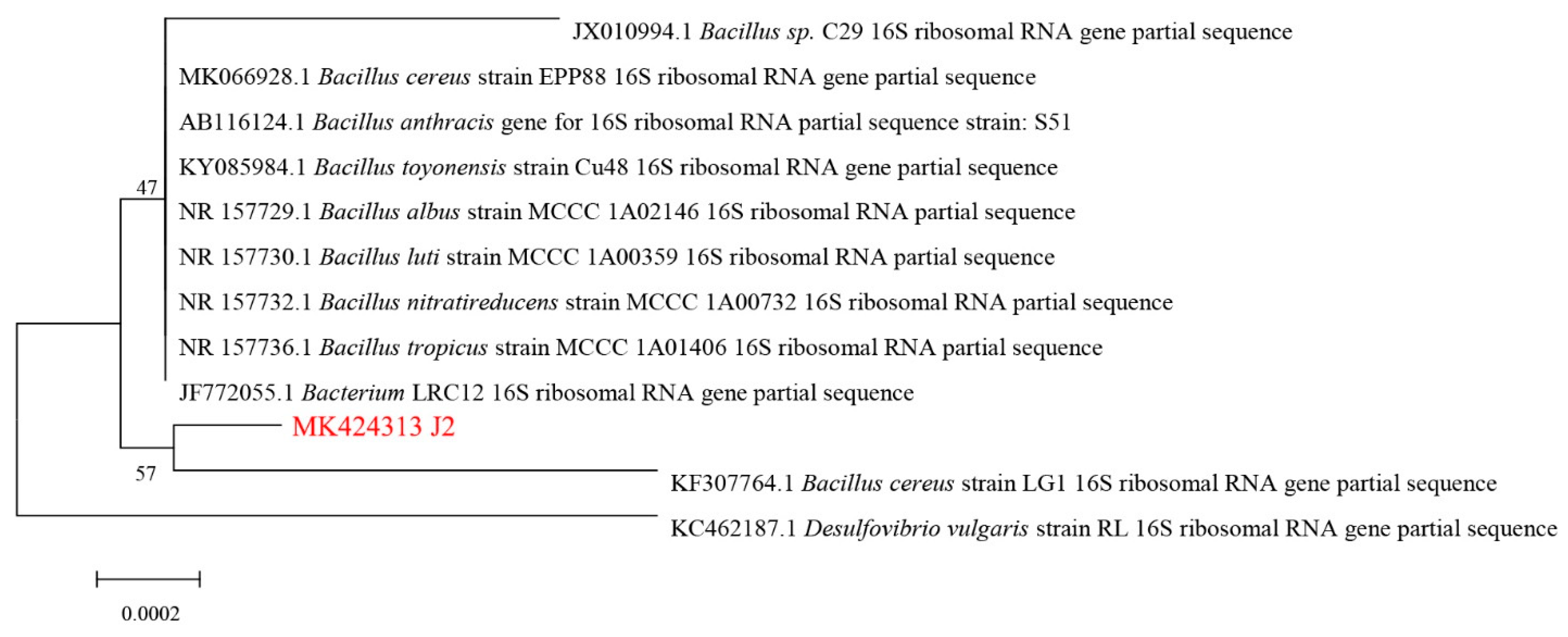
3.1. Isolation and Identification of the SM2-Degrading Strain

Under aerobic conditions, the activated sludge was acclimated with SM2 as the sole carbon source. The change in biomass and degradation of SM2 by the activated sludge during the acclimation process is shown in Figure. 1. After a 46-day acclimation period, the degradation rate of SM2 (50 mg/L) in activated sludge consistently reached almost 100% within 2 days. Following acclimation and selective enrichment with SM2, four morphologically distinct bacterial strains, designated J1-J4, were isolated.
The OD600 of strain J1 remained essentially unchanged, slightly decreasing in the later stages of the reaction, strains J2 and J3 showed a slight increase. The degradation of SM2 by J1 and J4 was 44.91% and 9.01%, whereas strains J2 and J3 had higher degradation efficiencies for SM2 within 72 h, reaching 94.21% and 80.84% (Figure 1). Strain J2, characterized by substantial growth and enhanced capability to degrade SM2, was isolated and chosen for additional study.
On the enrichment medium agar plate, strain J2 formed smooth, circular, non-pigmented colonies ranging from pink to creamy white, with diameters ranging from 2 to 4 mm following 24 h of incubation at 30 °C. The morphological and phenotypical characteristics of J2 are presented in Table 1, and sugar (alcohol, glycosides) fermentation experiments of strain J2 are summarized in Table 2. Light microscopy revealed that strain J2 was composed of motile, gram-positive, short rods. In 42 sugar fermentation experiments, strain J2 was able to utilize 19 types of sugars to produce acid, while 23 types of sugars did not result in acid production.
SEM revealed that strain J2 is a rod-shaped bacterium with approximate dimensions of 0.5 × 2 µm, flagellated, and featuring irregular surface protrusions (Figure 2). TEM indicated that strain J2 has a relatively thick cell wall, distinct bud scars, and longer flagella (with occasional flagellar detachment), some cells of strain J2 are undergoing binary fission (Figure 3).
Partial 16S rRNA sequencing of strain J2 (Genbank accession number MK424313) revealed a 99% similarity to the Bacillus cereus sequence (Figure 4). Therefore, the strain J2 was identified as Bacillus cereus. Bacillus cereus has been reported to degrade various organic pollutants, including chlortetracycline, pyrene, pyrethroid, low-density polyethylene (LDPE), and polystyrene [35,36,37,38,39]. Dong et al. [40] also discovered a Bacillus cereus strain capable of degrading SM2 in 2022, however, the degradation efficiency reported was significantly lower compared to that of strain J2.

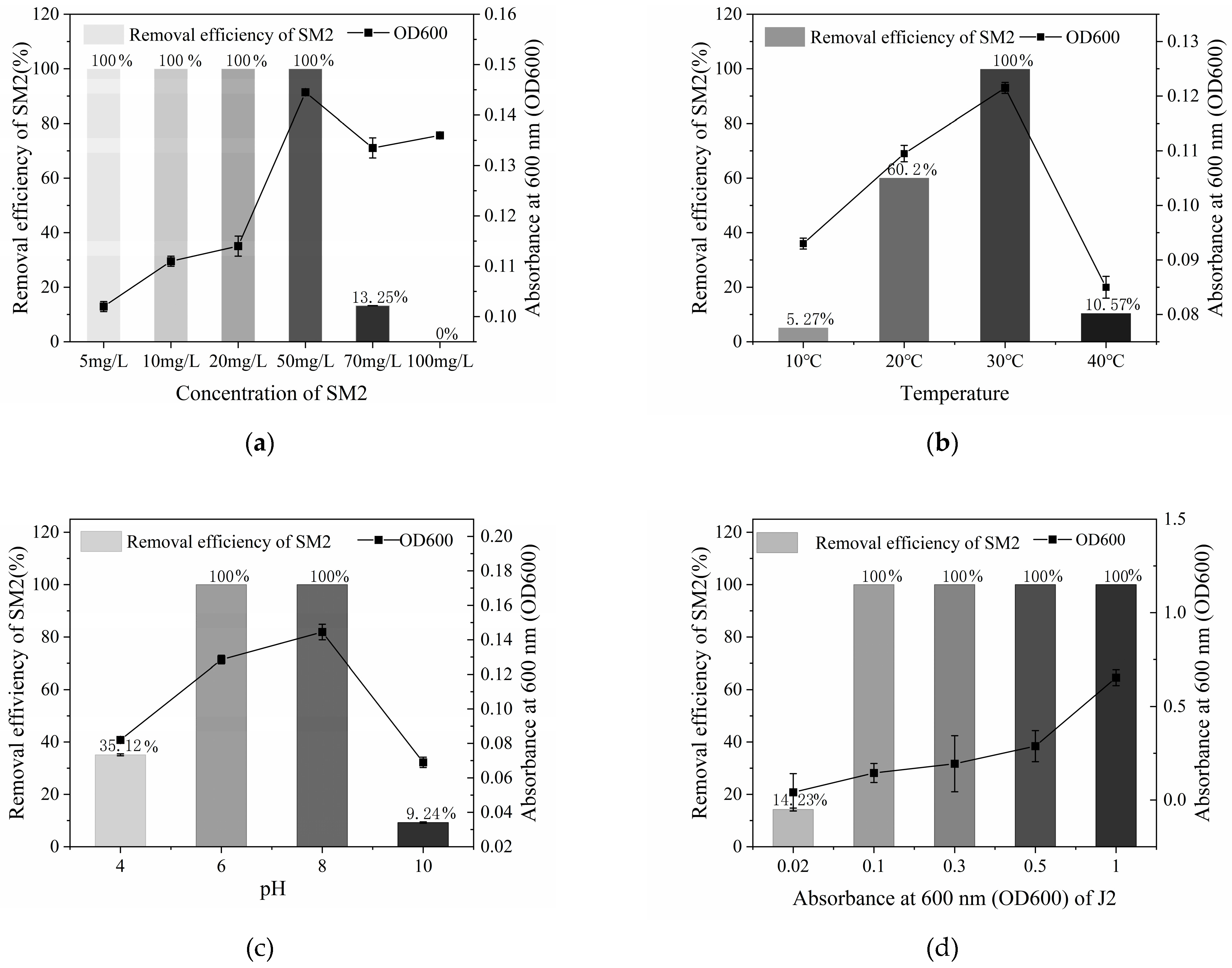
3.2. Optimal Conditions for SM2 Biodegradation by Strain J2

Several factors, including contaminant concentration, temperature, bacterial inoculum size, and pH significantly influence the degradation of contaminants. In order to achieve an optimal removal of SM2, effects of these factors was studied in this section.
Effect of SM2 concentration. As shown in Figure 5a, strain J2 achieved a 100% degradation rate within 36 h at initial SM2 concentrations ranging from 5 to 50 mg/L. The degradation rate at a concentration of 70 mg/L SM2 was 13.25%, and no significant degradation was observed at 100 mg/L within the same timeframe. An inverse relationship was observed between the initial SM2 concentration and the degradation rate, potentially attributable to the antibiotic’s inhibitory effect on bacterial activity; the higher the sulfonamide concentration, the more pronounced the negative impact on compound degradation [34]. This observation is corroborated by the study of Selvi et al. [41], which noted a similar phenomenon. In practical applications, it is necessary to ensure the degradation effect while handling higher concentrations of SM2. Consequently, an initial concentration of 50 mg/L was deemed an optimal starting point for strain J2 to effectively degrade SM2.
Effect of temperature. Temperature significantly influences the growth of strain J2 as well as the degradation of SM2, as shown in Figure 5b. Below 30 °C, the biomass of the strain (OD600) and the degradation increased with rising temperature. At 30 °C, 100% degradation was achieved within 36 h. However, when the cultivation temperature was further raised to 40 °C, the biomass of the strain decreased, and the degradation rate of SM2 also dropped, reaching only 10.57%. Temperature extremes were found to adversely affect strain J2′s degradation performance, either through enzyme inactivation at high temperatures or reduced enzymatic activity at low temperatures, both of which hinder normal metabolic degradation processes [42,43]. The optimal growth temperature for strain J2 was established at 30 °C.
Effect of pH. Figure 5c illustrates that at pH 6.0 and 8.0, strain J2 exhibited high OD600 values and achieved 100% SM2 degradation within 36 h, indicating optimal metabolic activity under these conditions. In contrast, at pH 4.0 and 10.0, the growth of strain J2 was inhibited, and SM2 degradation rates were significantly reduced to 35.12% and 9.24%, respectively. Changes in pH can alter the structure of microbial cell membranes and influence enzyme activity. Moreover, changes in pH may affect the ionization of molecules [34]. When the initial pH is 6.0 or 8.0, the growth condition of strain J2 is the best, and the degradation efficiency towards SM2 is the highest. Due to the fact that sulfonamides dissolve readily in weak alkaline environments [44], the optimal pH for strain J2 was determined to be pH 8.0.
Effect of Bacterial Inoculum Size (OD600). As shown in Figure 5d, increasing the cell concentration enhanced SM2 degradation, with strain J2 achieving 100% degradation rate at initial OD600 ranging from 0.1 to 1.0. The amount of substrate consumed by the metabolism of strain J2 also increases, thereby accelerating the degradation of the carbon source SM2. However, at higher cell concentrations (OD600 = 0.5 or 1.0), the substrate limitation reducing the advantage of strain J2 in degrading SM2. To balance degradation efficiency and cost-effectiveness, an initial OD600 of 0.1 was identified as the optimal starting biomass for strain J2 to degrade SM2.

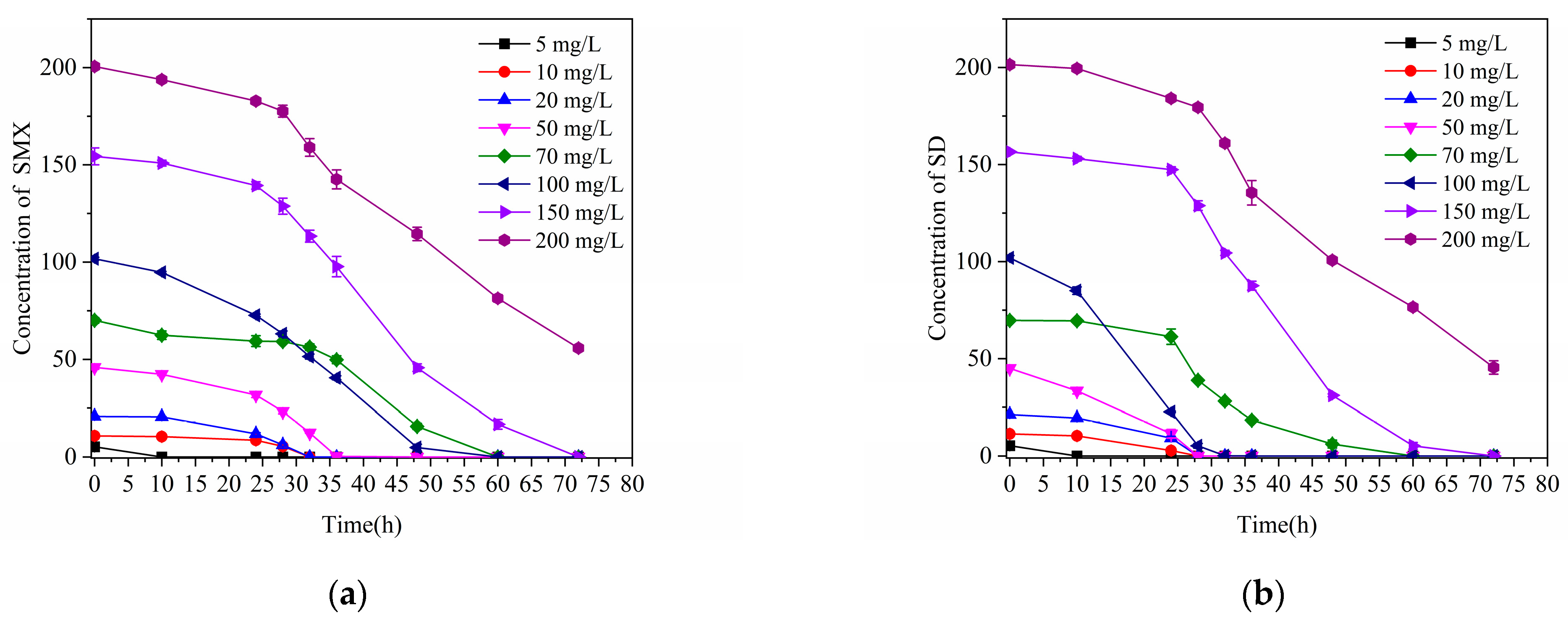
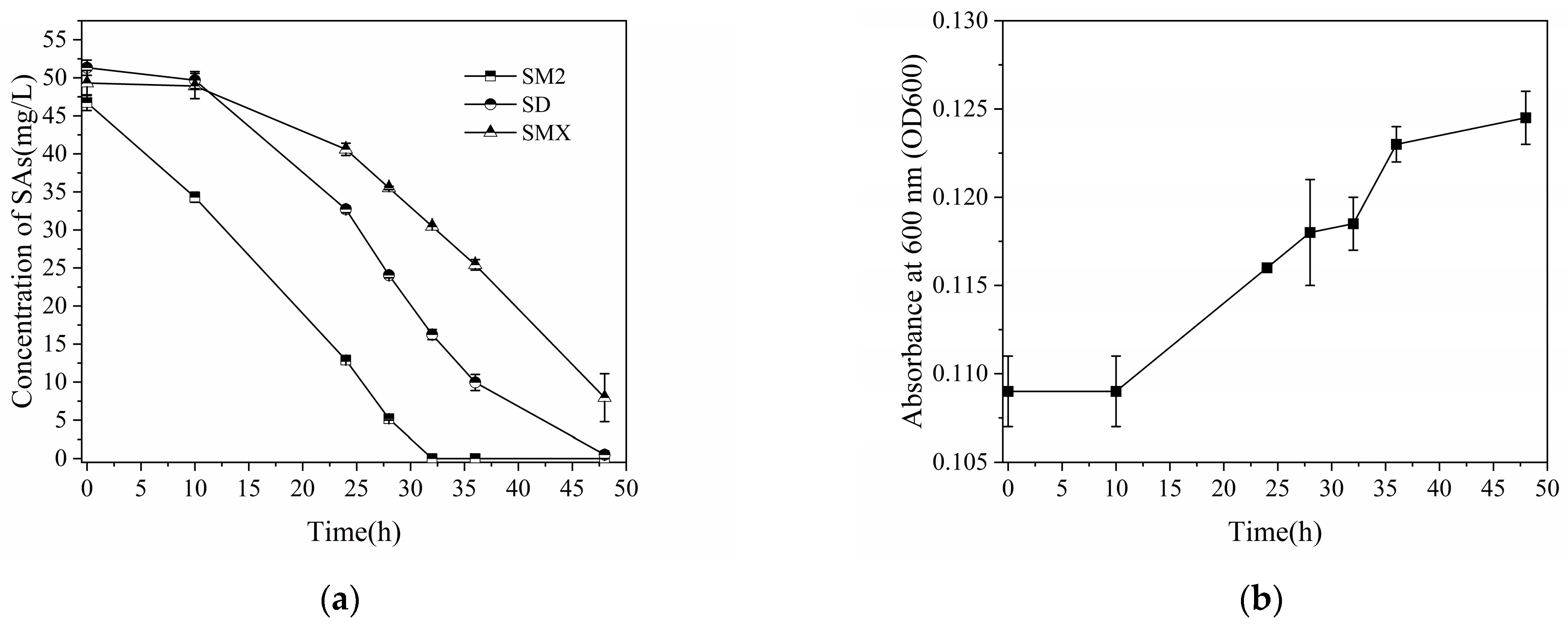
3.3. Degradation of Other Sulfonamides by Strain J2

SMX, SD, and SM2 are common sulfonamide antibiotics detected in the environment [45,46,47]. This section evaluated the degradation efficiency of strain J2 for different concentrations of SMX and SD. Strain J2 exhibited excellent degradation performance for both compounds. With increasing compound concentrations, the lag phase lengthened. Nevertheless, strain J2 was capable of completely degrading 150 mg/L of SMX and SD within 72 h. After a longer lag phase, it achieved degradation efficiencies of 72.18% for SMX and 77.44% for SD at a concentration of 200 mg/L (Figure 6).
In natural environments, sulfonamides typically coexist [48]. Thus, this study also assessed the degradation and growth performance of strain J2 in the presence of all three sulfonamides in a mixed system. As shown in Figure 7, strain J2 was able to grow normally and degrade all three compounds. SM2 degradation reached 100% within 32 h, while the degradation efficiencies of SMX and SD were slightly reduced. Within 48 h, strain J2 degraded 83.87% of SMX and 99.03% of SD. Furthermore, the biomass of strain J2 followed an “S”-shaped growth curve in the mixed system, demonstrating its ability to utilize sulfonamide compounds as the sole carbon source for growth and proliferation.

3.4. SM2 Biodegradation and Cell Growth Kinetics Determination

The growth curves of strain J2 at different SM2 concentrations, fitted using the Logistic model, are presented in Figure 8 and Table 3. At an initial SM2 concentration of 5 mg/L, the strain quickly entered the logarithmic phase. However, due to substrate limitation, it quickly transitioned into the stationary phase. When the initial SM2 concentration exceeded 20 mg/L, strain J2 exhibited an adaptation period of approximately 12 h (Figure 8). This adaptation period may be due to the strain requiring time to adjust to the relatively lower substrate availability and the new culture environment as SM2 concentrations increased. At low SM2 concentrations, the strain could sustain growth by degrading SM2 directly. In contrast, at higher concentrations, the toxicity of SM2 might also contribute to the slower initial growth phase [49,50].
Among all tested concentrations, the best growth performance was observed at 50 mg/L SM2, where the logarithmic growth phase lasted the longest. This may be attributed to the strain effectively utilizing SM2 as a carbon source. However, as the cultivation progressed, substrate consumption and the accumulation of toxic intermediates led to the death phase, reducing the number of viable cells. It is worth noting that OD600 measurements reflect both live and dead cells. As a result, the decrease in OD values during the death phase was not significant [33]. The R2 values of the fitted Logistic model ranged from 0.94 to 0.99, indicating a strong correlation between the model predictions and experimental data. These results confirm that the Logistic model can accurately describe the biomass growth trends of strain J2 over time at different SM2 concentrations (Table 3).
The growth kinetics of bacterial strains on inhibitory organic substrates were analyzed using the Haldane equation [49,51]. The specific growth rate (µ) of strain J2 in relation to SM2 concentration was effectively represented by the Haldane equation, as depicted in Figure 9. The kinetic parameters for strain J2 during the degradation of SM2 were as follows: µmax, 0.066 h−1, Ks, 186.667 mg/L, Ki, 14.127 mg/L.
The maximum specific growth rate was observed at an initial SM2 concentration of 51.35 mg/L, indicating that strain J2 exhibits a certain level of resistance to SM2. However, when the SM2 concentration surpassed 51.35 mg/L, a marked reduction in growth was noted, likely due to the increasing inhibitory effect of SM2 at higher concentrations.
Comparison with previous studies. Research on the biodegradation of SM2 has gained attention, with most studies conducted in the last five years (Table 4). However, the current understanding of SM2 biodegradation remains limited. Pure bacterial strains capable of degrading SM2 often require the addition of an external co-metabolizable carbon source. Furthermore, as shown in Table 4, the degradation efficiency of previously isolated pure strains using SM2 as the sole carbon source is generally unsatisfactory. Over a cultivation period of 26 to 168 h, the degradation rate ranges from 18.53% to 100%, influenced by factors such as strain type, SM2 concentration, and environmental conditions.
In contrast, strain J2 demonstrated superior performance compared to most previously reported pure strains. It achieved a maximum specific growth rate (μmax = 0.066 h−1) at an initial SM2 concentration of 51.35 mg/L. Strain J2 could completely degrade 50 mg/L of SM2 within 36 h. Additionally, it exhibited a remarkable tolerance to SM2, withstanding concentrations up to 100 mg/L—far exceeding the 1.5 mg/L to 50 mg/L tolerance levels of other strains. This high tolerance and resistance to the inhibitory effects of organic compounds are critical for its sustained, stable, and efficient degradation of SM2 [52].
In summary, strain J2, isolated in this study, effectively utilizes SM2 as the sole carbon source and exhibits a strong degradation capability. Its high tolerance and efficiency suggest significant potential for application in the treatment of wastewater and soils contaminated with sulfonamide antibiotics.
Table 4. Comparison of SM2 degradation effect and parameters.
MicroorganismCarbon SourceConc. Range
(mg/L)
Conc.at μm (mg/L)T
(℃)
pHIncubation Time (h)Degradation Rate (%)μm
(h−1)
Ref
Geobacillus sp. S-07glucose1010706.0-98.00-[26]
Trametes versicolorglucose9925-48100-[53]
Achromobacter sp. S-3glucose55308.02433.00-[54]
S. oneidensis MR-1SM222307.012023.00 ± 4.12-[34]
Shewanella sp. MR-4SM222307.012033.00 ± 2.58-[34]
Klebsiella Trevisan SX5glucose2020307.06050.000.0115[55]
AcinetobacerSM20.05~5.00.05107.09642.00−0.0089[56]
Fusarium solaniSM21.51.5307.016818.53-[57]
Paenarthrobacter sp. A01SM210~500100257.89696.7-[58]
Bacillus licheniformis ATCC 14580SM240-306.9100 0.2~7.8-[59]
Achromobacter mucicolens JD417SM25051367-460.088[18]
Paenarthrobacter ureafaciens strain YL1. SM2100-3077298-[60]
Bacillus thuringiensis H38SM25~20102573697.3-[61]
Bacillus cereus H38SM25-257.096100-[40]
Irpex lacteus WRF-ILSM20~2010354.52697.1-[62]
Bacillus cereus J2SM25~10051.35308.0361000.066This study
Note(s): Conc. = concentration, T, temperature, -, data not available. The Haldane model (Equation (2)) was used to calculate kinetic coefficients.

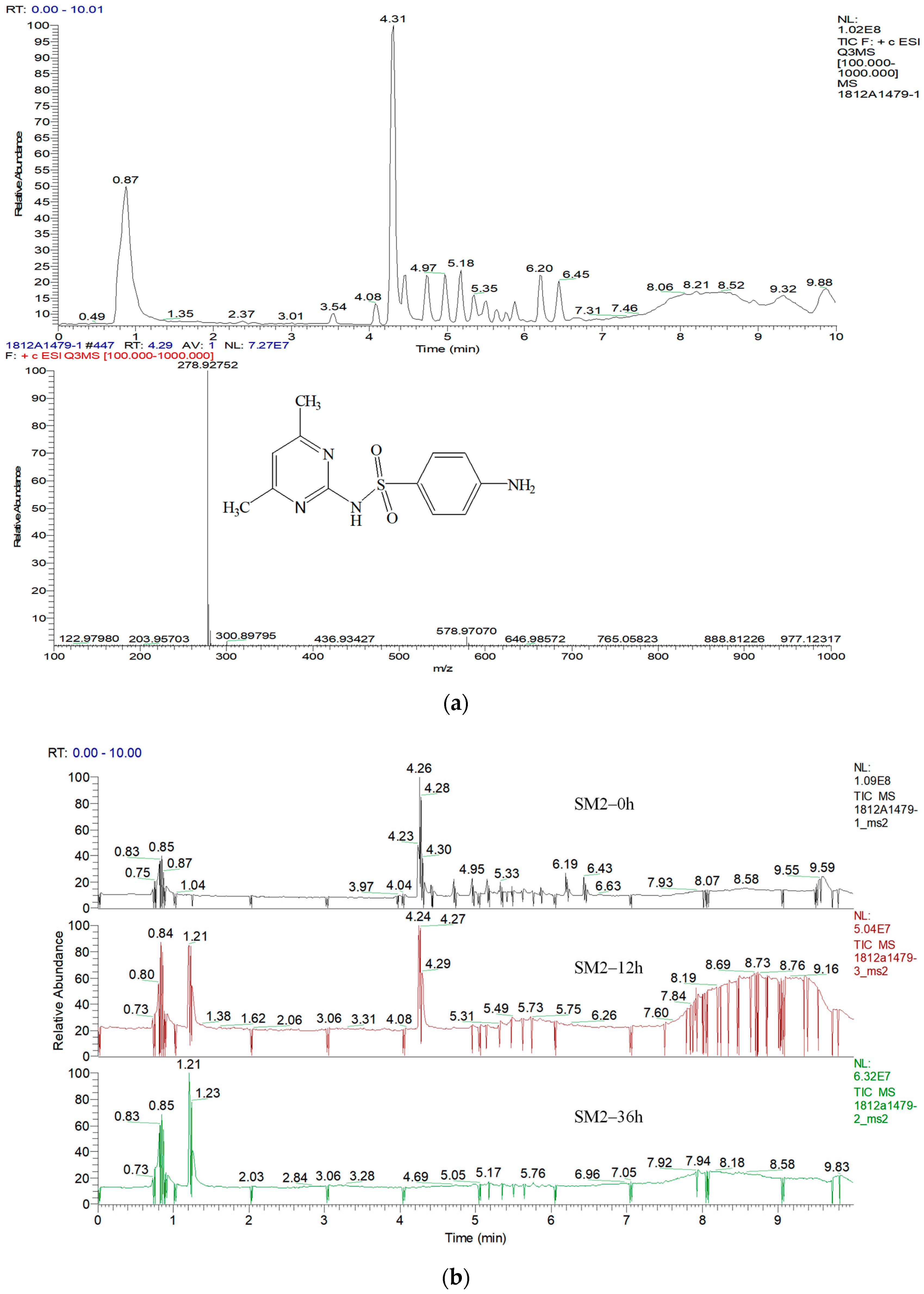
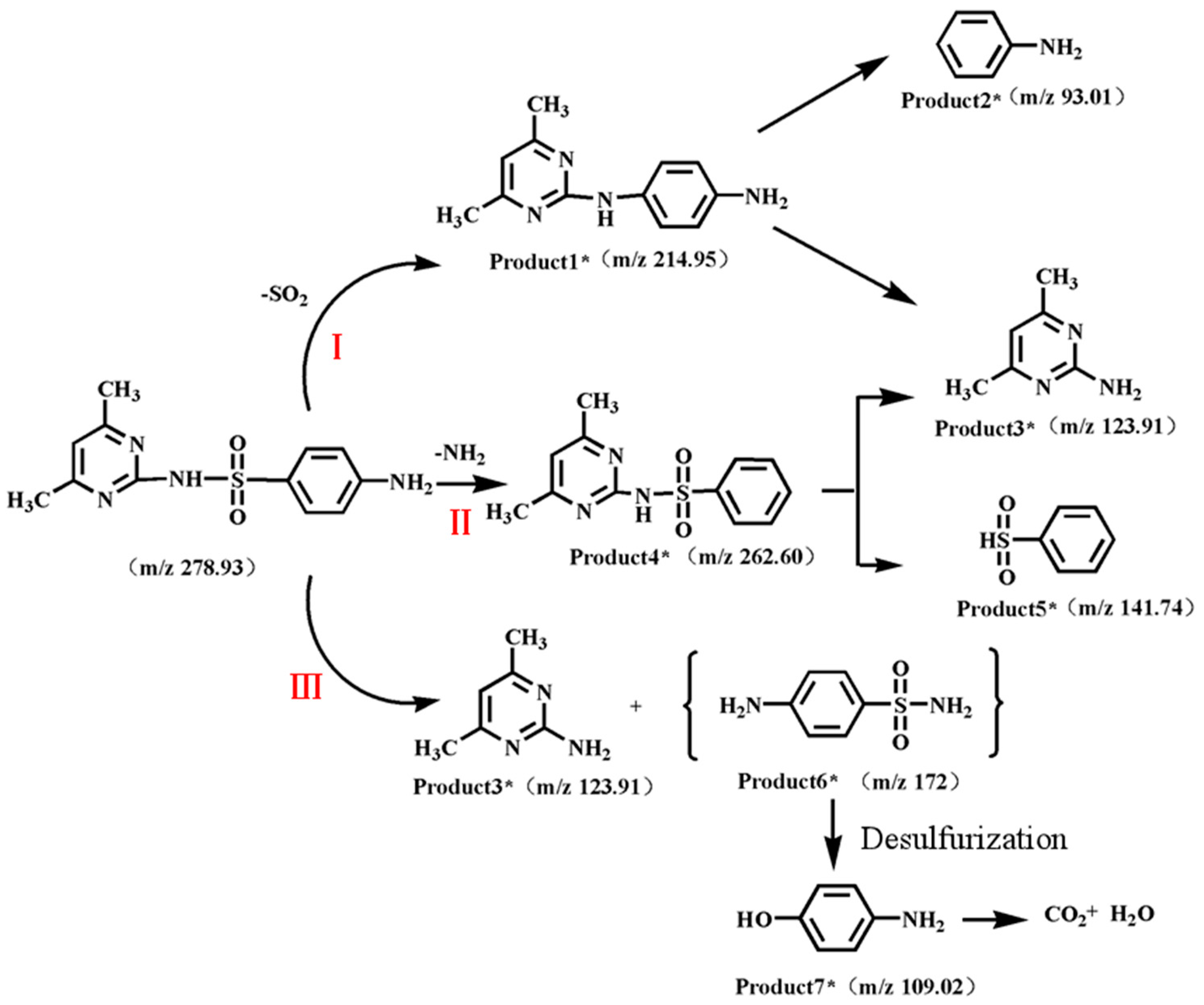
3.5. Products and Pathways of Sulfonamide Degradation

The standard chromatogram and mass spectrum of SM2 measured by TQ-LCMS are shown in Figure 10a, with the retention time of SM2 being 4.31 min and the mass-to-charge ratio (m/z) being 278.92752. Figure 10b shows the chromatograms spectrogram of degrading SM2 in different time. Based on the m/z values from the first and second mass spectrometry analyses, six intermediate products were identified. It is inferred that one additional intermediate product was not detected. The seven intermediate products, including the inferred one, are summarized in Table A1. In the seven intermediate products, including the desulfurized product (7), deaminated product (4), inter-ring coupling product (1), and bond-cleavage products (2, 3, 5, 6), it is evident that the degradation of SM2 primarily proceeds through desulfurization, reactive oxygen species oxidation, and hydrolytic bond-cleavage pathways, with the degradation of SM2 being accomplished through the intersection of multiple pathways. Three pathways by which strain J2 degrades SM2 are illustrated in Figure 11.
2-amino-4,6-dimethylpyrimidine (3) is a major degradation product of SM2, which is consistent with previous reports [16,33,53,63]. However, its concentration did not decrease as the reaction proceeded, suggesting that it may not be degraded by strain J2 and thus persists in the system. In the product mass spectrum, 4-amino-hydroxybenzenesulfonamide (6) was not detected, possibly due to the extreme instability of this compound [64]. In the mass spectrum at 12 h of the reaction, p-aminophenol (7) was present, but it was not detectable after 36 h of reaction. It is speculated that strain J2 can mineralize p-aminophenol into CO2 and H2O.

4. Conclusions

This study successfully isolated a highly efficiency SM2-degrading bacterial strain, identified as Bacillus cereus J2. The optimal conditions for J2 to degrade SM2 were a pH of 8, a temperature of 30 °C, and an inoculation amount of 0.1 OD600. The degradation rate of 50 mg/L SM2 reached 100% after 36 h. Strain J2 exhibits a high degradation efficiency towards SD and SMX, capable of completely degrading concentrations of 150 mg/L within 72 h. In a mixed system containing three sulfonamide compounds, strain J2 was able to grow normally and degrade each compound effectively. Growth kinetic parameters indicate that, compared to most of previously isolated pure strains, strain J2 exhibits strong SM2 removal capability and high tolerance to SM2. The potential metabolic pathway of SM2 by strain J2 was investigated using TQ-LCMS analysis. However, the biodegradation mechanism of SM2 is complex, and the functional genes and associated enzymes involved in the degradation of SM2 by strain J2 remain unclear, necessitating further research. The biodegradation products of SM2 are diverse, and further analysis of the metabolic products using carbon labeling or isotope labeling methods could provide deeper insights into the degradation pathways of this strain. Additionally, it is necessary to conduct quantitative analysis of the intermediate products of SM2 degradation as the reaction progresses, as well as to analyze the toxicity of both individual degradation products and their mixtures in order to determine their structures and potential impacts.

Author Contributions

Conceptualization, T.L. and J.Z.; methodology, J.Z. and Y.T.; software, Z.L.; validation, J.Z. and X.Z.; data curation, J.Z.; writing—original draft preparation, J.Z.; writing—review and editing, Z.W.; supervision, Z.W.; project administration, Z.W.; funding acquisition, T.L., X.Z. and Z.W. All authors have read and agreed to the published version of the manuscript.

Funding

This research was funded by National Natural Science Foundation of China (No. 42407164); the Key Research and Development Plans in Yunnan Province for the “Green governance and development technical research and application demonstration in Chishui River Basin” (No. 202203AC100001); the Science and Technology Program of Guangdong (No. 2024B1212040001); Guangdong-Hong Kong Joint Laboratory for Water Security (No. 2020B1212030005); Zhuhai Science and Technology Program for Social Development (No. 2420004000307).

Institutional Review Board Statement

Not applicable.

Data Availability Statement

The data presented in this study are available on request from the corresponding author.

Conflicts of Interest

The authors declare no conflicts of interest.

Appendix A

Table A1. The products of SM2-degrading.
ProductsFormulaRetention Timem/zStructural FormulaReferences
1C12H14N41.24214Water 17 00468 i001[53,63]
2C6H7N9.7893Water 17 00468 i002-
3C6H9N31.21123Water 17 00468 i003[16,33,53,63]
4C12H13N3O2S0.89263Water 17 00468 i004[53]
5C6H6O2S9.81142Water 17 00468 i005[53]
6C6H8N2O2S-172Water 17 00468 i006[51]
7C6H7NO7.84109Water 17 00468 i007[33]
Note(s): {} denotes that the compound was not detected.

References

  1. Jeśman, C.; Młudzik, A.; Cybulska, M. History of antibiotics and sulphonamides discoveries. Environ. Sci. Pollut. Res. 2011, 30, 320–322. [Google Scholar]
  2. Dmitrienko, S.G.; Kochuk, E.V.; Apyari, V.V.; Tolmacheva, V.V.; Zolotov, Y.A. Recent Advances in Sample Preparation Techniques and Methods of Sulfonamides Detection—A Review. Anal. Chim. Acta 2014, 850, 6–25. [Google Scholar] [CrossRef] [PubMed]
  3. Baran, W.; Adamek, E.; Ziemiańska, J.; Sobczak, A. Effects of the Presence of Sulfonamides in the Environment and Their Influence on Human Health. J. Hazard. Mater. 2011, 196, 1–15. [Google Scholar] [CrossRef] [PubMed]
  4. Ottosen, C.F.; Bjerg, P.L.; Kümmel, S.; Richnow, H.H.; Middeldorp, P.; Draborg, H.; Lemaire, G.G.; Broholm, M.M. Natural Attenuation of Sulfonamides and Metabolites in Contaminated Groundwater—Review, Advantages and Challenges of Current Documentation Techniques. Water Res. 2024, 254, 121416. [Google Scholar] [CrossRef]
  5. Duan, W.; Cui, H.; Jia, X.; Huang, X. Occurrence and Ecotoxicity of Sulfonamides in the Aquatic Environment: A Review. Sci. Total Environ. 2022, 820, 153178. [Google Scholar] [CrossRef] [PubMed]
  6. Yang, Q.; Lu, T.; Zhang, Q.; Farooq, U.; Wang, B.; Qi, Z.; Miao, R. Transport of Sulfanilamide in Saturated Porous Media under Different Solution Chemistry Conditions: Role of Physicochemical Characteristics of Soils. Environ. Sci. Pollut. Res. 2024, 31, 11622–11632. [Google Scholar] [CrossRef]
  7. Kim, Y.R.; Park, S.; Kim, J.Y.; Choi, J.-D.; Moon, G.-I. Simultaneous Determination of 31 Sulfonamide Residues in Various Livestock Matrices Using Liquid Chromatography-Tandem Mass Spectrometry. Appl. Biol. Chem. 2024, 67, 13. [Google Scholar] [CrossRef]
  8. Kim, D.-W.; Thawng, C.N.; Lee, K.; Wellington, E.M.H.; Cha, C.-J. A Novel Sulfonamide Resistance Mechanism by Two-Component Flavin-Dependent Monooxygenase System in Sulfonamide-Degrading Actinobacteria. Environ. Int. 2019, 127, 206–215. [Google Scholar] [CrossRef]
  9. Ren, T.-T.; Li, X.-Y.; Wang, Y.; Zou, Y.-D.; Liao, X.-D.; Liang, J.-B.; Wu, Y.-B. Effect of Different Sulfadimidine Addition Methods on Its Degradation Behaviour in Swine Manure. Environ. Sci. Pollut. Res. 2017, 24, 7253–7263. [Google Scholar] [CrossRef]
  10. Lin, T.; Yu, S.; Chen, Y.; Chen, W. Integrated Biomarker Responses in Zebrafish Exposed to Sulfonamides. Environ. Toxicol. Pharmacol. 2014, 38, 444–452. [Google Scholar] [CrossRef]
  11. Moore, J.E.; Moore, P.J.A.; Millar, B.C.; Goldsmith, C.E.; Loughrey, A.; Rooney, P.J.; Rao, J.R. The Presence of Antibiotic Resistant Bacteria along the River Lagan. Agric. Water Manag. 2010, 98, 217–221. [Google Scholar] [CrossRef]
  12. Luo, Y.; Mao, D.; Rysz, M.; Zhang, H.; Xu, L.; Alvarez, P.J.J. Trends in Antibiotic Resistance Genes Occurrence in the Haihe River, China. Environ. Sci. Technol. 2010, 44, 7220–7225. [Google Scholar] [CrossRef]
  13. Huang, H.; Liu, S.; Kang, Z.; Zhu, Y.; Zhang, C.; Xiang, E.; Lin, Z.; Liu, W. Effects of Atmosphere and Stepwise Pyrolysis on the Pyrolysis Behavior, Product Characteristics, and N/S Migration Mechanism of Vancomycin Fermentation Residue. Chem. Eng. J. 2024, 498, 155012. [Google Scholar] [CrossRef]
  14. Loganathan, P.; Vigneswaran, S.; Kandasamy, J.; Cuprys, A.K.; Maletskyi, Z.; Ratnaweera, H. Treatment Trends and Combined Methods in Removing Pharmaceuticals and Personal Care Products from Wastewater—A Review. Membranes 2023, 13, 158. [Google Scholar] [CrossRef]
  15. Shi, B.-J.; Wang, Y.; Geng, Y.-K.; Liu, R.-D.; Pan, X.-R.; Li, W.-W.; Sheng, G.-P. Application of Membrane Bioreactor for Sulfamethazine-Contained Wastewater Treatment. Chemosphere 2018, 193, 840–846. [Google Scholar] [CrossRef]
  16. Yang, N.; Wan, J.; Zhao, S.; Wang, Y. Removal of Concentrated Sulfamethazine by Acclimatized Aerobic Sludge and Possible Metabolic Products. PeerJ 2015, 3, e1359. [Google Scholar] [CrossRef]
  17. Xu, B.; Lin, Z.; Li, F.; Tao, T.; Zhang, G.; Wang, Y. Local O2 Concentrating Boosts the Electro-Fenton Process for Energy-Efficient Water Remediation. Proc. Natl. Acad. Sci. USA 2024, 121, e2317702121. [Google Scholar] [CrossRef]
  18. Zeng, L.; Du, H.; Lin, X.; Liao, R.; Man, Y.; Fang, H.; Yang, Y.; Tao, R. Isolation, Identification and Whole-Genome Analysis of an Achromobacter Strain with a Novel Sulfamethazine Resistance Gene and Sulfamethazine Degradation Gene Cluster. Bioresour. Technol. 2024, 399, 130598. [Google Scholar] [CrossRef]
  19. An, X.; Wang, Y.; Yu, C.; Hu, X. Biochar-Bacteria Coupling System Enhanced the Bioremediation of Phenol Wastewater-Based on Life Cycle Assessment and Environmental Safety Analysis. J. Hazard. Mater. 2024, 480, 136414. [Google Scholar] [CrossRef]
  20. Zhuo, T.; He, L.; Chai, B.; Zhou, S.; Wan, Q.; Lei, X.; Zhou, Z.; Chen, B. Micro-Pressure Promotes Endogenous Phosphorus Release in a Deep Reservoir by Favouring Microbial Phosphate Mineralisation and Solubilisation Coupled with Sulphate Reduction. Water Res. 2023, 245, 120647. [Google Scholar] [CrossRef]
  21. Li, N.; Liu, Q.; Zhou, G.-Q.; Dai, M.-X.; Kong, Q. Contaminant Removal and Microorganism Response of Activated Sludge in Sulfamethazine Wastewater Treatment. Int. Biodeterior. Biodegrad. 2019, 143, 104705. [Google Scholar] [CrossRef]
  22. Lin, B.; Lai, Y.; Ke, Y.; Huang, Y.; Tao, Y.; Han, X.; Ma, J. Recent Advances in Biofilm Technologies for Breeding Wastewater Treatment: Fundamentals, Performance and Impacts of Antibiotics. J. Water Process Eng. 2024, 62, 105429. [Google Scholar] [CrossRef]
  23. Meng, L.; Liang, L.; Shi, Y.; Yin, H.; Li, L.; Xiao, J.; Huang, N.; Zhao, A.; Xia, Y.; Hou, J. Biofilms in Plastisphere from Freshwater Wetlands: Biofilm Formation, Bacterial Community Assembly, and Biogeochemical Cycles. J. Hazard. Mater. 2024, 476, 134930. [Google Scholar] [CrossRef]
  24. Chen, J.; Xie, S. Overview of Sulfonamide Biodegradation and the Relevant Pathways and Microorganisms. Sci. Total Environ. 2018, 640–641, 1465–1477. [Google Scholar] [CrossRef]
  25. Gauthier, H.; Yargeau, V.; Cooper, D.G. Biodegradation of Pharmaceuticals by Rhodococcus rhodochrous and Aspergillus niger by Co-Metabolism. Sci. Total Environ. 2010, 408, 1701–1706. [Google Scholar] [CrossRef]
  26. Pan, L.; Tang, X.; Li, C.; Yu, G.; Wang, Y. Biodegradation of Sulfamethazine by an Isolated Thermophile–Geobacillus sp. S-07. World J. Microbiol. Biotechnol. 2017, 33, 85. [Google Scholar] [CrossRef]
  27. Birkigt, J.; Gilevska, T.; Ricken, B.; Richnow, H.-H.; Vione, D.; Corvini, P.F.-X.; Nijenhuis, I.; Cichocka, D. Carbon Stable Isotope Fractionation of Sulfamethoxazole during Biodegradation by Microbacterium sp. Strain BR1 and upon Direct Photolysis. Environ. Sci. Technol. 2015, 49, 6029–6036. [Google Scholar] [CrossRef]
  28. Tappe, W.; Hofmann, D.; Disko, U.; Koeppchen, S.; Kummer, S.; Vereecken, H. A Novel Isolated Terrabacter-like Bacterium Can Mineralize 2-Aminopyrimidine, the Principal Metabolite of Microbial Sulfadiazine Degradation. Biodegradation 2015, 26, 139–150. [Google Scholar] [CrossRef]
  29. Peng, X.; Li, T.; Zheng, Q.; Lu, Y.; He, Y.; Tang, Y.; Qiu, R. Citrobacter sp. Y3 Harbouring Novel Gene HBCD-Hd-1 Mineralizes Hexabromocyclododecane via New Metabolic Pathways According to Multi-Omics Characterization. J. Hazard. Mater. 2023, 442, 130071. [Google Scholar] [CrossRef]
  30. Pan, X.-R.; Shang-Guan, P.-K.; Li, S.-H.; Zhang, C.-H.; Lou, J.-M.; Guo, L.; Liu, L.; Lu, Y. The Influence of Carbon Dioxide on Fermentation Products, Microbial Community, and Functional Gene in Food Waste Fermentation with Uncontrol pH. Environ. Research. 2025, 267, 120645. [Google Scholar] [CrossRef]
  31. Zhuo, T.; Yu, K.; Chai, B.; Tang, Q.; Gao, X.; Wang, J.; He, L.; Lei, X.; Li, Y.; Meng, Y.; et al. Microplastics Increase the Microbial Functional Potential of Greenhouse Gas Emissions and Water Pollution in a Freshwater Lake: A Metagenomic Study. Environ. Res. 2024, 257, 119250. [Google Scholar] [CrossRef]
  32. Zheng, H.; Huang, S.; Huang, J.; Zeng, H.; Xu, M.; Cai, A.; Zhou, S.; Ma, X.; Deng, J. Unveiling the Optical and Molecular Characteristics of Aging Microplastics Derived Dissolved Organic Matter Transformed by UV/Chlor(Am)Ine Oxidation and Its Potential for Disinfection Byproducts Formation. J. Hazard. Mater. 2024, 480, 136440. [Google Scholar] [CrossRef] [PubMed]
  33. Fu, Y. Isolation And Degradation Characteristic of A New Sulfamethazine-Degrading Bacterium. Master’s Thesis, Harbin Institute of Technonogy, Harbin, China, 2019. [Google Scholar]
  34. Mao, F.; Liu, X.; Wu, K.; Zhou, C.; Si, Y. Biodegradation of Sulfonamides by Shewanella oneidensis MR-1 and Shewanella sp. Strain MR-4. Biodegradation 2018, 29, 129–140. [Google Scholar] [CrossRef]
  35. Zhang, S.; Wang, J. Biodegradation of Chlortetracycline by Bacillus cereus LZ01: Performance, Degradative Pathway and Possible Genes Involved. J. Hazard. Mater. 2022, 434, 128941. [Google Scholar] [CrossRef]
  36. Huang, Y.; Yang, L.; Pan, K.; Yang, Z.; Yang, H.; Liu, J.; Zhong, G.; Lu, Q. Heavy Metal-Tolerant Bacteria Bacillus Cereus BCS1 Degrades Pyrethroid in a Soil–Plant System. J. Hazard. Mater. 2024, 461, 132594. [Google Scholar] [CrossRef]
  37. Karale, M.A.; Kadam, T.A.; Bhosale, H.J.; More, R.A. A Plasmid Induces Biodegradation of Pyrene by Bacillus Cereus C7 and Identification of Its Degradation Metabolites. Indian J. Microbiol. 2024, 1–11. [Google Scholar] [CrossRef]
  38. Jebashalomi, V.; Emmanuel Charles, P.; Rajaram, R. Microbial Degradation of Low-Density Polyethylene (LDPE) and Polystyrene Using Bacillus cereus (OR268710) Isolated from Plastic-Polluted Tropical Coastal Environment. Sci. Total Environ. 2024, 924, 171580. [Google Scholar] [CrossRef]
  39. Xie, H.; Liu, H.; Liu, G.; Li, X. Degradation of Organic Compounds by a Novel Bacillus cereus BX16 in Starch Waste. Process Biochem. 2024, 144, 89–96. [Google Scholar] [CrossRef]
  40. Dong, Z.; Yan, X.; Wang, J.; Zhu, L.; Wang, J.; Li, C.; Zhang, W.; Wen, S.; Kim, Y.M. Mechanism for Biodegradation of Sulfamethazine by Bacillus cereus H38. Sci. Total Environ. 2022, 809, 152237. [Google Scholar] [CrossRef]
  41. Selvi, A.; Salam, J.A.; Das, N. Biodegradation of Cefdinir by a Novel Yeast Strain, Ustilago sp. SMN03 Isolated from Pharmaceutical Wastewater. World J. Microbiol. Biotechnol. 2014, 30, 2839–2850. [Google Scholar] [CrossRef] [PubMed]
  42. Peng, X.; Zhang, Z.; Luo, W.; Jia, X. Biodegradation of Tetrabromobisphenol A by a Novel Comamonas sp. Strain, JXS-2-02, Isolated from Anaerobic Sludge. Bioresour. Technol. 2013, 128, 173–179. [Google Scholar] [CrossRef]
  43. Fu, B.; Chen, L.; Cai, T.; Yang, Q.; Ding, D. Isolation and characterization of norfloxacin-degrading bacterium strain NOR-36. Acta Sci. Circumstantiae 2017, 37, 576–584. [Google Scholar] [CrossRef]
  44. Xu, M.; Liu, W.; Bai, Y.; Tu, Y. Complexation of Sulfamethazine with Typical Dissolved Organic Matter. J. Environ. Sci. 2018, 38, 4103–4112. [Google Scholar] [CrossRef]
  45. Li, S.; Wang, R.; Zhang, Y.; Zheng, H.; Dong, X.; Leong, Y.K.; Chang, J.-S. Ecological Risks of Sulfonamides and Quinolones Degradation Intermediates: Toxicity, Microbial Community, and Antibiotic Resistance Genes. Bioresour. Technol. 2024, 418, 131967. [Google Scholar] [CrossRef]
  46. Wang, Q.; Wang, H.; Lv, M.; Wang, X.; Chen, L. Sulfamethoxazole Degradation by Aeromonas caviae and Co-Metabolism by the Mixed Bacteria. Chemosphere 2023, 317, 137882. [Google Scholar] [CrossRef]
  47. Luo, Y.; Xu, L.; Rysz, M.; Wang, Y.; Zhang, H.; Alvarez, P.J.J. Occurrence and Transport of Tetracycline, Sulfonamide, Quinolone, and Macrolide Antibiotics in the Haihe River Basin, China. Environ. Sci. Technol. 2011, 45, 1827–1833. [Google Scholar] [CrossRef]
  48. Zhang, Y.; Xu, J.; Zhong, Z.; Li, L.; He, Y.; Chen, Y. Degradation of Sulfonamides Antibiotics in Lake Water and Sediment. Environ. Sci. Pollut. Res. Int. 2012, 20, 2372–2380. [Google Scholar] [CrossRef]
  49. Arutchelvan, V.; Kanakasabai, V.; Elangovan, R.; Nagarajan, S.; Muralikrishnan, V. Kinetics of High Strength Phenol Degradation Using Bacillus brevis. J. Hazard. Mater. 2006, 129, 216–222. [Google Scholar] [CrossRef]
  50. Banerjee, I.; Modak, J.M.; Bandopadhyay, K.; Das, D.; Maiti, B.R. Mathematical Model for Evaluation of Mass Transfer Limitations in Phenol Biodegradation by Immobilized Pseudomonas putida. J. Biotechnol. 2001, 87, 211–223. [Google Scholar] [CrossRef]
  51. Deng, Y.; Mao, Y.; Li, B.; Yang, C.; Zhang, T. Aerobic Degradation of Sulfadiazine by Arthrobacter spp.: Kinetics, Pathways, and Genomic Characterization. Environ. Sci. Technol. 2016, 50, 9566–9575. [Google Scholar] [CrossRef]
  52. Batchu, S.R.; Panditi, V.R.; O’Shea, K.E.; Gardinali, P.R. Photodegradation of Antibiotics under Simulated Solar Radiation: Implications for Their Environmental Fate. Sci. Total Environ. 2014, 470–471, 299–310. [Google Scholar] [CrossRef]
  53. García-Galán, M.J.; Rodríguez-Rodríguez, C.E.; Vicent, T.; Caminal, G.; Díaz-Cruz, M.S.; Barceló, D. Biodegradation of Sulfamethazine by Trametes versicolor: Removal from Sewage Sludge and Identification of Intermediate Products by UPLC–QqTOF-MS. Sci. Total Environ. 2011, 409, 5505–5512. [Google Scholar] [CrossRef]
  54. Huang, M.; Tian, S.; Chen, D.; Zhang, W.; Wu, J.; Chen, L. Removal of Sulfamethazine Antibiotics by Aerobic Sludge and an Isolated Achromobacter sp. S-3. J. Environ. Sci. 2012, 24, 1594–1599. [Google Scholar] [CrossRef] [PubMed]
  55. Zhao, F. The Degradation of Sulfamethazine by Microwave and Microorganism. Master’s Thesis, Zhengzhou University, Zhengzhou, China, 2012. [Google Scholar]
  56. Wang, J.; Zhou, A.; Zhang, Y.; Si, C.; Chen, Z.; Qian, H.; Zhao, Z. Research on the Adsorption and Migration of Sulfa Antibiotics in Underground Environment. Environ. Earth Sci. 2016, 75, 1252. [Google Scholar] [CrossRef]
  57. Wang, Q.; Zhu, P.; Xia, Z.; Wang, Y.; Zeng, Y.; Hou, Y. Screening and degradation properties of three kinds of agricultural antibiotics degrading fungi. J. Agric. Resour. Environ. 2018, 35, 533–539. [Google Scholar] [CrossRef]
  58. Cao, L.; Zhang, J.; Zhao, R.; Deng, Y.; Liu, J.; Fu, W.; Lei, Y.; Zhang, T.; Li, X.; Li, B. Genomic Characterization, Kinetics, and Pathways of Sulfamethazine Biodegradation by Paenarthrobacter sp. A01. Environ. Int. 2019, 131, 104961. [Google Scholar] [CrossRef]
  59. Islas-Espinoza, M.; Reid, B.J.; Wexler, M.; Bond, P.L. Soil Bacterial Consortia and Previous Exposure Enhance the Biodegradation of Sulfonamides from Pig Manure. Microb. Ecol. 2012, 64, 140–151. [Google Scholar] [CrossRef] [PubMed]
  60. Yu, L.; Wang, Y.; Su, X.; Fu, Y.; Ma, F.; Guo, H. Biodiversity, Isolation and Genome Analysis of Sulfamethazine-Degrading Bacteria Using High-Throughput Analysis. Bioprocess Biosyst. Eng. 2020, 43, 1521–1531. [Google Scholar] [CrossRef] [PubMed]
  61. Wen, S.; Liu, H.; Yang, R.; Wang, L.; Zhu, L.; Wang, J.; Kim, Y.M.; Wang, J. Immobilization of Bacillus thuringiensis and Applicability in Removal of Sulfamethazine from Soil. Environ. Pollut. 2023, 333, 122080. [Google Scholar] [CrossRef] [PubMed]
  62. Cao, Y.; Wang, L.; Wang, Y.; Wang, X.; Wei, J.; Yu, T.; Ma, F. Functional Fungal Pellets Self-Immobilized by Mycelium Fragments of Irpex Lacteus WRF-IL for Efficient Degradation of Sulfamethazine as the Sole Carbon Source. Bioresour. Technol. 2023, 385, 129376. [Google Scholar] [CrossRef] [PubMed]
  63. Xiao, H. Study On Photochemical Behavior and Photocatalytic Degradation of Sulfamethazine in Aqueous Environment. Ph.D. Thesis, Guangdong University of Technology, Guangzhou, China, 2016. [Google Scholar]
  64. Jiang, B. Characteristic and Sulfamethoxazole From Aqueous Solution Degradation Mechanism of Pseudomonas psychrophila HA-4. Ph.D. Thesis, Harbin Institute of Technonogy, Harbin, China, 2015. [Google Scholar]
Figure 1. (a) Growth curve of the four strains in the selective medium containing 50 mg/L SM2, showing the absorbance at 600 nm (OD600) over time to monitor cell growth; (b) the degradation rates of 50 mg/L SM2 by four bacterial strains.
Water 17 00468 g001
Figure 2. Scanning electron microscopy (SEM) images of J2, The images ware acquired at an accelerating voltage (HV) of 5 kV with a magnification of 5.00 k× to 20.00 k×. The scale bar represents 1 µm and 200 nm. The SEM image reveals the surface morphology of strain J2.
Water 17 00468 g002
Figure 3. Transmission electron microscope (TEM) images of J2. The images ware acquired at an accelerating voltage (HV) of 120 kV with a magnification of 3000× to 6000. The scale bar represents 1 µm and 500 nm. The TEM image clearly shows the presence of cell wall thickness and membrane morphology in strain J2.
Water 17 00468 g003
Figure 4. Phylogenetic tree of strain J2 and associated strains.
Water 17 00468 g004
Figure 5. Effect of concentration of SM2 (a), temperature (b), pH (c), absorbance at 600 nm (OD600) (d) on the growth of strain J2 and the degradation of SM2 (36 h). Data are presented as the average value of triplicate measurements with standard deviation.
Water 17 00468 g005
Figure 6. Degradation of SMX (a), and SD (b) with different concentrations by strain J2.
Water 17 00468 g006
Figure 7. (a) The effect of three sulfonamides cues on the removal rate of them by J2, (b) the growth curve of J2 based on three sulfonamides coexist.
Water 17 00468 g007
Figure 8. The growth curve fitting results of strain J2 at different SM2 concentrations (a. 5 mg/L b. 10 mg/L c. 20 mg/L d. 50 mg/L e. 70mg/L f. 100 mg/L).
Water 17 00468 g008
Figure 9. Haldane model fitting of the specific growth rate of strain J2 versus SM2 concentration.
Water 17 00468 g009
Figure 10. (a) Standard chromatogram spectrogram of SM2 and standard mass spectrogram of SM2, (b) the chromatogram spectrogram of SM2 degradation in different time
Water 17 00468 g010
Figure 11. Proposed mechanism of SM2 degradation by J2 ({} indicates undetected compounds).
Water 17 00468 g011
Table 1. Physiological characteristics of strain J2.
Analytical ParametersResultsAnalytical ParametersResults
cell morphologyshort rodsflagellumflagellated
motilitymotilemethyl red test-
gram test+phenylalanine dehydrogenase test-
catalase test+glucose acid salt test-
Note(s): +, positive reaction.; -, negative reaction.
Table 2. Sugar (alcohol, glycosides) fermentation experiments of strain J2.
Analytical ParametersResults (48 h)Analytical ParametersResults (48 h)
Erythritol+Salicin+
D-Arabinose-Cellobiose-
L-Arabinose+Maltose+
Ribose+Melibiose+
D-Xylose+Sucrose+
L-Xylose-Trehalose+
Chrysanthemel Alcohol+Inulin-
Methyl β-D-Xyloside-Raffinose-
Lactose-Melezitose-
Glucose+Starch-
D-Fructose +Glycogen-
Mannose+Xylitol-
Sorbose-Lactose-
Rhamnose-Tagatose-
Sorbitol-D-Fucose+
Inositol+L-Fucose-
Mannitol-D-Arabitol-
Dulcitol+L-Arabitol+
Methyl α-D-Mannose-Sodium Gluconate-
Methyl α-D-Glucoside-Amygdalin-
N-Acetylglucosamine+Arbutin+
Note(s): +, the strain utilizes the sugar to produce acid; -, the strain utilizes the sugar without acid production.
Table 3. Growth kinetics equation of strain J2 at different SM2 concentrations.
Concentration of
SM2 (mg/L)
Logistic EquationµR2
5 y = 0.102 0.002 1 + x 3.008 2.314 0.0024690.96482
10 y = 0.112 0.008 1 + x 8.349 3.326 0.0032890.9807
20 y = 0.115 0.013 1 + x 8.956 1.473 0.0049980.93607
50 y = 0.168 0.078 1 + x 33.961 1.434 0.0084380.97789
70 y = 0.134 0.049 1 + x 16.842 2.855 0.0074260.99927
100 y = 0.143 0.051 1 + x 27.501 2.244 0.0065140.99484
Disclaimer/Publisher’s Note: The statements, opinions and data contained in all publications are solely those of the individual author(s) and contributor(s) and not of MDPI and/or the editor(s). MDPI and/or the editor(s) disclaim responsibility for any injury to people or property resulting from any ideas, methods, instructions or products referred to in the content.

