Article

Glucose as a Metabolic Enhancer: Promoting Nonylphenol Detoxification by Chlorella pyrenoidosa

by
Jinrui Yuan
1,
Lin Zhao
1,
Yanting Li
1,
Guodong Xing
1,
Danning Chen
1,2 and
Yongkui Yang
1,*
1
School of Environmental Science and Engineering, Tianjin University, Tianjin 300072, China
2
Greenland Environmental Technology Co., Ltd., Tianjin 300193, China
*
Author to whom correspondence should be addressed.
Water 2025, 17(2), 244; https://doi.org/10.3390/w17020244
Submission received: 17 December 2024 / Revised: 10 January 2025 / Accepted: 14 January 2025 / Published: 16 January 2025

Abstract

:
The environmental treatment of endocrine-disrupting compounds (EDCs) has attracted significant attention. Nonylphenol (NP), a highly toxic EDC with widespread distribution, presents an urgent challenge requiring effective removal strategies. Although microalgae-based treatments offer environmentally friendly and cost-effective solutions, the high toxicity level of NP impedes this process. Analysis was conducted on cell biomass, cell morphology, extracellular polymeric substances (EPSs), and the degradation of nonylphenol in Chlorella pyrenoidosa treated with nonylphenol and glucose. Glucose restored the algal biomass to 2.23 times its original level, reduced cellular damage, and maintained normal physiological activities. Glucose also stimulated algal metabolism and promoted the secretion of EPSs. The polysaccharide content of soluble EPSs (S-EPSs) increased by 32.7%, whereas that of the bound EPSs (B-EPSs) increased by 55.5%. The three-dimensional excitation–emission matrix fluorescence spectroscopy of B-EPS indicated that glucose enhanced tryptophan secretion. Glucose showed great potential as a biostimulant to enhance NP bioremediation efficiency in aquatic ecosystems. This finding indicates that the nonylphenol remediation of wastewater can be integrated with microalgal biomass recovery, creating opportunities for revenue generation.

1. Introduction

Nonylphenol (NP), an endocrine-disrupting chemical, has been extensively used as a nonionic surfactant in various industries [1,2,3]. NP is also a chemically essential raw material for NP ethoxylates and is widely used as an emulsifier, an antistatic agent, and an agricultural product [4]. Consequently, NP is ubiquitously detected in the environment, including aquatic ecosystems, such as rivers, lakes, and oceans, as well as in soil [5]. Concentrations ranging from 1.3 μg/L to 10.9 μg/L have been reported in surface waters in China and South Korea [6,7]. Similar findings have been observed in South Africa and Portugal, where the NP concentrations reached 2.6 μg/L and 0.7 μg/L, respectively [8,9]. This compound adversely affects sperm production and male reproductive health [10,11] and has been linked to various cancers, including ovarian, uterine, pituitary, and testicular cancers [12]. NP poses significant risks to animal reproductive systems and exhibits acute toxicity, developmental toxicity, estrogenic effects, and reproductive toxicity in aquatic organisms [1,13,14]. In response, many countries have implemented policies to curtail the production and release of NP [11,15,16].
The high toxicity level and persistence of NP necessitate the development of effective methods for its removal from the environment. Various physical and chemical methods have been proposed, including membrane filtration, photocatalysis, and advanced chemical or electrochemical oxidation [17]. However, these methods are often expensive and may generate toxic byproducts. In contrast, bioremediation using microorganisms, particularly microalgae, is a cost-effective, eco-friendly, and sustainable alternative [18]. Microalgae exhibit high processing efficiencies and low operational costs, making them suitable for removing toxic organic and inorganic contaminants from wastewater [19,20,21]. Other studies have evaluated the NP-degrading capabilities of several marine microalgae, including Phaeocystis globosa, Nannochloropsis oculata, Dunaliella salina, and Platymonas subcordiformis. These studies demonstrated varying degrees of biodegradation efficiency within 120 h, ranging from 43.4% to 90.9% [22]. The current bioremediation technologies for NP primarily focus on removal using microalgae or algal–bacterial consortia [22,23,24,25,26]. Although these methods achieve high removal rates, portions of the removed NP are attributed to biosorption, bioaccumulation, and biodegradation, with biosorption and degradation playing significant roles. However, the excessive intracellular accumulation of NP can inhibit the morphology, physiology, and biochemistry of algae, thereby reducing their biodegradation capacity [27,28,29]. Co-metabolism, a form of biodegradation, involves the use of biodegradable materials (e.g., glucose or carbonates) to degrade or transform organic pollutants. In this process, NP is degraded alongside other substrates, making it a promising strategy [27,30]. Utilizing carbon sources to mitigate NP toxicity in microalgae and enhance their co-metabolic capabilities is crucial for improving NP degradation efficiency.
The role of carbon sources in the degradation of organic pollutants by algae has garnered considerable attention, leading to various experimental efforts. For example, NaHCO₃ can promote the breakdown of phenol and p-cresol by Chlorella within coking wastewater [31], accelerate the degradation of NP by Dictyosphaerium sp., and enhance the removal efficiency of antibiotics [32,33]. Although most heterotrophic microorganisms use various carbon sources, glucose is often preferred [34,35]. The previous investigations on glucose and algae have mainly focused on its effects on algal biomass and the production of high-value compounds, such as lipids, pigments, and other metabolites [36,37,38,39,40]. Some studies have explored the gene expression responses of microalgae to different glucose concentrations [41,42]. Some have investigated how glucose affects the degradation of phenols and antibiotics [43,44]. However, the role of glucose in mitigating NP toxicity and assisting in its removal and metabolism remains unclear.
In the present study, the effects of glucose supplementation were investigated on NP toxicity in Chlorella pyrenoidosa. The influence of NP and glucose was observed on algal growth by monitoring the changes in cell biomass. Cell morphologies were then compared using scanning electron microscopy (SEM). A metabolic pathway for NP degradation was proposed in the algal cells. The composition of extracellular polymeric substances (EPSs), specifically polysaccharides (PSs) and proteins (PNs), was explored. Three-dimensional excitation and emission matrix (3D-EEM) fluorescence was used to elucidate the role of glucose in algal responses to NP exposure. Glucose supplementation effectively mitigated the toxic effects of NP on microalgae. Specifically, the addition of glucose promoted the recovery of algal growth, increased EPS secretion, and accelerated NP degradation. These findings provide valuable insights into the application of algae to reduce the potential dangers of NP in aquatic ecosystems.

2. Materials and Methods

2.1. Algal Strain and Culture Conditions

Freshwater microalga Chlorella pyrenoidosa FACHB-5 (hereafter referred to as C. pyrenoidosa) was purchased from the Freshwater Algae Culture Collection at the Institute of Hydrobiology (FACHB), the National Aquatic Biological Resource Center. C. pyrenoidosa was grown in BG-11 medium [45,46]. The prepared medium was placed in 1000 mL wide-mouth flasks and autoclaved at 121 °C for 30 min. After sterilization, an appropriate volume of algal inoculum was added to initiate culture. The culture conditions were set at 25 ± 1 °C, with a light intensity of 2000 Lux, under a light/dark cycle of 12/12 h. Once the microalgae entered the logarithmic growth phase, they were inoculated. Strict aseptic conditions were maintained throughout inoculation. Nonylphenol (NP) with a purity of ≥99% was sourced from Macklin Chemical Company located in Shanghai, China. Chromatography-grade solvents, such as acetonitrile, dichloromethane, and methanol, were obtained from Kemiou Chemical Reagent Co., Ltd. (Tianjin, China). A stock solution with a concentration of 10.0 g/L was prepared by dissolving NP in methanol (Kemiou Chemical Reagent Co., Ltd., Tianjin, China). All the chemicals used were of analytical grade.

2.2. Experimental Design of Exposure and Degradation

In the present study, a comprehensive and systematic investigation was conducted to determine the effects of glucose and different concentrations of NP on the growth of C. pyrenoidosa. The NP removal rates at different concentrations of C. pyrenoidosa were also studied. The NP concentrations for the treatments were 0.0, 0.4, 1.0, 2.0, and 4.0 mg/L, with the non-biological control set at a concentration of 4.0 mg/L NP. Subsequently, the effect of glucose on the degradation of NP by C. pyrenoidosa under high-concentration exposure (4.0 mg/L) was examined. Finally, EPS secretion by C. pyrenoidosa was explored. Algal culture experiments were conducted in 100 mL flasks, each filled with 50 mL of BG-11 medium and cultured for 5 d. The concentration of added glucose was 0.4 g/L based on widely used concentrations in other published works [30,44,47]. The algae in the exponential phase were inoculated into medium at an initial density of 106 cells/mL. The pH of the algal solution ranged from 6.9 and 7.1. All cultures were manually shaken three times daily during incubation to facilitate gas exchange and prevent cell sedimentation [48]. Each experiment was performed in triplicate.

2.3. Analysis of Algal Growth, Morphologic Properties, and EPS

2.3.1. Algal Growth

Microalgal growth can be characterized by cell density, which is measured using the blood cell counting plate method. Algal biomass was measured every 24 h. The specific growth rate for a given period was determined by calculating the logarithmic increase in biomass using an equation for each individual vessel, both for the control and the treatment.
μ = ln X j ln X 0 t j t 0
where µ (1/d) is the specific growth rate from the initial time (t0) to time j(tj); X0 is the biomass at the initial time (t0); and Xj is the biomass at time j(tj).

2.3.2. Morphologic Properties

The morphology of C. pyrenoidosa was examined using scanning electron microscopy (SEM) (Hitachi Regulus 8100, Tokyo, Japan). To prepare the samples, an adequate volume of algal suspension was centrifuged at 1400× g for 5 min to collect algal cells. The cells were then fixed with a 2.5% glutaraldehyde solution and incubated at 4 °C for 12 h. The sample was then rinsed three times with phosphate-buffered saline (PBS) (0.1 M, pH 7.2). Subsequently, 1% osmium tetroxide was added to further fix the cells for 1 h at 4 °C, followed by another three rinses with PBS (0.1 M, pH 7.2). The samples were then dehydrated using a series of ethanol solutions of increasing concentrations (30%, 50%, 70%, 80%, 90%, 95%, and 100%) for 20 min at room temperature. Finally, the samples were lyophilized and coated with gold for SEM.

2.3.3. Extraction and Analysis of EPSs

EPSs were extracted from algae using a centrifugation method [49]. To separate the medium, 30 mL of algal samples underwent centrifugation at a speed of 2000× g for 10 min under a temperature condition of 4 °C. The resulting algal pellet was resuspended in 6 mL of 0.6% NaCl (Kemiou Chemical Reagent Co., Ltd., Tianjin, China) solution and centrifuged again at 4000× g for a duration of 15 min, with the temperature maintained at 4 °C. The collected supernatant was made to pass through a 0.45 μm cellulose acetate membrane, thus acquiring soluble EPSs (S-EPSs). After being resuspended in 6 mL of 0.6% NaCl solution, the algal cells were heated for 30 min in a water bath maintained at 60 °C, cooled, and then centrifuged at 10,000× g for 15 min at 4 °C. In this step, the supernatant was collected and used as bound EPSs (B-EPSs). Before analysis, the EPS solutions were made to pass through a 0.45 μm cellulose acetate filter for filtration. The PS and PN contents were measured. Specifically, the anthrone-sulfuric acid method was employed to quantify the PS content, and the Coomassie Brilliant Blue method was used to determine the PN content [50,51].
Samples were collected on days 2 and 5 for the three-dimensional fluorescence spectroscopy of the algal B-EPSs using a LengGuang F97XP fluorescence spectrophotometer (Shanghai, China) [27]. Fluorescence spectra were recorded at excitation wavelengths ranging from 220 to 450 nm (Ex) and emission wavelengths ranging from 250 to 550 nm (Em). The photomultiplier tube voltage was set to 900 V. The data were processed by subtracting blanks, eliminating Rayleigh and Raman scattering, and correcting for inner filter effects.

2.4. Extraction and Detection of NP

2.4.1. Extraction of NP from Culture Medium, Adsorbed on Algal Cell Walls, and Absorbed Within Algal Cells

(1)
Extraction of NP from Culture Medium
To extract NP from the culture solution, 5 mL of the algal suspension was centrifuged at 2000× g for 5 min. Once the supernatant was collected, it was combined with 3 mL of dichloromethane. The mixture was vortexed thoroughly and subjected to ultrasonic extraction three times for 15 min each, followed by incubation for 5 min. Subsequently, 2 mL of the bottom organic layer was suctioned, gently dried to a near-dry state with nitrogen, redissolved in 2 mL of acetonitrile, and passed through a 0.22 μm polyvinylidene fluoride (PVDF) membrane into a sample vial using a high-performance liquid chromatography (HPLC) system for the detection of the residual NP concentration in culture medium [25]. NP concentrations in the different treatments were determined based on a calibration curve derived from known concentrations of methanol–NP solutions. The removal rate of NP by the algae was calculated as follows:
R r e m o v a l   ( % ) = C 0 C t C 0 × 100
where Rremoval is the NP removal rate, C0 is the initial concentration of NP in the culture medium, and Ct is the concentration of NP in the culture medium at time t, all measured in mg/L.
(2)
Extraction of NP adsorbed on cell surfaces
The retained algal cells, as described in Section 2.4.1(1) were washed three times with ultrapure water to remove any residual medium. Five milliliters of 10% methanol was added and mixed for approximately 60 s, and NP present in the mixture was considered to be adsorbed on the cells’ surface [52]. The ultrasonic extraction procedure was repeated thrice, and the extracted solution was processed by nitrogen evaporation, dissolution, and filtration before HPLC detection of the NP concentration. The NP adsorption rates were calculated using the following equation:
R a d s o r p t i o n   ( % ) = C w × V 1 V 2 × C 0 × 100
where Radsorption is the NP adsorption rate, Cw is the concentration of NP in the reconstitution solution adsorbed by the algae at time t (mg/L), V1 is the volume of acetonitrile used for reconstitution (mL), V2 is the volume of the sampled algal suspension (mL), and C0 is the initial NP concentration in the culture medium (mg/L).
(3)
Extraction of NP absorbed by Algal Cells
The cells obtained in Section 2.4.1(2) were disrupted by freeze–thaw cycles; the biomass was put back into suspension with 5 mL of water, subsequently refrigerated at −20 °C for 20 min, and then defrosted at ambient temperature for 5 min. This cycle was promptly reiterated three times. After cell wall disruption, NP was extracted with a dichloromethane–methanol mixture (volume ratio 1:2) [22,52] and centrifuged at 3500× g for 5 min, after which the supernatant was collected. NP present in the mixture was considered absorbed by cells. The ultrasonic extraction operation was replicated three times consecutively, and the lower phase was processed by nitrogen evaporation, dissolution, and filtration using a 0.22 μm PVDF membrane prior to the HPLC analysis of NP content.
R a b s o r p t i o n   ( % ) = C c × V 1 V 2 × C 0 × 100
where Rabsorption is the NP absorption rate, Cc is the concentration of NP in the reconstitution solution absorbed by algae at time t (mg/L), V1 is the volume of acetonitrile used for reconstitution (mL), V2 is the volume of the algal suspension sampled (mL), and C0 is the initial NP concentration in the culture medium (mg/L).
Rdegradation (%) = RremovalRadsorptionRabsorption
where Rdegradation is the NP degradation rate.

2.4.2. Detection of NP and Degradation Metabolites

The HPLC system was furnished with an Eclipse C18 chromatographic column (250 × 4.6 mm, 5 μm) and a UV detector (Agilent Technologies, Santa Clara, CA, USA). The mobile phase used was an 8:2 (by volume) acetonitrile–water mixture, which flowed at a rate of 1.0 mL/min. The column temperature was kept steady at 30 °C, and the detection wavelength was set to 277 nm [25]. To detect NP metabolites in the algae, an Agilent 1290 Infinity liquid chromatograph outfitted with an Agilent 6550 quadrupole-time-of-flight mass spectrometer (UPLC—QTOF—MS) and a BEH C18 chromatographic column (100 × 2.1 mm, 1.7 μm) was put to use. The samples were analyzed in negative electrospray ionization (ESI) mode. When the samples were injected, the flow rate of the mobile phase (acetonitrile/water = 8:2, v:v) was configured as 1.0 mL/min [53], and the temperature was set at 25 °C. Full mass scanning was carried out under the circumstances, where the gas temperature was 180 °C and the flow rate was 6.0 L/min. The nebulizer pressure of the ESI ion source was set to 40 psi, the fragmentor voltage was set to 100 V, and the collision energy was set within an interval of 15–20 V.

2.5. Kinetic Equations for NP Degradation by C. pyrenoidosa

During the metabolic processes of algae involving NP, co-metabolism has been shown to have a significant impact on the metabolic processes of algae involving NP. The co-metabolic rates of NP can be described using pseudo-first-order kinetics as follows [54]:
C t = C 0 × e k t
where k is the first-order rate constant (1/d), Ct is the NP concentration (mg/L) in the culture medium at time t (mg/L), and C0 is the initial NP concentration in the culture medium (mg/L).

2.6. Statistical Analysis

Each experiment was repeated three times. One-way analysis of variance (ANOVA) was performed using SPSS 22 for Windows to analyze the data and test for differences. Statistical significance was defined at p < 0.05, and it was defined as statistically significant.

3. Results and Discussion

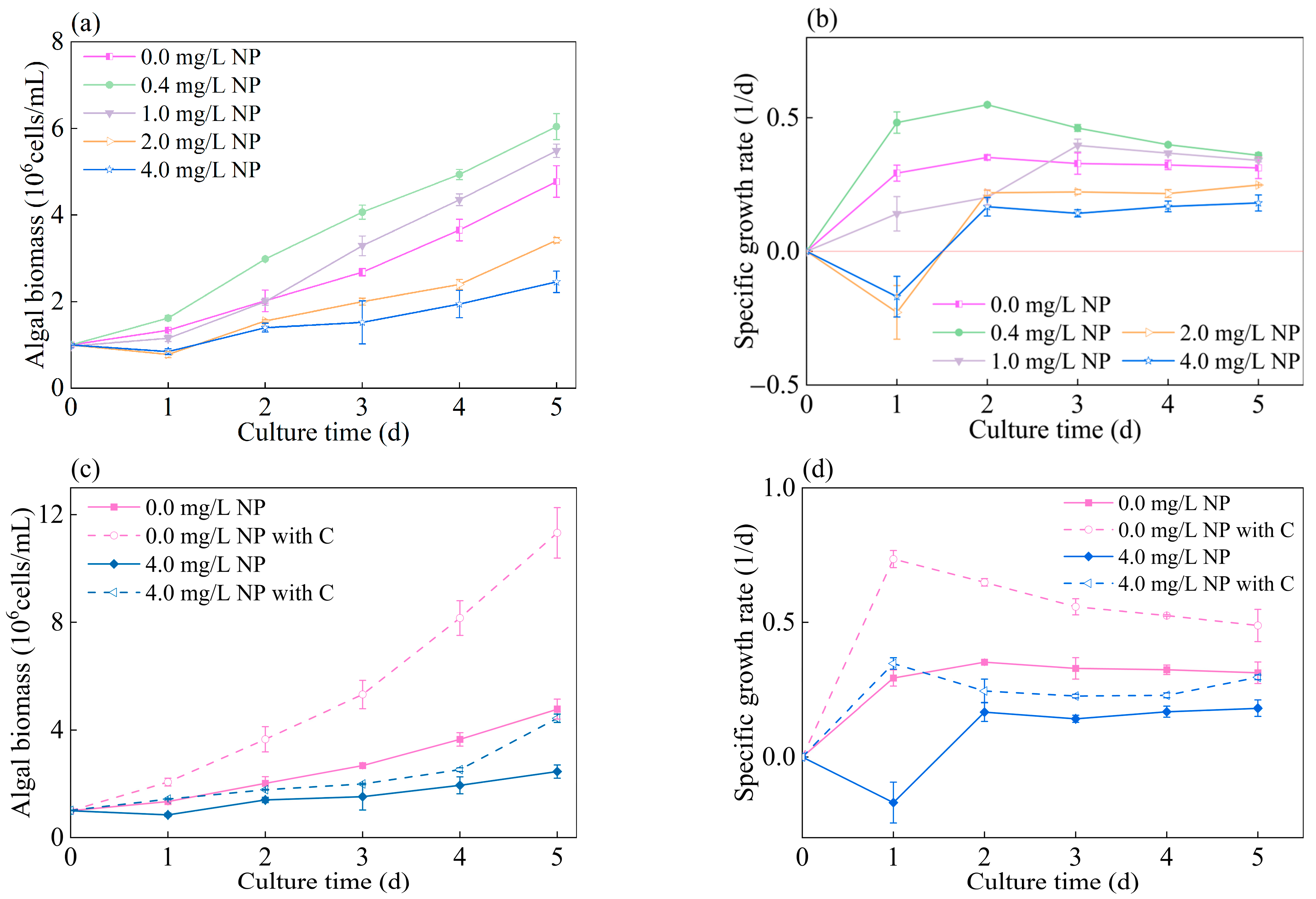
3.1. Detoxification Effect of Glucose on Growth of Algae Exposed to NP

Figure 1a,b illustrates the effects of various concentrations of NP on the growth of C. pyrenoidosa. Exposure to 0.4 mg/L NP resulted in the best growth performance throughout the cultivation period. By the end of the cultivation period, the biomass reached 6.0 × 10⁶ cells/mL, which was 1.26 times higher than that of the control group without NP (p < 0.05). The specific growth rate was consistently higher than that in the control group, peaking at 0.55 1/d on day 3. At low concentrations, certain xenobiotics can promote growth [23]. For the algae exposed to 1.0 mg/L NP, growth showed an initial inhibitory effect, followed by a promotional trend after day 2. The specific growth rate peaked on day 3, exceeding 0.40 1/d in both the cases, with the final biomass reaching 5.5 × 10⁶ cells/mL. This pattern can be attributed to the initial toxicity of NP at higher concentrations, which inhibited growth. However, as the incubation time progressed, the algae adapted to NP toxicity and degraded a portion of the compound, effectively converting the moderate concentration into a low-toxicity environment that subsequently promoted algal growth [52]. The algae exposed to NP concentrations above 1.0 mg/L exhibited significant inhibition throughout the cultivation period, with the inhibitory effect becoming more pronounced as the NP concentration increased. Moreover, the higher the NP concentration was, the lower the peak specific growth rate was. This observation is consistent with the understanding that elevated NP concentrations overwhelm the detoxification mechanisms of algae, leading to cellular damage and reduced growth [25,26,52]. Under high NP concentrations, the growth and performance of C. pyrenoidosa are significantly impeded [52], which undermines the utility of algae for the bioremediation of environments heavily contaminated with NP.
To overcome this limitation, glucose supplementation has been suggested as a strategy to improve the tolerance of C. pyrenoidosa to high NP concentrations [32,55], thereby enhancing its efficacy in the detoxification of polluted ecosystems. As shown in Figure 1c,d, the addition of glucose significantly enhanced the growth of C. pyrenoidosa. At high NP concentrations, the C. pyrenoidosa cells supplemented with glucose displayed notably superior growth compared to those of C. pyrenoidosa without glucose supplementation. By the end of the cultivation period, the algal cells treated with 4.0 mg/L NP and 0.4 g/L glucose resumed growth, achieving a specific growth rate comparable to that of the original C. pyrenoidosa (without NP and glucose), with a cell count of 4.4 × 10⁶ cells/mL. Moreover, a markedly higher specific growth rate was observed in C. pyrenoidosa with 0.0 mg/L NP and 0.4 g/L glucose, peaking at 0.74 1/d on day 3. By the end of the cultivation period, the biomass was 2.3 times greater that of the original C. pyrenoidosa. These findings suggest that glucose significantly stimulates algal growth and mitigates NP toxicity.

3.2. Effect of Glucose on Structural Characterization of Algal Cells Exposed to NP

SEM was conducted to investigate the effects of glucose addition on C. pyrenoidosa exposed to high NP concentrations. As shown in Figure 2, the original C. pyrenoidosa without the addition of NP or glucose exhibited a smooth and regular spherical shape (Figure 2a). After exposure to 4.0 mg/L of NP, the algae showed varying degrees of wrinkling, deformation, and even cell rupture (Figure 2b), indicating the high toxicity level of NP and the significant damage it caused to C. pyrenoidosa, which inhibited its growth. However, C. pyrenoidosa exposed to 4.0 mg/L NP and cultured with glucose, although showing signs of suppression, predominantly had better conditions than those cultured without glucose, with some cells showing less damage (Figure 2c). This is consistent with the growth results, suggesting that glucose enhanced the tolerance of C. pyrenoidosa to NP toxicity.

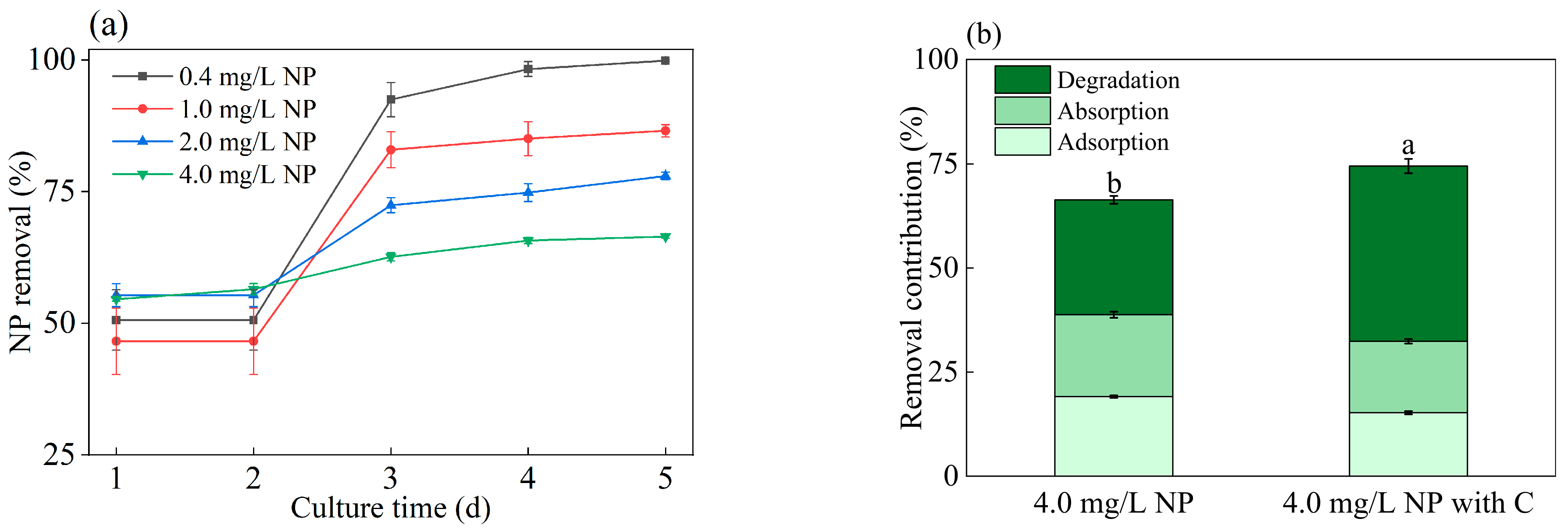
3.3. Glucose Improved NP-Removal Capacity of Algae

In the present study, after 120 h, the NP removal rate in the non-biological control group ranged from 2.5% to 4.1%, indicating that the loss caused by abiotic factors was insignificant. As shown in Figure 3a, the removal efficiency of C. pyrenoidosa is presented. At an NP concentration of 0.4 mg/L, C. pyrenoidosa achieved a removal rate of 99.9% for NP, but this rate decreased as the NP concentration increased. When the NP concentration was set at 4.0 mg/L, the removal rate of NP was found to be 33.6% less than that of the NP concentration at 0.4 mg/L (p < 0.05). Figure 3b illustrates the rates of adsorption, absorption, and degradation of NP by C. pyrenoidosa treated with 4.0 mg/L NP. Interestingly, by the end of the cultivation stage, the degradation efficiency of NP in the presence of glucose increased to 42.1%, which was approximately 14.5% higher than that of C. pyrenoidosa treated with 4.0 mg/L NP alone (p < 0.05). Cheng et al. found that after 120 h of cultivation, the degradation rate of 4.0 mg/L NP by Dictyosphaerium sp. was 16.8% [32]. He et al. reported that four species of freshwater microalgae degraded 0.5–2.5 mg/L NP at rates ranging from 15% to 40% [26]. Similar to our findings, Kong et al. found that appropriate glucose addition promoted phenol removal by up to 12% [43]. Our findings suggest that NP removal by C. pyrenoidosa is not solely a physical adsorption process, but also involves biological degradation [32], with glucose supplementation enhancing the NP-degrading capability of algae.
First-order kinetic models are widely used to describe algal degradation processes, primarily to characterize the degradation profile over time [56]. These models are particularly pertinent for evaluating the degradation capacities of microalgae [57]. Table S1 presents a first-order kinetic model for NP degradation by C. pyrenoidosa without glucose under various conditions. Specifically, the kinetic constant for C. pyrenoidosa with 0.4 mg/L NP is 1.51 1/d. However, as the NP concentration increased, the kinetic constant decreases and reaches only 0.08 1/d of 4.0 mg/L NP. According to the benchmark established by Joss [58], a metabolic rate constant (k) below 0.10 1/d indicates minimal NP removal. In contrast, after the addition of glucose, the degradation rate of NP in C. pyrenoidosa increased to 42.9%, with an enhanced k value of 0.17. Glucose acts as an electron donor, promoting the co-metabolism of various pollutants in algae [29,59,60]. The observed increase in the kinetic constant upon glucose supplementation suggests a significantly enhanced removal capability of C. pyrenoidosa.

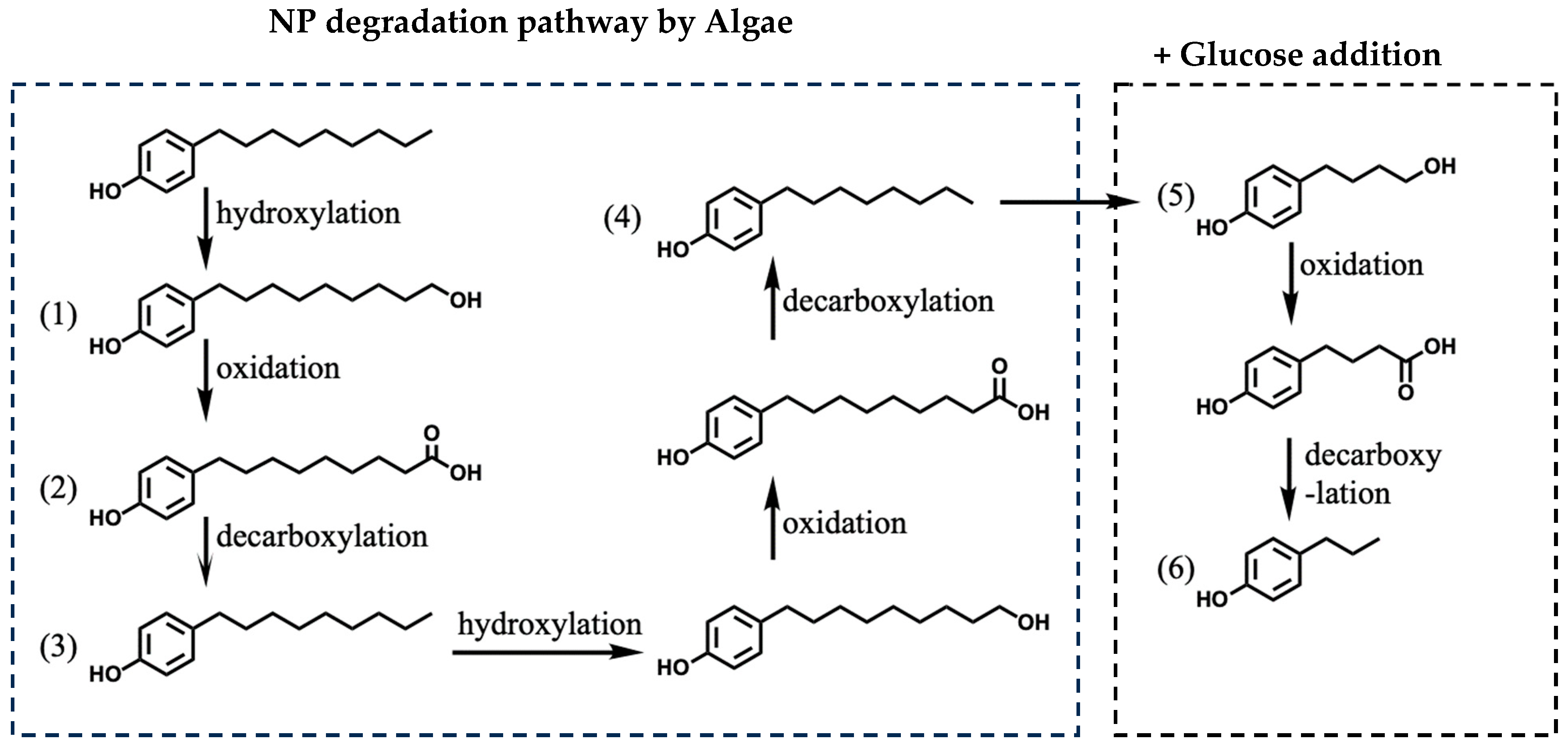
3.4. Effect of Glucose on NP-Degradation Pathway in Algae

The influence of glucose on the degradation pathway of NP in algae was investigated using UPLC-QTOF-MS. Based on their mass-to-charge ratios (m/z), six degradation byproducts were identified, and their potential molecular formulae are listed in Table S2. The degradation of NP begins with hydroxylation of the terminal end of the alkyl chain to generate 9-(4-hydroxyphenyl)nonanol (Figure 4). This hydroxyl group is subsequently oxidized to a carboxyl group, yielding 9-(4-hydroxyphenyl)nonanoic acid. Hydroxylation, oxidation, and decarboxylation shorten the carbon chain. Although the degradation pathway of NP in the algae was not significantly altered by the addition of glucose, two metabolites, 4-(4-hydroxyphenyl)butanol (m/z 165.0) and 4-n-propenylphenol (m/z 134.9), were detected in the glucose-supplemented treatment, suggesting an accelerated biodegradation process. The shortening of the linear chain also reduces the toxicity of the molecule to the ecosystem [61]. Therefore, glucose accelerates the degradation of NP and reduces their risk to aquatic ecosystems.

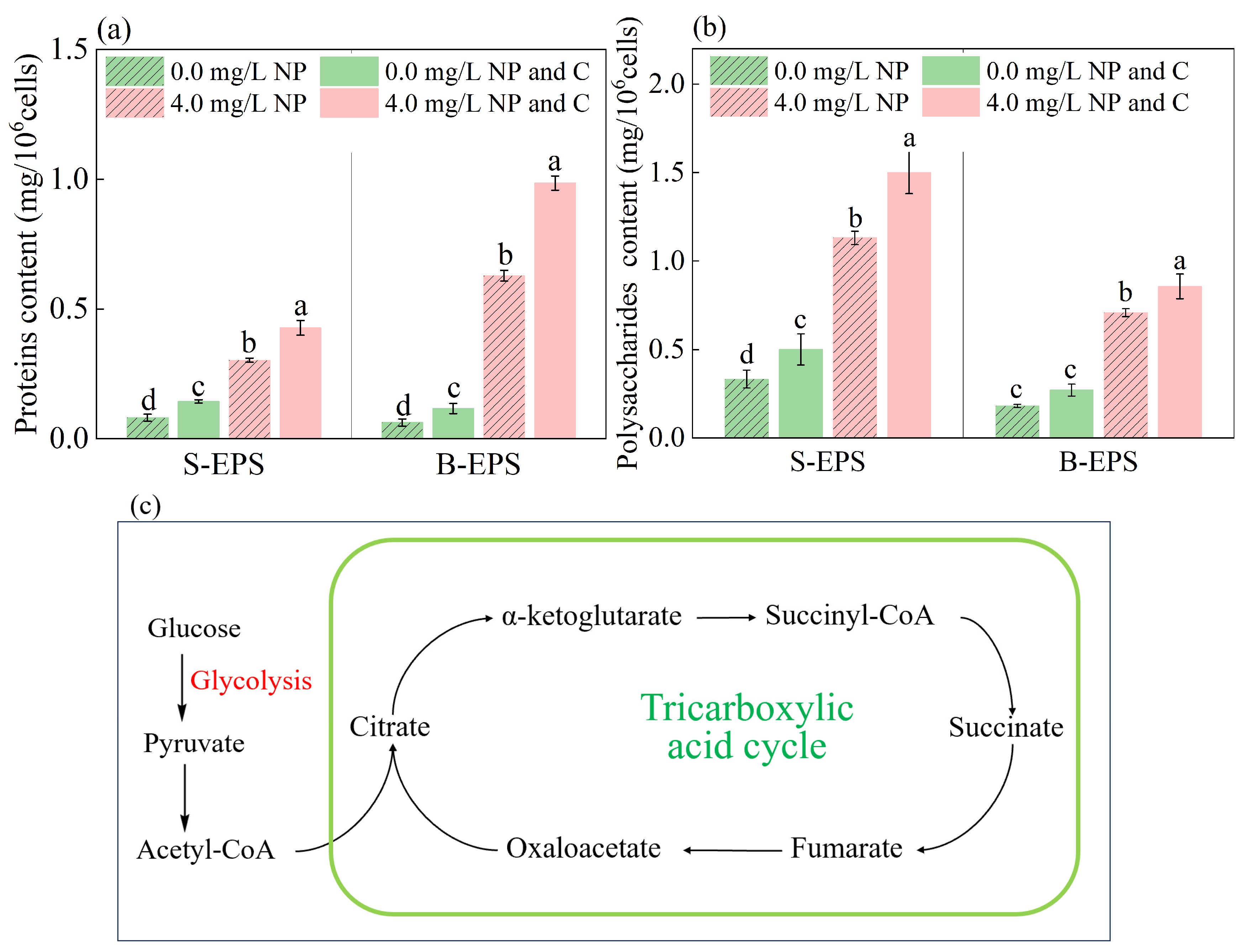
3.5. Glucose Stimulated EPS Secretion in Algae Exposed to NP

The secretion of EPSs by C. pyrenoidosa has been extensively investigated, with PS and PN identified as the primary functional components of EPSs related to protection [62]. As illustrated in Figure 5, exposure to 4.0 mg/L NP resulted in a significant increase in EPS secretion by the algae. Interestingly, the variations in the different types of EPS were not uniform. When exposed to 4.0 mg/L NP, the change in PN within the B-EPSs was more pronounced (Figure 5a), with glucose stimulating an additional 55.5% secretion by C. pyrenoidosa. In contrast, the variation in PS within the S-EPSs was more significant (Figure 5b), where glucose promoted an additional 32.7% secretion by C. pyrenoidosa (p < 0.05). It is possible that PS predominates in S-EPSs, whereas PN predominates in B-EPSs [27]. An EPS functions as a physical barrier surrounding algal cells. The greater the EPS content is, the thicker the protective layer is, which can more effectively prevent NP from contacting the cell body. Glucose further enhances EPS secretion, strengthening its protective effect, reducing the cytotoxic effects of NP, and promoting the co-metabolism of NP by microalgae [63]. This response may serve as a self-protective mechanism against NP exposure [23,27].
Glucose provides a significant energy source to cells, allowing C. pyrenoidosa to ensure its own growth and metabolic needs when exposed to NP, thereby facilitating the generation of a higher EPS content for self-protection [30]. As shown in Figure 5c, microalgae metabolize glucose through the tricarboxylic acid cycle to produce adenosine triphosphate (ATP), which can efficiently convert sugar molecules into ATP phosphoanhydride bonds [30]. Supplementing with glucose stimulates microalgae to generate and store more energy, promoting co-metabolism and further enhancing the degradation efficiency of NP. These results indicate that glucose stimulates the secretion of EPSs by algae to mitigate the stress imposed by NP on the cell body, thereby enhancing their tolerance to NP.
As shown in Figure 6, the 3D-EEM fluorescence spectrum of the B-EPSs from C. pyrenoidosa revealed distinct peaks corresponding to (I) tyrosine-like (Ex/Em: 260–280 nm/295–312 nm) and (II) tryptophan-like (Ex/Em: 270–280 nm/330–350 nm) compounds [52,64]. Additionally, the broad fluorescence band at Ex/Em wavelengths of 250–400 nm/380–500 nm was attributed to humic-like substances secreted by the algal cells [49,64], highlighting the diverse chemical composition of EPSs. On day 2, under all the three cultivation conditions, the intensity of peak I in the B-EPSs secreted by C. pyrenoidosa exceeded that of peak II (Figure 6a–c). However, this pattern changed by the end of the cultivation period. In the absence of NP and glucose, the intensity of peak II surpassed that of peak I (Figure 6). By contrast, exposure to NP inhibited this transformation. In the B-EPSs secreted by C. pyrenoidosa exposed to 4.0 mg/L NP, the intensity of peak II did not exceed that of peak I (Figure 6e). However, the addition of glucose reversed this situation, so the intensity of peak II was significantly higher than that of peak I (Figure 6f). The peak intensity was proportional to the protein content [65], with a higher intensity of peak II, indicating a greater content of tryptophan-like substances. These substances are abundant in functional groups that may help mitigate pollution [66,67]. Other studies have indicated that the secretion of tryptophan-like substances increases, a mechanism by which microbes alleviate chemical stress caused by sulfadiazine [68]. Tryptophan is a crucial amino acid that is transformed into various signaling molecules in algae [69,70,71]. Upon NP exposure, the upregulation of the tryptophan metabolic pathway augments production of tryptophan and its derivatives, serving as a protective mechanism against NP-induced growth inhibition and other adverse effects [72,73,74]. Our results confirm that glucose promotes the production of tryptophan, which might enhance the degradation of NP by the algae, further strengthening the protective effect of EPS on C. pyrenoidosa exposed to NP.

3.6. Practical Applications

Our study demonstrates the potential of algae in remediating wastewater containing NP and proposes a method to enhance degradation rates by adding glucose. Glucose supplementation increases the tolerance of algae to NP, promotes co-metabolism, and enhances the degradation rate of NP by algae. This also leads to the production of large quantities of biomass and EPS. Biomass can generate additional economic value, whereas EPSs can be reused for applications such as heavy metal adsorption [75]. However, increasing the glucose levels in aquatic environments can increase the risk of eutrophication, distort food webs, and alter nutrient cycles [32]. These risks should be carefully considered before practical application.

4. Conclusions

The current understanding of the role of carbon sources in algal responses to NP stress remains limited. Glucose effectively mitigates the toxic effects of NP on algae. Glucose provides energy to sustain vital algal activities, facilitating the co-metabolism of NP. Additionally, glucose stimulates the production of more stress-resistant substances, namely EPSs, which protect cells and mitigate cellular damage. As a result, glucose intensifies material and energy metabolism, as well as cellular synthesis, thereby enhancing the capacity of algae to degrade and detoxify NP. These findings significantly contribute to our understanding of NP bioremediation strategies and highlight the importance of metabolic enhancers such as glucose in optimizing the detoxification processes of algae in polluted water bodies.

Supplementary Materials

The following supporting information can be downloaded at: https://www.mdpi.com/article/10.3390/w17020244/s1. Table S1: Kinetic equations for NP degradation by Chlorella pyrenoidosa under different conditions; Table S2: Metabolites of NP degradation by Chlorella pyrenoidosa determined using UPLC-QTOF-MS analysis.

Author Contributions

Methodology and writing—original draft preparation, J.Y.; validation, Y.L.; data curation, D.C. and G.X.; supervision, writing—review and editing, Y.Y.; supervision, project administration, and funding acquisition, L.Z. All authors have read and agreed to the published version of the manuscript.

Funding

This research was funded by the Tianjin Key Research and Development Program, Tianjin City, China (21YFSNSN00160), and the Key Program of “Science and Technology Helping Economy 2020”, Tianjin City, China (SQ2020YFF0412145).

Data Availability Statement

The data presented in this study are available upon request from the corresponding author.

Conflicts of Interest

Author Danning Chen was employed by the company Greenland Environmental Technology Co., Ltd. The remaining authors declare that the research was conducted in the absence of any commercial or financial relationships that could be construed as a potential conflict of interest.

References

  1. Bhandari, G.; Bagheri, A.R.; Bhatt, P.; Bilal, M. Occurrence, Potential Ecological Risks, and Degradation of Endocrine Disrupter, Nonylphenol, from the Aqueous Environment. Chemosphere 2021, 275, 130013. [Google Scholar] [CrossRef] [PubMed]
  2. Ding, F.; Zhao, Q.; Wang, L.; Ma, J.; Song, L.; Huang, D. Adsorption Behavior of Nonylphenol on Polystyrene Microplastics and Their Cytotoxicity in Human Caco-2 Cells. Water 2022, 14, 3288. [Google Scholar] [CrossRef]
  3. Rahmani, F.; Sajjadi, N.; Dehghani, M.H.; Zaeimdar, M. Modelling and Optimization of Nonylphenol Biosorption by Novel Low-Cost Magnetic Chlorella Vulgaris. Emerg. Contam. 2024, 10, 100310. [Google Scholar] [CrossRef]
  4. De La Parra-Guerra, A.; Olivero-Verbel, J. Toxicity of Nonylphenol and Nonylphenol Ethoxylate on Caenorhabditis Elegans. Ecotoxicol. Environ. Saf. 2020, 187, 109709. [Google Scholar] [CrossRef]
  5. Yang, W.; Gao, X.; Wu, Y.; Wan, L.; Tan, L.; Yuan, S.; Ding, H.; Zhang, W. The Combined Toxicity Influence of Microplastics and Nonylphenol on Microalgae Chlorella Pyrenoidosa. Ecotoxicol. Environ. Saf. 2020, 195, 110484. [Google Scholar] [CrossRef] [PubMed]
  6. Li, D.; Kim, M.; Shim, W.J.; Yim, U.H.; Oh, J.-R.; Kwon, Y.-J. Seasonal Flux of Nonylphenol in Han River, Korea. Chemosphere 2004, 56, 1–6. [Google Scholar] [CrossRef]
  7. Yu, Y.; Zhai, H.; Hou, S.; Sun, H. Nonylphenol Ethoxylates and Their Metabolites in Sewage Treatment Plants and Rivers of Tianjin, China. Chemosphere 2009, 77, 1–7. [Google Scholar] [CrossRef]
  8. Sibali, L.L.; Okwonkwo, J.O.; McCrindle, R.I. Levels of Selected Alkylphenol Ethoxylates (APEs) in Water and Sediment Samples from the Jukskei River Catchment Area in Gauteng, South Africa. Water SA 2010, 36, 229–238. [Google Scholar]
  9. Rocha, M.J.; Silva, F.; Rocha, E. Annual Evaluation of 17 Oestrogenic Endocrine Disruptors and Hazard Indexes in the Douro River Estuary—The Atlantic Discharge of the Highest-Flow River of Southwestern Europe. Water 2022, 14, 2046. [Google Scholar] [CrossRef]
  10. Li, J.; Zhang, H.; Li, J.; Qin, N.; Wei, Q.; Li, Y.; Peng, Y.; Li, H. Assessment of Nonylphenol Exposure Based on Global Urinary Concentration Data and Its Risk Analysis. Environ. Res. 2024, 244, 117903. [Google Scholar] [CrossRef] [PubMed]
  11. Zhao, Y.; Ji, J.; Wu, Y.; Chen, S.; Xu, M.; Cao, X.; Liu, H.; Wang, Z.; Bi, H.; Guan, G.; et al. Nonylphenol and Its Derivatives: Environmental Distribution, Treatment Strategy, Management and Future Perspectives. Chemosphere 2024, 352, 141377. [Google Scholar] [CrossRef]
  12. Noorimotlagh, Z.; Mirzaee, S.A.; Martinez, S.S.; Rachoń, D.; Hoseinzadeh, M.; Jaafarzadeh, N. Environmental Exposure to Nonylphenol and Cancer Progression Risk–A Systematic Review. Environ. Res. 2020, 184, 109263. [Google Scholar] [CrossRef] [PubMed]
  13. Bodziach, K.; Staniszewska, M.; Nehring, I.; Ożarowska, A.; Zaniewicz, G.; Meissner, W. Endocrine Disrupting Bisphenol A, 4-Tert-Octylphenol and 4-Nonylphenol in Gonads of Long-Tailed Ducks Clangula Hyemalis Wintering in the Southern Baltic. Environ. Res. 2024, 243, 117772. [Google Scholar] [CrossRef] [PubMed]
  14. Ho, H.; Watanabe, T. Distribution and Removal of Nonylphenol Ethoxylates and Nonylphenol from Textile Wastewater—A Comparison of a Cotton and a Synthetic Fiber Factory in Vietnam. Water 2017, 9, 386. [Google Scholar] [CrossRef]
  15. Lalonde, B.; Garron, C. Nonylphenol, Octylphenol, and Nonylphenol Ethoxylates Dissemination in the Canadian Freshwater Environment. Arch. Environ. Contam. Toxicol. 2021, 80, 319–330. [Google Scholar] [CrossRef] [PubMed]
  16. Ryu, H.-D.; Han, H.; Park, T.-J.; Park, J.-H.; Kim, Y.S. New Findings on the Occurrence, Removal, and Risk Assessment of Nonylphenol and Octylphenol in Industrial Wastewater Treatment Plants in Korea. J. Hazard. Mater. 2024, 461, 132615. [Google Scholar] [CrossRef] [PubMed]
  17. Babu, D.S.; Srivastava, V.; Nidheesh, P.V.; Kumar, M.S. Detoxification of Water and Wastewater by Advanced Oxidation Processes. Sci. Total Environ. 2019, 696, 133961. [Google Scholar] [CrossRef]
  18. Bilal, M.; Rasheed, T.; Nabeel, F.; Iqbal, H.M.N.; Zhao, Y. Hazardous Contaminants in the Environment and Their Laccase-Assisted Degradation—A Review. J. Environ. Manag. 2019, 234, 253–264. [Google Scholar] [CrossRef]
  19. Al-Khiat, S.H.; Bukhari, N.A.; Ameen, F.; Abdel-Raouf, N. Comparison of the Microalgae Phormidium Tenue and Chlorella Vulgaris as Biosorbents of Cd and Zn from Aqueous Environments. Environ. Res. 2023, 235, 116675. [Google Scholar] [CrossRef] [PubMed]
  20. Chang, Y.-L.; Nagarajan, D.; Chen, J.-H.; Yen Chen, C.; Wu, Y.-J.; Whang, L.-M.; Lee, D.-J.; Chang, J.-S. Microalgae-Bacteria Consortia for the Treatment of Raw Dairy Manure Wastewater Using a Novel Two-Stage Process: Process Optimization and Bacterial Community Analysis. Chem. Eng. J. 2023, 473, 145388. [Google Scholar] [CrossRef]
  21. Ma, Y.; Lin, S.; Guo, T.; Guo, C.; Li, Y.; Hou, Y.; Gao, Y.; Dong, R.; Liu, S. Exploring the Influence of Sulfadiazine-Induced Stress on Antibiotic Removal and Transformation Pathway Using Microalgae Chlorella sp. Environ. Res. 2024, 256, 119225. [Google Scholar] [CrossRef] [PubMed]
  22. Wang, L.; Xiao, H.; He, N.; Sun, D.; Duan, S. Biosorption and Biodegradation of the Environmental Hormone Nonylphenol By Four Marine Microalgae. Sci. Rep. 2019, 9, 5277. [Google Scholar] [CrossRef] [PubMed]
  23. Yang, W.; Gao, X.; Wu, Y.; Wan, L.; Lu, C.; Huang, J.; Chen, H.; Yang, Y.; Ding, H.; Zhang, W. Chemical- and Species-Specific Toxicity of Nonylphenol and Octylphenol to Microalgae Chlorella Pyrenoidosa and Scenedesmus Obliquus. Environ. Toxicol. Pharmacol. 2021, 81, 103517. [Google Scholar] [CrossRef] [PubMed]
  24. López-Pacheco, I.Y.; Salinas-Salazar, C.; Silva-Núñez, A.; Rodas-Zuluaga, L.I.; Donoso-Quezada, J.; Ayala-Mar, S.; Barceló, D.; Iqbal, H.M.N.; Parra-Saldívar, R. Removal and Biotransformation of 4-Nonylphenol by Arthrospira Maxima and Chlorella Vulgaris Consortium. Environ. Res. 2019, 179, 108848. [Google Scholar] [CrossRef] [PubMed]
  25. Feng, Y.; Wang, A.; Fu, W.; Song, D. Growth Performance, Antioxidant Response, Biodegradation and Transcriptome Analysis of Chlorella Pyrenoidosa after Nonylphenol Exposure. Sci. Total Environ. 2022, 806, 150507. [Google Scholar] [CrossRef] [PubMed]
  26. He, N.; Sun, X.; Zhong, Y.; Sun, K.; Liu, W.; Duan, S. Removal and Biodegradation of Nonylphenol by Four Freshwater Microalgae. Int. J. Environ. Res. Public. Health 2016, 13, 1239. [Google Scholar] [CrossRef]
  27. Cheng, Q.; Jiang, Y.; Jin, Z.; Hui, C.; Xu, L.; Zhou, Q.; Zhao, Y.; Du, L.; Jiang, H. Enhanced Excretion of Extracellular Polymeric Substances Associated with Nonylphenol Tolerance in Dictyosphaerium sp. J. Hazard. Mater. 2020, 395, 122644. [Google Scholar] [CrossRef] [PubMed]
  28. Li, X.; Li, W.; Zhai, J.; Wei, H. Effect of Nitrogen Limitation on Biochemical Composition and Photosynthetic Performance for Fed-Batch Mixotrophic Cultivation of Microalga Spirulina Platensis. Bioresour. Technol. 2018, 263, 555–561. [Google Scholar] [CrossRef] [PubMed]
  29. Xiong, J.-Q.; Kurade, M.B.; Kim, J.R.; Roh, H.-S.; Jeon, B.-H. Ciprofloxacin Toxicity and Its Co-Metabolic Removal by a Freshwater Microalga Chlamydomonas Mexicana. J. Hazard. Mater. 2017, 323, 212–219. [Google Scholar] [CrossRef] [PubMed]
  30. Vo, H.N.P.; Ngo, H.H.; Guo, W.; Nguyen, K.H.; Chang, S.W.; Nguyen, D.D.; Liu, Y.; Liu, Y.; Ding, A.; Bui, X.T. Micropollutants Cometabolism of Microalgae for Wastewater Remediation: Effect of Carbon Sources to Cometabolism and Degradation Products. Water Res. 2020, 183, 115974. [Google Scholar] [CrossRef] [PubMed]
  31. Xiao, M.; Ma, H.; Sun, M.; Yin, X.; Feng, Q.; Song, H.; Gai, H. Characterization of Cometabolic Degradation of P-Cresol with Phenol as Growth Substrate by Chlorella Vulgaris. Bioresour. Technol. 2019, 281, 296–302. [Google Scholar] [CrossRef]
  32. Cheng, Q.; Du, L.; Xu, L.; Zhao, Y.; Ma, J.; Lin, H. Toxicity Alleviation and Metabolism Enhancement of Nonylphenol in Green Algae Dictyosphaerium sp. by NaHCO3. Sci. Total Environ. 2022, 848, 157698. [Google Scholar] [CrossRef] [PubMed]
  33. Zhang, L.; Guo, R.; Li, H.; Du, Q.; Lu, J.; Huang, Y.; Yan, Z.; Chen, J. Mechanism Analysis for the Process-Dependent Driven Mode of NaHCO3 in Algal Antibiotic Removal: Efficiency, Degradation Pathway and Metabolic Response. J. Hazard. Mater. 2020, 394, 122531. [Google Scholar] [CrossRef]
  34. Perez-Garcia, O.; Escalante, F.M.E.; de-Bashan, L.E.; Bashan, Y. Heterotrophic Cultures of Microalgae: Metabolism and Potential Products. Water Res. 2011, 45, 11–36. [Google Scholar] [CrossRef] [PubMed]
  35. Wang, S.; Wu, Y.; Wang, X. Heterotrophic Cultivation of Chlorella Pyrenoidosa Using Sucrose as the Sole Carbon Source by Co-Culture with Rhodotorula Glutinis. Bioresour. Technol. 2016, 220, 615–620. [Google Scholar] [CrossRef]
  36. Liu, N.; Guo, B.; Cao, Y.; Wang, H.; Yang, S.; Huo, H.; Kong, W.; Zhang, A.; Niu, S. Effects of Organic Carbon Sources on the Biomass and Lipid Production by the Novel Microalga Micractinium Reisseri FM1 under Batch and Fed-Batch Cultivation. South Afr. J. Bot. 2021, 139, 329–337. [Google Scholar] [CrossRef]
  37. Mao, X.; Wang, X.; Ge, M.; Chen, F.; Liu, B. New Insights into Xanthophylls and Lipidomic Profile Changes Induced by Glucose Supplementation in the Marine Diatom Nitzschia Laevis. Mar. Drugs 2022, 20, 456. [Google Scholar] [CrossRef] [PubMed]
  38. Přibyl, P.; Cepák, V. Screening for Heterotrophy in Microalgae of Various Taxonomic Positions and Potential of Mixotrophy for Production of High-Value Compounds. J. Appl. Phycol. 2019, 31, 1555–1564. [Google Scholar] [CrossRef]
  39. Wang, Z.; Luo, F.; Wang, Z.; Zhou, R.; Tang, Y.; Li, Y. The Potential Growth and Lipid Accumulation in Coccomyxa Subellipsoidea Triggered by Glucose Combining with Sodium Acetate. World J. Microbiol. Biotechnol. 2019, 35, 110. [Google Scholar] [CrossRef]
  40. Zhang, Z.; Sun, D.; Zhang, Y.; Chen, F. Glucose Triggers Cell Structure Changes and Regulates Astaxanthin Biosynthesis in Chromochloris Zofingiensis. Algal Res. 2019, 39, 101455. [Google Scholar] [CrossRef]
  41. Joun, J.; Sirohi, R.; Sim, S.J. The Effects of Acetate and Glucose on Carbon Fixation and Carbon Utilization in Mixotrophy of Haematococcus Pluvialis. Bioresour. Technol. 2023, 367, 128218. [Google Scholar] [CrossRef] [PubMed]
  42. Lv, J.; Zhao, F.; Feng, J.; Liu, Q.; Nan, F.; Liu, X. Transcriptomic Analysis Reveals the Mechanism on the Response of Chlorococcum sp. GD to Glucose Concentration in Mixotrophic Cultivation. Bioresour. Technol. 2019, 288, 121568. [Google Scholar] [CrossRef] [PubMed]
  43. Kong, W.; Yang, S.; Guo, B.; Wang, H.; Huo, H.; Zhang, A.; Niu, S. Growth Behavior, Glucose Consumption and Phenol Removal Efficiency of Chlorella Vulgaris under the Synergistic Effects of Glucose and Phenol. Ecotoxicol. Environ. Saf. 2019, 186, 109762. [Google Scholar] [CrossRef] [PubMed]
  44. Du, Y.; Wang, J.; Wang, Z.; Torres, O.L.; Guo, R.; Chen, J. Exogenous Organic Carbon as an Artificial Enhancement Method to Assist the Algal Antibiotic Treatment System. J. Clean. Prod. 2018, 194, 624–634. [Google Scholar] [CrossRef]
  45. Han, C.; Ren, J.; Wang, B.; Wang, Z.; Yin, H.; Ke, F.; Xu, D.; Zhang, L.; Si, X.; Shen, Q. Ignored Effects of Phosphite (P+III) on the Growth Responses of Three Typical Algae Species. Environ. Pollut. 2022, 294, 118672. [Google Scholar] [CrossRef] [PubMed]
  46. Zhou, Y.; Zhu, Y.; Wu, F.; Pan, X.; Li, W.; Han, J. Transcriptomics Revealed the Key Molecular Mechanisms of Ofloxacin-Induced Hormesis in Chlorella Pyrenoidosa at Environmentally Relevant Concentration. Environ. Pollut. 2024, 361, 124887. [Google Scholar] [CrossRef]
  47. Li, Q.; Xu, Y.; Liang, C.; Peng, L.; Zhou, Y. Nitrogen Removal by Algal-Bacterial Consortium during Mainstream Wastewater Treatment: Transformation Mechanisms and Potential N2O Mitigation. Water Res. 2023, 235, 119890. [Google Scholar] [CrossRef]
  48. Shang, Z.; Li, M.; Ma, Y.; Si, B.; Wei, Z.; Zhang, X. Deciphering Metabolomic Insights into Benzoic Acid-Mediated Nutrient Enrichment in Chlorella Pyrenoidosa. Food Biosci. 2024, 59, 104157. [Google Scholar] [CrossRef]
  49. Qu, F.; Liang, H.; He, J.; Ma, J.; Wang, Z.; Yu, H.; Li, G. Characterization of Dissolved Extracellular Organic Matter (dEOM) and Bound Extracellular Organic Matter (bEOM) of Microcystis Aeruginosa and Their Impacts on UF Membrane Fouling. Water Res. 2012, 46, 2881–2890. [Google Scholar] [CrossRef] [PubMed]
  50. Chen, B.; Li, F.; Liu, N.; Ge, F.; Xiao, H.; Yang, Y. Role of Extracellular Polymeric Substances from Chlorella Vulgaris in the Removal of Ammonium and Orthophosphate under the Stress of Cadmium. Bioresour. Technol. 2015, 190, 299–306. [Google Scholar] [CrossRef]
  51. Morris, D.L. Quantitative Determination of Carbohydrates With Dreywood’s Anthrone Reagent. Science 1948, 107, 254–255. [Google Scholar] [CrossRef] [PubMed]
  52. Cheng, Q.; Zhou, Q.; Jin, Z.; Jiang, Y.; Xu, L.; Jiang, H.; Zhao, Y. Bioaccumulation, Growth Performance, and Transcriptomic Response of Dictyosphaerium sp. after Exposure to Nonylphenol. Sci. Total Environ. 2019, 687, 416–422. [Google Scholar] [CrossRef] [PubMed]
  53. Yang, Z.; Shi, Y.; Zhang, Y.; Cheng, Q.; Li, X.; Zhao, C.; Zhang, D. Different Pathways for 4-n-Nonylphenol Biodegradation by Two Aspergillus Strains Derived from Estuary Sediment: Evidence from Metabolites Determination and Key-Gene Identification. J. Hazard. Mater. 2018, 359, 203–212. [Google Scholar] [CrossRef]
  54. Park, J.; Yamashita, N.; Wu, G.; Tanaka, H. Removal of Pharmaceuticals and Personal Care Products by Ammonia Oxidizing Bacteria Acclimated in a Membrane Bioreactor: Contributions of Cometabolism and Endogenous Respiration. Sci. Total Environ. 2017, 605–606, 18–25. [Google Scholar] [CrossRef] [PubMed]
  55. Vo, H.N.P.; Ngo, H.H.; Guo, W.; Liu, Y.; Woong Chang, S.; Nguyen, D.D.; Zhang, X.; Liang, H.; Xue, S. Selective Carbon Sources and Salinities Enhance Enzymes and Extracellular Polymeric Substances Extrusion of Chlorella sp. for Potential Co-Metabolism. Bioresour. Technol. 2020, 303, 122877. [Google Scholar] [CrossRef] [PubMed]
  56. Li, S.; Show, P.L.; Ngo, H.H.; Ho, S.-H. Algae-Mediated Antibiotic Wastewater Treatment: A Critical Review. Environ. Sci. Ecotechnol. 2022, 9, 100145. [Google Scholar] [CrossRef]
  57. Qian, Z.; Na, L.; Bao-Long, W.; Tao, Z.; Peng-Fei, M.; Wei-Xiao, Z.; Sraboni, N.Z.; Zheng, M.; Ying-Qi, Z.; Liu, Y. Capabilities and Mechanisms of Microalgae on Nutrients and Florfenicol Removing from Marine Aquaculture Wastewater. J. Environ. Manag. 2022, 320, 115673. [Google Scholar] [CrossRef] [PubMed]
  58. Joss, A.; Zabczynski, S.; Göbel, A.; Hoffmann, B.; Löffler, D.; McArdell, C.S.; Ternes, T.A.; Thomsen, A.; Siegrist, H. Biological Degradation of Pharmaceuticals in Municipal Wastewater Treatment: Proposing a Classification Scheme. Water Res. 2006, 40, 1686–1696. [Google Scholar] [CrossRef] [PubMed]
  59. Leng, L.; Wei, L.; Xiong, Q.; Xu, S.; Li, W.; Lv, S.; Lu, Q.; Wan, L.; Wen, Z.; Zhou, W. Use of Microalgae Based Technology for the Removal of Antibiotics from Wastewater: A Review. Chemosphere 2020, 238, 124680. [Google Scholar] [CrossRef] [PubMed]
  60. Lu, Q.; Chen, P.; Addy, M.; Zhang, R.; Deng, X.; Ma, Y.; Cheng, Y.; Hussain, F.; Chen, C.; Liu, Y.; et al. Carbon-Dependent Alleviation of Ammonia Toxicity for Algae Cultivation and Associated Mechanisms Exploration. Bioresour. Technol. 2018, 249, 99–107. [Google Scholar] [CrossRef] [PubMed]
  61. MtibaÃ, R.; Ezzanad, A.; Aranda, E.; Pozo, C.; Ghariani, B.; Moraga, J.; Nasri, M. Biodegradation and Toxicity Reduction of Nonylphenol, 4-Tert-Octylphenol and 2,4-Dichlorophenol by the Ascomycetous Fungus Thielavia Sp HJ22: Identification of Fungal Metabolites and Proposal of a Putative Pathway. Sci. Total Environ. 2020, 708, 135129. [Google Scholar] [CrossRef] [PubMed]
  62. Sheng, G.-P.; Yu, H.-Q.; Li, X.-Y. Extracellular Polymeric Substances (EPS) of Microbial Aggregates in Biological Wastewater Treatment Systems: A Review. Biotechnol. Adv. 2010, 28, 882–894. [Google Scholar] [CrossRef] [PubMed]
  63. Lv, J.; Zhao, F.; Feng, J.; Liu, Q.; Nan, F.; Xie, S. Extraction of Extracellular Polymeric Substances (EPS) from a Newly Isolated Self-Flocculating Microalga Neocystis Mucosa SX with Different Methods. Algal Res. 2019, 40, 101479. [Google Scholar] [CrossRef]
  64. Guo, X.; Yuan, D.; Li, Q.; Jiang, J.; Chen, F.; Zhang, H. Spectroscopic Techniques for Quantitative Characterization of Cu (II) and Hg (II) Complexation by Dissolved Organic Matter from Lake Sediment in Arid and Semi-Arid Region. Ecotoxicol. Environ. Saf. 2012, 85, 144–150. [Google Scholar] [CrossRef] [PubMed]
  65. You, G.; Hou, J.; Xu, Y.; Wang, C.; Wang, P.; Miao, L.; Ao, Y.; Li, Y.; Lv, B. Effects of CeO2 Nanoparticles on Production and Physicochemical Characteristics of Extracellular Polymeric Substances in Biofilms in Sequencing Batch Biofilm Reactor. Bioresour. Technol. 2015, 194, 91–98. [Google Scholar] [CrossRef]
  66. Xin, K.; Chen, X.; Zhang, Z.; Zhang, Z.; Pang, H.; Yang, J.; Jiang, H.; Lu, J. Trace Antibiotics Increase the Risk of Antibiotic Resistance Genes Transmission by Regulating the Biofilm Extracellular Polymeric Substances and Microbial Community in the Sewer. J. Hazard. Mater. 2022, 432, 128634. [Google Scholar] [CrossRef]
  67. Xu, L.-Z.-J.; Wu, J.; Xia, W.-J.; Jin, L.-Y.; Zhao, Y.-H.; Fan, N.-S.; Huang, B.-C.; Jin, R.-C. Adaption and Restoration of Anammox Biomass to Cd(II) Stress: Performance, Extracellular Polymeric Substance and Microbial Community. Bioresour. Technol. 2019, 290, 121766. [Google Scholar] [CrossRef] [PubMed]
  68. Qiu, L.; Wu, J.; Du, W.; Nafees, M.; Yin, Y.; Ji, R.; Banwart, S.A.; Guo, H. Response of Soil Bacterial Communities to Sulfadiazine Present in Manure: Protection and Adaptation Mechanisms of Extracellular Polymeric Substances. J. Hazard. Mater. 2021, 408, 124887. [Google Scholar] [CrossRef] [PubMed]
  69. Dao, G.-H.; Wu, G.-X.; Wang, X.-X.; Zhang, T.-Y.; Zhan, X.-M.; Hu, H.-Y. Enhanced Microalgae Growth through Stimulated Secretion of Indole Acetic Acid by Symbiotic Bacteria. Algal Res. 2018, 33, 345–351. [Google Scholar] [CrossRef]
  70. Lin, H.; Li, Y.; Hill, R.T. Microalgal and Bacterial Auxin Biosynthesis: Implications for Algal Biotechnology. Curr. Opin. Biotechnol. 2022, 73, 300–307. [Google Scholar] [CrossRef]
  71. Shasaltaneh, M.D.; Moosavi-Nejad, Z.; Gharavi, S.; Fooladi, J. Cane Molasses as a Source of Precursors in the Bioproduction of Tryptophan by Bacillus Subtilis. Iran. J. Microbiol. 2013, 5, 285–292. [Google Scholar]
  72. Cao, X.; Wang, X.; Chen, H.; Li, H.; Tariq, M.; Wang, C.; Zhou, Y.; Liu, Y. Neurotoxicity of Nonylphenol Exposure on Caenorhabditis Elegans Induced by Reactive Oxidative Species and Disturbance Synthesis of Serotonin. Environ. Pollut. 2019, 244, 947–957. [Google Scholar] [CrossRef] [PubMed]
  73. Ge, C.; Luo, X.; Lv, Y.; Wu, L.; Hu, Z.; Huang, W.; Zhan, S.; Shen, X.; Hui, C.; Yu, D.; et al. Essential Oils Ameliorate the Intestinal Damages Induced by Nonylphenol Exposure by Modulating Tryptophan Metabolism and Activating Aryl Hydrocarbon Receptor via Gut Microbiota Regulation. Chemosphere 2024, 362, 142571. [Google Scholar] [CrossRef] [PubMed]
  74. Zhao, Y.; Wang, H.-P.; Han, B.; Yu, X. Coupling of Abiotic Stresses and Phytohormones for the Production of Lipids and High-Value by-Products by Microalgae: A Review. Bioresour. Technol. 2019, 274, 549–556. [Google Scholar] [CrossRef] [PubMed]
  75. Tyagi, B.; Gupta, B.; Thakur, I.S. Biosorption of Cr (VI) from Aqueous Solution by Extracellular Polymeric Substances (EPS) Produced by Parapedobacter sp. ISTM3 Strain Isolated from Mawsmai Cave, Meghalaya, India. Environ. Res. 2020, 191, 110064. [Google Scholar] [CrossRef]
Figure 1. (a,c) Algal biomass of various treatments throughout 5-day culture period. (b,d) Specific growth rate of different treatments throughout 5-day culture period.
Water 17 00244 g001
Figure 2. Scanning electron microscopy images of C. pyrenoidosa under different conditions: (a) original C. pyrenoidosa without addition of NP and glucose; (b) C. pyrenoidosa with 4.0 mg/L NP; (c) C. pyrenoidosa with 4.0 mg/L NP and 0.4 g/L glucose at end of cultivation period.
Water 17 00244 g002
Figure 3. (a) NP removal rate at different concentrations using C. pyrenoidosa during 5-day culture period. (b) Contributions of adsorption, absorption, and degradation by algae to NP removal. C represents 0.4 g/L glucose. Different letters (a, b) indicate statistically significant disparities between groups. p < 0.05.
Water 17 00244 g003
Figure 4. Degradation pathway of NP in Chlorella pyrenoidosa. The compounds (1)–(6) were degradation byproducts detected by UPLC—QTOF—MS.
Water 17 00244 g004
Figure 5. (a) Protein content of EPS in C. pyrenoidosa. (b) Polysaccharide contents of EPS in C. pyrenoidosa. C represents 0.4 g/L glucose. (c) Tricarboxylic acid cycle in microalgae. Different letters (a, b, c, d) indicate statistically significant disparities between groups. p < 0.05.
Water 17 00244 g005
Figure 6. (ac) Three-dimensional excitation and emission matrix fluorescence spectra of B-EPS in C. pyrenoidosa on day 2: (a) 0.0 mg/L NP; (b) 4.0 mg/L NP; (c) 4.0 mg/L NP with C. (df) Three-dimensional excitation and emission matrix fluorescence spectra of B-EPS of C. pyrenoidosa at cultured end-stage: (d) 0.0 mg/L NP; (e) 4.0 mg/L NP; (f) 4.0 mg/L NP with C. C represents 0.4 g/L glucose. (I) indicates tyrosine-like (Ex/Em: 260–280 nm/295–312 nm) compounds and (II) indicates tryptophan-like (Ex/Em: 270–280 nm/330–350 nm) compounds.
Water 17 00244 g006
Disclaimer/Publisher’s Note: The statements, opinions and data contained in all publications are solely those of the individual author(s) and contributor(s) and not of MDPI and/or the editor(s). MDPI and/or the editor(s) disclaim responsibility for any injury to people or property resulting from any ideas, methods, instructions or products referred to in the content.

